Article

Microencapsulation of an Extract of Sechium edule (Jacq.) Sw., with Antineoplastic Activity

by
Sandra Salazar-Aguilar
1,
Lucero del Mar Ruiz-Posadas
2,
Jorge Cadena-Iñiguez
3,*,
Marcos Soto-Hernández
2,
Edelmiro Santiago-Osorio
1,
Itzen Aguiñiga-Sánchez
1,4,
Víctor M. Cisneros-Solano
5,
Juan F. Aguirre-Medina
6 and
Álvaro D. Olivera
7
1
Hematopoiesis and Leukemia Laboratory, Research Unit on Cell Differentiation and Cancer, Facultad de Estudios Superiores Zaragoza, Universidad Nacional Autónoma de México, Mexico City CP. 09230, Mexico
2
Campus Montecillo, Colegio de Postgraduados, Montecillo, Texcoco CP. 56230, Mexico
3
Campus San Luis Potosí, Colegio de Postgraduados, Salinas de Hidalgo CP. 78600, Mexico
4
Department of Biomedical Sciences, School of Medicine, Autónoma de México, Facultad de Estudios Superiores Zaragoza, Mexico City CP. 09230, Mexico
5
Centro Regional Universitario Oriente, Universidad Autónoma Chapingo, Huatusco, Veracruz CP. 94100, Mexico
6
Facultad de Ciencias Agrícolas, Universidad Autónoma de Chiapas, Huehuetán CP. 30660, Mexico
7
Department de Desarrollo Tecnológico, Centro Universitario Regional Este Universidad de la República, Rocha CP. 27000, Uruguay
*
Author to whom correspondence should be addressed.
Appl. Sci. 2022, 12(1), 24; https://doi.org/10.3390/app12010024
Submission received: 22 October 2021 / Revised: 8 December 2021 / Accepted: 15 December 2021 / Published: 21 December 2021
(This article belongs to the Special Issue Bioactive Molecules in Food)

Abstract

:
Microencapsulation is a technique used in pharmaceuticals as an administration vehicle. Encapsulating secondary metabolites for therapeutic purposes has been promoted recently. Microencapsulation based on chitosan was developed for the methanol extract of cv. Perla negra (S. edule (Jacq.) Sw.) (Cucurbitaceae) fruits to evaluate its viability as an administration vehicle and to assess the possible negative interaction between the extract and chitosan. Microencapsulation was performed by coacervation, implementing a method with constant sonication. The microparticles obtained were registered by means of Scanning Electron Microscopy. The presence of the bioactive in aqueous medium was recorded for release tests, measuring with spectrophotometry its concentration as a function of time. The assessment of the biological effect of the microencapsulated extract was done on the HeLa cell line and control cells (lymphocytes). Microspheres with an average size of 20 µm and a loading capacity of 98% were obtained. The highest concentration of released extract was 24 µg mL−1 at 23 h. The mainly chitosan-based microspheres did not affect the antiproliferative activity of the extract of cv. Perla negra and proved to be a potential vehicle for its therapeutic administration. The empty microspheres made with chitosan also showed to have an antiproliferative effect, and those loaded with extract showed cellular inhibition (statistical IC50) of 8 µg mL−1 without affecting the lymphocytes. Chitosan does not interfere with the biological activity of the metabolites incorporated into the microspheres since they retain their inhibitory activity on proliferation in tumor cells, thus constituting a potential vehicle for the therapeutic administration of fruit extract.

1. Introduction

Microencapsulation is a technique by which coating and preserving bioactive compounds is attempted to prevent changes in their physical-chemical properties from oxidation or reaction with other chemical substances, with the aim of prolonging their useful life, forming particles of micrometric size [1,2,3,4,5,6,7,8]. In pharmaceuticals, it is used with the purpose of having an administration vehicle for drugs, improving their efficiency [9]. One of the elements used to achieve encapsulation is chitosan, a polymer that is obtained from the partial deacetylation of chitin, substituting the acetyl group for an amino group, making it a cationic linear polymer, liberating the amino group of carbon two [3,10,11,12,13,14].
Chitosan is a derivative of chitin. It is the second most abundant polysaccharide in nature, after cellulose, isolated from exoskeletons of different species of crustaceans (shrimp, prawns, prawns, and crabs). Chitosan is obtained from the partial deacetylation of chitin, substituting the acetyl group for amino group, using strong alkalis. It is a linear cationic polymer, with high molecular weight β(1–4)-2 acetamido-2-deoxy-β-d-glucose and β(1–4)-2-amino-2-deoxy-β-d-glucose, leaving the amino group of carbon 2 free [3,10,11,12,13,15].
The physicochemical properties of chitosan are given by the degree of deacetylation in which the chitosan is found. It can vary from 30 to 95%, and the lower the degree of deacetylation, the higher the viscosity and vice versa [16]. This characteristic also affects its average molecular weight and its charge. Its pKa 6.3 value makes it insoluble in aqueous solutions above pH 7; however, at pH 6 the amino groups of glucosamine give it a protonated character, which makes it soluble in dilute acids [12], such as formic, acetic, tartaric and citric acids [14]. It also provides biodegradability, biocompatibility, mucoadhesion, filmogenic and hemostatic capacity, absorption promotion and antimicrobial, anticolesterolemic, antioxidant [13], antiviral and antifungal activity [17].
The positive electric charge of the polymer interacts with the negative charges of mucin (glycoprotein that contains sialic acid) present in the mucous tissue [18]. The nature of this interaction increases the permanence time of microparticles next to the tissue, allowing the discharge of the bioactive in the suitable site and benefiting its capture for internalization into the cell through a process of endocytosis [19]. The chitosan microspheres are an adequate vehicle to release drugs or other bioactives into tissues that have a mucous layer, such as the ocular [20], nasal, oral, vaginal, parodontal, pulmonary and rectal tissues [11,19,21,22,23,24], favoring the maximum absorption of the compounds with minimum adverse reactions, for example in the stomach and intestine.
The pharmaceutical industry has conducted a variety of studies with chitosan as a diluent for direct compression of tablets, a binder for wet granulation, a filler in extrusion (tablet manufacture) and as a biomaterial with potential in the development of release systems for drugs through nanoparticles, microspheres, membranes, sponges and fibrils [13,18,25]. This is possible due to its mucoadhesion capacity given by the positive charges it possesses. And allows it to adhere electrostatically to mucin (glycoprotein containing sialic acid), which has negative charges and is present in mucosal tissue [18].
The chitosan shows important properties of biodegradability, biocompatibility, mucoadhesion, filmogenic and hemostatic capacity [13], absorption promotion and antimicrobial [26], anticholesterolemic, antioxidant [13], antiviral and anti-fungi activity [17]. Because of this, it is frequently used to elaborate excipients, including microencapsulation for different drugs [11].
Microencapsulation of the methanol extract from the cv. Perla negra (S. edule (Jacq.) Sw.) (Cucurbitaceae) fruit was developed in this study because this cultivar showed outstanding antiproliferative activity on the cancerous HeLa cell line (IC50 of 1.85 µg mL−1) without effect on the normal cells (lymphocytes) [27]. Choosing chitosan for the manufacturing of microparticles involved assessing its possible interference with the extract’s activity, with the objective of having an appropriate administration vehicle, while assessing its biological effect on the same cancerous cell line and lymphocytes.

2. Materials and Methods

2.1. Obtaining the Extract

Fruits from cv. Perla negra (Reg. C.P. 1896) of Sechium edule (Jacq.) Sw., were used at horticultural maturity [28,29]. The fruits were collected from the National Sechium edule Germplasm Bank (BANGESe) in Veracruz, Mexico (19°08′48″ N and 97°57′00″ W) on 23 October 2019. The vegetation type is mountain cloud forest (1340 m of altitude) with average annual temperature of 19–22 °C, relative humidity of 85–90% and average annual rainfall of 2250 mm. The soils are vitreous luvisols, rich in organic matter, low in calcium and high in nutrients of iron, manganese and zinc, with moderate fertility, coarse texture and fragments of volcanic glass (pH 4.3–6.5).
The fruits were finely chopped and dried in an oven with air circulation at 45 °C, and later a discontinuous extraction with methanol (99.8%, ACS certificate, Merck, Kenilworth, NJ, USA) was done for 48 h at room temperature (20 ± 2 °C) until the solvent did not show the typical green color of the fruit. Later, the sample was evaporated until a constant weight was recorded at a reduced pressure at 45 °C [30,31,32]. Obtaining the methanol extract and manufacturing the microspheres and the release tests were carried out in the Phytochemistry Laboratory and Stomata Physiology Laboratory of Colegio de Postgraduados. The biological assessment with cells (HeLa and lymphocytes) was done in the Hematopoiesis and Leukemia Laboratory, at the Zaragoza Higher Studies School of the National Autonomous University in Mexico (UNAM, Mexico City, Mexico).

2.2. Manufacturing the Microspheres

The method suggested by [33] and modified by Olivera [34] was used; it is a coacervation method [5] based on the formation of a water and oil (w/o) emulsion, using chitosan (mean molecular weight, 75–85% deacetylation Sigma-Aldrich, St. Louis, MI, USA) as an encapsulating agent and glutaraldehyde (25% glutaraldehyde in aqueous solution, J. T. BAKER, Mexico) as a crossover agent, implementing sonication in the entire process (Branson Sonicator 8510, SPW Industrial, Laguna Hills, Ca, E.U.A, Germany). Chitosan was prepared at 3% in acetic acid at 5% (p/v) in continuous agitation for 8 h using a magnetic agitator and 12 h with a digital agitator (Caframo Mod. BDC250R, Cole-Parmer Instrument Company 625 E, Vernon Hills, IL, USA); this solution was filtered with a vacuum pump using a pressured filter (6 cm of mercury) in a Kitazato flask connected to a vacuum pump, and filter paper was used (Wattman No. 3). Empty microspheres were made and loaded with extract. For the latter, 72.1 mg of the extract was weighed and diluted with 300 µL of ethanol at 96% and 700 µL of a phosphate buffering solution (PBS). It was centrifuged for 2 min at 10,000 rpm. The supernatant was taken and mixed with 9 mL of chitosan and was agitated in a vortex until the mixture was homogenous and left to rest. In a sample of 300 mL of corn oil (Zea mays L.) (Mazola), 3.75 mL of Span 80 (Sorbitan Monoolate, Sigma Aldrich E.U.A.) was added as a surfactant, and agitation was started at 1100 rpm in the digital agitator. A total of 10 mL of the chitosan mixture and the drip extract (one drop every 10 s) were added. Later, 15 mL of glutaraldehyde was added dropwise (one drop every 10 s) at 0, 15, 30, 45 and 60 min (3 mL every 15 min). The agitation was maintained for 2 additional hours, and after the agitation time, the mixture was left to stand for 1 h.

2.3. Washing and Drying the Microspheres

Washing was performed by intercalating petroleum ether (35–60 °C RA ACS, J.T. Baker, Mexico) and distilled water. The washing was not stopped until no residues of any type were observed in either of the liquids. This process was done under pressure (6 cm mercury) in a Kitazato flask connected to a vacuum pump, and filter paper was used (Wattman No. 3). The spheres were dried at room temperature (22 °C ± 2 and 68% relative humidity).

2.4. Characterization and Release Tests of the Microspheres

The characterization was made through Scanning Electron Microscopy (SEM) (JOEL JSM-6390 USA and FEI QUANTA FEG 250, Amiron Machinery, Oxnard, CA, USA). To carry out the release tests, 50 mg of spheres was weighed, and 10 mL of phosphate buffer saline (PBS) was added. They were left standing, and a sample of 3 mL was taken for its reading in the spectrophotometer (SPECTRONIC Genesys 5, Wotol, Machines & Equipmnet, Brisbane, CA, USA). The absorbance was measured at 320 nm, every 10 min for 1 h, then at 22, 23, 24, 46, 47 and 48 h. The physiological serum (pH 7.2) and a solution of 20 µm mL−1 of the Perla negra methanol extract were used as control.

2.5. Assessment of the Biological Activity

Bioassays were carried out with empty microspheres and microspheres loaded with the extract to assess the biological activity on the cervical cancer HeLa cell line and normal cells (human lymphocytes NOM-253-SSA1-2012) [35]. Cells were seeded in 96-well plates at a density of 2 × 10⁴ and 4 × 10⁵ cells per milliliter, respectively, and incubated for 3 h prior to stimulation with different concentrations of loaded or empty microspheres (5, 10, 15, 20 µg mL−1), and they were maintained for 72 and 48 h, respectively, with the stimulus for the evaluation. Cell proliferation was evaluated through the violet crystal technique [36] as an indirect method to quantify the cell number [37]. The optical density of the cells was evaluated at 570 nm in a plate spectrophotometer (Spectra Tecan Image, Austin, TX 78701, EE. UU, Austria). For all the bioassays, there was a negative control and a positive control based on the commercial antineoplastic Cisplatine (0.72 µg mL−1). All the experiments were carried out in triplicate independently with three repetitions each, obtaining n = 9 data for each condition. Statistical analysis was carried out through analysis of variance (ANOVA) performed for comparison of means with Tukey’s test (p ≤ 0.05) and comparing it against the control (0 mg mL−1).

3. Results

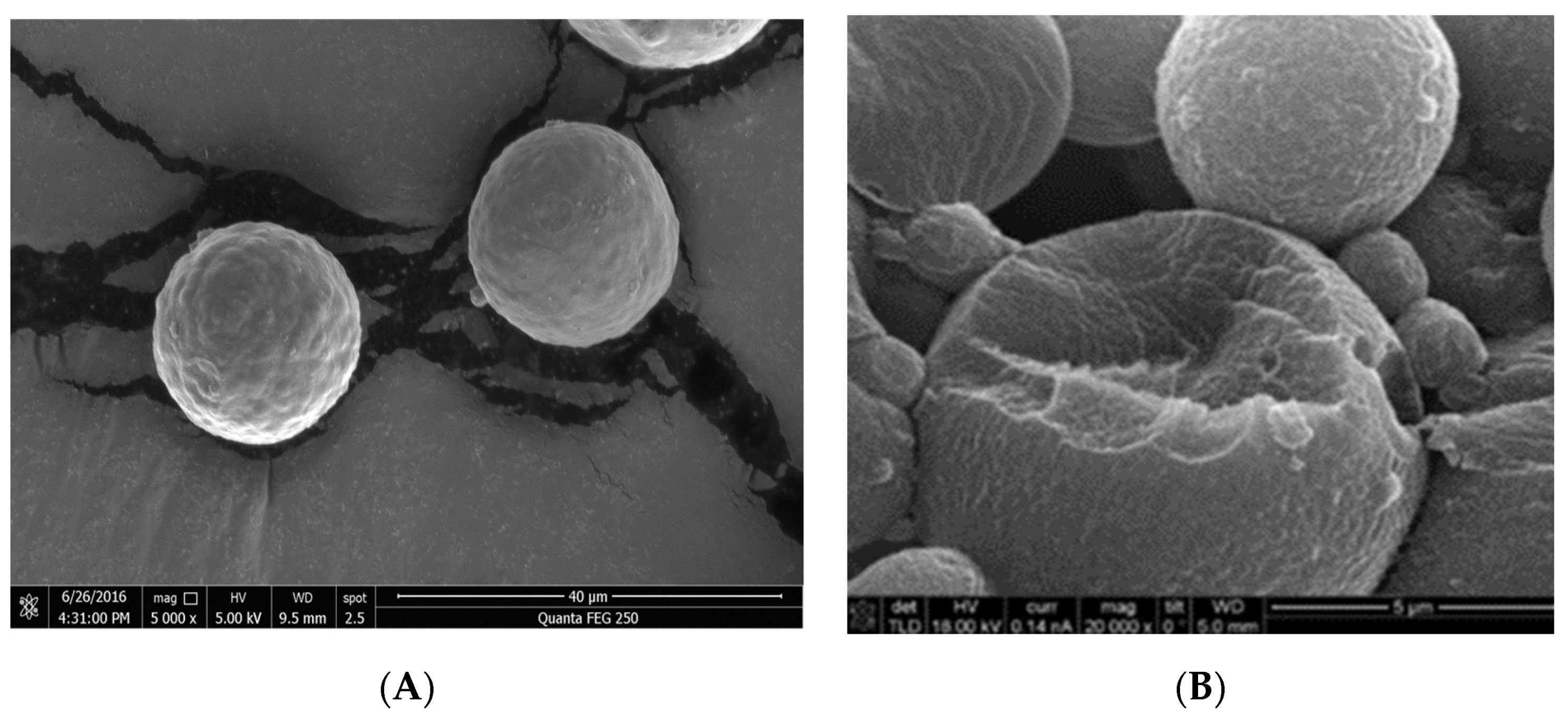
3.1. Characterization of the Microencapsulated Product
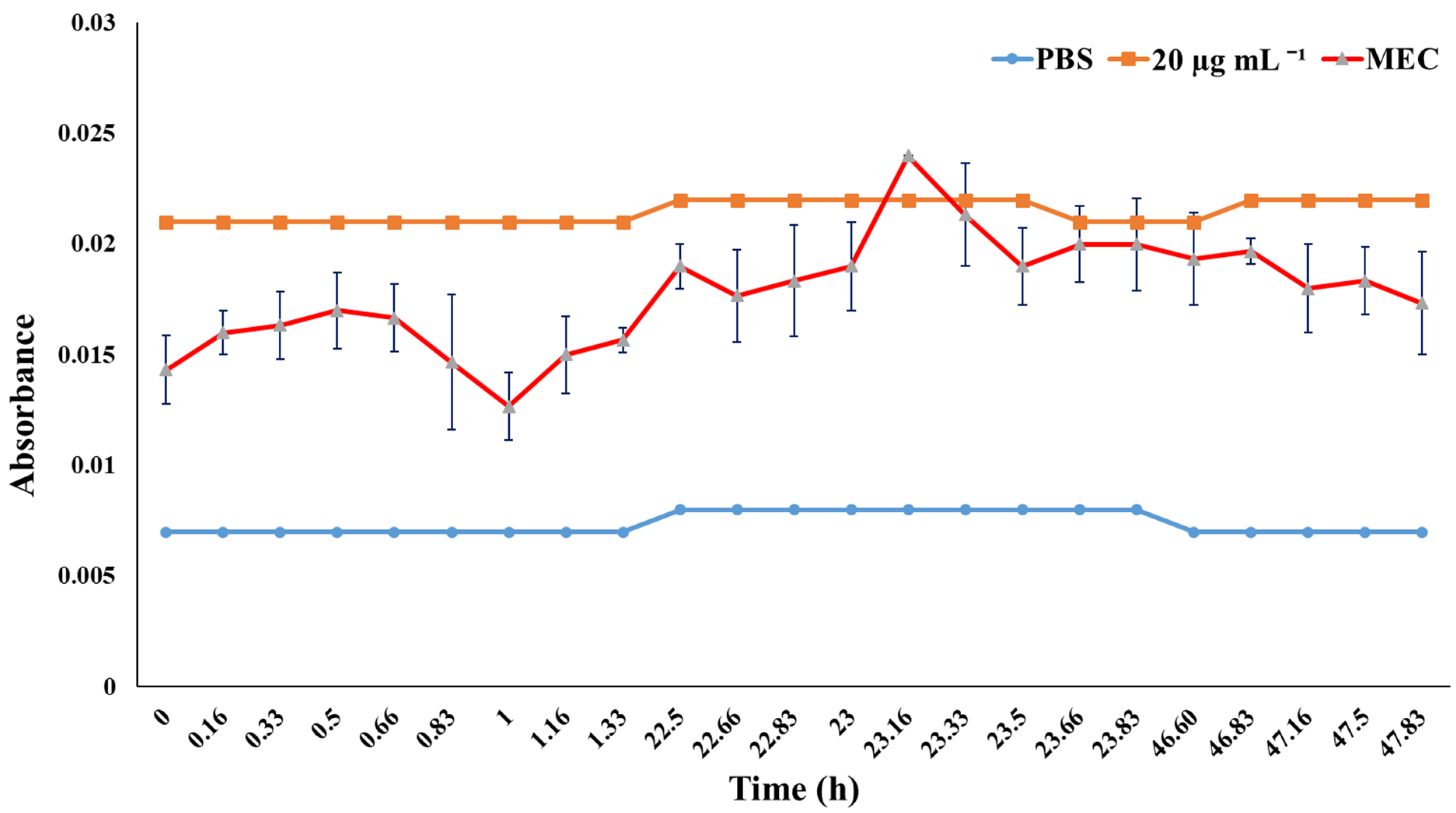
A total microsphere weight of 1740 g was obtained, equivalent to a yield of 15.81%. The microspheres had a well-defined spherical shape, compact surface with small round bumps, low clumping, and their internal structure formed a matrix of the encapsulating material (Figure 1A,B). The mean size was 20 µm, and it is likely that the difference in size is due to the speed of agitation, as well as the smooth part of the interior produced by sonication in the manufacturing process.

3.2. Release Tests

The concentration released from the Perla negra extract was 8.5 µg mL−1 during the first hour, and the highest was 24 µg mL−1 after 23 h (1380 min) (Figure 2) with a load efficiency of microspheres of 98%.

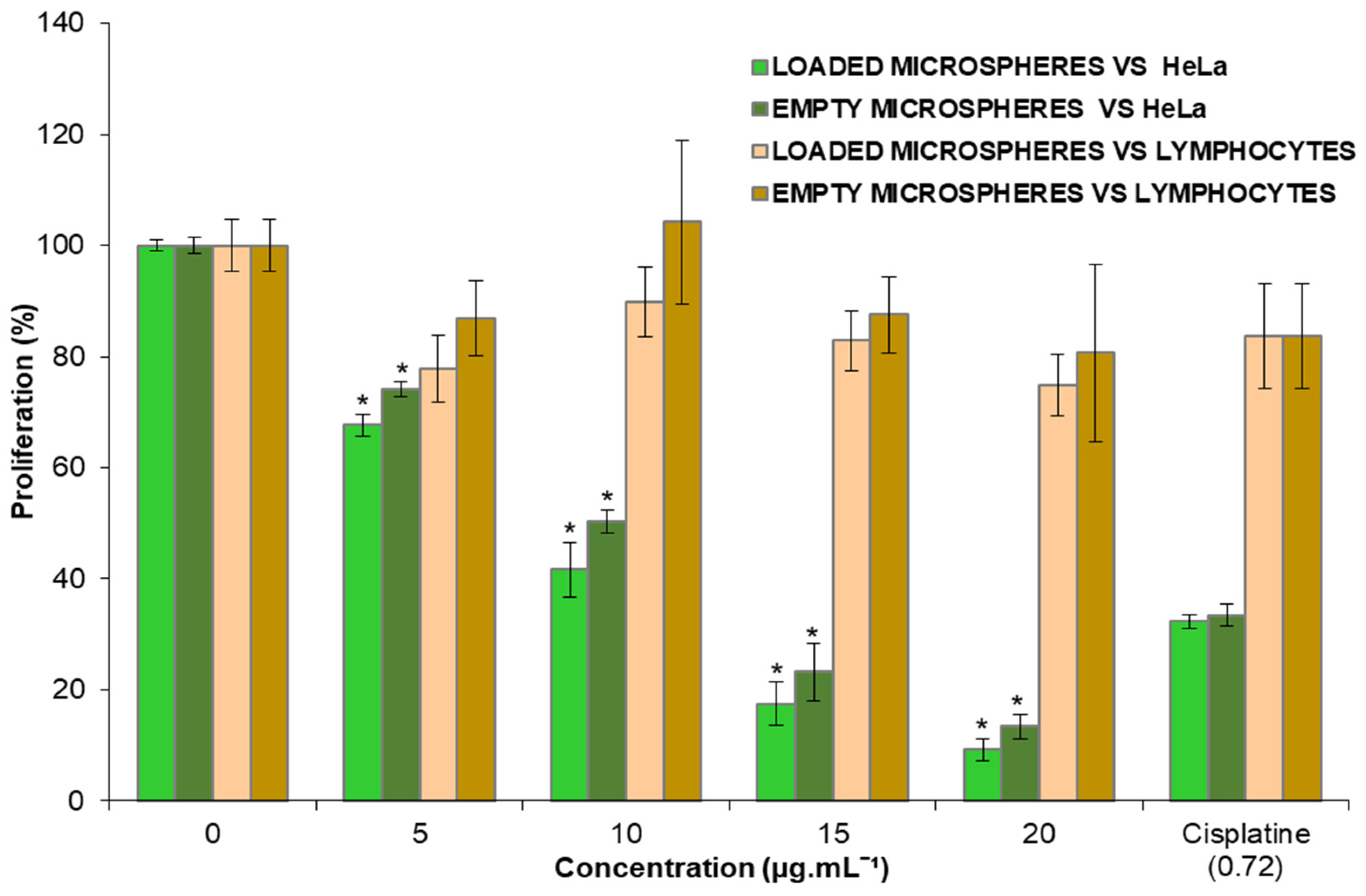
3.3. Effect of the Microspheres on HeLa and Lymphocytes

Both the loaded and the empty microparticles showed inhibition in the proliferation of the cancerous HeLa cell line, with a dose–response relationship without significant effect on the proliferation of human lymphocytes (normal cells), which suggests that they have a certain specificity of action (Figure 3). The empty microspheres exerted a percentage of inhibition of 26 to 87%, while the loaded ones showed inhibition values of 33 to 91% due to the release of the Perla negra extract (Figure 3), without presenting a significant difference, attributed to the lower initial concentration of extract. The statistical mean inhibitory concentration (IC50) of the loaded microspheres was 8 µg mL−1 obtained through linear regression. The analysis (ANOVA) for comparison of means with Tukey’s test, applying a (p ≤ 0.05) compared with the control (0 mg mL−1), showed a significant difference in the HeLa cell line with the different treatments, while there was no significant difference in lymphocytes with no treatment; therefore, there is no significance (*) in any brown bar (Figure 3).
Table 1 shows the metabolites that make up the encapsulated extract of cv. Perla negra, corroborating its content in the microspheres. The foregoing suggests its biological activity in therapeutic use.

4. Discussion

The study evaluated chitosan due to its harmless properties as a material for the microencapsulation of cv. Perla negra, whose antiproliferative effect has been registered [27]. The coacervation method with sonication was used, obtaining microspheres with an average size of 20 µm with an extract release concentration of 24 µg mL−1 at 23 h. They showed antiproliferative activity in HeLa without damage to lymphocytes. The same activity was observed with the empty microspheres (chitosan only), showing that there is no interference between the encapsulating material and the extract and suggesting its use as an administration vehicle.
Authors such as [3,34,38] obtained microspheres with a range of size of 1–30 µm, following the same method of microencapsulation. It is likely that the difference in size is due to the agitation speed (Figure 1). Olivera et al. [34] mention that the release concentration they obtained was 0.0056 µg mL−1 at 140 min, releasing DNA from cow thymus (Bos taurus L.) with 85% of load efficiency, while Olivera et al. [38] mentioned a release of 1.30 mg mL−1 at 120 min of bovine serum albumin (BSA) without recording the load efficiency. It is possible that the difference observed in the release time of the cv. Perla negra extract, compared to the release time of protein and DNA recorded by Olivera et al. [34,38] as the load efficiency, is due to the nature of the encapsulated product, and mainly the morphology of the microparticles obtained. These authors obtained particles with a more compact internal structure than the internal structure obtained in this study, with greater definition of the matrix of the encapsulating material (Figure 1).
Studies that have been carried out to evaluate the effects of terpenoids have reported antiviral, antibacterial, anti-hypertensive, anti-hyperglycemic, and anticancer activities against parasites as well as control of central nervous system disorders [39]. The effects of the flavonoids are diverse and include antiallergy, anti-depressive, antibacterial, anti-inflammatory, antioxidant, antiviral, and anticancer activities; flavonoids also can treat thrombosis, diabetes mellitus, rheumatoid diseases, cardiovascular diseases and gastric ulcers, and they offer hepatoprotection [40,41,42]. Table 1 shows that the secondary metabolites (terpenoids) were microencapsulated, which suggests that the antiproliferative activity is not at risk.
Regarding the inhibitory effect of proliferation of empty microspheres on HeLa cells, Xie et al. [43] mention that chitosan has antitumor, antiulcer, immune-stimulating, and antibacterial activities. It has also been reported that water-soluble chitosan has a low molecular weight in the growth of Sarcoma 180 (S180) tumor cells in mice. It had the highest value of inhibition at 64.2%, with a dose of 200 mg kg−1 applied via intraperitoneal injection; 33.7% with a dose of 200 mg kg−1 applied through oral administration [44]; 50% with a dose of 50 mg kg−1 applied via intraperitoneal injection; and 31% with a dose of 400 mg kg−1 via oral administration [45].
Another study evaluated the antimetastatic property of chitosan oligosaccharides (COS) on human breast carcinoma cells (MDA-MB-231), obtaining a response–dose relation and showing that cell migration through a membrane covered with Matrigel is inhibited, and the activity and amount of MMP9 protein decreased [46].
Authors such as [47] explored the effects of the degree of deacetylation and the molecular mass of different oligosaccharides of chitosan obtained from the enzymatic hydrolysis of high-molecular-weight chitosan in in vitro conditions. Authors assessed the antitumor activity in the PC3 (human prostate cancer cells), A549 (human lung cancer cell) and HepG2 (hepatocellular carcinoma cell) cell lines, showing that the COS had mean cytotoxic concentrations (CC50) of 25, 5 and 12.5 µg mL−1 of inhibition, respectively, and defining that the relatively smaller size (molecular weight), higher solubility and lower degree of deacetylation are promising factors for the development of potential pharmaceutical or nutraceutical products with this material.
Studies carried out with chitosan nanospheres at 85% deacetylation, with mean molecular weight and 40 nm diameter, showed a growth inhibition effect on Sarcoma 180 (S180) and mouse Hepatoma (H22) cells in vivo, finding that 2.5 mg kg−1 oral administration led to 53% and 59% cellular inhibition, respectively [48]. These same nanospheres have also been evaluated in human hepatocellular carcinoma (BEL7402) cells in vivo (mice) via oral administration with 1 mg kg−1 per day, obtaining 61.69% tumor growth inhibition, while in in vitro conditions they showed an IC50 of 0.94 µg mL−1 in 72 h [45].
Chitosan has proven to have inhibitory activity in different cancerous cell lines in vitro and in vivo [44,45,47,48,49], and its effect depends on different factors such as molecular weight, degree of deacetylation, and even how it is administered, whether orally or via intraperitoneal injection. This can be observed in the assessment of the effect of nanospheres on BEL7402 reported by [49]. The microspheres evaluated on HeLa cells had a statistical IC50 of 8 µg mL−1, and in the nanospheres evaluated by [49] it was 0.94 µg mL−1; that is, the latter showed the same effect with a dose 8.5 times lower than the microspheres. This is probably due to their size since the nanospheres are 500 times smaller, which can facilitate their absorption in the cell membrane [19] and influence the inhibitory effect.
It is possible the microspheres obtained in this study have the same mode of action as the nanospheres on BEL7402 that were manufactured with chitosan with the same degree of deacetylation, which causes disruption of the cell membrane in cancer cells, decreases the superficial negative load, decreases the potential of the mitochondrial membrane, induces lipid oxidation and disrupts the fatty acids of the membrane and DNA fragmentation [49].
For this effect, it is suggested that the amino groups interact with negatively charged phospholipid components in the membranes, which leads to the release of proteins and other intracellular constituents, accelerating the exchange with the medium, in addition to forming chelates and inhibiting some enzymes [26,50].
This effect is perhaps not as evident in the lymphocytes because of the very precise function that they have. Since these are cells with a very heterogeneous population and are highly specific, every type of lymphocyte presents in its membrane a single type of very specific receptor, which is why the recognition of antigens is quite broad, and they have the capacity of responding to any pathogen or strange substance that enters an organism. When there is an antigen, only the lymphocytes that recognize it are activated and proliferate, eliminating it quickly [51]; therefore, it is likely that this mechanism inhibits the effect of chitosan on these cells.
The amino group that has the chitosan makes it possible for it to be soluble in water because of its load, which makes it bioadhesive; this improves the permeation through negatively loaded surfaces, for example the mucous and basal membranes, inducing chitosan to ease the bioavailability of polar drugs and their transport through the epithelial surfaces [52]. This is a property that has led to chitosan being frequently used as an excipient and encapsulating agent for microencapsulation in a large diversity of pharmaceutical products, with different methods [11,19] and different administration vehicles [24].
The relevance of the study showed that the antiproliferative activity of the encapsulated extract did not change (it was not decreased or enhanced by the encapsulating material). Chitosan has been reported to have antiproliferative activity, which was demonstrated in the empty microspheres and did not interfere with the activity registered of the extract (Figure 3). This is very important for the administration of the metabolites of the cv extract Perla negra, whose nature and content are shown in Table 1 and which, in addition to acting as an antiproliferative, has other properties previously mentioned, such as nutraceutical, anti-inflammatory, antioxidant, etc. Even though this study did not evaluate the synergistic effect between the extract and chitosan, it is possible to suggest that in the absence of interference between them, the matrix is beneficial for use in the treatment of diseases of public interest such as cervical uterine cancer.
Based on the results, it can be said that chitosan microspheres are a good administration vehicle for Perla negra extract because the microspheres do not counteract the inhibitory action [27], although it would be pertinent to increase the concentration of the extract. The results suggest the possibility of microencapsulation of other extracts of Sechium sp., which have shown antiproliferative properties not only in HeLa but in other cancer cell lines, such as mouse lung fibrosarcoma (L929) and mouse macrophage leukemia (P388) [53,54], broadening the treatment alternatives to different types of cancer.

5. Conclusions

The chitosan-based microspheres did not affect the antiproliferative activity of the cv. Perla negra (Sechium edule (Jacq.) Sw.) fruit extract and are a potential vehicle for therapeutic administration. The empty microspheres elaborated with chitosan have an antiproliferative effect, and loaded they showed cell inhibition with a statistical IC50 of 8 µg mL−1, without affecting the lymphocytes. It is confirmed that the chitosan microspheres made under our conditions have characteristics similar to those reported in the literature, including their ability to inhibit the proliferation and inclusion of bioactive compounds. For the first time, it is shown that microspheres can store and release complex compounds such as Sechium ethanol extract, including four cucurbitacins and seven flavonoids. The compounds incorporated into the microspheres retain their proliferation inhibitory activity in tumor cells; therefore, they constitute a potential vehicle for the therapeutic administration of the cv. Perla negra (Sechium edule (Jacq.) Sw.) fruit extract.

Supplementary Materials

The following supporting information can be downloaded at: https://www.mdpi.com/article/10.3390/app12010024/s1, Data supplemental: Norm of the National Commission of Bioethics-Mexico to the main text.

Author Contributions

Conceptualization, M.S.-H. and J.C.-I.; genetic improvement of cv. Perla negra: J.C.-I. and V.M.C.-S.; Phitochemistry: S.S.-A. and M.S.-H.; Microencapsulation: L.d.M.R.-P. and Á.D.O.; Cellular evaluation: E.S.-O. and I.A.-S.; Software, Formal analysis, and Data curation: J.F.A.-M.; Writing—original draft: S.S.-A., J.C.-I. and I.A.-S. All authors have read and agreed to the published version of the manuscript.

Funding

This research received no external funding.

Institutional Review Board Statement

The study was conducted according to the guidelines of the Declaration of Helsinki, and approved by the Institutional Review Board (or Ethics Committee) of Hematopoiesis and Leukemia Laboratory, Research Unit on Cell Differentiation an Cancer, Facultad de Estudios Superiores Zaragoza, Universidad Nacional Autónoma de México (protocol: Conbioetica-salud.gob.mx, approved on 11 May 2020).

Informed Consent Statement

Informed consent was obtained from all subjects involved in the study. The bioethics standard is attached as supplementary material.

Data Availability Statement

Not report any data.

Acknowledgments

To the National Science and Technology Council (Consejo Nacional de Ciencia y Tecnología, CONACyT). To Interdisciplinary Research Group at Sechium edule in México, A.C. (GISeM) for the biological material provided and support given. To M.Sc. Luis Martínez for the support in the performance of bioassays, in the Hematopoiesis and Leukemia laboratory of FES Zaragoza UNAM.

Conflicts of Interest

The authors declare no conflict of interest.

References

  1. Pedraz, J.L.; Orive, G. La microencapsulación de células. Una nueva alternativa terapéutica? An. Real Acad. Nac. Farm 2004, 70, 777–787. [Google Scholar]
  2. López, H.O.; Márquez, T.; Mayo, C.O.; Toledo, A.C.S.; Pérez, Y.E.S. Características del aceite de semillas de Cucurbita pepo L. microencapsulado mediante secado por aspersión con maltodextrina y goma arábiga. Lat. Am. J. Pharm. 2009, 28, 628–632. [Google Scholar]
  3. Lopretti, M.; Barreiro, F.; Fernandes, I.; Damboriarena, A.; Ottati, C.; Olivera, A. Microencapsulación de compuestos de actividad biológica. INNOTEC 2011, 2, 19–23. [Google Scholar] [CrossRef]
  4. Sáez, V.; Hernández, E.; Sanz, A.L.; Katime, I. Liberación controlada fármacos. Micropartículas. Rev. Iberoam. Polim. 2004, 5, 87–101. [Google Scholar]
  5. Martín, V.M.J.; Morales, H.M.E.; Gallardo, L.V.; Ruiz, M.M.A. Técnicas de microencapsulación: Una propuesta para microencapsular probióticos. Ars Pharm. 2009, 50, 43–50. [Google Scholar]
  6. Cabanè, P.; Alvo, A.; Neira-Carrillo, A.; Caviedes, P.; Gac, P. Microencapsulación de células y tejido para terapia celular. Rev. Cir. 2011, 63, 110–113. [Google Scholar] [CrossRef] [Green Version]
  7. Marte, Y.A.; Marte, Y.A.; Tejada, A.E.; Berruezo, G.R. Effect of different concentrations of pulverized mesocarp of Citrus paradisi Macf. on the bromatological characteristics of spray-dried lemon juice powder. Food Sci. Nutr. 2018, 6, 1261–1268. [Google Scholar] [CrossRef] [PubMed] [Green Version]
  8. López, H.O.D. Microencapsulación de sustancias oleosas mediante secado por aspersión. Rev. Cuba. Farm 2010, 44, 381–389. [Google Scholar]
  9. López-Hernández, O.D.; Gómez-Carril, M.; Padrón-Yaquis, A.S.; Toledo-Rivero, A.; Toledo-Sanchez, C.A.; Reyes-Sanchez, E.; Reguera-Ruiz, E.; Duque-Rodríguez, J.; López-Pelaez, B. Microencapsulación de tramadol en ácido poliláctico mediante secado por aspersión. Rev. Cuba. Farm 2010, 44, 432–442. [Google Scholar]
  10. Lárez, V.C. Algunos usos del quitosano en sistemas acuosos. Rev. Iberoam. Polim. 2003, 4, 91–109. [Google Scholar]
  11. Agnihotri, S.A.; Mallikarjuna, N.; Amninabhavi, T.M. Recent advances on chitosan-based micro-and nanoparticles in drug delivery. J. Control. Release 2004, 100, 5–28. [Google Scholar] [CrossRef] [PubMed]
  12. Acosta, N.; Heras, A. New Drug Delivery Systems Based on Chitosan. Curr. Drug Discov. Technol. 2008, 5, 333–341. [Google Scholar] [CrossRef]
  13. Expósito, R. Quitosano, un Biopolímero con Aplicaciones en Sistemas de Liberación Controlada de Fármacos. Ph.D. Thesis, Universidad Complutense de Madrid, Facultad de Ciencias Biológica, Departamento de Bioquímica y Molecular I, Madrid, Spain, 2010; p. 171. Available online: https://eprints.ucm.es/id/eprint/11160/1/T32051.pdf (accessed on 30 November 2021).
  14. Mármol, Z.; Páez, G.; Rincón, M.; Araujo, K.; Aiello, C.; Chandler, C.; Gutiérrez, E. Quitina y Quitosano polímeros amigable. Una revisión de sus aplicaciones. Rev. Tecnocient. 2011, 1, 53–58. [Google Scholar]
  15. Patel, V.; Patel, M.; Patel, R. Chitosan: A Unique Pharmaceutical Excipient. Drug Deliv. Technol. 2005, 6, 30–40. [Google Scholar]
  16. Veiga, O.M.D.; Ruiz, C.R. El Quitosano: Usos farmacéuticos y biológicos. Rev. OFIL 2004, 14, 33–42. Available online: https://ilaphar.org/wp-content/uploads/2014/01/OFILn142.pdf (accessed on 17 August 2021).
  17. Rodríguez-Pedroso, A.T.; Ramírez-Arrebato, M.A.; Rivero-González, D.; Bosquez-Molina, E.; Barrera-Necha, L.L.; Bautista-Baños, S. Propiedades químico-estructurales y actividad biológica de la quitosana en microorganismos fitopatógenos. Rev. Chapingo Ser. Hortic. 2009, 15, 307–317. [Google Scholar]
  18. Khanvilkar, K.; Donovan, M.D.; Flanagan, D.R. Drug transfer through mucus. Adv. Drug Deliv. Rev. 2001, 48, 173–193. [Google Scholar] [CrossRef]
  19. Tiyaboonchai, W. Chitosan Nanoparticles. A Promising System for Drug Delivery. Naresuan Univ. J. 2003, 11, 51–66. [Google Scholar]
  20. De Enríquez, S.A.; Diebold, Y.; Calonge, M.; García-Vazquez, C.; Callejo, S.; Vila, A.; Alonso, M.J. Chitosan nanoparticles as a potential drug delivery system for the ocular surface: Toxicity, uptake mechanism and in vivo tolerance. Investig. Ophthalmol. Vis. Sci. 2006, 47, 1416–1425. [Google Scholar] [CrossRef]
  21. Genta, I.; Costantini, M.; Asti, A.; Conti, B.; Montanari, L. Influence of glutaraldehyde on drug release and mucoadhesive properties of chitosan microspheres. Carbohydr. Polym. 1998, 36, 81–88. [Google Scholar] [CrossRef]
  22. Baki, D.E.; Ottenbrite, R.M. Perspectives on: Chitosan drug delivery systems based on their geometries. J. Bioact. Compat. Polym. 2006, 21, 351–368. [Google Scholar] [CrossRef]
  23. Amidi, M.; Mastrobattista, E.; Jiskoot, W.; Henninknnin, W.E. Chitosan-based delivery systems for protein therapeutics and antigens. Adv. Drug Deliv. Rev. 2010, 62, 59–82. [Google Scholar] [CrossRef] [PubMed]
  24. Shaji, J.; Jain, V.; Lodha, S. Chitosan: A novel pharmaceutical excipient. International. J. Pharm. Sci. 2010, 1, 11–28. [Google Scholar]
  25. Morris, G.A.; Samil, K.M.; Harding, S.E.; Adams, G.G. Polysaccharide drug delivery systems based on pectin and chitosan. Biotechnol. Genet. Eng. Rev. 2010, 27, 257–284. Available online: https://www.tandfonline.com/doi/pdf/10.1080/02648725.2010.10648153 (accessed on 20 August 2021). [CrossRef]
  26. Chen, C.-S.; Liau, W.-Y.; Tsai, G.-J. Antibacterial Effects of N-Sulfonated and N-Sulfobenzoyl Chitosan and Application to Oyster Preservation. J. Food Prot. 1998, 61, 1124–1128. [Google Scholar] [CrossRef] [PubMed]
  27. Salazar-Aguilar, S.; Ruiz-Posadas, L.D.M.; Cadena-Iñiguez, J.; Soto-Hernández, M.; Santiago-Osorio, E.; Aguiñiga-Sánchez, I.; Rivera-Martínez, A.R.; Aguirre-Medina, J.F. Sechium edule (Jacq.) Swartz, a New Cultivar with Antiproliferative Potential in a Human Cervical Cancer HeLa Cell Line. Nutricients 2017, 9, 798. [Google Scholar] [CrossRef] [Green Version]
  28. Cadena-Iñiguez, J.; Arévalo-Galarza, L.; Avendaño-Arrazate, C.H.; Soto-Hernández, R.M.; Ruiz-Posadas, L.M.; Santiago-Osorio, E.; Acosta-Ramos, M.; Cisneros-Solano, V.M.; Aguirre-Medina, J.F.; Ochoa-Martínez, D. Production, genetics, postharvest management, and pharmacological characteristics of Sechium edule (Jacq.) Sw. Fresh Prod. 2007, 1, 41–53. [Google Scholar]
  29. Cadena-Iñiguez, J.; Soto, H.M.; Arevalo, G.M.L.; Avendaño, A.C.H.; Aguirre, M.J.F.; Ruiz, P.L.M. Caracterización bioquímica de variedades domesticadas de Sechium edule (Jacq.) Sw. comparadas con parientes silvestres. Rev. Chapingo Ser. Hortic. 2011, 17, 45–55. [Google Scholar]
  30. Che, C.; Fang, X.; Phoebe, C.H.; Kiunghorn, A.D.; Farnswworth, N.R. High-field 1H-NRM spectral analysis of some cucurbitacins. J. Nat. Prod. 1985, 48, 429–434. [Google Scholar] [CrossRef]
  31. Afifi, M.S.; Ross, S.A.; ElSohly, M.A.; Naeem, Z.E.; Halaweish, F.T. Cucurbitacins of Cucumis prophetarum and Cucumis prophetarum. J. Chem. Ecol. 1999, 25, 847–859. [Google Scholar] [CrossRef]
  32. Cadena-Iñiguez, J. Caracterización Morfoestructural, Fisiológica, Química y Genética de Diferentes Tipos de Chayote (Sechium edule (Jacq.) Sw). Ph.D. Thesis, Colegio de Postgraduados, Texcoco, Mexico, 2005; p. 156. Available online: www.colpos.mx (accessed on 12 July 2021).
  33. Akbuğa, J.; Bergişadi, N. 5-Fluorouracil-loaded chitosan microspheres: Preparation and release characteristics. J. Microencapsul. 1996, 13, 161–168. [Google Scholar] [CrossRef]
  34. Olivera, A.D.; Barreiro, M.F.; Lopretti, M. Microesferas de quitosano como potenciales transportadores de ácidos nucleicos y otros bioactivos. Rev. Iberoam. Polim. 2012, 13, 238–244. [Google Scholar]
  35. NOM-253-SSA1-2012. Para la Disposición de Sangre Humana y sus Componentes con Fines Terapéuticos. Diario Oficial. Tercera Sección Poder Ejecutivo. Secretaria de Salud. 2012. Available online: http://www.cnts.salud.gob.mx/descargas/NOM-253-SSA1-2012.pdf (accessed on 30 November 2021).
  36. Kueng, W.; Silver, E.; Eppnberg, V. Quantification of cell cultured on 96-wels plates. Anal. Biochem. 1989, 186, 16–19. [Google Scholar] [CrossRef]
  37. Gillies, R.; Didier, N.; Denton, M. Determination of cell number in monolayer cultures. Anal. Biochem. 1986, 159, 109–113. [Google Scholar] [CrossRef]
  38. Olivera, A.D.; Barreiro, M.F.; Lopretti, M. Proteínas de actividad bilógica microencapsuladas, ultraestructura y distribución de carga. Rev. Iberoam. Polim. 2014, 15, 219–228. [Google Scholar]
  39. Brahmkshatriya, P.P.; Brahmkshatriya, P.S. Terpenes: Chemistry, Biological Role, and Therapeutic Applications; Ramawat, K., Mérillon, J.M., Eds.; Springer: Berlin/Heidelberg, Germany, 2013. [Google Scholar] [CrossRef]
  40. Agrawal, A.D. Pharmacological Activities of Flavonoids: A Review. Int. J. Pharm. Sci. Nanotechnol. 2011, 4, 1394–1398. [Google Scholar] [CrossRef]
  41. Kaur, S.H.; Kumar, B.; Prasher, S.; Tiwari, P.; Salhan, M.; Sharma, P. A Review of phytochemistry and pharmacology of flavonoids. Sci. Pharm. 2011, 1, 25–41. [Google Scholar]
  42. Kumar, S.; Pandey, A.K. Chemistry and biological activities of flavonoids: An overview. Hindawi Sci. World J. 2013, 1–16. [Google Scholar] [CrossRef] [Green Version]
  43. Xie, W.; Xu, P.; Liu, Q. Antioxidant activity of water-soluble chitosan derivatives. Bioorg. Med. Chem. Lett. 2001, 11, 1699–1701. [Google Scholar] [CrossRef]
  44. Qin, C.; Du, Y.; Xiao, L.; Li, Z.; Gao, X. Enzymic preparation of water-soluble chitosan and their antitumor activity. Int. J. Biol. Macromol. 2002, 31, 111–117. [Google Scholar] [CrossRef]
  45. Qin, C.; Zhou, B.; Zeng, L.; Zhang, Z.; Liu, Y.; Du, Y.; Xiao, L. The physicochemical properties and antitumor activity of cellulase-treated chitosan. J. Agric. Food Chem. 2004, 84, 107–115. [Google Scholar] [CrossRef]
  46. Nam, K.S.; Shon, Y.H. Suppression of metastasis of human breast cancer cells by chitosan oligosaccharides. J. Microbiol. Biotechnol. 2009, 19, 629–633. [Google Scholar] [CrossRef]
  47. Park, J.K.; Chung, M.J.; Na Choi, H.; Park, Y.I. Effects of the Molecular Weight and the Degree of Deacetylation of Chitosan Oligosaccharides on Antitumor Activity. Int. J. Mol. Sci. 2011, 12, 266–277. [Google Scholar] [CrossRef] [PubMed] [Green Version]
  48. Qi, L.; Xu, Z. In vivo antitumor activity of chitosan nanoparticles. Bioorg. Med. Chem. Lett. 2006, 16, 4243–4245. [Google Scholar] [CrossRef]
  49. Qi, L.; Xu, Z.; Chen, M. In vitro and in vivo suppression of hepatocellular carcinoma growth by chitosan nanoparticles. Eur. J. Cancer 2007, 43, 184–193. [Google Scholar] [CrossRef] [PubMed]
  50. Ayala, A.; Colina, M.; Molina, J.; Vargas, J.; Rincon, D.; Medina, J.; Rosales, L.; Cardenas, H. Evaluación de la actividad antifúngica del quitosano contra el hongo Mycosphaerella fijiensis Morelet que produce la sigatoka negra que ataca el plátano. Rev. Iberoam. Polim. 2014, 15, 312–338. [Google Scholar]
  51. González-Fernández, E.; Fernández, M.S.; Lorenzo, A. Linfocitos T y B. Clasificación. Receptores. Generación de diversidad: Mecanismos moleculares. Capacidades funcionales. Rev. Med. 2005, 9, 1–24. Available online: https://dialnet.unirioja.es/servlet/articulo?codigo=1252689 (accessed on 27 August 2021).
  52. Thanou, M.; Verhoef, J.C.; Junginger, H.E. Chitosan and its derivatives as intestinal absorption enhancers. Adv. Drug Deliv. Rev. 2001, 50, 91–101. [Google Scholar] [CrossRef]
  53. Cadena-Iñiguez, J.; Soto-Hernandez, M.; Torres-Salas, A.; Aguiñiga-Sánchez, I.; Ruiz-Posadas, L.; Rivera-Martínez, A.R.; Avendaño-Arrazate, C.; Santiago-Osorio, E. The antiproliferative effect of chayote varieties (Sechium edule (Jacq.) Sw.) on tumour cell lines. J. Med. Plant. 2013, 7, 455–460. [Google Scholar] [CrossRef]
  54. Aguiñiga-Sánchez, I.; Cadena-Íñiguez, J.; Santiago-Osorio, E.; Gómez-García, G.; Mendoza-Núñez, V.M.; Rosado-Pérez, J.; Ruíz-Ramos, M.; Cisneros-Solano, V.M.; Ledesma-Martínez, E.; Delgado-Bordonave, A.D.J.; et al. Chemical analyses and in vitro and in vivo toxicity of fruit methanol extract of Sechium edule var. nigrum spinosum. Pharm. Biol. 2017, 55, 1638–1645. [Google Scholar] [CrossRef]
Figure 1. Morphology of the microspheres with methanol extract of Sechium edule cv. Perla negra. (A,B): View of internal structure of the microspheres obtained in this study implementing sonication.
Applsci 12 00024 g001
Figure 2. Release curves of the methanol extract of cv. Perla negra (Sechium edule (Jacq.) Sw.) contained in the microspheres. The bars indicate ± standard deviation of n = 6. (MEC = loaded microspheres; PBS = phosphate buffer saline).
Applsci 12 00024 g002
Figure 3. Inhibition of the proliferation of HeLa cells and lymphocytes, as an effect of the application of empty microspheres and loaded microspheres with cv. Perla negra (Sechium edule (Jacq.) Sw.) methanol extract. The asterisk indicates the level of statistical significance (p ≤ 0.05). The bars indicate ± standard error of n = 9.
Applsci 12 00024 g003
Table 1. Composition of the microencapsuled extract of cv. Perla Negra (Sechium edule (Jacq.) Sw.).
Metabolite (Triterpenoid)Concentration
(µg mg−1)
FlavonoidConcentration
(µg mg−1)
Cucurbitacin B137.00Rutin0.34
Cucurbitacin D935.94Myricetin1.85
Cucurbitacin E98.21Quercetin0.25
Cucurbitacin I40.37Naringenin2.26
Phloretin4.72
Galangin0.43
Apigenin0.18
Publisher’s Note: MDPI stays neutral with regard to jurisdictional claims in published maps and institutional affiliations.

