Article

Carotenoid Accumulation and the Expression of Carotenoid Metabolic Genes in Mango during Fruit Development and Ripening

1
The United Graduate School of Agricultural Science, Gifu University, 1-1 Yanagido, Gifu-shi, Gifu 501-1193, Japan
2
School of Bioresources and Technology, King Mongkut’s University of Technology Thonburi, 49 Soi Thian Thale 25, Bang Khun Thian Chai Thale Road, Tha Kham, Bang Khun Thian, Bangkok 10150, Thailand
3
Department of Biological and Environmental Sciences, Faculty of Agriculture, Shizuoka University, 836 Ohya, Suruga, Shizuoka 422-8529, Japan
4
Department of Plant Science, Faculty of Technology and Community Development, Thaksin University, Phatthalung 93110, Thailand
*
Authors to whom correspondence should be addressed.
These authors contributed equally to this work.
Current affiliation: Horticulture Research Institute, Department of Agriculture, Chatuchak, Bangkok 10900, Thailand.
Appl. Sci. 2021, 11(9), 4249; https://doi.org/10.3390/app11094249
Submission received: 21 March 2021 / Revised: 26 April 2021 / Accepted: 5 May 2021 / Published: 7 May 2021

Abstract

:
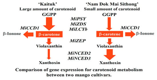
Carotenoids are considered to be important components in mango fruits. However, there is a lack of understanding about the regulation of carotenoids in mango. To gain an insight into the carotenoid metabolism pathway, carotenoid content and the expression of carotenoid metabolic genes were investigated in the peel and pulp of mango during fruit development and ripening in three cultivars, ‘Kaituk’, ‘Nam Dok Mai No.4′, and ‘Nam Dok Mai Sithong’, which are different in color. The highest carotenoid content was observed in ‘Kaituk’, followed by ‘Nam Dok Mai No.4′ and ‘Nam Dok Mai Sithong’, with the major carotenoid being β-carotene. The gene expression analysis found that carotenoid metabolism in mango fruit was primarily regulated at the transcriptional level. The changing patterns of carotenoid biosynthetic gene expression (MiPSY, MiPDS, MiZDS, MiCRTISO, MiLCYb, MiLCYe, MiHYb, and MiZEP) were similar to carotenoid accumulation, and ‘Kaituk’ exhibited a higher expression level than the other two cultivars. In addition, the differential regulation of carotenoid catabolic genes was found to be a mechanism responsible for variability in carotenoid content among the three mango cultivars. The expression of carotenoid catabolic genes (MiCCD1, MiNCED2, and MiNCED3) more rapidly decreased in ‘Kaituk’, resulting in a larger amount of carotenoids in ‘Kaituk’ than the other two cultivars.

Graphical Abstract

1. Introduction

Mango (Mangifera indica L.) is one of the world’s most famous tropical fruits because of its tasty and attractive color. The mango color is a major fruit quality factor influencing consumer satisfaction and determining the appropriate maturity for harvest. The pigmentation of mango is mainly determined by carotenoid content and compositions, which contribute to many shades of yellow color fruits [1]. It has been well documented that mango is a rich source of dietary carotenoids for humans. Carotenoids provide many benefits to human health because of their antioxidant properties. The consumption of carotenoids enhances the immune system and is associated with lowered risk factors of some cancers and heart disease [2].
Due to the crucial roles of carotenoids in plants and humans, the carotenoid metabolic pathway has been well studied in a number of fruits and vegetables, such as citrus, melon, strawberry, apple, and chili [3,4,5,6,7,8]. As shown in Figure 1, C20-geranylgeranyl diphosphate (GGPP) is used as a substrate in the carotenoid biosynthesis pathway and then catalyzed by phytoene synthase (PSY) to produce colorless C40-phytoene. A subsequent series of desaturation and isomerization results in the conversion, catalyzed by phytoene desaturase (PDS), ζ-carotene desaturase (ZDS), and carotenoid isomerase (CRTISO). Then, the two critical branching steps are carried forward to yield diverse carotenoids. The cyclization of lycopene with one ε-ring and one β-ring produces α-carotene by lycopene ε-cyclase (LCYe) and lycopene β-cyclase (LCYb). α-Carotene is subsequently converted into lutein by β-ring hydroxylase (HYb) and ε-ring hydroxylase (HYe). Another branch of lycopene is cyclized with two β-rings by lycopene β-cyclase (LCYb) to yield β-carotene. Then, β-carotene is converted into β-cryptoxanthin and zeaxanthin by β-ring hydroxylase (HYb). Zeaxanthin is then converted into violaxanthin by zeaxanthin epoxidase (ZEP). Furthermore, carotenoid catabolism can be catalyzed by a group of enzymes, called carotenoid cleavage dioxygenases (CCDs). They cleave a variety of carotenoids at different cleavage positions depending on the carotenoid substrate. The CCDs can cleave the double bonds of several carotenoid substrates to generate diverse products of apocarotenoids. 9-Cis-epoxycarotenoid dioxygenases (NCEDs), members of the CCD family, enzymatically catabolize violaxanthin to yield C25 epoxy-apocarotenoid, which is then modified to abscisic acid (ABA).
The earlier studies on carotenoid metabolism demonstrated that carotenoid content and composition could be varied among fruit development stages and ripening stages, plant species/cultivars, and plant tissues [9]. In various fruits and vegetables, carotenoid content continuously increased during fruit development together with the appearance of yellow/orange color in ripe fruit, such as melon, tomato, and citrus [3,10,11]. Contrastingly, the carotenoid content dramatically decreased during fruit development in apple, grape, and some berry fruits due to the anthocyanin biosynthesis for red pigment formation [12,13,14,15]. Carotenoid biosynthesis is a crucial biochemical change during mango fruit development and ripening. In recent years, the regulatory mechanism of carotenoid metabolism has been evaluated in a great number of fruits and vegetables during the pre/postharvest process. However, limited information is currently available on mango fruits.
Therefore, the objective of this research was to elucidate carotenoid accumulation and the expression of carotenogenic genes during fruit development and ripening in both the peel and pulp of three mango cultivars with different fruit colors, namely: ‘Kaituk’, ‘Nam Dok Mai No.4′, and ‘Nam Dok Mai Sithong’. These three mango cultivars were considered as an economically important fruit in Thailand with great production and export potential. With an increasing demand and increasing percentage of these mango cultivars that are being exported worldwide, they are different in yellow color development. Thus, it is interesting to use these cultivars as plant materials in this study. The diversities in carotenoid accumulation among different fruit developmental stages, plant tissues, and mango cultivars were discussed and the regulatory mechanism of carotenoid metabolism during mango fruit development was clarified at the molecular level.

2. Materials and Methods

2.1. Plant Materials

The three commercial mango cultivars in Thailand were used as plant materials, namely: ‘Nam Dok Mai Sithong’, ‘Nam Dok Mai No.4′, and ‘Kaituk’. The uniform and blemish-free fruits were randomly harvested from the mango trees in commercial orchards at different developmental stages from 45, 59, 74, 88, and 98 days after full bloom. Mango peel was separated from its pulp, directly frozen in liquid nitrogen and vacuum-freeze-dried, then stored at −30 °C until used.

2.2. Determination of Fruit Color

The fruit color was evaluated in peel and pulp at different developmental stages as L* (lightness), a* (red/green value), b* (blue/yellow value), and hue values using a Minolta chromameter (Model CR-300, Minolta, Osaka, Japan). The color readings were taken in 3 fruits per sample at the equatorial of the fruit.

2.3. Determination of Ethylene Production and Respiration Rate

Ethylene production was evaluated by using gas chromatography with a flame ionization detector (FID) equipped with an 80/100 mesh Porapack-Q column with nitrogen as a carrier gas (GC B14; Shimadzu: Kyoto, Japan). Fruit samples were kept in a sealed plastic container, and then incubated at ambient temperature for 1 h. A 1 mL gas sample was withdrawn from the head space for ethylene measurement in the three replications. The data are expressed as μL C2H4 kg−1 h−1.
Respiration rates were measured using gas chromatography with an 80/100 mesh Porapack-Q column and a thermal conductivity detector (GC A8; Shimadzu; Kyoto, Japan) with the same procedure as described in ethylene production. There were three replications of each maturity stage. The data are expressed as mg CO2 kg−1 h−1.

2.4. Analysis of Carotenoid Content and Composition

Carotenoid extraction and quantification in mango fruits were performed with the method previously described by Kato et al. [10]. The contents of all-trans-violaxanthin, 9-cis-violaxanthin, lutein, α-carotene, β-carotene, and β-cryptoxanthin in the peel and pulp of mango were measured by HPLC in the three replications.
Carotenoids were extracted from samples using hexane–acetone–ethanol (50:25:25, v/v/v) mixture containing magnesium carbonate basic and centrifuged at 4000 rpm for 20 min. The supernatant containing the pigments and hexane was evaporated to dryness. The dry samples were re-dissolved in 12 mL of diethyl ether containing 0.1% (w/v) butylated hydroxytoluene (BHT) and saponified overnight using 20% (w/v) methanolic KOH. After saponification, NaCl-saturated water was used to remove water soluble impurities. Anhydrous Na2SO4 was subsequently added to remove residual water from the extract. The retained carotenoids were eluted from anhydrous Na2SO4 by diethyl ether, and subsequently evaporated to dryness. Carotenoid residues were then re-dissolved in tert-butyl methyl ether (TBME): methanol (1:1, v/v) containing 0.5% (w/v) BHT.
The quantification of carotenoids was carried out with a reverse-phase HPLC system (Jasco, Tokyo, Japan) fit with a YMC Carotenoid S-5 column of 250 × 4.6-mm-i.d. (Waters, Milford, MA, USA) at a flow rate of 1 mL min−1. The eluent was detected by a photodiode array detector (MD-910, Jasco). Three different gradient elution schedules were used to assess carotenoids in the samples according to a previously described method [10]. Peaks were identified by comparing their retention times and absorption spectra with authentic standards and expressed as µg g−1 fresh weight.

2.5. Isolation and Sequence Analysis of Genes Involved in Carotenoid Metabolism Pathway

The total RNA extraction in mango fruits was performed by a modified method from the previously described method of Ikoma et al. [16]. Total RNA was cleaned up by the RNeasy Mini Kit (Qiagen, Hilden, Germany) in accordance with the manufacturer’s instruction guide and stored as ethanol precipitation at −80 °C. Two micrograms of purified RNA was used to synthesize first strand cDNA using TaqMan Reverse Transcription Reagents (Applied Biosystems).
The cDNA fragments of genes related to the carotenoid metabolism pathway (MiPSY, MiPDS, MiZDS, MiCRTISO, MiLCYb, MiLCYe, MiHYb, MiZEP, MiCCD1, MiNCED2, and MiNCED3) were amplified by PCR using a cDNA template and set of primers shown in Table 1. The primers were designed based on the known sequences of carotenogenic genes in Mangifera indica cultivars ‘Jinhuang’, ‘Hongmang NO.6’, ‘Kensington Pride’, and ‘Zill’. The amplified cDNAs were confirmed by sequencing.

2.6. Real Time Quantitative Reverse Transcription Polymerase Chain Reaction (qRT-PCR)

qRT-PCR was conducted in the three replications for each sample. TaqMan MGB probes and the set of primers for MiPSY, MiPDS, MiZDS, MiCRTISO, MiLCYb, MiLCYe, MiHYb, MiZEP, MiCCD1, MiNCED2, and MiNCED3 were designed on the basis of common sequences among the three mango varieties for each gene using Primer Express software (Applied Biosystems) shown in Table 2. The TaqMan Ribosomal RNA Control Reagents VIC Probe (Applied Biosystems) was used for the endogenous control.
Gene expression was analyzed by StepOnePlusTM Real-Time PCR System (Applied Biosystems) in accordance with the manufacturer’s instruction guide. In the Real-Time PCR reaction mixture, each reaction contained 900 nM of primers, 250 nM of TaqMan MGB probe, and the cDNA template. The cycling protocol was of 95 °C for 10 min, then 40 cycles of 95 °C for 15 s and 60 °C for 60 s. The results were calculated using StepOnePlusTM Real-Time PCR System Software (Applied Biosystems) and normalized with the results of 18S ribosomal RNA.

2.7. Data Analysis

Data presented in this study were expressed as the mean ± standard error (SE) for three replications.

3. Results

3.1. Changes in Respiration Rate, Ethylene Production, Peel and Pulp Color during Mango Fruit Development and Ripening

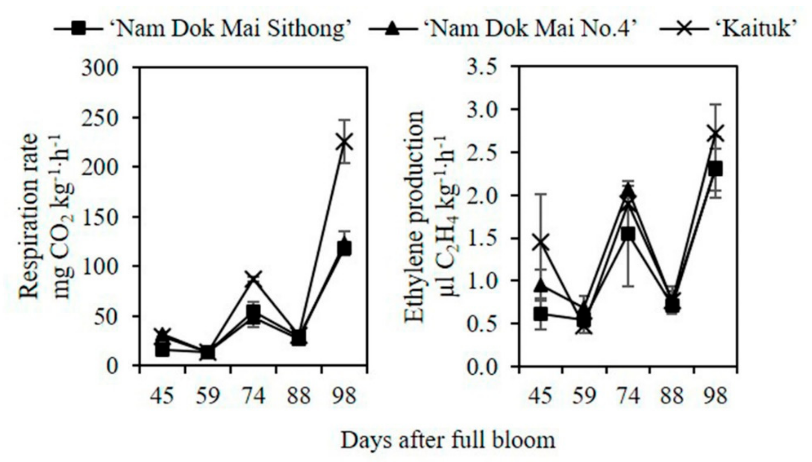
In this study, three cultivars of mango fruits were collected at different maturity stages. The respiration rate was unsteady in the early developmental stage and suddenly reached a peak at the full ripening. The increase in respiration rate was more rapid in ‘Kaituk’ than in the other two cultivars. Similar trends were also observed in ethylene production. The ethylene production was unstable at a low level in the early developmental stage before it immediately increased in the full ripening stage, and higher ethylene production was found in ‘Kaituk’ than in the other two cultivars (Figure 2).
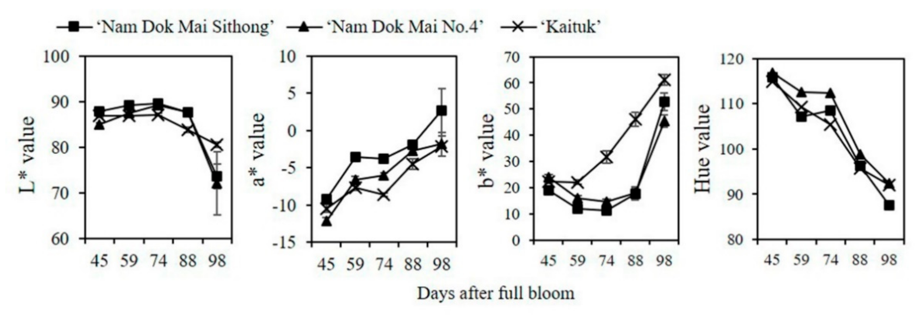
The differences in the peel and pulp color were determined by chroma and hue values, and color was expressed as the four values, including L* for the lightness (0–100), a* for green (−) to red (+), b* for blue (−) to yellow (+), and hue for yellow (90°) to green (180°). In mango peel, L* values continuously increased and then remained constant at the ripening stage from 74–98 days after full bloom in the three cultivars. The a* value continuously increased over fruit development and ripening in all cultivars. The b* values increased more rapidly in ‘Kaituk’ than other cultivars. In the meantime, the b* values continuously decreased and then slightly increased at the full ripening stage in ‘Nam Dok Mai Sithong’ and ‘Nam Dok Mai No.4′. The hue values notably decreased during fruit development and ripening until close to 90° at the full ripening stage in the three cultivars (Figure 3).
In mango pulp, the L* values were unchanged at the early fruit developmental stage before they dramatically dropped at the full ripening stage. The a* and b* values continuously increased over fruit development and ripening. The increase in b* value was more rapid in ‘Kaituk’ than the other two cultivars over fruit development and ripening. Contrastingly, the hue values greatly declined during fruit development until close to 90° at the full ripening stage in the three cultivars (Figure 4).

3.2. Carotenoid Content and Composition in Mango Peel during Fruit Development and Ripening

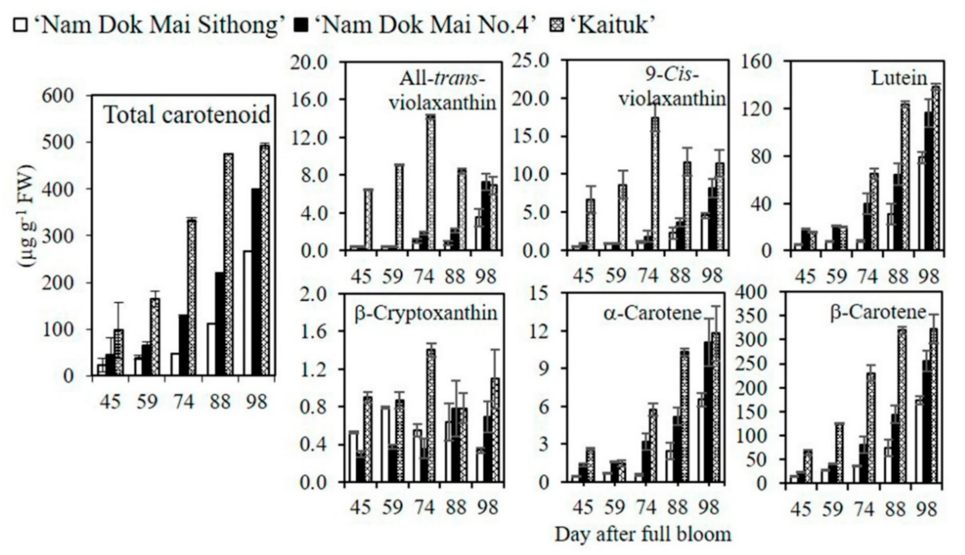
In this study, three cultivars of mango were collected at different fruit maturity stages, then the variations in carotenoid content and composition were investigated by HPLC. In mango peel, six carotenoids were detected. The major carotenoid presented in peel was β-carotene, which accounted for 70% of total carotenoids. The minor carotenoids were all-trans-violaxanthin, 9-cis-violaxanthin, lutein, β-cryptoxanthin, and α-carotene, which were presented at low levels. The total carotenoid content was progressively accumulated during fruit ripening in the three cultivars. The accumulation of total carotenoids was low at the early developmental stage. Then, it was continuously increased and reached the peak at the full ripening stage (98 days after full bloom). ‘Kaituk’ had the largest amount of total carotenoid content in the peel in all stages among three cultivars, followed by ‘Nam Dok Mai No.4′ and ‘Nam Dok Mai Sithong’. At the full ripening stage, the total carotenoid content in ‘Kaituk’ was 1.5 and 2 times higher than ‘Nam Dok Mai No.4′ and ‘Nam Dok Mai Sithong’, respectively (Figure 5).

3.3. Carotenoid Content and Composition in Mango Pulp during Fruit Development and Ripening

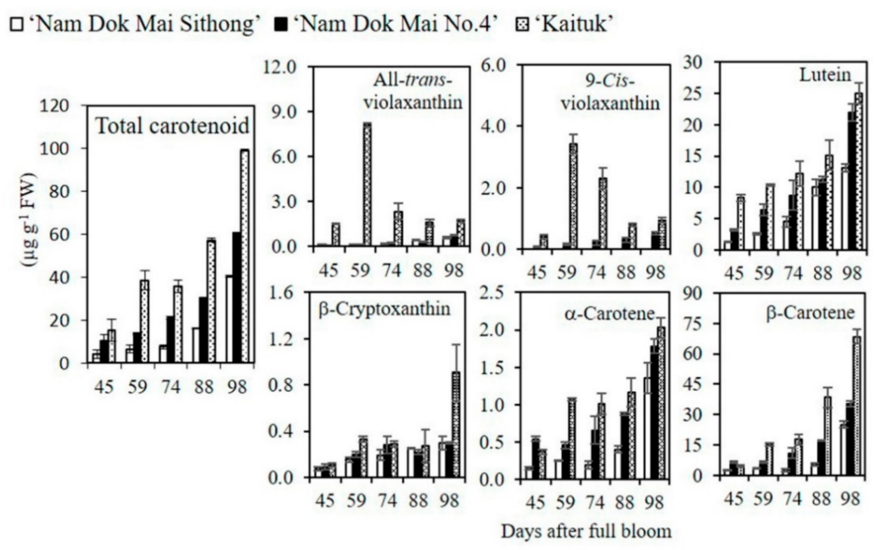
In mango pulp, six carotenoids were also detected. The major carotenoid accumulated in pulp was β-carotene, which accounted for 70% of total carotenoids. The minor carotenoids were all-trans-violaxanthin, 9-cis-violaxanthin, lutein, β-cryptoxanthin, and α-carotene, which accumulated at low levels during fruit development and ripening. The changing patterns of carotenoids accumulated in the pulp were similar to the peel, but a smaller amount of carotenoids was found in the pulp. The total carotenoid content was continuously increased toward fruit development and achieved the maximum at the full ripening stage (98 days after full bloom). Among three cultivars, ‘Kaituk’ had the highest total carotenoid content in the pulp in all stages. At the full ripening stage, the total carotenoid content in ‘Kaituk’ was 1.6 and 2.5 times higher than ‘Nam Dok Mai No.4′ and ‘Nam Dok Mai Sithong’, respectively (Figure 6).

3.4. The Expression of Genes Involved in Carotenoid Metabolism Pathway in Mango Peel during Fruit Development and Ripening

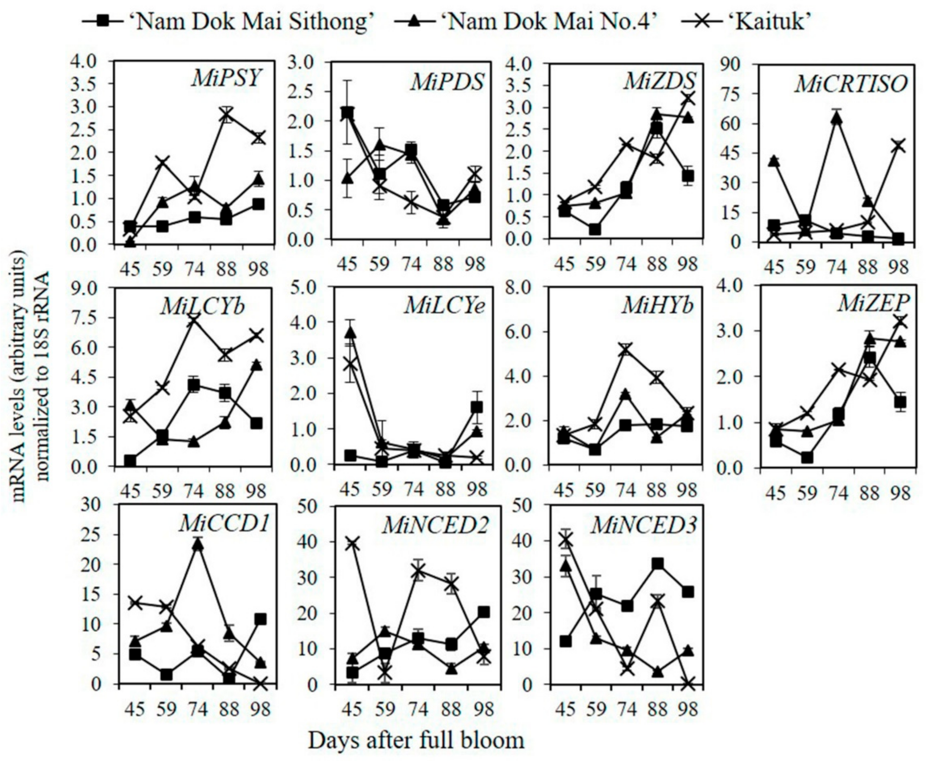
To gain an understanding of the regulatory mechanism of carotenoid metabolism in mango, the expression of eight carotenoid biosynthesis genes (MiPSY, MiPDS, MiZDS, MiCRTISO, MiLCYb, MiLCYe, MiHYb, and MiZEP) and three carotenoid catabolic genes (MiCCD1, MiNCED2, and MiNCED3) was analyzed in the peel of three mango cultivars during fruit development and ripening. For carotenoid biosynthetic genes, as shown in Figure 7, the expression of MiPSY, MiZDS, MiLCYb, and MiZEP progressively increased during fruit development in the three mango cultivars. ‘Kaituk’ exhibited a higher expression level of those genes during fruit development and ripening compared to that of two cultivars. Contrastingly, the changes in the expression of MiPDS, MiCRTISO, MiLCYe, and MiHYb fluctuated in the three cultivars (Figure 7).
For carotenoid catabolic genes, it was found that the expression of MiCCD1 continuously declined in ‘Kaituk’ over fruit development and ripening. In the meantime, the expression of MiCCD1 increased in the early development stage and then decreased in the ripening stage in ‘Nam Dok Mai No.4′. Contrastingly, the expression of MiCCD1 fluctuated and rapidly increased in ‘Nam Dok Mai Sithong’. The expression of MiNCED2 was unsteady before it dramatically dropped to a low level in ‘Kaituk’, while it gradually increased in ‘Nam Dok Mai No.4′ and ‘Nam Dok Mai Sithong’. The expression of MiNCED3 was unsteady before it markedly decreased in ‘Kaituk’ and continuously decreased over fruit development and ripening in ‘Nam Dok Mai No.4′. In contrast, the expression of MiNCED3 continuously increased during fruit development and ripening in ‘Nam Dok Mai Sithong’ (Figure 7).

3.5. The Expression of Genes Involved in Carotenoid Metabolism Pathway in Mango Pulp during Fruit Development and Ripening

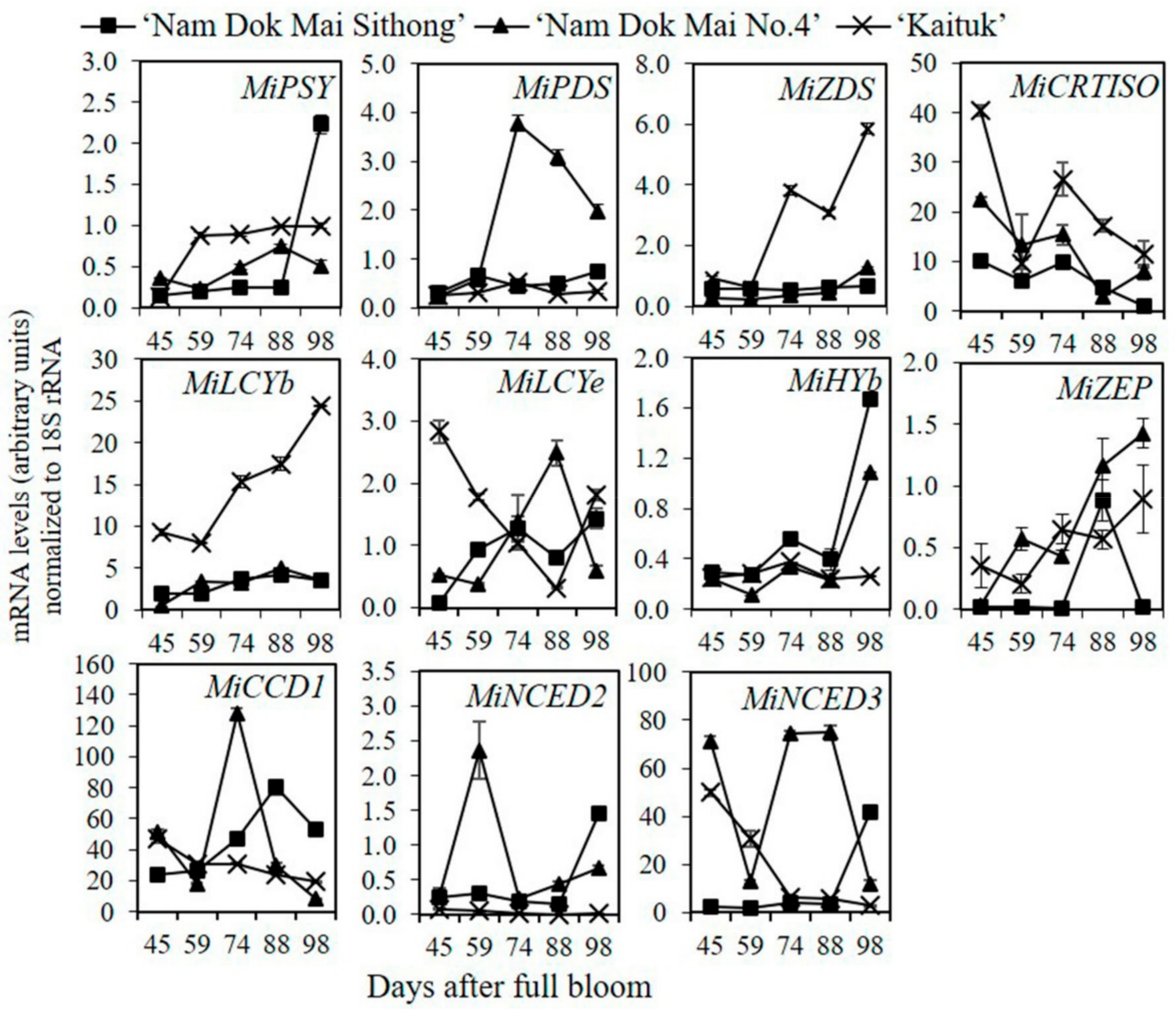
The expression of carotenoid metabolic genes was performed in the pulp of three mango cultivars. For carotenoid biosynthetic genes, as shown in Figure 8, the expression of MiZDS and MiLCYb considerably increased during fruit development and ripening. The expression of those two genes was higher in ‘Kaituk’ than in the other two cultivars in all developmental stages. Contrastingly, the expression of other carotenoid biosynthetic genes exhibited a fluctuating trend among different fruit developmental stages and mango cultivars (Figure 8).
For carotenoid catabolic genes, the expression of MiCCD1 progressively decreased in ‘Kaituk’ while it fluctuated in the other two cultivars toward fruit development and ripening. The expression of MiNCED2 remained constant at a very low level during fruit development and ripening in ‘Kaituk’, while it fluctuated and slightly increased at the full ripening stage in ‘Nam Dok Mai No.4′ and ‘Nam Dok Mai Sithong’. The expression of MiNCED3 dramatically fell to near zero in ‘Kaituk’, while it fluctuated in ‘Nam Dok Mai No.4′ and sharply increased in ‘Nam Dok Mai Sithong’ during fruit development (Figure 8).

4. Discussion

Carotenoid regulation in plants is a complex mechanism due to the high variation among the different developmental stages, species/cultivars, and plant tissues [17,18,19]. In this study, three commercial mango cultivars in Thailand with different colors were used as plant materials. To gain an understanding of the regulatory mechanism of carotenoid accumulation in mango, the carotenoid profiles, the expression of carotenoid metabolic genes, and relevant metabolic changes were investigated at different developmental stages in the peel and pulp of mango.
Various physiological and biochemical processes progressively changed during fruit ripening, particularly plant respiration rate, ethylene production, and fruit pigmentation as well [20,21,22,23]. In climacteric fruits, the early changes during the ripening process were the onset of ethylene production and the rise in cellular respiration [24,25]. In this study, the patterns of fruit respiration, ethylene production, and color development were examined at different stages of fruit development. It was found that mango substantially produced ethylene and increased respiration rate during fruit development and ripening. In the meantime, carotenoid content considerably increased, accompanied by a lower hue value and higher a* and b* values, which enhanced the degradation of green pigment and the presence of yellow pigment in mango fruits. It might be concluded that those changes were particular to the typical climacteric pattern of ripening.
The results in carotenoid identification and quantification illustrated that six carotenoids were identified, of which β-carotene was largely accumulated in all developmental stages and accounted for almost 70% of the total carotenoid content in both peel and pulp. It might be proposed that the increase in β-carotene played a very huge part in elevating the level of total carotenoid content in mango. These results corresponded with the previous studies in several fruits, which were found in yellow/orange colored fruits such as carrot [26], loquat [27], melon [28], and persimmon [29].
Carotenoid accumulation was described as a dynamic process during fruit development and ripening [30]. In these results, it was found that carotenoid accumulation in mango fruits was directly influenced by the stage of fruit development from immature to full ripening. In the early developmental stage, carotenoid accumulation was maintained at a low level. Then, it increasingly accumulated until it reached the maximum in the full ripening stage, where ‘Kaituk’ contained the highest carotenoid content in all developmental stages, followed by ‘Nam Dok Mai No.4′ and ‘Nam Dok Mai Sithong’. The present results were similar to previous studies in mango cv. ‘Tainong’, ‘Hongyu’, and ‘Jinhuang’. It was reported that the differences in carotenoid content and the development of yellow coloration were associated with the masking of carotenoids by chlorophyll and unmasking by chlorophyll degradation during fruit development and ripening [5,31,32,33]. It was also observed that the distinct level in carotenoid concentration influenced fruit color and color differences among cultivars and developmental stages. Fruits and vegetables triggered a deeper yellow/orange/red coloration by increasing carotenoid content [34,35,36]. This might suggest that the largest amount of carotenoids in ‘Kaituk’, particularly β-carotene, contributed to a more yellowish color in ‘Kaituk’ than in ‘Nam Dok Mai No.4′ and ‘Nam Dok Mai Sithong’ in all developmental stages, which was evidenced by a lower hue value and higher b* value evaluated in ‘Kaituk’ compared to those of the other two cultivars.
In addition to the differences in fruit development and cultivar, it is well documented that carotenoid accumulation varied significantly among plant tissues [37]. In this study, the differential accumulation of carotenoids in both tissues was detected, with a greater amount of carotenoids in the peel than in the pulp. These results corresponded with previous findings in various fruits. For example, in citrus, carotenoid accumulation in the peel was 5–10 times higher than in the pulp [38]. Similar results were also found in apple, where carotenoid accumulation in the peel was 3–5 times higher than in the pulp [14]. Apart from that, the carotenoid compositions could also vary between plant tissues [39]. However, the variation in carotenoid contents and compositions in plant tissues could not always be described by the transcriptional regulation. The total amount of carotenoids was considered to be associated with the sink capacity of plastids, where carotenoids were synthesized. In some plants, carotenoid accumulation could be influenced by the presence/absence of chloroplasts/chromoplasts [9]. In cauliflower orange (Or) mutant, enhancing the sink capacity by activating the differentiation of chloroplasts or other colorless plastids into chromoplasts enabled an increase in carotenoid accumulation in various tissues [40]. This might suggest that the regulatory mechanism of carotenoids was different in the peel and pulp. The accumulation of carotenoids and their regulatory mechanism should be separately investigated.
Due to the critical importance of carotenoids in plants and human health, the regulatory mechanism of carotenoids has been characterized in a variety of plant species at multiple molecular levels, including transcription, post-transcription, and post-translation [41]. However, the understanding of carotenoid regulation has not been achieved in mango fruits. To clarify the carotenoid metabolism pathway in mango, the expression of crucial regulatory genes in the carotenoid biosynthesis pathway (MiPSY, MiPDS, MiZDS, MiCRTISO, MiLCYb, MiLCYe, MiHYb, and MiZEP) and degradation pathway (MiCCD1, MiNCED2, and MiNCED3) was determined in the three mango cultivars during fruit development and ripening.
In this study, the relationship between the expression of carotenoid biosynthesis genes and the accumulation of carotenoids was observed. In mango peel, the expression of MiPSY, MiZDS, MiLCYb, and MiZEP rapidly increased during fruit development and ripening. Meanwhile, in mango pulp, the expression of MiZDS and MiLCYb continuously increased during fruit development and ripening. The highest transcript level of those genes was detected in ‘Kaituk’ during fruit development and ripening, followed by ‘Nam Dok Mai No.4′ and ‘Nam Dok Mai Sithong’ in both peel and pulp. These results indicated that the variations in carotenoid content in developmental stages and mango cultivars might be attributed to the expression of MiPSY, MiZDS, MiLCYb, and MiZEP in peel and MiZDS, and MiLCYb in pulp.
The expression of PSY has been described as a crucial step influencing carotenoid accumulation in higher plants [42]. To clarify the role of PSY in regulating carotenoid biosynthesis, tomato fruits were transformed with an antisense PSY1, which reduced carotenoid accumulation. It was found that carotenoid biosynthesis was inhibited and resulted in ripe fruit with white color. This indicated that PSY was a rate-limiting gene in regulating carotenoid biosynthesis in plants. The considerable increment of PSY expression during ripening was described in many fruits and vegetables, such as banana [43], citrus [4], and pepper [7]. It was described that the increment in the expression of the PSY gene and carotenoid content coincidentally occurred with the elevated expression of relevant genes in the ethylene biosynthesis pathway [44].
Different metabolic steps also presented as being related to carotenoid biosynthesis. The expression of MiZDS and MiLCYb genes corresponded with carotenoid accumulation in the peel and pulp of mango during fruit development and ripening. The expression levels of those two genes were higher in ‘Kaituk’ than the other two cultivars. It was previously found that ZDS and LCYb played a vital role in carotenoid biosynthesis in fruit mainly accumulating β-carotene. ZDS catalyzed the dehydrogenation of ζ-carotenoid to pro-lycopene, which was subsequently involved in β-carotene biosynthesis. In apricot and tomato, ZDS was considered to be one of the key determining genes for carotenoid regulation [45]. LCYb played a significant role in controlling the β,β-carotenoid synthesis. In loquat and kiwi, the expression of LCYb during the ripening process was closely related to the accumulation of carotenoids [46,47]. Those observations were similar to the present results in mango, which predominately accumulated β-carotene, accounting for almost 60% of total carotenoids in the fruits [48,49]. This suggested that the coordinated expression of MiZDS and MiLCYb in the carotenoid biosynthesis pathway might be a crucial step to control carotenoid accumulation in the three mango cultivars.
Beside carotenoid biosynthetic genes, carotenoid catabolic genes also played a vital role in regulating carotenoid accumulation during fruit maturation [50]. This has illustrated that the carotenoid pool was controlled by the rate of carotenoid catabolism. The accumulation of carotenoids in various plants has been shown to be inversely correlated with the expression of carotenoid catabolic genes, particularly in members of NCED, CCD1, and CCD4 subfamilies [51]. The high CCDs transcript level indicated that most of the carotenoids accumulated in plants were degraded. In the previous findings, the role of carotenoid catabolism in controlling carotenoid accumulation in several plants was clearly described. In chrysanthemum, the levels of carotenoid biosynthesis gene expression in white and yellow petals were similar. In the meantime, CCD4 was highly expressed in white petal to degrade carotenoids, but almost undetectable in the yellow petal [52]. In white and yellow fleshed peach, the differences in carotenoid biosynthetic genes were undetected, but the high transcript level of the CCD4 gene in white flesh peach was found, accounting for the higher carotenoid degradation [53]. In strawberry, which contained a small amount of carotenoids, the induction of CCD1 transcript level enhanced the degradation of carotenoids nearly 50% during fruit ripening. In mango, little information is available about the role of carotenoid catabolism in fruit pigmentation compared to carotenoid biosynthesis. In this study, the results were similar to the earlier study. It was found that the transcript level of MiCCD1, MiNCED2, and MiNCED3 was lower in ‘Kaituk’ than the other two mango cultivars in both tissues. This might have contributed to the low carotenoid degradation and led to the largest amount of carotenoids accumulated in ‘Kaituk’. A similar mechanism of carotenoid regulation was also found in mango cv. ‘Jinhuang’. The high expression of CCD was closely related to the degradation of carotenoids synthesized by ‘Jinhuang’ [5]. These results might conclude that the differential regulation of MiCCD1, MiNCED2, and MiNCED3 appeared to be a critical mechanism responsible for determining the carotenoid accumulation in different mango cultivars.

5. Conclusions

Carotenoid profiles, the transcriptional regulation of carotenogenic genes and some relevant changes associated with carotenoid regulation were investigated in peel and pulp during mango fruit development and ripening among three cultivars. The results showed that carotenoid accumulation in both tissues increased continuously over fruit development and ripening in the three cultivars. The highest carotenoid content was observed in ‘Kaituk’, followed by ‘Nam Dok Mai NO.4′ and ‘Nam Dok Mai Sithong’, in which β-carotene was a major carotenoid found in both tissues. The transcriptional abundance differed among fruit developmental stages and mango cultivars. The coordinated expression of MiPSY, MiZDS, MiLCYb, and MiZEP in peel and MiZDS and MiLCYb in pulp highly regulated carotenoid accumulation during fruit development and ripening. In addition, the transcriptional regulation of carotenoid catabolic genes was observed to be associated with carotenoid accumulation in mango. The differential expression of MiCCD1, MiNCED2, and MiNCED3 was described as a mechanism determining the diversity in carotenoid profiles among mango cultivars. The results contributed to the better understanding of the transcriptional regulation of carotenoids in mango, which will be beneficial for improving mango nutritional quality.

Author Contributions

Conceptualization, P.J.; Data curation, G.M. and L.Z.; Formal analysis, W.Y. and T.T.V.; Funding acquisition, P.J.; Investigation, T.T.V. and N.T.; Methodology, M.K.; Project administration, T.T.V.; Resources, A.U., P.J. and M.K.; Supervision, M.K.; Validation, A.U., G.M., L.Z., S.K., P.J. and M.K.; Visualization, A.U., G.M. and M.K.; Writing—original draft, W.Y.; Writing—review & editing, W.Y. All authors have read and agreed to the published version of the manuscript.

Funding

This research was partially funded by Petch Pra Jom Klao Ph.D.’s Degree Research Scholarship from King Mongkut’s University of Technology Thonburi.

Institutional Review Board Statement

Not applicable.

Informed Consent Statement

Not applicable.

Data Availability Statement

Not applicable.

Conflicts of Interest

The authors declare no conflict of interest.

References

  1. Ornelas-Paz, D.; Yahia, J.; Gardea, A. Changes in external and internal color during postharvest ripening of ‘Manila’and ‘Ataulfo’mango fruit and relationship with carotenoid content determined by liquid chromatography–APcI+-time-of-flight mass spectrometry. Postharvest Biol. Technol. 2008, 50, 145–152. [Google Scholar] [CrossRef]
  2. Cooperstone, J.L.; Schwartz, S.J. Recent Insights into Health Benefits of Carotenoids. In Handbook on Natural Pigments in Food and Beverages; Woodhead Publishing: Cambridge, UK, 2016; pp. 473–497. [Google Scholar]
  3. Tadmor, Y.; Burger, J.; Yaakov, I.; Feder, A.; Libhaber, S.E.; Portnoy, V.; Meir, A.; Tzuri, G.; Sa’ar, U.; Rogachev, I.; et al. Genetics of flavonoid, carotenoid, and chlorophyll pigments in melon fruit rinds. J. Agric. Food Chem. 2010, 58, 10722–10728. [Google Scholar] [CrossRef] [PubMed]
  4. Kato, M. Mechanism of carotenoid accumulation in citrus fruit. J. Jpn. Soc. Hortic. Sci. 2012, 81, 219–233. [Google Scholar] [CrossRef] [Green Version]
  5. Liang, M.; Su, X.; Yang, Z.; Deng, H.; Yang, Z.; Liang, R.; Huang, J. Carotenoid composition and expression of carotenogenic genes in the peel and pulp of commercial mango fruit cultivars. Sci. Hortic. 2020, 263, 109072. [Google Scholar] [CrossRef]
  6. Delgado-Pelayo, R.; Gallardo-Guerrero, L.; Hornero-Méndez, D. Chlorophyll and carotenoid pigments in the peel and flesh of commercial apple fruit varieties. Food Res. Int. 2014, 65, 272–281. [Google Scholar] [CrossRef] [Green Version]
  7. Rodriguez-Uribe, L.; Guzman, I.; Rajapakse, W.; Richins, R.D.; O’Connell, M.A. Carotenoid accumulation in orange-pigmented Capsicum annuum fruit, regulated at multiple levels. J. Exp. Bot. 2012, 63, 517–526. [Google Scholar] [CrossRef] [PubMed] [Green Version]
  8. Zhu, H.; Chen, M.; Wen, Q.; Li, Y. Isolation and characterization of the carotenoid biosynthetic genes LCYB, LCYE and CHXB from strawberry and their relation to carotenoid accumulation. Sci. Hortic. 2015, 182, 134–144. [Google Scholar] [CrossRef]
  9. Lado, J.; Zacarías, L.; Rodrigo, M.J. Regulation of Carotenoid Biosynthesis during Fruit Development. In Carotenoids in Nature; Springer: New York, NY, USA, 2016; pp. 161–198. [Google Scholar]
  10. Kato, M.; Ikoma, Y.; Matsumoto, H.; Sugiura, M.; Hyodo, H.; Yano, M. Accumulation of Carotenoids and Expression of Carotenoid Biosynthetic Genes during Maturation in Citrus. Plant Physiol. 2004, 134, 824–837. [Google Scholar] [CrossRef] [Green Version]
  11. Sun, L.; Yuan, B.; Zhang, M.; Wang, L.; Cui, M.; Wang, Q.; Leng, P. Fruit-specific RNAi-mediated suppression of SlNCED1 increases both lycopene and β-carotene contents in tomato fruit. J. Exp. Bot. 2012, 63, 3097–3108. [Google Scholar] [CrossRef]
  12. Beekwilder, J.; Van Der Meer, I.M.; Simic, A.; Uitdewilligen, J.; Van Arkel, J.; De Vos, R.C.; Jonker, H.; Verstappen, F.W.; Bouwmeester, H.J.; Sibbesen, O.; et al. Metabolism of carotenoids and apocarotenoids during ripening of raspberry fruit. Biofactors 2008, 34, 57–66. [Google Scholar]
  13. García-Limones, C.; Schnábele, K.; Blanco-Portales, R.; Luz Bellido, M.; Caballero, J.L.; Schwab, W.; Muñoz-Blanco, J. Functional characterization of FaCCD1: A carotenoid cleavage dioxygenase from strawberry involved in lutein degradation during fruit ripening. J. Agric. Food Chem. 2008, 56, 9277–9285. [Google Scholar] [CrossRef]
  14. Ampomah-Dwamena, C.; Dejnoprat, S.; Lewis, D.; Sutherland, P.; Volz, R.K.; Allan, A.C. Metabolic and gene expression analysis of apple (Malus × domestica) carotenogenesis. J. Exp. Bot. 2012, 63, 4497–4511. [Google Scholar] [CrossRef]
  15. Young, P.R.; Lashbrooke, J.G.; Alexandersson, E.; Jacobson, D.; Moser, C.; Velasco, R.; Vivier, M.A. The genes and enzymes of the carotenoid metabolic pathway in Vitis vinifera L. BMC Genom. 2012, 13, 243. [Google Scholar] [CrossRef] [Green Version]
  16. Ikoma, Y.; Yano, M.; Ogawa, K.; Yoshioka, T.; Xu, Z.C.; Hisada, S.; Omura, M.; Moriguchi, T. Isolation and Evaluation of RNA from Polysaccharide-rich Tissues in Fruit for Quality by cDNA Library Construction and RT-PCR. Jpn. Soc. Hortic. Sci. 1996, 64, 809–814. [Google Scholar] [CrossRef] [Green Version]
  17. Zhang, L.; Ma, G.; Kato, M.; Yamawaki, K.; Takagi, T.; Kiriiwa, Y.; Ikoma, Y.; Matsumoto, H.; Yoshioka, T.; Nesumi, H. Regulation of Carotenoid Accumulation and the Expression of Carotenoid Metabolic Genes in Citrus Juice Sacs in vitro. J. Exp. Bot. 2012, 63, 871–886. [Google Scholar] [CrossRef] [Green Version]
  18. Bramley, P.M. Carotenoid Biosynthesis and Chlorophyll Degradation. In The Molecular Biology and Biochemistry of Fruit Ripening; John Wiley & Sons: New York, NY, USA, 2013; pp. 75–116. [Google Scholar]
  19. Ohmiya, A.; Kato, M.; Shimada, T.; Nashima, K.; Kishimoto, S.; Nagata, M. Molecular basis of carotenoid accumulation in horticultural crops. Hortic. J. 2019, 88, 135–149. [Google Scholar] [CrossRef] [Green Version]
  20. Barry, C.S.; Giovannoni, J.J. Ethylene and fruit ripening. J. Plant Growth Regul. 2007, 26, 143–159. [Google Scholar] [CrossRef]
  21. Yin, X.R.; Chen, K.S.; Allan, A.C.; Wu, R.M.; Zhang, B.; Lallu, N.; Ferguson, I.B. Ethylene-induced modulation of genes associated with the ethylene signalling pathway in ripening kiwifruit. J. Exp. Bot. 2008, 59, 2097–2108. [Google Scholar] [CrossRef] [Green Version]
  22. Honda, C.; Bessho, H.; Murai, M.; Iwanami, H.; Moriya, S.; Abe, K.; Wada, M.; Moriya-Tanaka, Y.; Hayama, H.; Tatsuki, M. Effect of temperature on anthocyanin synthesis and ethylene production in the fruit of early-and medium-maturing apple cultivars during ripening stages. HortScience 2014, 49, 1510–1517. [Google Scholar] [CrossRef]
  23. Charoenchongsuk, N.; Matsumoto, D.; Itai, A.; Murayama, H. Ripening characteristics and pigment changes in russeted pear fruit in response to ethylene and 1-MCP. Horticulturae 2018, 4, 22. [Google Scholar] [CrossRef] [Green Version]
  24. Amorós, A.; Zapata, P.; Pretel, M.T.; Botella, M.A.; Serrano, M. Physico-chemical and physiological changes during fruit development and ripening of five loquat (Eriobotrya japonica Lindl.) cultivars. Food Sci. Technol. Int. 2003, 9, 43–51. [Google Scholar] [CrossRef]
  25. Abbas, M.E.F.; Fandi, B.S. Respiration rate, ethylene production and biochemical changes during fruit development and maturation of jujube (Ziziphus mauritiana Lamk). J. Sci. Food Agric. 2002, 82, 1472–1476. [Google Scholar] [CrossRef]
  26. Rodriguez-Concepcion, M.; Stange, C. Biosynthesis of carotenoids in carrot: An underground story comes to light. Arch. Biochem. Biophys. 2013, 539, 110–116. [Google Scholar] [CrossRef] [PubMed]
  27. Zhou, C.H.; Xu, C.J.; Sun, C.D.; Li, X.; Chen, K.S. Carotenoids in white-and red-fleshed loquat fruits. J. Agric. Food Chem. 2007, 55, 7822–7830. [Google Scholar] [CrossRef]
  28. Feder, A.; Chayut, N.; Gur, A.; Freiman, Z.; Tzuri, G.; Meir, A.; Gal-On, A.; Shnaider, Y.; Wolf, D.; Katzir, N.; et al. The role of the carotenogenic metabolic flux in carotenoid accumulation and chromoplast differentiation: Lessons from the melon fruit. Front. Plant Sci. 2019, 10, 1250. [Google Scholar] [CrossRef]
  29. Qi, Y.; Liu, X.; Zhang, Q.; Wu, H.; Yan, D.; Liu, Y.; Zhu, X.; Ren, X.; Yang, Y. Carotenoid accumulation and gene expression in fruit skins of three differently colored persimmon cultivars during fruit growth and ripening. Sci. Hortic. 2019, 248, 282–290. [Google Scholar] [CrossRef]
  30. Yuan, H.; Zhang, J.; Nageswaran, D.; Li, L. Carotenoid Metabolism and Regulation in Horticultural Crops. Hortic. Res. 2015, 2, 15036. [Google Scholar] [CrossRef] [Green Version]
  31. Ma, X.; Zheng, B.; Ma, Y.; Xu, W.; Wu, H.; Wang, S. Carotenoid accumulation and expression of carotenoid biosynthesis genes in mango flesh during fruit development and ripening. Sci. Hortic. 2018, 237, 201–206. [Google Scholar] [CrossRef]
  32. Matile, P.; Hortensteiner, S.; Thomas, H. Chlorophyll degradation. Annu. Rev. Plant Physiol. Plant Mol. Biol. 1999, 50, 67–95. [Google Scholar] [CrossRef] [Green Version]
  33. Zhang, J.Y.; Pan, D.L.; Jia, Z.H.; Wang, T.; Wang, G.; Guo, Z.R. Chlorophyll, carotenoid and vitamin C metabolism regulation in Actinidia chinensis’ Hongyang’outer pericarp during fruit development. PLoS ONE 2018, 13, e0194835. [Google Scholar]
  34. Bramley, P.M. Regulation of carotenoid formation during tomato fruit ripening and development. J. Exp. Bot. 2002, 53, 2107–2113. [Google Scholar] [CrossRef] [Green Version]
  35. Khoo, H.E.; Ismail, A.; Mohd-Esa, N.; Idris, S. Carotenoid content of underutilized tropical fruits. Plant Food Hum. Nutr. 2008, 63, 170–175. [Google Scholar] [CrossRef]
  36. Sagawa, J.M.; Stanley, L.E.; LaFountain, A.M.; Frank, H.A.; Liu, C.; Yuan, Y.W. An R2R3MYB transcription factor regulates carotenoid pigmentation in Mimulus lewisii flowers. New Phytol. 2016, 209, 1049–1057. [Google Scholar] [CrossRef]
  37. Cazzonelli, C.I.; Pogson, B.J. Source to Sink: Regulation of Carotenoid Biosynthesis in Plants. Trends Plant Sci. 2010, 15, 266–274. [Google Scholar] [CrossRef]
  38. Alquezar, B.; Rodrigo, M.J.; Zacarías, L. Carotenoid biosynthesis and their regulation in citrus fruits. Tree For. Sci. Biotech. 2008, 2, 23–37. [Google Scholar]
  39. Shen, Y.H.; Yang, F.Y.; Lu, B.G.; Zhao, W.W.; Jiang, T.; Feng, L.; Ming, R. Exploring the differential mechanisms of carotenoid biosynthesis in the yellow peel and red flesh of papaya. BMC Genom. 2019, 20, 49. [Google Scholar] [CrossRef] [PubMed] [Green Version]
  40. Lu, S.; Van Eck, J.; Zhou, X. The cauliflower Or gene encodes a DnaJ cysteine-rich domain-containing protein that mediates high levels of beta-carotene accumulation. Plant Cell. 2006, 18, 3594–3605. [Google Scholar] [CrossRef] [Green Version]
  41. Stanley, L.; Yuan, Y.W. Transcriptional regulation of carotenoid biosynthesis in plants: So many regulators, so little consensus. Front. Plant Sci. 2019, 10, 1017. [Google Scholar] [CrossRef] [Green Version]
  42. Liu, L.; Shao, Z.; Zhang, M.; Wang, Q. Regulation of carotenoid metabolism in tomato. Mol. Plant. 2015, 8, 28–39. [Google Scholar] [CrossRef] [Green Version]
  43. Mlalazi, B.; Welsch, R.; Namanya, P.; Khanna, H.; Geijskes, R.J.; Harrison, M.D.; Harding, R.; Dale, J.L.; Bateson, M. Isolation and functional characterisation of banana phytoenesynthase genes as potential cis-genes. Planta 2012, 236, 1585–1598. [Google Scholar] [CrossRef] [Green Version]
  44. Dong, T.; Hu, Z.; Deng, L.; Wang, Y.; Zhu, M.; Zhang, J.; Chen, G. A tomato MADS-box transcription factor, SlMADS1, acts as a negative regulator of fruit ripening. Plant Physiol. 2013, 163, 1026–1036. [Google Scholar] [CrossRef] [PubMed] [Green Version]
  45. Marty, I.; Bureau, S.; Sarkissian, G.; Gouble, B.; Audergon, J.M.; Albagnac, G. Ethylene regulation of carotenoid accumulation and carotenogenic gene expression in colour-contrasted apricot varieties (Prunus armeniaca). J. Exp. Bot. 2005, 56, 1877–1886. [Google Scholar] [CrossRef] [PubMed] [Green Version]
  46. Zhang, L.; Ma, G.; Shirai, Y.; Kato, M.; Yamawaki, K.; Ikoma, Y.; Matsumoto, H. Expression and functional analysis of two lycopene β-cyclases from citrus fruits. Planta 2012, 236, 1315–1325. [Google Scholar] [CrossRef] [PubMed] [Green Version]
  47. Ma, G.; Zhang, L.; Yungyuen, W.; Tsukamoto, I.; Iijima, N.; Oikawa, M.; Kato, M. Expression and functional analysis of citrus carotene hydroxylases: Unravelling the xanthophyll biosynthesis in citrus fruits. BMC Plant Biol. 2016, 16, 1–12. [Google Scholar] [CrossRef] [Green Version]
  48. Fu, X.; Kong, W.; Peng, G.; Zhou, J.; Azam, M.; Xu, C.; Grierson, D.; Chen, K. Plastid structure and carotenogenic gene expression in red-and white-fleshed loquat (Eriobotrya japonica) fruits. J. Exp. Bot. 2012, 63, 341–354. [Google Scholar] [CrossRef] [Green Version]
  49. Ampomah-Dwamena, C.; McGhie, T.; Wibisono, R.; Montefiori, M.; Hellens, R.P.; Allan, A.C. The kiwifruit lycopene beta-cyclase plays a significant role in carotenoid accumulation in fruit. J. Exp. Bot. 2009, 60, 3765–3779. [Google Scholar] [CrossRef] [Green Version]
  50. Kato, M.; Matsumoto, H.; Ikoma, Y.; Okuda, H.; Yano, M. The role of carotenoid cleavage dioxygenases in the regulation of carotenoid profiles during maturation in citrus fruit. J. Exp. Bot. 2006, 57, 2153–2164. [Google Scholar] [CrossRef] [Green Version]
  51. Gayen, D.; Ali, N.; Sarkar, S.N.; Datta, S.K.; Datta, K. Down-regulation of lipoxygenase gene reduces degradation of carotenoids of golden rice during storage. Planta 2015, 242, 353–363. [Google Scholar] [CrossRef]
  52. Ohmiya, A.; Kishimoto, S.; Aida, R.; Yoshioka, S.; Sumitomo, K. Carotenoid cleavage dioxygenase (CmCCD4a) contributes to white color formation in chrysanthemum petals. Plant Physiol. 2006, 142, 1193–1201. [Google Scholar] [CrossRef] [Green Version]
  53. Brandi, F.; Bar, E.; Mourgues, F.; Horváth, G.; Turcsi, E.; Giuliano, G.; Liverani, A.; Tartarini, S.; Lewinsohn, E.; Rosati, C. Study of ‘Red haven’ peach and its white-fleshed mutant suggests a key role of CCD4 carotenoid dioxygenase in carotenoid and nor isoprenoid volatile metabolism. BMC Plant Biol. 2011, 11, 24. [Google Scholar] [CrossRef] [Green Version]
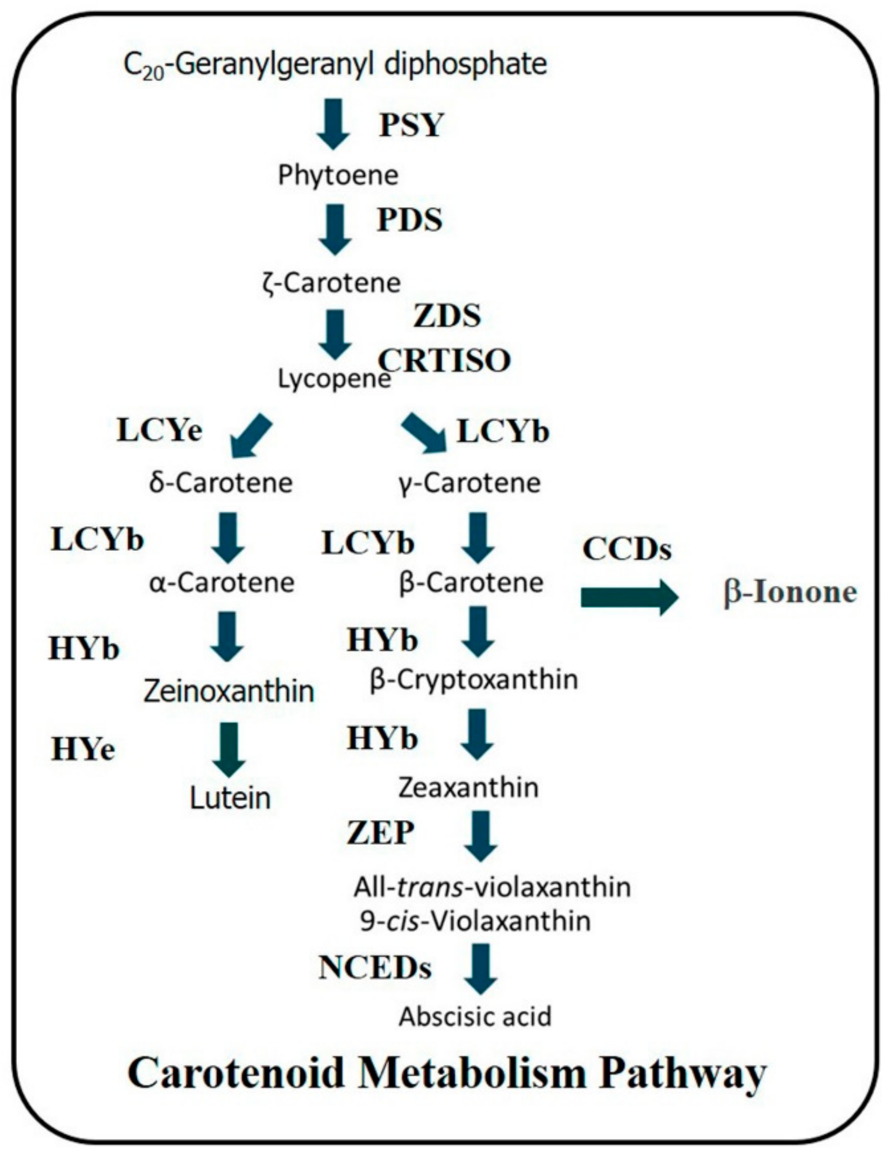
Figure 1. The principal pathway of carotenoid metabolism in higher plants. Abbreviations of carotenoid metabolic enzymes: PSY, phytoene synthase; PDS, phytoene desaturase; ZDS, ζ-carotene desaturase; CRISO, carotenoid isomerase; LCYe, lycopene ε-cyclase; LCYb, lycopene β-cyclase; HYe, ε-ring hydroxylase; HYb, β-ring hydroxylase; ZEP, zeaxanthin epoxidase; CCDs, carotenoid cleavage dioxygenases; NCEDs, 9-cis-epoxycarotenoid dioxygenases.
Applsci 11 04249 g001
Figure 2. Changes in respiration rate and ethylene production of mango fruit cv. ‘Nam Dok Mai Sithong’, ‘Nam Dok Mai No. 4′ and ‘Kaituk’ during fruit development (45–88 days) and full ripening stage (98 days). The vertical bars represent the means with SE for three replications.
Applsci 11 04249 g002
Figure 3. L*, a*, b*, and hue values in peel of mango fruit cv. ‘Nam Dok Mai Sithong’, ‘Nam Dok Mai No. 4′ and ‘Kaituk’ during fruit development (45–88 days) and full ripening stage (98 days). The vertical bars represent the means with SE for three replications.
Applsci 11 04249 g003
Figure 4. L*, a*, b*, and hue values in pulp of mango fruit cv. ‘Nam Dok Mai Sithong’, ‘Nam Dok Mai No. 4′ and ‘Kaituk’ during fruit development (45–88 days) and full ripening stage (98 days). The vertical bars represent the means with SE for three replications.
Applsci 11 04249 g004
Figure 5. Changes in carotenoid content and compositions in peel of mango fruit cv. ‘Nam Dok Mai Sithong’, ‘Nam Dok Mai No. 4′ and ‘Kaituk’ during fruit development (45-88 days) and full ripening stage (98 days). The vertical bars represent the means with SE for three replications.
Applsci 11 04249 g005
Figure 6. Changes in carotenoid content and compositions in pulp of mango fruit cv. ‘Nam Dok Mai Sithong’, ‘Nam Dok Mai No. 4′ and ‘Kaituk’ during fruit development (45-88 days) and full ripening stage (98 days). The vertical bars represent the means with SE for three replications.
Applsci 11 04249 g006
Figure 7. The expression of genes involved in carotenoid metabolism in peel of mango fruit cv. ‘Nam Dok Mai Sithong’, ‘Nam Dok Mai No. 4′ and ‘Kaituk’ during fruit development (45-88 days) and full ripening stage (98 days). The vertical bars represent the means with SE for three replications.
Applsci 11 04249 g007
Figure 8. The expression of genes involved in carotenoid metabolism in pulp of mango fruit cv. ‘Nam Dok Mai Sithong’, ‘Nam Dok Mai No. 4′ and ‘Kaituk’ during fruit development (45–88 days) and full ripening stage (98 days). The vertical bars represent the means with SE for three replications.
Applsci 11 04249 g008
Table 1. Primers used for RT-PCR and cDNA lengths of genes involved in carotenoid metabolism pathway.
cDNAForward Primer (F) and Reverse Primer (R) 5’- 3’Length (bp)
MiPSYF: GTGGTATTGAAGCAGGCAGCCTTGGTTA
R: GCATAGGAGACTGGCAATGCAGCTATCT
943
MiPDSF: TCTGGCAGATGCAGGCCACAAACCT
R: CTGTTGTCACACGCTCAGGTACTCC
448
MiZDSF: AGAAACGCTCTGGCTCTTGCTCTAAGTC
R: CACTGTGACAACAGGCACGCCAACTAAT
550
MiCRTISOF: TGTTAAAGCTGAGGTTCTGCCACCCGAT
R: ACAGCTATAACGCCTTGTCCTGGAAACG
449
MiLCYbF: CTGGTCTGGCGCAGTTGTTTACATTGAC
R: CCGCGCTACAAGTGAAGTTTCCTCAAGA
483
MiLCYeF: AGTTGTGAGATCACTGTCAGAGGCTCCA
R: CTCCTTCCAGCAATCCGATATAGAACCG
477
MiHYbF: GCGTGGCTGAGAAGTTAGCGAGGAA
R: CTTCTCTGGGTCTGTGGTGAGACTC
284
MiZEPF: CAAGCAAAAGCTCTAGTGAGGAACTGGC
R: GCAGAAGTGATCCGACAAGATTTGCACG
360
MiCCD1F: CTGGAGAATCCAGATCTGGACATGGTCA
R: GTAATTCCACACTGCAACAGGATCTGGC
491
MiNCED2F: CGAACGTTCCATGAAAACCGTACGGTAC
R: CACTCTCCTTCAGTCCTTCATTTCCCGA
1578
MiNCED3F: GCATGGCCAGGTCTTAAACCCAGTTCTA
R: CAAGGCAGCCTAAGGATAGCTTTACCTC
1732
Table 2. TaqMan MGB probes and primers used for qRT-PCR of genes involved in carotenoid metabolism pathway.
cDNATaqMan MGB ProbesPrimer Sequences
MiPSYACAGGCAACGACAGAGAF: CATGGGAATTGCACCTGGAT
R: GCCAAGGCAGCATTGTAGACA
MiPDSTGCTATTGGACTTCTTCF: TTGGCCCGAGAAAGTCAAGT
R: GATTGTCCACCAAGCATTGCT
MiZDSTCCCTGGAATTAAAAGATF: TGCATATGTTGCTGCATGTGAT
R: CCCTCCACGACAATGGAAGT
MiCRTISOCTATGATGCAAAGAAGGF: GGCAGGGAATGTCTCAAAAGG
R: CCAATTATTTCATCTGCCACAAGT
MiLCYbTGATGCAACTGGGTTTTF: ACAATTCAGGCTGCTGTGGTT
R: GCTTATCATACTGAACAAGGCACCTA
MiLCYeTTTCTTCCGGTTACCCAAAF: GGCATCAGGACATTTTTCCATT
R: CGAGAAATCCCTGCCACATC
MiHYbCTCTCTCTGAAATGTTCGF: CAAATGGAGGGTGGTGAGGTT
R: CAGCACCAACAGAGAGTGCAA
MiZEPATGCCTGGCACCCAAF: GAGGAAGAGTTGTGCTGGTAGGA
R: CCTTGCCCCAGATTTGGA
MiCCD1TTGGAGGAAATGTTAAAGGF: GGCTGGAAAGACAAAGCTTGA
R: TGGGCCCAGGTCGAAGAT
MiNCED2ATGAAGTTGTTGTTATAGGATCF: GGGAGGAGCCGGAATCC
R: GTCAGCCGGTGTCATGCA
MiNCED3TGATAGCTCACCCAAAAGF: CGACGGTCAGCTTAATTCAACA
R: TCGCCTGAAACGGGATCA
18sProprietary (Applied Biosystems, Foster City, CA, USA)Proprietary (Applied Biosystems)
Publisher’s Note: MDPI stays neutral with regard to jurisdictional claims in published maps and institutional affiliations.