Article

Electric Stimulation of Astaxanthin Biosynthesis in Haematococcus pluvialis

1
Gwangju Bio/Energy R&D Center, Korea Institute of Energy Research, Gwangju 61003, Korea
2
Climate Change Research Division, Korea Institute of Energy Research, Daejeon 34129, Korea
3
School of Chemical Engineering, and Institute for Environment & Energy, Pusan National University, Busan 46241, Korea
*
Author to whom correspondence should be addressed.
These authors contributed equally to this work.
Appl. Sci. 2021, 11(8), 3348; https://doi.org/10.3390/app11083348
Submission received: 17 March 2021 / Revised: 5 April 2021 / Accepted: 7 April 2021 / Published: 8 April 2021
(This article belongs to the Special Issue Algal Biorefinery and Microbial Fuel Cells 2021)

Abstract

:
The green microalga Haematococcus pluvialis accumulates astaxanthin, a potent antioxidant pigment, as a defense mechanism against environmental stresses. In this study, we investigated the technical feasibility of a stress-based method for inducing astaxanthin biosynthesis in H. pluvialis using electric stimulation in a two-chamber bioelectrochemical system. When a cathodic (reduction) current of 3 mA (voltage: 2 V) was applied to H. pluvialis cells for two days, considerable lysis and breakage of algal cells were observed, possibly owing to the formation of excess reactive oxygen species at the cathode. Conversely, in the absence of cell breakage, the application of anodic (oxidation) current effectively stimulated astaxanthin biosynthesis at a voltage range of 2–6 V, whereas the same could not be induced in the untreated control. At an optimal voltage of 4 V (anodic current: 30 mA), the astaxanthin content in the cells electro-treated for 2 h was 36.9% higher than that in untreated cells. Our findings suggest that electric treatment can be used to improve astaxanthin production in H. pluvialis culture if bioelectrochemical parameters, such as electric strength and duration, are regulated properly.

1. Introduction

Astaxanthin (3,3’-dihydroxy-β-carotene-4,4’-dione) is a common high-value ketocarotenoid pigment (~US $7150 per kg) involved in various metabolic functions, including anti-oxidative, anti-inflammatory, and immune-stimulatory functions [1,2]. The green microalga Haematococcus pluvialis produces astaxanthin in large quantities (~4% of dry cell weight) at the dormant aplanospore (cyst) stage and has been studied extensively as a model strain in research settings as well as in industrial applications [3,4].
Algal astaxanthin synthesis is usually induced as a defense mechanism against environmental oxidative stress [2,5,6]. Various stress-inducing conditions, such as nutrient starvation [7,8], exposure to intense light [9,10], high temperatures [11,12], high salinity [13,14], and nanomaterial addition to culture [15], have been applied to increase the content and productivity of algal astaxanthin. Among them, nitrogen-nutrient depletion has been employed extensively for effective astaxanthin induction in H. pluvialis cultures [2,16]. However, this approach is time-consuming and associated with a risk of biocontamination from external sources; moreover, it significantly reduces the biomass productivity of H. pluvialis [17]. Therefore, the development of an effective astaxanthin induction strategy remains a major challenge in H. pluvialis biorefinery approaches.
Electric stimulation or induction, a relatively new technique in algal biorefinery approaches, involves the application of an electric current to live algal cells through the anode and cathode of a bioelectrochemical system. Recently, Kim et al. [18] treated H. pluvialis seed cells at 100 mA (voltage: 25 V) for a short duration (1 min) in a single-chamber electrochemical system. After cultivation for seven days, the cell number density of the electro-treated cells increased by 20%, a considerable increase compared to the cell number density of untreated cells, owing to the enhancement of nitrogen consumption and chlorophyll biosynthesis in H. pluvialis cells. However, the intracellular astaxanthin content remained almost constant regardless of the treatment. Recently, we reported the effective induction of triacylglycerols (TAGs) and total fatty acids in Chlorella species by the application of a mild cathodic current of 31 mA for 4 h, which is a relatively long duration, in a two-chamber bioelectrochemical system [19]. Compared to that in the untreated control, the TAG and total fatty acid contents in electro-treated Chlorella cells increased by 210% and 24%, respectively. Of note, the astaxanthin and fatty acid biosynthesis pathways are closely related in H. pluvialis, and astaxanthin is present in TAG bodies mostly in mono- and di-ester forms with fatty acids [2,20]. In connection with the previous study on algal lipids [19], in this study, we aimed to investigate the technical feasibility of using electric stimulation to improve astaxanthin accumulation in H. pluvialis using a two-chamber bioelectrochemical system.

2. Materials and Methods

2.1. Microalga and Photosynthetic Cultivation

H. pluvialis NIES-144, provided by the National Institute of Environmental Studies (NIES, University of Tokyo, Tokyo, Japan), was used in this study. The NIES-C medium with the following composition (per liter) was used: 0.15 g Ca(NO3)2, 0.10 g KNO3, 0.05 g β-glycerophosphoric acid disodium salt pentahydrate, 0.04 g MgSO4·7H2O, 0.50 g tris-aminomethane, 0.01 mg thiamine, 0.10 μg biotin, 0.10 μg vitamin B12, and 3.0 mL PIV metal solution [21]. One liter of the PIV metal solution contains 1.0 g Na2EDTA, 0.196 g FeCl3·6H2O, 36.0 mg MnCl2·4H2O, 22.0 mg ZnSO4·7H2O, 4.0 mg CoCl2·6H2O, and 2.5 mg Na2MoO4·2H2O. After adjusting the pH to 7.5, the medium was filter-sterilized using a 0.2 μm membrane (Satorius Stedium Biotech., Göttingen, Germany). For the seed culture, one colony was collected from an agar plate culture of H. pluvialis and inoculated in the medium in a 250 mL-scale Erlenmeyer flask with a porous silicon stopper (working volume, 100 mL) in a shaking incubator (IS-971RF, Lab Companion, Daejeon, Korea; 25 °C and 150 rpm) under continuous illumination (light intensity, 40 μmol photons m−2 s−1) [15].
A primary photoautotrophic culture of H. pluvialis was established using a 250 mL-scale funnel-type photobioreactor (f-PBR; working vol., 150 mL) using a method optimized previously [22]. H. pluvialis cyst cells collected from the seed flask culture were inoculated in the medium contained in the f-PBR till an optical density (OD) of 0.05 was achieved at 660 nm, as measured using a UV-VIS spectrophotometer (Optizen 2120UV, Mecasys Co., Daejeon, Korea). The f-PBR was continuously supplied with 5% (v/v) CO2 in air at a rate of 60 mL min−1 (0.4 vvm) from the bottom of the reactor. The f-PBR was incubated for 25 days in a plant growth chamber (GC-300, JEIO TECH, Korea; light intensity, 50 μmol photons m−2 s−1; 12 h light/dark cycle; 25 °C). The details of the culture conditions employed have been reported in a previous study [22].

2.2. Electric Stimulation

A two-chambered electrochemical reactor (constructed using Lexan polycarbonate materials, each of 110 mm × 110 mm × 25 mm; working volume, 120 mL) was used in this study (Figure 1). Carbon cloth electrodes (45 mm × 45 mm; FuelCell Co., Brazos County, TX, USA) were used as both the anode and cathode and were connected by a titanium wire. A proton-exchange membrane (Nafion 117, DuPont, Newark, DE, USA) was used to separate the anodic and cathodic chambers. For the electric stimulation experiment, green vegetative H. pluvialis cells cultured for 5 days in the f-PBR (cell density, ca. 15 × 104 mL−1) were harvested by centrifugation (4000 rpm, 4 °C, 5 min; MICRO 17TR, Hanil Science Inc., Daejeon, Korea), washed in 0.1 M sodium phosphate buffer (pH 7.0), resuspended in the same buffer, and then transferred to each compartment of the electrochemical reactor using a sterile disposable hypodermic syringe. The voltage between the anode and cathode was controlled using a DC power supply (GPDX303S, Good Will Instrument Co., New Taipei City, Taiwan). In the cathodic current experiment, the voltage was fixed at 2 V for 2 days, whereas in the anodic current experiment three different 2, 4, and 6 V were examined for 1 and 2 days. The cathodic and anodic chambers were continuously aerated at a rate of 48 mL min−1 (0.4 vvm) using a sterile syringe filter of 0.2 μm (Satorius Stedium Biotech., Göttingen, Germany). All bioelectrochemical experiments were conducted in a temperature-controlled room (ca. 25 °C). It was speculated that photosynthetic growth (or cell division) of H. pluvialis could not be induced owing to the use of the phosphate buffer during electric stimulation. Details of the bioelectrochemical conditions have been published earlier [19].

2.3. Cyclic Voltammetry (CV) Analysis

Bioelectrochemical reactions between the carbon electrode and H. pluvialis cells were characterized using CV analysis at a constant temperature (ca. 25 °C). The CV measurements were performed in a 20 mL single-chamber electrochemical reactor (BASi® Analytical Instruments, West Lafayette, IN, USA) with three electrodes using a potentiostat (CHI 660E, CH Instruments Inc., Austin, TX, USA). Glassy carbon (3 mm diameter; CHI 104, CH Instruments Inc.), Ag/AgCl (3 M NaCl, sat.; Re-6, BASi Analytical Instruments), and a platinum wire (ALS Co., Tokyo, Japan) were used as the working, reference, and counter electrodes, respectively. The potential sweep range and scan rate were −0.5 V to +0.5 V and 10 mV s−1, respectively. Similar to that in the electric simulation experiment, green vegetative H. pluvialis cells cultured for 5 days in the f-PBR were used for CV measurements. The PBR algal cells were centrifuged at 4000 rpm (4 °C for 5 min; MICRO 17TR, Hanil Science Inc., Daejeon, Korea), washed in fresh NIES-C medium, resuspended in the same medium, and transferred to the electrochemical reactor using a sterile disposable hypodermic syringe. Before the experiment, oxygen dissolved in the medium was removed carefully by supplying the reactor with pure N2 gas (filtered through a 0.2 μm membrane) at a flow rate of 2.5 mL min−1 for 30 min.

2.4. Other Analytical Methods

H. pluvialis cell growth was monitored by measuring the OD and cell number using a UV-VIS spectrophotometer (660 nm; Optizen 2120UV, Mecasys Co., Daejeon, Korea) and a Coulter counter (MultisizerTM 4, Beckman Coulter, Indiana Polis, CA, USA), respectively. The final dry cell weight of the algal cells after cultivation for 25 days was determined by filtering 5 mL of the cell aliquots using pre-weighed GF/C glass microfiber filters (Whatman, UK) and drying at 60 °C overnight [15]. The morphological features of H. pluvialis cells were observed using bright-field microscopy (Axio Imager.A2, Carl Zeiss, Germany). The pH value was measured using a pH meter (DKK-TOA Co., Tokyo, Japan). The light intensity was measured using a quantum meter (Li-250A, Li-COR Inc., Lincoln, NE, USA).
For analyzing astaxanthin production, the H. pluvialis cells were harvested by centrifugation (4000 rpm for 10 min; Combi R515, Hanil Science Inc., Daejeon, Korea), double-washed with purified water, and freeze-dried for 3 days in a freeze-dryer lyophilizer (FD8508, IlShin BioBase Co., Yangju-si, Korea). The freeze-dried cells were stored in a vacuum bag at −20 °C until further use. The astaxanthin content was measured using a high-performance liquid chromatography (HPLC) instrument (Agilent 1260 Infinity, Hewlett-Packard, Santa Clara, CA, USA) equipped with a diode-array detector and a YMC Carotenoid column (250 mm × 4.6 mm, 5 μm, YMC Inc., Kyoto, Japan), as previously described [15,23]. Briefly, ca. 16 mg of freeze-dried H. pluvialis cells and 1.0 g of glass microbeads were mixed vigorously with 1 mL acetone for 10 min at room temperature (ca. 27 °C). The mixture was then separated by centrifugation at 8000 rpm for 5 min (MICRO 17TR, Hanil Science Ltd., Daejeon, Korea). The astaxanthin-containing acetone solution was collected and evaporated using a rotary vacuum evaporator (EZ2 PLUS, Genevac Ltd., Ipswich, UK) at 40 °C for 30 min. The astaxanthin extract was mixed with 0.2 mL dichloromethane, 0.8 mL methanol, and 1 mL 0.05 N NaOH solution (in freshly prepared methanol). For saponification, the sample was incubated overnight in dark at 4 °C and then analyzed using HPLC.

3. Results and Discussion

3.1. H. pluvialis Life Cycle and Astaxanthin Induction

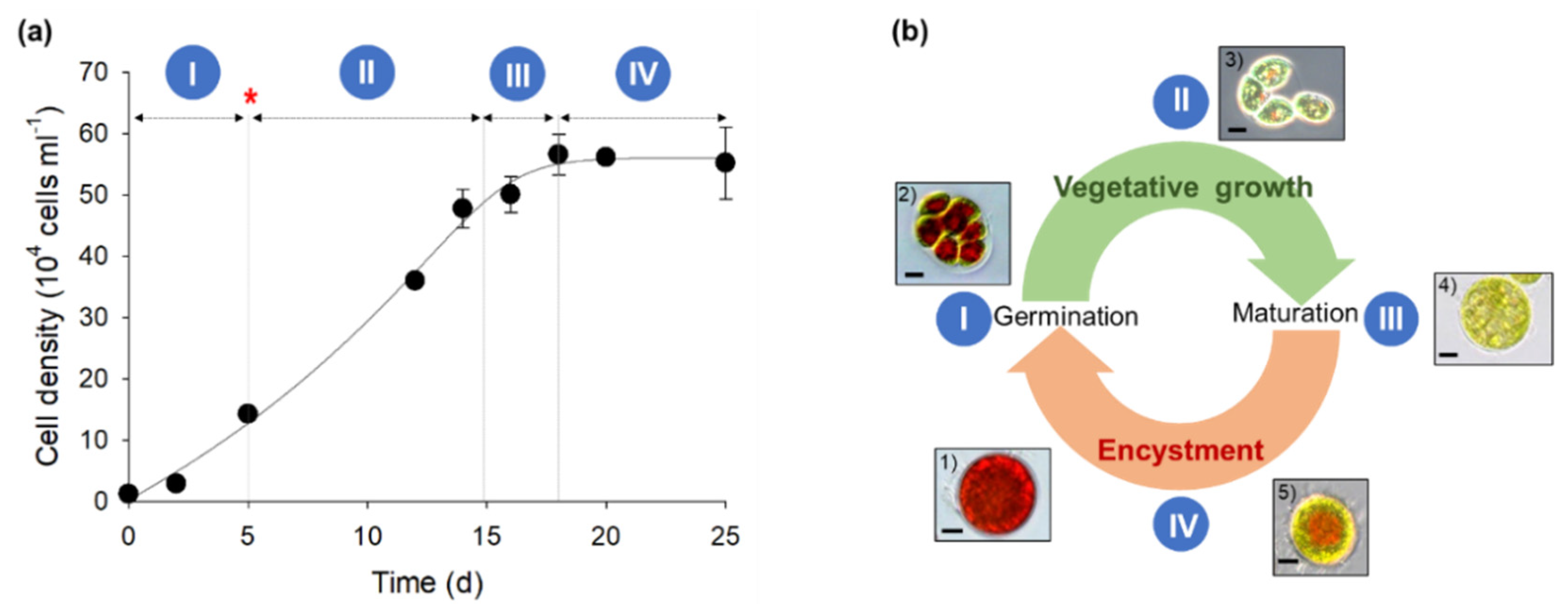
Figure 2 shows the changes in the cell number density and shape of H. pluvialis upon culturing for 25 days in the photosynthetic photobioreactor. To enhance astaxanthin production in H. pluvialis cells, 1 mM nitrate was used as the nitrogen source. The cell number density of H. pluvialis increased almost linearly with time after a short delay period of 2 days (Figure 2a). However, after 15 days of culture, as the nitrate content was depleted, the growth rate of H. pluvialis decreased considerably and the culture soon entered a stationary growth phase.
The growth pattern of H. pluvialis cells during the 25 day cultivation period could be largely divided into two different stages: vegetative growth (cell division and enlargement), and encystment with astaxanthin induction. More specifically, based on the morphological characteristics observed using optical microscopy, the life cycle of the microorganism was characterized by five distinct cell types (Figure 2b). When the red cysts (cell-type #1) harvested from the seed flask culture were inoculated in fresh medium contained in the photobioreactor, the cells began to germinate within 6 h. A dividing cyst (cell-type #2) with the internally differentiated zooids was split open, releasing daughter cells into the external environment. The biflagellate motile cells released (cell type #3) grew rapidly and became the predominant cell type during the vegetative growth stage. With cell aging, sequentially, the motile cells lost their flagella, expanded in volume, and formed non-motile palmella cells (cell-type #4). Under conditions of stress, such as nitrogen depletion, intracellular astaxanthin biosynthesis was induced in the palmella cells, which led to brownish-green coloration in the cells (cell-type #5). Eventually, these cells were transformed into asexual aplanospores enriched in astaxanthin with a characteristic bright red color (cell type #1).
After the 25 day cultivation period, the specific astaxanthin productivity of H. pluvialis in the photobioreactor was estimated to be 2.4 ± 0.5 mg·g-dcw−1·d−1. This result is comparable to that of H. pluvialis cells cultured in nitrogen-free medium for 14 days (ca. 2.1 mg·g-dcw−1·d−1) [24]. However, as specified above, nitrogen depletion requires a significantly long processing time and cannot be considered ideal for designing an economical H. pluvialis biorefinery process.

3.2. Electric Induction of Astaxanthin Production Using Cathodic and Anodic Currents

Microbial electro-stimulation can be performed by applying either a cathodic or anodic current in a two-chamber electrochemical system (Figure 1). Anodic current stimulation is useful for controlling intracellular redox balance, such as NAD(P)H/NAD(P)+ balance, in bacteria [25]. Meanwhile, cathodic current treatment has been reported to stimulate the accumulation of reduced biomolecules, such as TAGs and fatty acids (particularly in Chlorella), via electron transfer and/or reactive oxygen species (ROS) generation from the cathode [19].
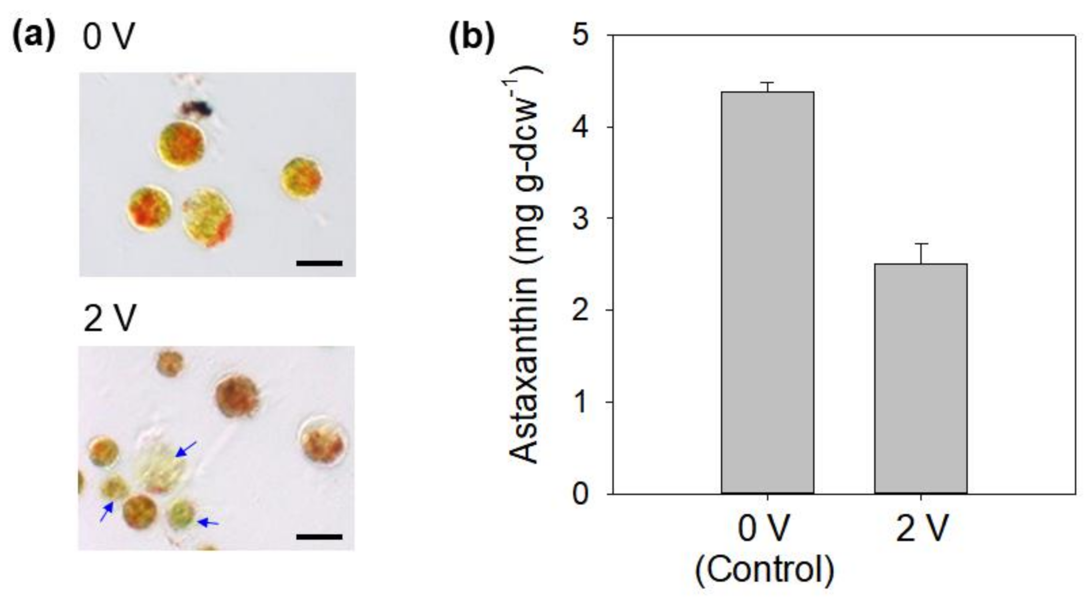
In this study, since a mild cathodic current was considered to act as a novel stress inducer for algal astaxanthin biosynthesis, alterations in the morphology and astaxanthin content of H. pluvialis were evaluated in the cathodic chamber of the bioelectrochemical system previously optimized for Chlorella lipids [19]. Biflagellate motile H. pluvialis cells cultured for 5 days in the photobioreactor were harvested, resuspended in 0.1 M phosphate buffer (pH 7.0), and electro-treated for 2 days at a cathodic current of 3 mA (voltage: 2 V). After incubation for 2 days, regardless of the electric treatment applied, most vegetative H. pluvialis cells were transformed into brown or brownish-green non-motile palmella cells, indicating that astaxanthin biosynthesis was induced, albeit at low levels, within the cells. However, the astaxanthin content of the cells treated at 2 V reduced by 57%, a significant reduction compared to that in the untreated control (4.4 ± 0.1 mg g-dcw−1). Furthermore, some H. pluvialis cells were damaged (Figure 3). This suggests that unlike in case of Chlorella [19], the cathodic current environment negatively affects the viability and astaxanthin-synthesizing activity of H. pluvialis cells.
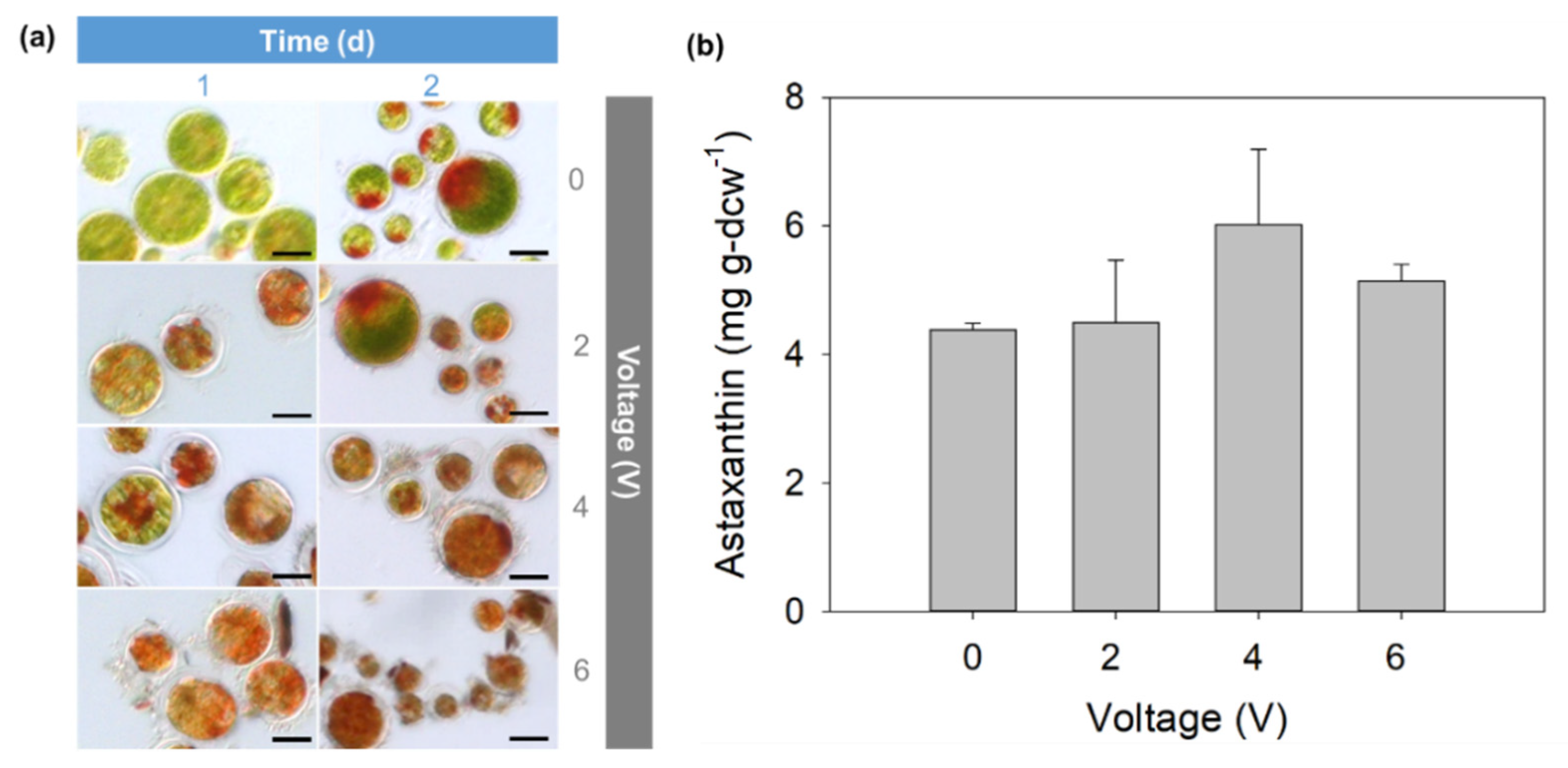
Considering the severe cell damage induced, we switched from cathodic to anodic current for electric treatment while maintaining other bioelectrochemical parameters. Figure 4 shows the changes in the optical morphology and astaxanthin content of H. pluvialis cells treated for 2 days under anodic current at 2, 4, and 6 V. The untreated H. pluvialis cells remained green for 1 day and turned partially brownish-green after 2 days. Conversely, the electro-treated H. pluvialis cells showed a significant color change to brown on day 1, indicating the relatively rapid induction of astaxanthin biosynthesis than that in the control. Contrary to the cathodic current results (Figure 3), no significant cell degradation and disintegration were observed at the applied voltages of 2–6 V. Instead, unique morphological changes, such as separation of the cell wall and cytoplasm and secretion of extracellular biomaterials (possibly cell wall lysates) occurred in the electro-treated H. pluvialis cells, which tended to worsen as the voltage increased from 2 to 6 V. Nevertheless, at an optimal voltage of 4 V (current intensity: 30 mA), the astaxanthin content was 36.9% higher (6.0 ± 1.1 mg g-dcw−1) than that in the untreated control. This result implies that anodic current treatment stimulated astaxanthin accumulation in H. pluvialis. However, at a higher voltage of 6 V (current intensity: 81 mA), the astaxanthin content of H. pluvialis cells decreased compared to that achieved at 4 V, indicating that an excessively high current does not necessarily induce astaxanthin production in H. pluvialis cells.
The anodic stimulation results (Figure 4) further suggested that algal astaxanthin induction is highly dependent on the current intensity and duration of treatment. Current-duration relationships and optimization (such as long treatment durations at low current intensities, short treatment durations at high current intensities, or a gradual increase/decrease in current intensity with the treatment duration) could be studied in greater detail in further investigation for designing practical induction processes. Of note, the response of microorganisms to electric current is strongly dependent on various bioelectrochemical parameters, such as electrode type/material, reactor design, electrolyte concentration, and the electron mediator used [19,25,26].

3.3. CV Analysis

To characterize the bioelectrochemical reactions occurring between the carbon electrode and the H. pluvialis cells, a cyclic voltammogram was constructed based on the reaction occurring in a single-chamber electrochemical reactor with three electrodes (i.e., counter, working, and reference) and analyzed using a potentiostat (Figure 5). Similar to that in the electric stimulation process described above, green vegetative H. pluvialis cells cultured for 5 days in the photobioreactor were tested. The current density–voltage relationship varied significantly in the presence of algal cells under the applied reduction/oxidation potentials. For the algal cells, both reduction and oxidation current densities showed an increasing trend relative to that in the cell-free solution (abiotic control) within the voltage range of −0.5 to +0.5 V (vs. Ag/AgCl). Particularly, the oxidation current density increased only slightly (from +0.1 (abiotic) to +0.15 µA cm−2 (biotic)) at +0.5 V, whereas the reduction current density increased significantly (from −0.1 (abiotic) to −0.4 µA cm−2 (biotic)) at −0.5 V. This suggests that even at the same voltage, significantly different oxidation and reduction environments can be created for the algal cells (Figure 3 and Figure 4). The lysis and degradation of H. pluvialis cells during the cathodic current treatment (Figure 3) might be attributed to the formation of strong ROS in excess at the carbon cathode. The in situ production of H2O2 via the cathodic 2 e oxygen reduction reaction (ORR, O2 + 2 H+ + 2 e → H2O2) generates potent hydroxyl radicals (·OH), which can damage algal cells [19,27]. Specifically, carbon-based electrodes have been reported to effectively induce the 2 e ORR reaction [28].
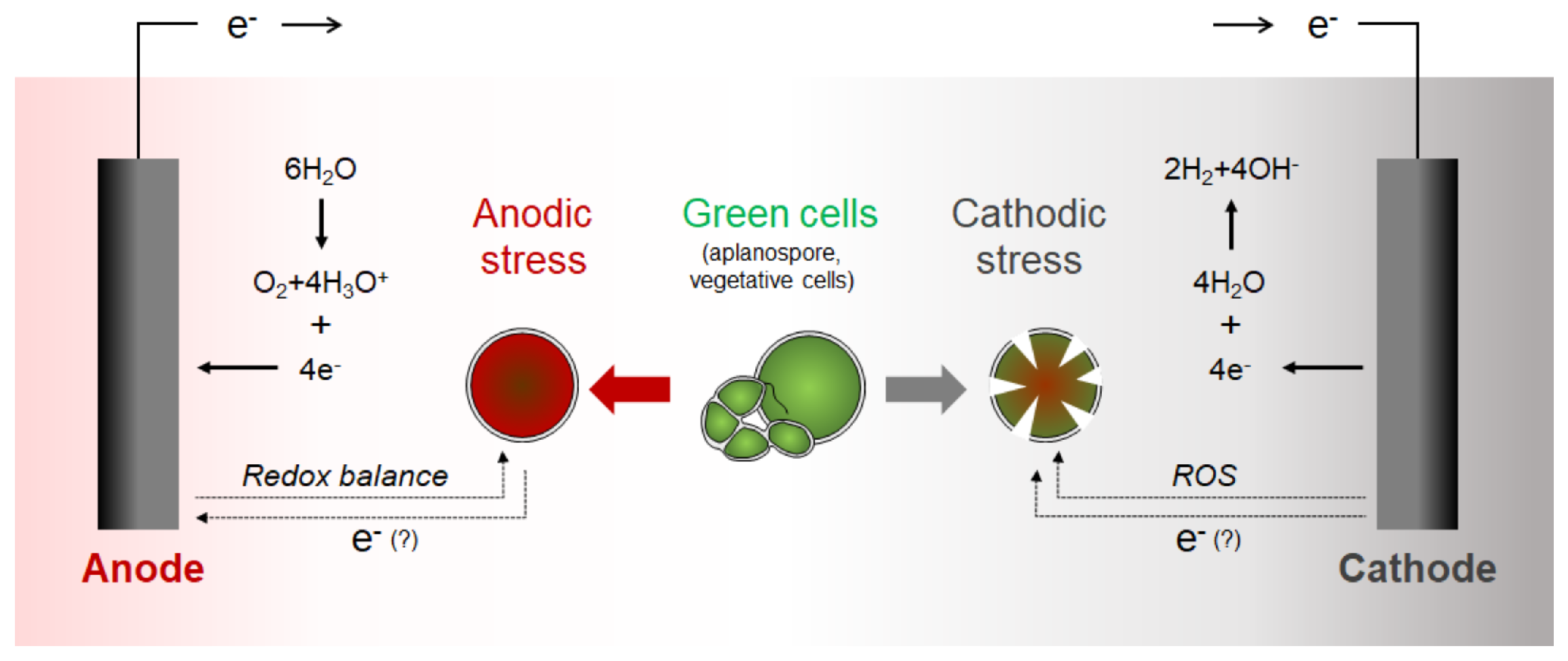
Figure 6 shows a hypothetical model for electric treatment of H. pluvialis in the two-chamber bioelectrochemical system. Only a limited number of studies have been conducted on the electric treatment of microalgae, and detailed bioelectrochemical mechanisms are yet to be identified [18,19,26,29]. Electrical stimulation can be used to induce the synthesis of various microbial products biologically by electron transfer from the electrode or non-biologically by oxidizing chemicals generated from the electrodes [25,30]. Biological stimulation involves the direct interaction between membrane-associated enzymes and the electrode surface or the indirect transfer of electrons to the microorganism through electron mediators such as hydrogen. As a non-biological stimulant, ROS is known to induce the biosynthesis of neutral lipids and/or astaxanthin in various algal species, including Chlorella and Haematococcus [2,19]. The characterization and identification of the mechanism underlying the electric stimulation of astaxanthin production in H. pluvialis should be investigated in future studies.

4. Conclusions

The electro-treatment of H. pluvialis cells for astaxanthin induction in the two-chamber bioelectrochemical system was found to be highly dependent on cathodic (reduction) and anodic (oxidation) current conditions. Cathodic current treatment led to significant lysis and breakage of algal cells, even when a low voltage (2 V; current, 3 mA) was applied. In contrast, astaxanthin biosynthesis was induced successfully in H. pluvialis cells using anodic current treatment, with approximately 36.9% improvement achieved compared to that in the control. This was also accompanied by distinct morphological changes in the cells. Cyclic voltammetry analysis showed that the oxidation and reduction environments were significantly different even at the same voltage. This study suggests that electrochemical stimulation can serve as a unique method for promoting astaxanthin accumulation in H. pluvialis. However, for practical applications, extensive research, including the characterization of the precise bioelectrochemical mechanisms, as well as the optimization of process components, such as the reactor, electrode material, and electron mediator, is necessary.

Author Contributions

Conceptualization, Y.-K.O. and S.-Y.L.; Methodology, H.-N.F., S.-Y.L., S.-A.C., and J.-Y.L.; Investigation, H.-N.F. and S.-Y.L.; Resources, J.-S.L.; Supervision, Y.-K.O. and J.-S.L.; Writing—original draft preparation, H.-N.F. and S.-Y.L.; Writing—review and editing, Y.-K.O. and B.-L.K.; Funding acquisition, Y.-K.O. All authors have read and agreed to the published version of the manuscript.

Funding

This work was supported by a 2-Year Research Grant of Pusan National University.

Institutional Review Board Statement

Not applicable.

Informed Consent Statement

Not applicable.

Data Availability Statement

Not applicable.

Conflicts of Interest

The authors declare no conflict of interest.

References

  1. Kim, D.Y.; Vijayan, D.; Praveenkumar, R.; Han, J.I.; Lee, K.; Park, J.Y.; Chang, W.S.; Lee, J.S.; Oh, Y.K. Cell-wall disruption and lipid/astaxanthin extraction from microalgae: Chlorella and Haematococcus. Bioresour. Technol. 2016, 199, 300–310. [Google Scholar] [CrossRef] [PubMed]
  2. Shah, M.M.R.; Liang, Y.; Cheng, J.J.; Daroch, M. Astaxanthin-producing green microalga Haematococcus pluvialis: From single cell to high value commercial products. Front. Plant Sci. 2016, 7, 531. [Google Scholar] [CrossRef] [Green Version]
  3. Lee, S.Y.; Cho, J.M.; Chang, Y.K.; Oh, Y.-K. Cell disruption and lipid extraction for microalgal biorefineries: A review. Bioresour. Technol. 2017, 244, 1317–1328. [Google Scholar] [CrossRef]
  4. Poonkum, W.; Powtongsook, S.; Pavasant, P. Astaxanthin induction in microalga H. pluvialis with flat panel airlift photobioreactors under indoor and outdoor conditions. Prep. Biochem. Biotechnol. 2015, 45, 1–17. [Google Scholar] [CrossRef]
  5. Han, D.; Li, Y.; Hu, Q. Astaxanthin in microalgae: Pathways, functions and biotechnological implications. Algae 2013, 28, 131–147. [Google Scholar] [CrossRef]
  6. Noh, M.H.; Cha, S.; Kim, M.; Jung, G.Y. Recent advances in microbial cell growth regulation strategies for metabolic engineering. Biotechnol. Bioprocess. Eng. 2020, 25, 810–828. [Google Scholar] [CrossRef]
  7. Liyanaarachchi, V.C.; Nishshanka, G.K.S.H.; Premaratne, R.G.M.M.; Ariyadasa, T.U.; Nimarshana, P.H.V.; Malik, A. Astaxanthin accumulation in the green microalga Haematococcus pluvialis: Effect of initial phosphate concentration and stepwise/continuous light stress. Biotechnol. Rep. 2020, 28, e00538. [Google Scholar] [CrossRef] [PubMed]
  8. Zhang, W.-W.; Zhou, X.-F.; Zhang, Y.-L.; Cheng, P.-F.; Ma, R.; Cheng, W.-L.; Chu, H.-Q. Enhancing astaxanthin accumulation in Haematococcus pluvialis by coupled light intensity and nitrogen starvation in column photobioreactors. J. Microbiol. Biotechnol. 2018, 28, 2019–2028. [Google Scholar] [CrossRef]
  9. Oslan, S.N.H.; Shoparwe, N.F.; Yusoff, A.H.; Rahim, A.A.; Chang, C.S.; Tan, J.S.; Oslan, S.N.; Arumugam, K.; Ariff, A.B.; Sulaiman, A.Z.; et al. A Review on Haematococcus pluvialis bioprocess optimization of green and red stage culture conditions for the production of natural astaxanthin. Biomolecules 2021, 11, 256. [Google Scholar] [CrossRef]
  10. Kiperstok, A.C.; Sebestyén, P.; Podola, B.; Melkonian, M. Biofilm cultivation of Haematococcus pluvialis enables a highly productive one-phase process for astaxanthin production using high light intensities. Algal Res. 2017, 21, 213–222. [Google Scholar] [CrossRef]
  11. Giannelli, L.; Yamada, H.; Katsuda, T.; Yamaji, H. Effects of temperature on the astaxanthin productivity and light harvesting characteristics of the green alga Haematococcus pluvialis. J. Biosci. Bioeng. 2015, 119, 345–350. [Google Scholar] [CrossRef]
  12. Hong, M.-E.; Hwang, S.K.; Chang, W.S.; Kim, B.W.; Lee, J.; Sim, S.J. Enhanced autotrophic astaxanthin production from Haematococcus pluvialis under high temperature via heat stress-driven Haber–Weiss reaction. Appl. Microbiol. Biotechnol. 2015, 99, 5203–5215. [Google Scholar] [CrossRef]
  13. Harker, M.; Tsavalos, A.J.; Young, A.J. Factors responsible for astaxanthin formation in the Chlorophyte Haematococcus pluvialis. Bioresour. Technol. 1996, 55, 207–214. [Google Scholar] [CrossRef]
  14. Kou, Y.; Liu, M.; Sun, P.; Dong, Z.; Liu, J. High light boosts salinity stress-induced biosynthesis of astaxanthin and lipids in the green alga Chromochloris zofingiensis. Algal Res. 2020, 50, 101976. [Google Scholar] [CrossRef]
  15. Kim, Y.-E.; Matter, I.A.; Lee, N.; Jung, M.; Lee, Y.-C.; Choi, S.-A.; Lee, S.Y.; Kim, J.R.; Oh, Y.-K. Enhancement of astaxanthin production by Haematococcus pluvialis using magnesium aminoclay nanoparticles. Bioresour. Technol. 2020, 307, 123270. [Google Scholar] [CrossRef]
  16. Grewe, C.; Griehl, C. Time- and media-dependent secondary carotenoid accumulation in Haematococcus pluvialis. Biotechnol. J. 2008, 3, 1232–1244. [Google Scholar] [CrossRef]
  17. Choi, Y.Y.; Joun, J.M.; Lee, J.; Hong, M.E.; Pham, H.-M.; Chang, W.S.; Sim, S.J. Development of large-scale and economic pH control system for outdoor cultivation of microalgae Haematococcus pluvialis using industrial flue gas. Bioresour. Technol. 2017, 244, 1235–1244. [Google Scholar] [CrossRef] [PubMed]
  18. Kim, J.Y.; Lee, C.; Jeon, M.S.; Park, J.; Choi, Y.E. Enhancement of microalga Haematococcus pluvialis growth and astaxanthin production by electrical treatment. Bioresour. Technol. 2018, 268, 815–819. [Google Scholar] [CrossRef] [PubMed]
  19. Choi, S.-A.; Lee, S.Y.; Lee, J.; Cho, J.M.; Lee, J.-S.; Kim, S.W.; Kim, D.-Y.; Park, S.-K.; Jin, C.-S.; Oh, Y.-K. Rapid induction of edible lipids in Chlorella by mild electric stimulation. Bioresour. Technol. 2019, 292, 121950. [Google Scholar] [CrossRef] [PubMed]
  20. Pick, U.; Zarka, A.; Boussiba, S.; Davidi, L. A hypothesis about the origin of carotenoid lipid droplets in the green algae Dunaliella and Haematococcus. Planta 2019, 249, 31–47. [Google Scholar] [CrossRef]
  21. Praveenkumar, R.; Lee, K.; Lee, J.; Oh, Y.-K. Breaking dormancy: An energy-efficient means of recovering astaxanthin from microalgae. Green Chem. 2015, 17, 1226–1234. [Google Scholar] [CrossRef]
  22. Choi, S.-A.; Jeong, Y.; Lee, J.; Huh, Y.H.; Choi, S.H.; Kim, H.-S.; Cho, D.-H.; Lee, J.-S.; Kim, H.; An, H.-R.; et al. Biocompatible liquid-type carbon nanodots (C-paints) as light delivery materials for cell growth and astaxanthin induction of Haematococcus pluvialis. Mater. Sci. Eng. C 2020, 109, 110500. [Google Scholar] [CrossRef] [PubMed]
  23. Moon, G.; Lee, N.; Kang, S.; Park, J.; Kim, Y.-E.; Lee, S.-A.; Chitumalla, R.K.; Jang, J.; Choe, Y.; Oh, Y.-K.; et al. Hydrothermal synthesis of novel two-dimensional α-quartz nanoplates and their applications in energy-saving, high-efficiency, microalgal biorefineries. Chem. Eng. J. 2020, 127467. [Google Scholar] [CrossRef]
  24. Imamoglu, E.; Dalay, M.C.; Sukan, F.V. Influences of different stress media and high light intensities on accumulation of astaxanthin in the green alga Haematococcus pluvialis. New Biotechnol. 2009, 26, 199–204. [Google Scholar] [CrossRef] [PubMed]
  25. Oh, Y.-K.; Hwang, K.-R.; Kim, C.; Kim, J.R.; Lee, J.-S. Recent developments and key barriers to advanced biofuels: A short review. Bioresour. Technol. 2018, 257, 320–333. [Google Scholar] [CrossRef]
  26. La, H.-J.; Choi, G.-G.; Cho, C.; Seo, S.-H.; Srivastava, A.; Jo, B.-H.; Lee, J.-Y.; Jin, Y.-S.; Oh, H.-M. Increased lipid productivity of Acutodesmus dimorphus using optimized pulsed electric field. J. Appl. Phycol. 2016, 28, 931–938. [Google Scholar] [CrossRef]
  27. Liang, J.; Tang, D.; Huang, L.; Chen, Y.; Ren, W.; Sun, J. High oxygen reduction reaction performance nitrogen-doped biochar cathode: A strategy for comprehensive utilizing nitrogen and carbon in water hyacinth. Bioresour. Technol. 2018, 267, 524–531. [Google Scholar] [CrossRef]
  28. Özcan, A.; Şahin, Y.; Savaş Koparal, A.; Oturan, M.A. Carbon sponge as a new cathode material for the electro-Fenton process: Comparison with carbon felt cathode and application to degradation of synthetic dye basic blue 3 in aqueous medium. J. Electroanal. Chem. 2008, 616, 71–78. [Google Scholar] [CrossRef]
  29. Wu, Y.; Guan, K.; Wang, Z.; Xu, B.; Zhao, F. Isolation, identification and characterization of an electrogenic microalgae strain. PLoS ONE 2013, 8, e73442. [Google Scholar] [CrossRef]
  30. Lee, S.Y.; Oh, Y.-K.; Lee, S.; Fitriana, H.N.; Moon, M.; Kim, M.-S.; Lee, J.; Min, K.; Park, G.W.; Lee, J.-P.; et al. Recent developments and key barriers to microbial CO2 electrobiorefinery. Bioresou. Technol. 2021, 320, 124350. [Google Scholar] [CrossRef]
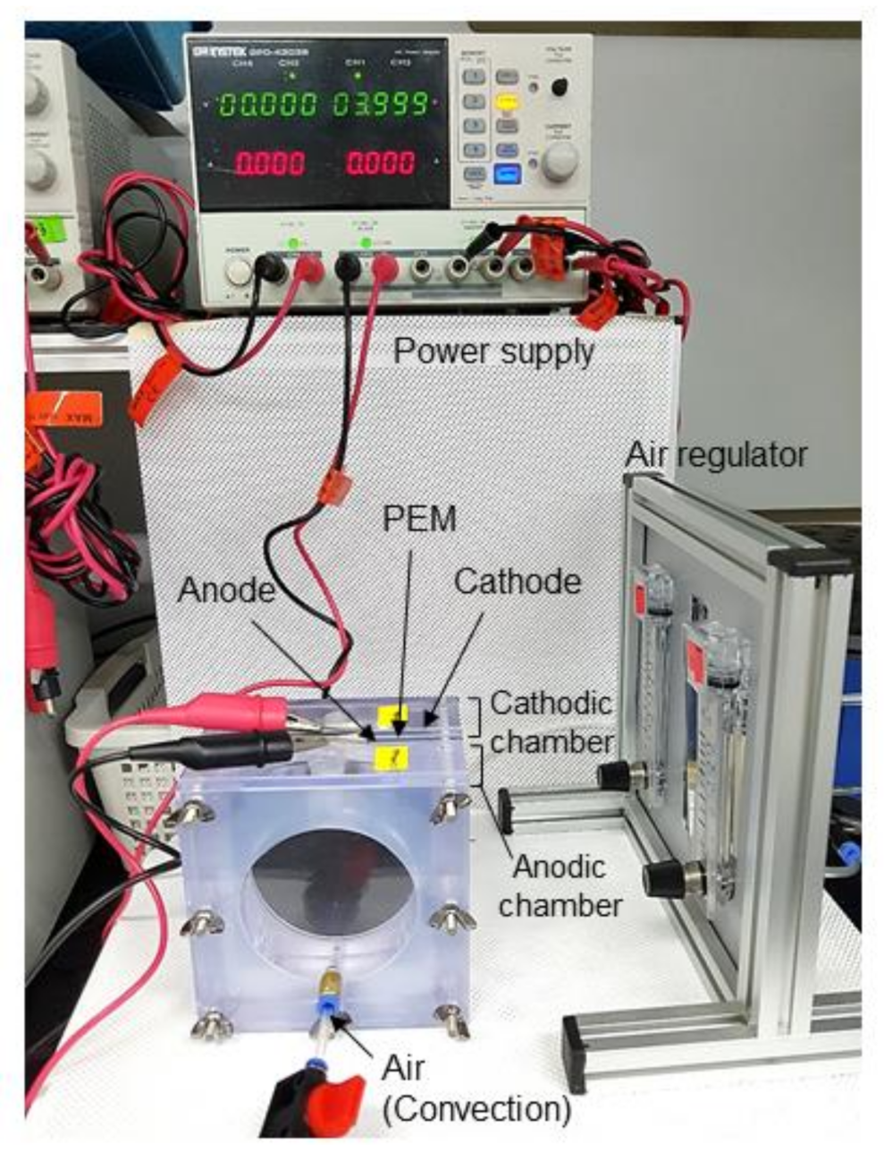
Figure 1. The two-chamber bioelectrochemical system equipped with DC power supply and an aeration control unit [19]. PEM, proton-exchange membrane.
Applsci 11 03348 g001
Figure 2. Time course profile of changes in the cell number density (a) and morphology (b) of Haematococcus pluvialis cells during photobioreactor cultivation for 25 days. The life cycle of H. pluvialis is characterized by five types of distinguishable cellular morphologies based on microscopic observation: (1) astaxanthin-rich asexual aplanospore; (2) dividing aplanospore with internal differentiated zooids; (3) green vegetative biflagellate cell; (4) green vegetative non-motile palmella; and (5) astaxanthin-accumulating palmella cell. The asterisk (*) indicates the growth stage of H. pluvialis cells used for the electric stimulation experiment. Scale bars in the optical images represent 10 µm.
Applsci 11 03348 g002
Figure 3. Microscopic image (a) and astaxanthin content (b) in Haematococcus pluvialis cells after electro-treatment at 2 V (current: 3 mA) for 2 days in the cathodic chamber of the two-chamber electrochemical reactor. The blue arrows in Figure 3a represent cells damaged or disrupted by electric stress. Scale bars in the optical images represent 50 μm.
Applsci 11 03348 g003
Figure 4. Effects of voltage intensity on the optical morphology (a) and astaxanthin content (b) of Haematococcus pluvialis cells in the anodic chamber of the two-chamber electrochemical reactor. The cellular astaxanthin levels were analyzed after the 2 day treatment. Scale bars in the optical images represent 15 μm.
Applsci 11 03348 g004
Figure 5. Cyclic voltammograms of the carbon electrode in the presence (biotic, red line) and absence (abiotic, black line) of Haematococcus pluvialis cells in a single-chamber electrochemical reactor with three electrodes. The scan rate was 10 mV s−1.
Applsci 11 03348 g005
Figure 6. Simple hypothetical schematic of the electric treatment of Haematococcus pluvialis in the two-chamber bioelectrochemical system. ROS, reactive oxygen species.
Applsci 11 03348 g006
Publisher’s Note: MDPI stays neutral with regard to jurisdictional claims in published maps and institutional affiliations.