Article

Enzymatic Synthesis of Glucose Fatty Acid Esters Using SCOs as Acyl Group-Donors and Their Biological Activities

1
Department of Clinical Biochemistry, College of Medicine, University of Jeddah, Jeddah 21589, Saudi Arabia
2
National Research Centre, Biochemistry Department, Genetic Engineering and Biotechnology Division, Cairo 12622, Egypt
3
Department of Biology, College of Science, University of Jeddah, Jeddah 21589, Saudi Arabia
4
National Research Centre, Department of Chemistry of Natural and Microbial Products, Division of Pharmaceutical and Drug Industries, Cairo 12622, Egypt
5
Department of Chemistry, College of Science, University of Jeddah, Jeddah 21589, Saudi Arabia
6
Unit of Microbiology, Division of Genetics, Cell and Developmental Biology, Department of Biology, University of Patras, 26504 Patras, Greece
7
Department of Biological Sciences, Faculty of Science, King Abdulaziz University, Jeddah 21589, Saudi Arabia
8
Department of Processes Design & Development, Egyptian Petroleum Research Institute (EPRI), Nasr City, Cairo 11727, Egypt
*
Author to whom correspondence should be addressed.
Both authors contributed equally to this paper.
Appl. Sci. 2021, 11(6), 2700; https://doi.org/10.3390/app11062700
Submission received: 24 February 2021 / Revised: 11 March 2021 / Accepted: 15 March 2021 / Published: 17 March 2021
(This article belongs to the Section Applied Biosciences and Bioengineering)

Abstract

:
Sugar fatty acid esters, especially glucose fatty acid esters (GEs), have broad applications in food, cosmetic and pharmaceutical industries. In this research, the fatty acid moieties derived from polyunsaturated fatty acids containing single-cell oils (SCOs) (i.e., those produced from Cunninghamella echinulata, Umbelopsis isabellina and Nannochloropsis gaditana, as well as from olive oil and an eicosapentaenoic acid (EPA) concentrate) were converted into GEs by enzymatic synthesis, using lipases as biocatalysts. The GE synthesis was monitored using thin-layer chromatography, FTIR and in situ NMR. It was found that GE synthesis carried out using immobilized Candida antarctica B lipase was very effective, reaching total conversion of reactants. It was shown that EPA-GEs were very effective against several pathogenic bacteria and their activity can be attributed to their high EPA content. Furthermore, C. echinulata-GEs were more effective against pathogens compared with U. isabellina-GEs, probably due to the presence of gamma linolenic acid (GLA) in the lipids of C. echinulata, which is known for its antimicrobial activity, in higher concentrations. C. echinulata-GEs also showed strong insecticidal activity against Aedes aegypti larvae, followed by EPA-GEs, olive oil-GEs and N. gaditana-GEs. All synthesized GEs induced apoptosis of the SKOV-3 ovarian cancer cell line, with the apoptotic rate increasing significantly after 48 h. A higher percentage of apoptosis was observed in the cells treated with EPA-GEs, followed by C. echinulata-GEs, U. isabellina-GEs and olive oil-GEs. We conclude that SCOs can be used in the synthesis of GEs with interesting biological properties.

1. Introduction

Sugar fatty acid esters, the so-called sugar esters (SEs), are biodegradable, odorless, non-irritating and non-toxic surfactants with broad applications in the food [1,2], cosmetic [3] and pharmaceutical [4] industries. Moreover, SEs have gained attention thanks to their anti-bacterial (e.g., against numerous pathogenic species of Gram-positive and Gram-negative bacteria) and antifungal activity [5], while they are also reported as insecticides and miticides [6].
SEs are synthesized from renewable resources, such as sugars and fatty acids (FAs). Different types of sugars (e.g., sucrose, fructose, glucose and lactose) can be used as acyl-acceptors to produce SEs by esterification with FAs or transesterification with FA esters used as acyl-donors [7,8,9]. Glucose (Glc), a cheap and broadly available carbohydrate, has only one primary hydroxyl group, which predetermines a highly regioselective synthesis of SEs called glucose fatty acid esters (GEs) [10,11]. Concerning acyl-donors, polyunsaturated fatty acids (PUFAs) can be used for this purpose, as it is known that PUFAs or PUFA derivatives have interesting biological properties, including their role in protecting against cardiovascular diseases, as well as antimicrobial and anticancer activities [12,13,14]. GEs can be synthesized both chemically and enzymatically [15,16]. Lately, the main efforts in the processing of GEs have been focused on enzymatic synthesis because this is an advantageous method, requiring less energy and reducing solvent toxicity [11,17], with lipases being the most important enzymes used for this synthesis [18,19]. Commercially available immobilized lipases, such as Candida antarctica B lipase, are used to catalyze the acylation of glucose with various FAs [20,21].
Different sources of PUFAs can be used as sources of acyl groups, in the form of either FAs or FA esters, for GE production. Whilst terrestrial plant oils have been used as a source of PUFAs [22,23], alternative and non-food supply sources, such as microalgae [24,25] and fungi [26] could be used, thanks to their ability to produce microbial lipids rich in PUFAs of medicinal and nutritional interest. The microbial lipids, the so-called single-cell oils (SCOs), are synthesized by oleaginous microorganisms that are capable of producing substantial amounts of lipid stored within their cells [27,28]. The genus of Nannochloropsis includes various marine microalgae species, such as N. salina [29], N. gaditana [30,31] and N. oculata [32], which are able to grow efficiently under a variety of culture conditions on low-quality waters, even on wastewaters, and accumulate lipids rich in PUFAs, such as eicosapentaenoic acid (EPA). Furthermore, oleaginous fungal species, such as Cunninghamella echinulata and Umbelopsis isabellina are able to synthesize PUFAs, especially γ-linolenic acid (GLA) [13,25], and are thus regarded as promising candidates for SCO production [33,34,35].
In a previous paper [36], we produced FA amides (FAAs) using lipids containing PUFAs as acyl group donors in different percentages and we concluded that FAAs can be used as bioactive compounds in various biological applications depending on their FA composition. The aim of the current paper was to enzymatically synthesize GEs using SCOs as acyl group donors similar to those used in FAA, either in the form of free fatty acids (FFAs) or in the form of fatty acid methyl esters (FAMEs). The reaction was carried out under various conditions, using two immobilized lipases as catalysts. SCOs, produced from various sources as described in El-Baz et al. [36], contained EPA, GLA or oleic acid in high percentages. The biological activity of the aforementioned GEs against important human pathogens, the larvae of Aedes aegypti and the SKOV-3 cancer cell line was studied and compared with GEs synthesized using olive oil and an EPA concentrate (i.e., a fish oil derivative containing EPA in very high percentages) as acyl group donors. We concluded that GEs derived from SCOs possess interesting biological activities and can therefore be used in the production of pharmaceuticals in the future.

2. Materials and Methods

2.1. Chemicals

D-glucose and molecular sieves (3 Å) were purchased from Acros Organics (Thermo Fisher Scientific, Waltham, MA). DMSO, tert-amyl alcohol, NaCl, MgSO4, immobilized Candida antarctica B lipase (enzymatic activity >5 units/mg) and immobilized C. rugosa lipase (enzymatic activity >0.1 units/mg) were purchased from Sigma Aldrich Co., St. Louis, MO, USA.

2.2. Biological Material and SCO Production

The fungal strains Cunninghamella echinulata ATHUM 4411 and Umbelopsis isabellina ATHUM 2935 (culture collection of National and Kapodistrian University of Athens, Greece) and the microalga strain Nannochloropsis gaditana (culture collection CCAP 849/5) were used as sources of SCOs. Additionally, a Greek virgin olive oil (Altis, Upfield Hellas) and an EPA concentrate (Dr Tolonen’s E-EPA, Probiotics International Limited, Lopen Head, Somerset, United Kingdom) containing 500 mg of EPA per capsule were used. Culture conditions, cell mass harvesting, lipid extraction and purification, FAME and FFA preparation and gas chromatography analysis were as described in El-Baz et al. [36]. Mass spectra were recorded on a Thermo ISQ Single Quadrupole GC-MS.

2.3. Enzymatic Synthesis of GEs

GEs were synthesized by esterification (or transesterification) of glucose served as an acyl acceptor, with FFAs (or FAMEs) served as acyl group donors. The reaction was performed in 50-mL Erlenmeyer flasks using FFAs or FAMEs at a concentration of 0.04 mmol/mL, and corresponding amounts of glucose were added to achieve 1:1, 1:2 and 1:3 molar ratios of FFA (or FAME) to glucose. The molar ratios were calculated considering the MS of the FFAs of the five substrates, i.e., olive oil: m/z (%): 284 (M+, 21); EPA concentrate: m/z (%): 302 (M+, 55); C. echinulata: m/z (%): 280 (M+, 39); U. isabellina: m/z (%): 302 (M+, 33) and N. gaditana: m/z (%): 302 (M+, 67). Τhe reactants were dissolved in 25 mL of a solvent mixture consisting of 80% DMSO and 20% tert-amyl alcohol to which 1 g of 3 Å molecular sieves was added and the mixture was sonicated for 20 min. The reaction was catalyzed by 0.25 g of immobilized C. antarctica B lipase or 0.25 g of immobilized C. rugosa lipase. The flasks were incubated at 50 ± 1 °C in a shaking incubator at 100 rpm for 50 h.
After the incubation period, the reaction mixture was filtered to remove the molecular sieves and the immobilized lipase, and the solvent was evaporated under reduced pressure. The reaction residue was separated in ethyl acetate and distilled water (25 mL each). The organic layer, containing the synthesized GEs, was washed with 10 mL saturated aqueous NaCl, dried over MgSO4 and gravity-filtered, and the solvent was removed under reduced pressure to get the crude product.

2.4. GE Analysis

2.4.1. Thin-Layer Chromatography and FTIR

Qualitative synthesis of GEs was monitored by thin-layer chromatography (TLC) as described in El-Baz et al. [36] for FAAs. Equally, FTIR spectra for FAMEs and the GE products were recorded as described in the aforementioned paper. FTIR spectra were used to detect the formation of the ester carbonyl group, thus confirming GE synthesis.

2.4.2. Quantitative Determination of the Enzymatic Conversion through In Situ NMR Monitoring

The conversion of FAMEs to GEs was determined during the reaction via in situ NMR monitoring. First, the proton NMR of both reactants individually was assigned and the progress of the reaction was monitored by 1H NMR at regular intervals of 10 h. The conversion was calculated according to the formula:
Conversion(%) = [Ip/(Ir + Ip)] × 100,
where Ip is the integration of the signal of the product and Ir is the integration of the signal of the reactant. Ip was represented by integration of signal due to CH2OCO and Ir by integration of signal due to the glucose protons of C6 signal. Heteronuclear multiple bond correlation (HMBC) spectroscopy was used to identify the ester product by determining the chemical shifts in carbon and hydrogen atoms and formation of the ester bond. NMR spectra were recorded at 298 K on a Bruker Avance III 400 (9.4 T, 400.13 MHz for 1H, 100.62 MHz for 13C) spectrometer (Bruker, Billerica, MA, USA) with a 5-mm BBFO probe. Chemical shifts (δ in ppm) were relative to the internal standard, DMSO-d6 (d 2.50), for 1H NMR.

2.4.3. Reusability of Candida antarctica Lipase

To check the reusability of Candida antarctica (CA) lipase for several reaction cycles, we used olive oil FAMEs and glucose as substrates under the optimized reaction conditions. After completion of the reaction, the enzyme was removed by filtration and washed with ethanol solvent in a Soxhlet extraction apparatus. Short-chain alcohols may deactivate lipase, but the use of ethanol here was necessary to remove any traces of FAMEs that may have been present. The regenerated enzyme was reused in a new transesterification reaction and the process was repeated three times.

2.5. Biological Activity of GEs

The antimicrobial, insecticidal and anticancer activities of the synthesized GEs were determined using the protocols described in El-Baz et al. [36]. Briefly, the antimicrobial activity of GEs (used at a concentration of 40 μg/mL) was evaluated in vitro using the agar well diffusion assay [37], the minimum inhibitory concentration (MIC) and the minimum bactericidal concentration (MBC) [38] against human pathogens including the Gram-negative Escherichia coli (ATCC 25922), Klebsiella pneumoniae (ATCC 700603), Pseudomonas aeruginosa (ATCC 15442) and Salmonella typhimurium (ATCC 14028); the Gram-positive bacteria Bacillus subtilis (ATCC 6633), MRSA Staphylococcus aureus (ATCC 4330) and S. aureus (ATCC 25923); and the unicellular fungus Candida albicans (ATCC 10221). The insecticidal activity was evaluated by exposing early 4th instar larvae of a field strain of Aedes aegypti to different concentrations of GEs (up to 100 ppm) for 48 h, in glass beakers containing 100 mL of tap water and GE solutions. Calculation of statistical parameters was performed using the Finney method [39]. The apoptotic activity of the SKOV-3 ovarian cancer cell line in response to the tested compounds (both FAMEs and GEs) was determined by Annexin FITC, as per the manufacturer’s instructions (BD Biosciences, San Jose, CA, USA).

2.6. Statistical Analysis

The acquired data were analyzed using the Statistical Package for the Social Sciences (SPSS), version 9.0 and the results are given as the mean ± SD of three replicates. The mean comparison between the various assessed groups was performed using one-way analysis of variance (ANOVA). Statistical significance was defined when p < 0.05.

3. Results and Discussion

3.1. Biomass and SCO Production

The performance of the oleaginous microorganisms cultivated in flasks or bioreactors, and the FA composition of the produced lipids were presented previously [36]. Here, the essential data are provided as Supplementary Materials to facilitate reading (Tables S1 and S2).

3.2. Optimization of the GE Synthesis

For GE synthesis, the olive oil-derived FFAs and FAMEs were used as model substrates to optimize the lipase-catalyzed reaction of esterification and transesterification (Figure 1). The reaction was carried out in a solvent system consisting of a tert-amyl alcohol and DMSO mixture as described elsewhere [11,40,41] for 50 h at 55 °C with shaking at 100 rpm, and the progress of the reaction was monitored by TLC analysis.
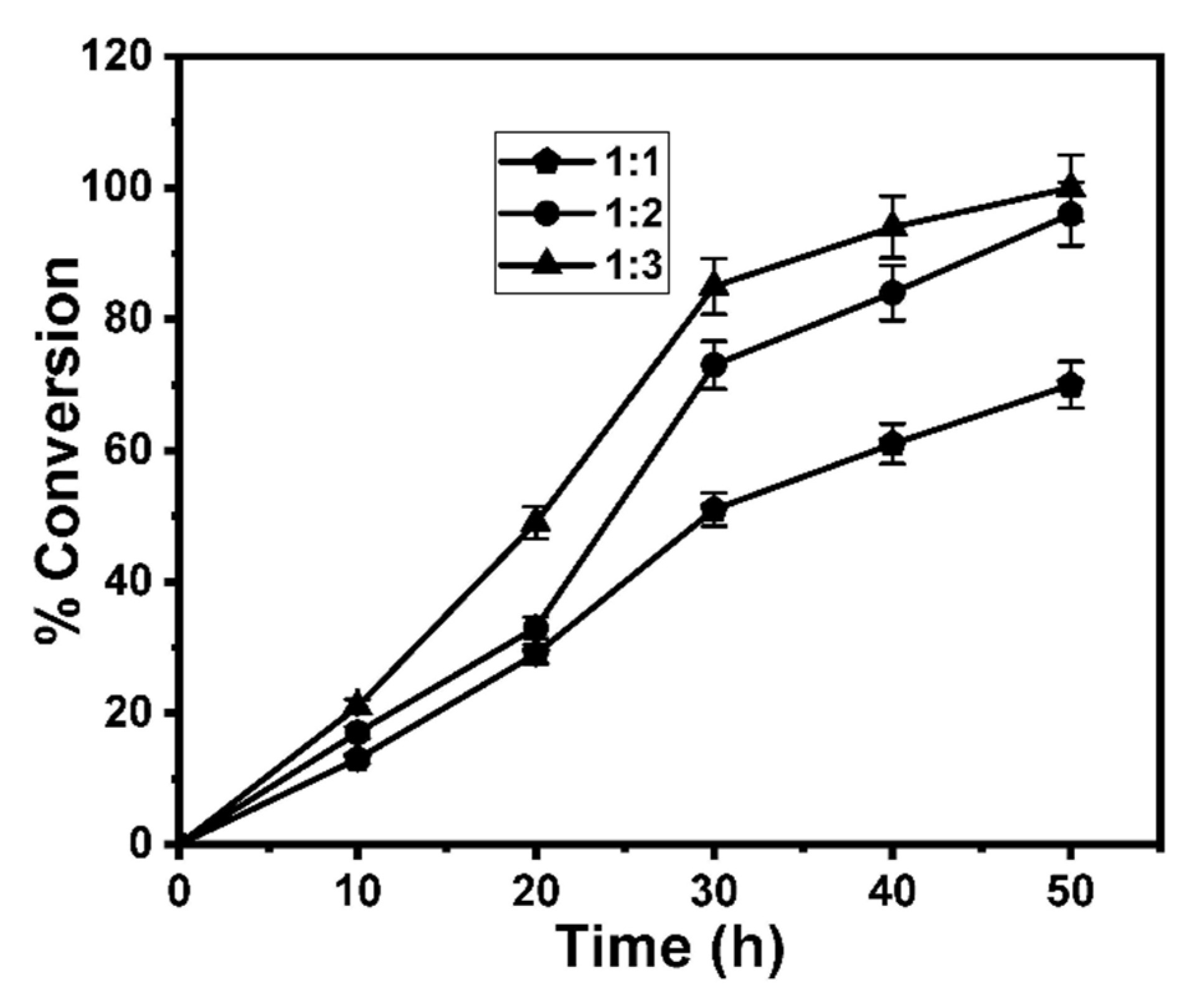
The effect of various reaction conditions, such as the molar ratio of the reactants, the reaction temperature and the type and the concentration of immobilized enzyme, on the conversion rate was studied. Two immobilized lipases, lipase from C. antarctica B (CA lipase) and lipase from C. rugosa (CR lipase), were used as catalysts for both esterification and transesterification reactions for GE production (Table 1). The use of immobilized lipases as catalysts for GE synthesis has been proposed by several researchers [9,21,42,43,44]. Moreover, different molar ratios of olive oil FFAs (or FAMEs) to glucose were tested (Table 1), showing that the conversion rate increased with an increasing concentration of glucose, attaining its maximum value with CA lipase as the catalyst and a FAMEs:glucose ratio of 1:3 (Entry 6, Table 1 and Figure 2). Glucose can be considered as a good acyl acceptor for SE synthesis in non-conventional media, ensuring a high conversion rate, due to its relatively higher solubility compared with other sugars [45,46,47]. On the contrary, the use of sugars with a higher degree of polymerization adversely affects the conversion rate as a result of their very low solubility in organic solvents.
Although the conversion rate obtained by utilizing FFAs as acyl donor was high, this was, in all cases, below to that obtained with FAMEs as the substrate, probably due to the water that was produced, which may react with GE, causing hydrolysis (Figure 1). On the contrary, the transesterification reaction carried out using FAMEs is almost irreversible, as methanol (produced as a byproduct) at elevated temperatures evaporates to prevent the reaction from reversing [48,49]. CR lipase showed a lower conversion rate (Entries 7–12, Table 1) than CA lipase. Furthermore, the reusability of CA lipase was checked for several reaction cycles for the synthesis of GE using olive oil FAMEs as substrate under the optimized reaction conditions. It was found that the conversion rate using the regenerated enzyme remained essentially the same for three reaction cycles, while in the fourth cycle, the conversion rate was reduced to 88% of the original, which is important for the sustainability of the process. The reaction conditions were further optimized using the lipase CA in different quantities as a catalyst, and it was found that a 100% conversion was obtained using 0.25 g of the lipase CA (Table 2, entry 4), while a higher enzyme quantity was not necessary.
The results recorded in this study emphasized the feasibility of enzymatic synthesis of GEs, the conversion yield of which reached 100% using CA lipase as a biocatalyst. These results are in agreement with those reported by Yan et al. [50], who demonstrated that glucose FA monoesters were synthesized (with up to 93% yields) using lipase B from C. antarctica. High yields for GE enzymatic synthesis were also reported by Sebatini et al. [51] using lipase-Fe3O4 nanoparticles as a catalyst. Furthermore, Findrik et al. [20] reported that the highest SE yield was achieved using CA lipase as a catalyst, and glucose and palmitic acid as substrates.

3.3. Product Identification and Quantitative Analysis

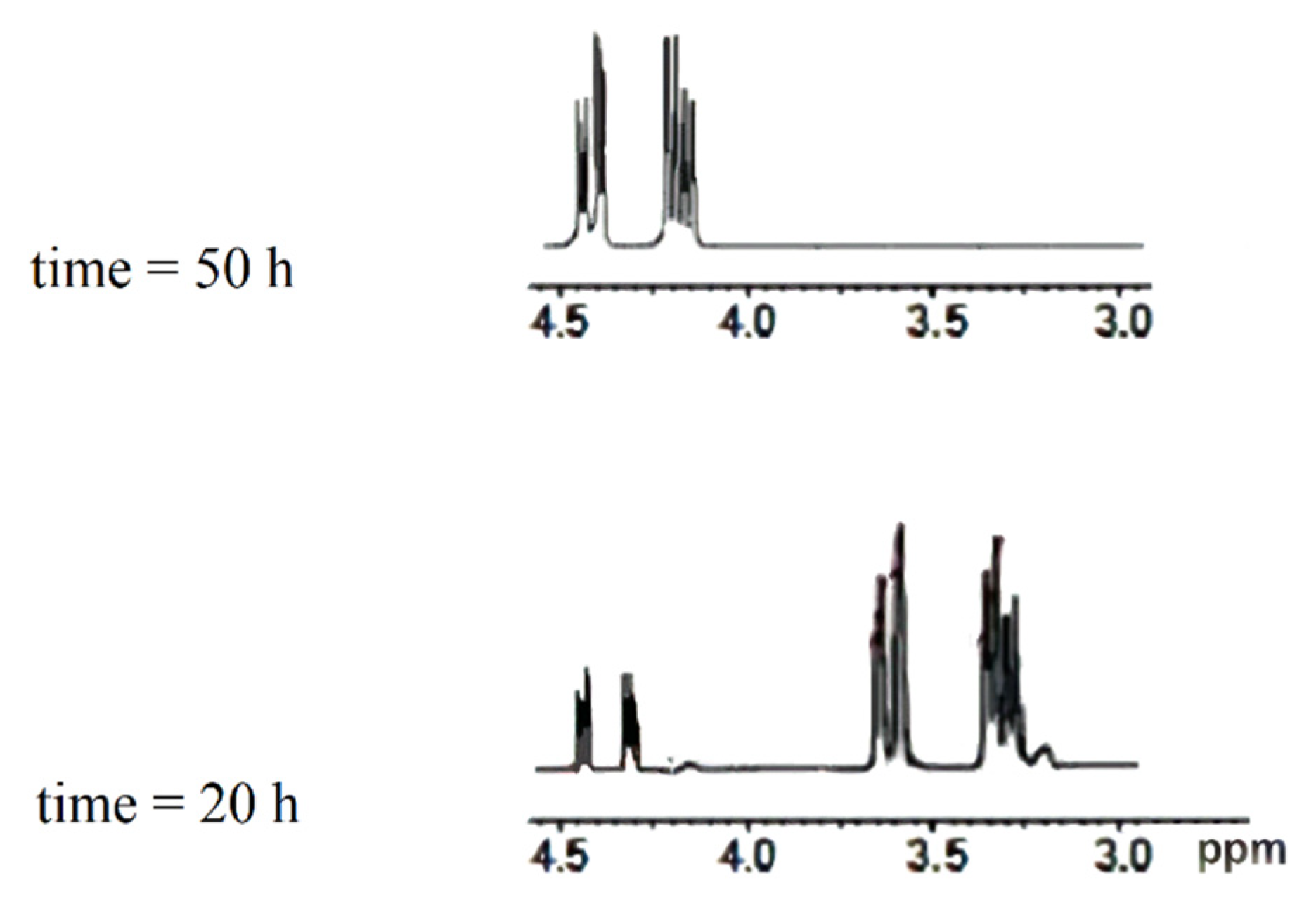
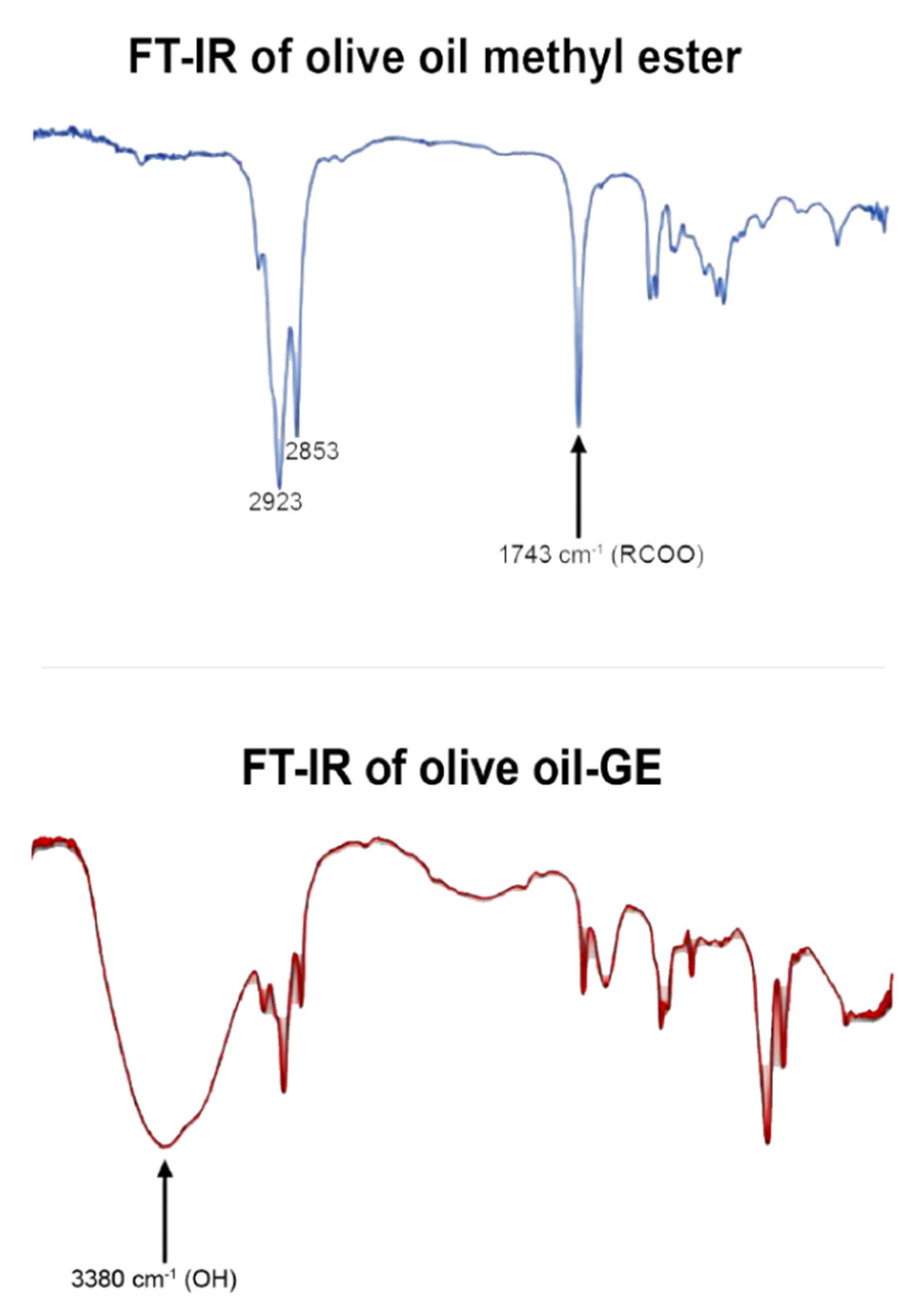
The percent conversion, which was taken as a scale to determine the optimum conditions, was quantified via in situ NMR monitoring (Figure 3). The 1H NMR noted new multiple signals at 4.17 and 4.41 ppm, which matched the CH2OCO and grew concurrently with a decline in the intensity of the glucose protons of C6 signals at 3.39 and 3.52 ppm. The latter signals disappeared after 50 h of reaction when the FAME:glucose 1:3 ratio was used, which means that 100% conversion was achieved (Figure 3). The main product obtained was identified as Glc (C-6)-OCOR, as indicated by 2D (1H-13C HMBC) NMR (Figure 4a), showing a correlation between the peak assigned to the proton C-6 glucose ester and the carbonyl function in olive oil FAMEs (Figure 4b). The structure of the obtained GEs was additionally confirmed by FTIR analysis, showing the appearance of a broad band at 3380 cm−1 due to the hydroxyl groups of glucose and the two stretching bands of O-C bond at 1316 cm−1 and 1015 cm−1, in parallel with the disappearance of the band at 1743 cm−1, due to the consumption of the carbonyl group of FAMEs (Figure 5).

3.4. GE Synthesis Using FAMEs from Different Origins

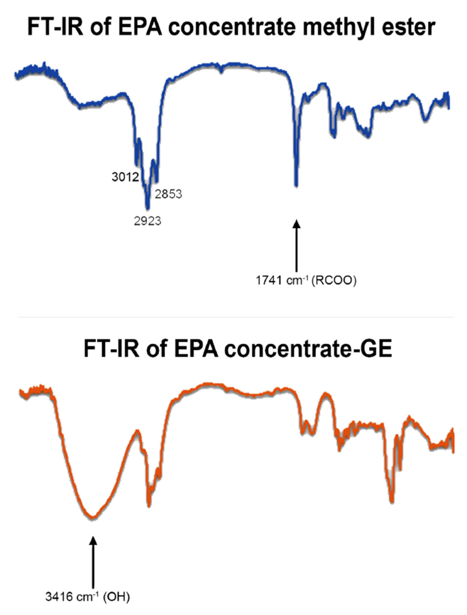
After optimization of the reaction conditions, GEs were synthesized using FAMEs derived from SCOs produced by C. echinulata, U. isabellina and N. gaditana and from an EPA concentrate (Table 3). The GE synthesis when EPA-FAMEs were used as substrate was excellent (i.e., 99% conversion), followed by N. gaditana-FAMEs, U. isabellina-FAMEs and C. echinulata-FAMEs (i.e., 86%, 85% and 80%, respectively) (Figure 6). The structures of the obtained GEs were confirmed on the basis of their FTIR spectra, in which the appearance of the broad and due to the hydroxyl groups of glucose was observed (Figure 7 and Figures S1–S5). According to the data presented above, we can conclude that the synthesis of GEs accomplished in this work was successful, while possible large-scale applications, using SCOs instead of traditional sources of PUFAs, will not interfere with the food supply chain.

3.5. Antimicrobial Activity of GEs

GEs derived from FAMEs of C. echinulata, U. isabellina, N. gaditana SCOs, olive oil and EPA concentrate were tested against various human pathogens for their antimicrobial activity by the agar well diffusion method, which resulted in the formation of a zone of inhibition with a variable diameter (Table 4).
GEs produced in this work, except for U. isabellina-GEs, showed moderate to strong inhibitory activity against all test organisms. In detail, U. isabellina-GEs showed weak antimicrobial activity (inhibition zone ≈ 6 mm) against S. typhimurium, P. aeruginosa and S. aureus. On the contrary, C. echinulata-GEs, N. gaditana-GEs and olive oil-GEs moderately inhibited all test organisms, while EPA-GEs showed the strongest antimicrobial activity against all test organisms, specifically against C. albicans (20.0 ± 0.1 mm), B. subtilis (17.0 ± 0.5 mm) and S. aureus (17.0 ± 0.2 mm). Finally, all GEs showed a higher antimicrobial activity against C. albicans than against bacteria.
It was shown that EPA-GEs were very effective against all bacteria tested and their activity can be attributed to their high EPA content. Furthermore, C. echinulata-GEs were more effective against pathogens compared with U. isabellina-GEs, probably due to the presence of GLA in the lipids of C. echinulata in enhanced concentrations, which is known for its antimicrobial activity [25]. Previous research showed that GLA or EPA containing FA potassium salts were also effective against several Gram-positive and Gram-negative bacteria, but resistance was observed in some cases, such as in the case of E. coli ATCC 25922 [14]. The fact that this strain is sensitive to GLA or EPA containing GEs indicates that, in addition to esterified FA, the polar group plays a role in the activity of the various FA preparations.
The MIC and MBC values were determined for selected pathogens (Table 5). All tested pathogenic strains were sensitive to all GEs, being inhibited at low MIC that ranged between 6.3 and 50 µg/mL, and destroyed at MBCs between 50 and 100 µg/mL. C. echinulata-GEs and EPA-GEs were more effective against all tested bacteria compared with N. gaditana-GEs and olive oil-GEs.
The variability of the inhibitory effect found in this paper is in agreement with previous papers reporting that SEs exhibited a variable effect on different bacterial species [52,53], while, depending on the conditions, SEs may inhibit Gram-negative [40,54] or Gram-positive bacteria [55,56]. Moreover, depending on the dose, SEs can be either bactericidal [57] or bacteriostatic [58].
Wagh et al. [59] reported that the inhibitory effect of SEs is dependent on the esterification level, type (e.g., the length of aliphatic chain) and number of esterified FAs on the sugar molecule and the nature of the carbohydrate. Furthermore, Karlová et al. [60] reported that the antimicrobial effects of fructose esters decreased as the aliphatic chain increased. It seems that the carbon chain length was the most important factor influencing the surface properties, whereas the degree of esterification and hydrophilic groups showed little effect [61].
The antimicrobial activity of glucose esters was tested against E. coli, B. subtilis, B. megaterium and B. cereus [51,56]. In addition, the unsaturated FAs’ lactose esters were shown to exhibit antimicrobial activity against Gram-positive and Gram-negative microorganisms and fungi [5]. The antimicrobial activity of SEs is due to autolysis caused by the interaction of the esters with cell membranes of bacteria. The lytic action is thought to be due to the activation of autolytic enzymes rather than the actual solubilization of the bacterial cell membrane [62].
The FAAs synthesized in El-Baz et al. [36] using lipids with a similar FA composition as acyl group-donors to those used in this paper also exhibited significant antimicrobial activity. However, contrary to the results reported here, the FAAs containing oleic acid in high percentages (i.e., derived from olive oil and U. isabellina oil) were more effective against human pathogens than other FAAs.

3.6. Insecticidal Activity of GEs

Aedes aegypti (the yellow fever mosquito) spreads dangerous human arboviruses including dengue, Zika and chikungunya. Consequently, control of yellow fever mosquitoes is a critical public health priority [63].
The susceptibility of A. aegypti larvae to GEs under laboratory conditions was tested by using dipping methods. The larvicidal activity of a compound is usually improved by increasing its concentration and exposure time, as Rodrigues et al. [64] reported for plant-derived bioactive products, such as essential oils, ethanol extracts and FAMEs. In the current study, C. echinulata-GEs showed strong insecticidal activity against A. aegypti larvae with a LC50 of 0.541 mg/L, which could be probably attributed to the presence of GLA in significant concentrations, followed by EPA-GEs, olive oil-GEs and N. gaditana-GEs, demonstrating a LC50 of 10.24, 12.88 and 16.92 mg/L, respectively. On the contrary, U. isabellina-GEs were less active, presenting a LC50 equal to 39.62 mg/L (Table 6, Figure 8). On the other hand, the RR values indicated that A. aegypti mosquitoes were much more susceptible to C. echinulata-GEs than to EPA-GEs, olive oil-GEs, N. gaditana-GEs and U. isabellina-GEs by about 20- to 70-fold. Overall, most of the GEs produced in this study, especially those of C. echinulata-GEs, had superior insecticidal activity. Likewise, FAAs synthesized using the GLA-rich lipids produced by C. echinulata displayed a superior insecticidal activity against the same organism [36], suggesting that this FA is probably a key molecule responsible for the bioactivity of preparations.
Chortyk [65] reported that SEs are useful as effective, environmentally safe pesticides for the control of soft-bodied arthropod pests. In addition, Puterka et al. [6] confirmed that the majority of the SEs exhibited higher insecticidal activity than insecticide soap. The nature of both the sugar and FA moieties determine the SEs’ physicochemical properties, such as the solubility in water and stability of emulsions, and their insecticidal activity. However, changing the sugar or FA components from lower to higher carbon chains or the sugar from a monosaccharide to a disaccharide does not follow a consistent relationship with insecticide activity. C18 FAs, such as oleic, elaidic, linoleic, and linoleic acids, inhibited proliferation of malarial parasites in mice infected with Plasmodium vinckei petteri or with Plasmodium yoelii nigeriensis [66].

3.7. Quantitative Analysis of Ovarian Cancer Cell Apoptosis Induced by GEs

The ability of both FAMEs and GEs produced in this study to induce SKOV-3 cell apoptosis was assessed by flow cytometry after Annexin FITC staining of cells (Figure 9). The results show that all GEs induced apoptosis of the SKOV-3 ovarian cancer cell line compared with untreated cells, with the apoptotic rate increasing significantly after 48 h. A higher percentage of apoptosis was observed in the cells treated with EPA-GEs (i.e., 43.1%), followed by C. echinulata-GEs, U. isabellina-GEs and olive oil-GEs (i.e., 39.2%, 34.0% and 33.5%, respectively). Similarly, FAAs containing EPA in their structures in high percentages displayed a strong anticancer activity against the SKOV-3 ovarian cancer cell line [36]. In the current paper, similar and in some cases higher apoptosis was observed in the SKOV-3 cells treated with FAMEs instead of GEs. Likewise, FA lithium salts derived from C. echinulata lipids were proved effective against HL-60 human leukemia cells [13].
Morin et al. [67] and Siena et al. [68] reported that a variety of modified FAs are promising molecules in the treatment of cancers. Furthermore, there have been several studies dealing with the anticancer, antimicrobial and anti-inflammatory activities of SE derivatives [69,70]. Our results correlated with those reported by An and Feng [71], who evaluated the antitumor activity of a series of glucosyl ester derivatives against three cancer cells, human breast adenocarcinoma (MCF-7), human colon carcinoma (K562) and human hepatoma (HepG2). They found that the glucosyl esters exhibited significant anticancer activity in a dose- and time-dependent fashion. The structure—activity relationship analysis revealed that lipophilic properties might be an essential parameter affecting their activity. Research to inhibit cancer cell proliferation has shown that SE activity is linked to the nature of both sugar and fatty acyl chains [71].

4. Conclusions

Two immobilized lipases, especially CA lipase, efficiently catalyzed the synthesis of SEs using glucose and FAMEs derived from lipids of different origin, including SCOs, as substrates. The reaction of GE synthesis can be completed under environmentally friendly conditions using a solvent mixture consisting of 80% DMSO and 20% tert-amyl alcohol in 24 h. The enzyme used in the synthesis can be recycled at least three times without losing its catalytic activity. The synthesized GEs displayed significant biological activities against important human pathogenic microorganisms, the larvae of A. aegypti and the SKOV-3 ovarian cancer cell line, which are related to their FA profile. Although the biological activity of some GEs has been determined in the past, in the current paper, we tested the activity of GEs containing different microbial PUFAs (such as the omega-6 GLA or the omega-3 EPA) or the monounsaturated omega-9 oleic acid, allowing us to compare the effect of the acyl group on the activity of the GEs. We can conclude that SCOs, characterized by a wide diversity in FA composition, can be considered as acyl group donors suitable for the production of GEs with different bioactivity.

Supplementary Materials

The following are available online at https://www.mdpi.com/2076-3417/11/6/2700/s1, Figure S1: FTIR analysis of Cunninghamella echinulata FAMEs and their glucose esters. Figure S2: FTIR analysis of Umbelopsis isabellina FAMEs and their glucose esters. Figure S3: FTIR analysis of Nannochloropsis gaditana FAMEs and their glucose esters. Figure S4: FTIR analysis of olive oil FAMEs and their glucose esters. Figure S5: FTIR analysis of EPA concentrate FAMEs and their glucose esters. Table S1: Biomass yield (x, g or mg/L) and lipid content (L/x, %) of the microorganisms used in this study as source of lipids. The cultures were performed in triplicate. Table S2: Fatty acid composition of the methyl ester mixtures used as acyl donors in the amide and GE synthesis. Analyses were performed in three independent samples.

Author Contributions

Conceptualization, G.A.; supervision, G.A.; carrying out experimental work H.A.E.-B., A.M.E., T.S.S., M.D., J.A.M., M.N.B. and H.R.M.; writing, G.A., A.M.E., T.S.S. and H.R.M.; funding acquisition, A.M.E. All authors have read and agreed to the published version of the manuscript.

Funding

This work was funded by the University of Jeddah, Saudi Arabia, under Grant No. (UJ-06-18-ICP). The authors, therefore, acknowledge with thanks the University’s technical and financial support.

Institutional Review Board Statement

Not applicable.

Informed Consent Statement

Not applicable.

Data Availability Statement

All data are included in the current manuscript. Biological material and chemicals synthesized during this research are stored in the authors’ laboratories.

Conflicts of Interest

The authors declare that there are no conflicts of interest/competing interests.

References

  1. Furukawa, S.; Akiyoshi, Y.; O’Toole, G.A.; Ogihara, H.; Morinaga, Y. Sugar fatty acid esters inhibit biofilm formation by food-borne pathogenic bacteria. Int. J. Food Microbiol. 2010, 138, 176–180. [Google Scholar] [CrossRef] [PubMed] [Green Version]
  2. Neta, N.S.; Teixeira, J.A.; Rodrigues, L.R. Sugar ester surfactants: Enzymatic synthesis and applications in food industry. Crit. Rev. Food Sci. Nutr. 2015, 55, 595–610. [Google Scholar] [CrossRef] [PubMed]
  3. Khan, N.R.; Rathod, V.K. Enzyme catalyzed synthesis of cosmetic esters and its intensification: A review. Process Biochem. 2015, 50, 1793–1806. [Google Scholar] [CrossRef]
  4. Li, W.; Das, S.; Ng, K.Y.; Heng, P.W.S. Formulation, biological and pharmacokinetic studies of sucrose ester-stabilized nanosuspensions of oleanolic acid. Pharm. Res. 2011, 28, 2020–2033. [Google Scholar] [CrossRef] [PubMed]
  5. Lucarini, S.; Fagioli, L.; Campana, R.; Cole, H.; Duranti, A.; Baffone, W.; Vllasaliu, D.; Casettari, L. Unsaturated fatty acids lactose esters: Cytotoxicity, permeability enhancement and antimicrobial activity. Eur. J. Pharm. Biopharm. 2016, 107, 88–96. [Google Scholar] [CrossRef] [PubMed]
  6. Puterka, G.J.; Farone, W.; Palmer, T.; Barrington, A. Structure-function relationships affecting the insecticidal and miticidal activity of sugar esters. J. Econ. Entomol. 2003, 96, 636–644. [Google Scholar] [CrossRef] [PubMed]
  7. Zhao, K.H.; Cai, Y.Z.; Lin, X.S.; Xiong, J.; Halling, P.J.; Yang, Z. Enzymatic synthesis of glucose-based fatty acid esters in bisolvent systems containing ionic liquids or deep eutectic solvents. Molecules 2016, 21, 1294. [Google Scholar] [CrossRef] [PubMed] [Green Version]
  8. De Lima, L.N.; Mendes, A.A.; Fernandez-Lafuente, R.; Tardioli, P.W.; de Lima Camargo Giordano, R. Performance of different immobilized lipases in the syntheses of short- and long-chain carboxylic acid esters by esterification reactions in organic media. Molecules 2018, 23, 766. [Google Scholar] [CrossRef] [PubMed] [Green Version]
  9. Liang, M.Y.; Chen, Y.; Banwell, M.G.; Wang, Y.; Lan, P. Enzymatic preparation of a homologous series of long-chain 6-O-acylglucose esters and their evaluation as emulsifiers. J. Agric. Food Chem. 2018, 66, 3949–3956. [Google Scholar] [CrossRef] [PubMed]
  10. Lin, X.S.; Wen, Q.; Huang, Z.L.; Cai, Y.Z.; Halling, P.J.; Yang, Z. Impacts of ionic liquids on enzymatic synthesis of glucose laurate and optimization with superior productivity by response surface methodology. Process Biochem. 2015, 50, 1852–1858. [Google Scholar] [CrossRef]
  11. Ren, K.; Lamsal, B.P. Synthesis of some glucose-fatty acid esters by lipase from Candida antarctica and their emulsion functions. Food Chem. 2017, 214, 556–563. [Google Scholar] [CrossRef] [Green Version]
  12. Mozaffarian, D.; Wu, J.H.Y. Omega-3 Fatty acids and cardiovascular disease. J. Am. Coll. Cardiol. 2011, 58, 2047–2067. [Google Scholar] [CrossRef] [PubMed] [Green Version]
  13. Alakhras, R.; Bellou, S.; Fotaki, G.; Stephanou, G.; Demopoulos, N.A.; Papanikolaou, S.; Aggelis, G. Fatty acid lithium salts from Cunninghamella echinulata have cytotoxic and genotoxic effects on HL-60 human leukemia cells. Eng. Life Sci. 2015, 15, 243–253. [Google Scholar] [CrossRef]
  14. Sayegh, F.; Elazzazy, A.; Bellou, S.; Moustogianni, A.; Elkady, A.I.; Baeshen, M.N.; Aggelis, G. Production of polyunsaturated single cell oils possessing antimicrobial and anticancer properties. Ann. Microbiol. 2016, 66, 937–948. [Google Scholar] [CrossRef]
  15. Gumel, A.M.; Annuar, M.S.M.; Heidelberg, T.; Chisti, Y. Lipase mediated synthesis of sugar fatty acid esters. Process Biochem. 2011, 46, 2079–2090. [Google Scholar] [CrossRef]
  16. Plou, F.J.; Cruces, M.A.; Ferrer, M.; Fuentes, G.; Pastor, E.; Bernabé, M.; Christensen, M.; Comelles, F.; Parra, J.L.; Ballesteros, A. Enzymatic acylation of di- and trisaccharides with fatty acids: Choosing the appropriate enzyme, support and solvent. J. Biotechnol. 2002, 96, 55–66. [Google Scholar] [CrossRef] [Green Version]
  17. Jegannathan, K.R.; Nielsen, P.H. Environmental assessment of enzyme use in industrial production—A literature review. J. Clean. Prod. 2013, 42, 228–240. [Google Scholar] [CrossRef] [Green Version]
  18. Abdulmalek, E.; Hamidon, N.F.; Abdul Rahman, M.B. Optimization and characterization of lipase catalysed synthesis of xylose caproate ester in organic solvents. J. Mol. Catal. B Enzym. 2016, 132, 1–4. [Google Scholar] [CrossRef]
  19. Vescovi, V.; dos Santos, J.B.C.; Tardioli, P.W. Porcine pancreatic lipase hydrophobically adsorbed on octyl-silica: A robust biocatalyst for syntheses of xylose fatty acid esters. Biocatal. Biotransform. 2017, 35, 298–305. [Google Scholar] [CrossRef]
  20. Findrik, Z.; Megyeri, G.; Gubicza, L.; Bélafi-Bakó, K.; Nemestóthy, N.; Sudar, M. Lipase catalyzed synthesis of glucose palmitate in ionic liquid. J. Clean. Prod. 2016, 112, 1106–1111. [Google Scholar] [CrossRef]
  21. Pöhnlein, M.; Ulrich, J.; Kirschhöfer, F.; Nusser, M.; Muhle-Goll, C.; Kannengiesser, B.; Brenner-Weiß, G.; Luy, B.; Liese, A.; Syldatk, C.; et al. Lipase-catalyzed synthesis of glucose-6-O-hexanoate in deep eutectic solvents. Eur. J. Lipid Sci. Technol. 2015, 117, 161–166. [Google Scholar] [CrossRef]
  22. Bell, J.G.; Pratoomyot, J.; Strachan, F.; Henderson, R.J.; Fontanillas, R.; Hebard, A.; Guy, D.R.; Hunter, D.; Tocher, D.R. Growth, flesh adiposity and fatty acid composition of Atlantic salmon (Salmo salar) families with contrasting flesh adiposity: Effects of replacement of dietary fish oil with vegetable oils. Aquaculture 2010, 306, 225–232. [Google Scholar] [CrossRef]
  23. Eroldoğan, T.; Turchini, G.M.; Yılmaz, A.H.; Taşbozan, O.; Engin, K.; Ölçülü, A.; Özşahinoğlu, I.; Mumoğullarında, P. Potential of cottonseed oil as fish oil replacer in European sea bass feed formulation. Turkish J. Fish. Aquat. Sci. 2012, 12, 787–797. [Google Scholar]
  24. Bellou, S.; Baeshen, M.N.; Elazzazy, A.M.; Aggeli, D.; Sayegh, F.; Aggelis, G. Microalgal lipids biochemistry and biotechnological perspectives. Biotechnol. Adv. 2014, 32, 1476–1493. [Google Scholar] [CrossRef]
  25. Kothri, M.; Mavromati, M.; Elazazy, A.M.; Baeshen, M.N.; Moussa, T.A.A.; Aggelis, G. Microbial sources of polyunsaturated fatty acids (PUFAs) and the prospect of organic residues and wastes as growth media for PUFA-producing microorganisms. FEMS Microbiol. Lett. 2020, 367, fnaa028. [Google Scholar] [CrossRef] [PubMed]
  26. Bellou, S.; Triantaphyllidou, I.E.; Aggeli, D.; Elazzazy, A.M.; Baeshen, M.N.; Aggelis, G. Microbial oils as food additives: Recent approaches for improving microbial oil production and its polyunsaturated fatty acid content. Curr. Opin. Biotechnol. 2016, 37, 24–35. [Google Scholar] [CrossRef] [PubMed]
  27. Papanikolaou, S.; Aggelis, G. Sources of microbial oils with emphasis to Mortierella (Umbelopsis) isabellina fungus. World J. Microbiol. Biotechnol. 2019, 35, 63. [Google Scholar] [CrossRef] [PubMed]
  28. Athenaki, M.; Gardeli, C.; Diamantopoulou, P.; Tchakouteu, S.S.; Sarris, D.; Philippoussis, A.; Papanikolaou, S. Lipids from yeasts and fungi: Physiology, production and analytical considerations. J. Appl. Microbiol. 2018, 124, 336–367. [Google Scholar] [CrossRef] [PubMed] [Green Version]
  29. Bellou, S.; Aggelis, G. Biochemical activities in Chlorella sp. and Nannochloropsis salina during lipid and sugar synthesis in a lab-scale open pond simulating reactor. J. Biotechnol. 2012, 164, 318–329. [Google Scholar] [CrossRef] [PubMed]
  30. Dourou, M.; Tsolcha, O.N.; Tekerlekopoulou, A.G.; Bokas, D.; Aggelis, G. Fish farm effluents are suitable growth media for Nannochloropsis gaditana, a polyunsaturated fatty acid producing microalga. Eng. Life Sci. 2018, 18, 851–860. [Google Scholar] [CrossRef] [Green Version]
  31. Rocha, J.M.S.; Garcia, J.E.C.; Henriques, M.H.F. Growth aspects of the marine microalga Nannochloropsis gaditana. Biomol. Eng. 2003, 20, 237–242. [Google Scholar] [CrossRef] [Green Version]
  32. Campaña-Torres, A.; Martínez-Córdova, L.R.; Martínez-Porchas, M.; López-Elías, J.A.; Porchas-Cornejo, M.A. Productive response of Nannochloropsis oculata, cultured in different media and their efficiency as food for the rotifer Brachionus rotundiformis. Phyton 2012, 81, 45–50. [Google Scholar]
  33. Ruan, Z.; Hollinshead, W.; Isaguirre, C.; Tang, Y.J.; Liao, W.; Liu, Y. Effects of inhibitory compounds in lignocellulosic hydrolysates on Mortierella isabellina growth and carbon utilization. Bioresour. Technol. 2015, 183, 18–24. [Google Scholar] [CrossRef]
  34. Harde, S.M.; Wang, Z.; Horne, M.; Zhu, J.Y.; Pan, X. Microbial lipid production from SPORL-pretreated Douglas fir by Mortierella isabellina. Fuel 2016, 175, 64–74. [Google Scholar] [CrossRef]
  35. Gardeli, C.; Athenaki, M.; Xenopoulos, E.; Mallouchos, A.; Koutinas, A.A.; Aggelis, G.; Papanikolaou, S. Lipid production and characterization by Mortierella (Umbelopsis) isabellina cultivated on lignocellulosic sugars. J. Appl. Microbiol. 2017, 123, 1461–1477. [Google Scholar] [CrossRef] [PubMed]
  36. El-Baz, H.A.; Ahmed, M.E.; Saleh, T.S.; Dritsas, P.; Mahyoub, J.A.; Baeshen, M.N.; Madian, H.R.; Alkhaled, M.; Aggelis, G. Singe cell oil (SCO) based bioactive compounds: Enzymatic synthesis of fatty acid amides using microbial lipids as acyl group-donors and their biological activities. Appl. Biochem. Biotechnol. 2020, 193, 822–845. [Google Scholar] [CrossRef] [PubMed]
  37. Perez, C.; Pauli, M.; Bazerque, P. An antibiotic assay by the agar well diffusion method. Acta Biol. Med. Exp. 1990, 15, 113–115. [Google Scholar]
  38. Wiegand, I.; Hilpert, K.; Hancock, R.E.W. Agar and broth dilution methods to determine the minimal inhibitory concentration (MIC) of antimicrobial substances. Nat. Protoc. 2008, 3, 163–175. [Google Scholar] [CrossRef]
  39. Finney, D.J. Probit Analysis, 3rd ed.; Cambridge University Press: Cambridge, UK, 1971. [Google Scholar]
  40. Ferrer, M.; Soliveri, J.; Plou, F.J.; López-Cortés, N.; Reyes-Duarte, D.; Christensen, M.; Copa-Patiño, J.L.; Ballesteros, A. Synthesis of sugar esters in solvent mixtures by lipases from Thermomyces lanuginosus and Candida antarctica B, and their antimicrobial properties. Enzyme Microb. Technol. 2005, 36, 391–398. [Google Scholar] [CrossRef] [Green Version]
  41. Ferrer, M.; Plou, F.J.; Fuentes, G.; Cruces, M.A.; Andersen, L.; Kirk, O.; Christensen, M.; Ballesteros, A. Effect of the immobilization method of lipase from Thermomyces lanuginosus on sucrose acylation. Biocatal. Biotransform. 2002, 20, 63–71. [Google Scholar] [CrossRef]
  42. Habulin, M.; Šabeder, S.; Knez, Ž. Enzymatic synthesis of sugar fatty acid esters in organic solvent and in supercritical carbon dioxide and their antimicrobial activity. J. Supercrit. Fluids 2008, 45, 338–345. [Google Scholar] [CrossRef]
  43. Ganske, F.; Bornscheuer, U.T. Optimization of lipase-catalyzed glucose fatty acid ester synthesis in a two-phase system containing ionic liquids and t-BuOH. J. Mol. Catal. B Enzym. 2005, 36, 40–42. [Google Scholar] [CrossRef]
  44. Yu, J.; Zhang, J.; Zhao, A.; Ma, X. Study of glucose ester synthesis by immobilized lipase from Candida sp. Catal. Commun. 2008, 9, 1369–1374. [Google Scholar] [CrossRef]
  45. Degn, P.; Zimmermann, W. Optimization of carbohydrate fatty acid ester synthesis in organic media by a lipase from Candida antarctica. Biotechnol. Bioeng. 2001, 74, 483–491. [Google Scholar] [CrossRef] [PubMed]
  46. H-Kittikun, A.; Prasertsan, P.; Zimmermann, W.; Seesuriyachan, P.; Chaiyaso, T. Sugar ester synthesis by thermostable lipase from Streptomyces thermocarboxydus ME168. Appl. Biochem. Biotechnol. 2012, 166, 1969–1982. [Google Scholar] [CrossRef] [PubMed]
  47. Chaiyaso, T.; H-kittikim, A.; Zimmermann, W. Biocatalytic acylation of carbohydrates with fatty acids from palm fatty acid distillates. J. Ind. Microbiol. Biotechnol. 2006, 33, 338–342. [Google Scholar] [CrossRef]
  48. Davis, R.A.; Lin, C.H.; Gervay-Hague, J. Chemoenzymatic synthesis of cholesteryl-6-O-tetradecanoyl-α-d-glucopyranoside: A product of host cholesterol efflux promoted by Helicobacter pylori. Chem. Commun. 2012, 48, 9083. [Google Scholar] [CrossRef] [Green Version]
  49. Yang, Z.; Huang, Z.L. Enzymatic synthesis of sugar fatty acid esters in ionic liquids. Catal. Sci. Technol. 2012, 2, 1767–1775. [Google Scholar] [CrossRef]
  50. Yan, Y.; Bornscheuer, U.T.; Stadler, G.; Lutz-Wahl, S.; Reuss, M.; Schmid, R.D. Production of sugar fatty acid esters by enzymatic esterification in a stirred-tank membrane reactor: Optimization of parameters by response surface methodology. J. Am. Oil Chem. Soc. 2001, 78, 147–153. [Google Scholar] [CrossRef]
  51. Sebatini, A.M.; Jain, M.; Radha, P.; Kiruthika, S.; Tamilarasan, K. Immobilized lipase catalyzing glucose stearate synthesis and their surfactant properties analysis. 3 Biotech 2016, 6, 184. [Google Scholar] [CrossRef] [Green Version]
  52. Zhao, L.; Zhang, H.; Hao, T.; Li, S. In vitro antibacterial activities and mechanism of sugar fatty acid esters against five food-related bacteria. Food Chem. 2015, 187, 370–377. [Google Scholar] [CrossRef] [PubMed]
  53. Watanabe, T.; Katayama, S.; Matsubara, M.; Honda, Y.; Kuwahara, M. Antibacterial carbohydrate monoesters suppressing cell growth of Streptococcus mutans in the presence of sucrose. Curr. Microbiol. 2000, 41, 210–213. [Google Scholar] [CrossRef]
  54. Xiao, D.; Ye, R.; Davidson, P.M.; Hayes, D.G.; Golden, D.A.; Zhong, Q. Sucrose monolaurate improves the efficacy of sodium hypochlorite against Escherichia coli O157: H7 on spinach. Int. J. Food Microbiol. 2011, 145, 64–68. [Google Scholar] [CrossRef] [PubMed]
  55. Devulapalle, K.S.; Gómez de Segura, A.; Ferrer, M.; Alcalde, M.; Mooser, G.; Plou, F.J. Effect of carbohydrate fatty acid esters on Streptococcus sobrinus and glucosyltransferase activity. Carbohydr. Res. 2004, 339, 1029–1034. [Google Scholar] [CrossRef] [PubMed]
  56. Piao, J.; Kawahara-Aoyama, Y.; Inoue, T.; Adachi, S. Bacteriostatic activities of monoacyl sugar alcohols against thermophilic sporeformers. Biosci. Biotechnol. Biochem. 2006, 70, 263–265. [Google Scholar] [CrossRef] [PubMed] [Green Version]
  57. De Lamo-Castellvi, S.; Ratphitagsanti, W.; Balasubramaniam, V.M.; Yousef, A.E. Inactivation of Bacillus amyloliquefaciens spores by a combination of sucrose laurate and pressure-assisted thermal processing. J. Food Prot. 2010, 73, 2043–2052. [Google Scholar] [CrossRef]
  58. Shearer, A.E.H.; Dunne, C.P.; Sikes, A.; Hoover, D.G. Bacterial spore inhibition and inactivation in foods by pressure, chemical preservatives, and mild heat. J. Food Prot. 2000, 63, 1503–1510. [Google Scholar] [CrossRef]
  59. Wagh, A.; Shen, S.; Shen, F.A.; Miller, C.D.; Walsha, M.K. Effect of lactose monolaurate on pathogenic and nonpathogenic bacteria. Appl. Environ. Microbiol. 2012, 78, 3465–3468. [Google Scholar] [CrossRef] [PubMed] [Green Version]
  60. Karlová, T.; Poláková, L.; Šmidrkal, J.; Filip, V. Antimicrobial effects of fatty acid fructose esters. Czech J. Food Sci. 2010, 28, 146–149. [Google Scholar] [CrossRef] [Green Version]
  61. Zhang, X.; Wei, W.; Cao, X.; Feng, F. Characterization of enzymatically prepared sugar medium-chain fatty acid monoesters. J. Sci. Food Agric. 2015, 95, 1631–1637. [Google Scholar] [CrossRef] [PubMed]
  62. Wang, Y.J. Saccharides: Modifications and applications. In Chemical and Functional Properties of Food Saccharides; CRC Press: Boca Raton, FL, USA, 2004; ISBN 9780203495728. [Google Scholar]
  63. Powell, J.R. Perspective piece mosquito-borne human viral diseases: Why Aedes aegypti? Am. J. Trop. Med. Hyg. 2018, 98, 1563–1565. [Google Scholar] [CrossRef] [PubMed]
  64. Rodrigues, A.M.; Sampaio, C. de G.; de Souza, J.S.N.; Campos, A.R.; da Silva, A.B.R.; de Morais, S.M.; Martins, V.E.P. Different susceptibilities of Aedes aegypti and Aedes albopictus larvae to plant-derived products. Rev. Soc. Bras. Med. Trop. 2019, 52, e20180197. [Google Scholar] [CrossRef] [PubMed] [Green Version]
  65. Chortyk, O.T. Chemically synthesized sugar esters for the control of soft-bodied arthropods. United States Department of Agriculture 2003. U.S. Patent Application No. 6,605,598 B1. Available online: https://patentscope2.wipo.int/search/en/detail.jsf?docId=WO1996001832 (accessed on 12 March 2021).
  66. Krugliak, M.; Deharo, E.; Shalmiev, G.; Sauvain, M.; Moretti, C.; Ginsburg, H. Antimalarial effects of C18 fatty acids on Plasmodium falciparum in culture and on Plasmodium vinckei petteri and Plasmodium yoelii nigeriensis in vivo. Exp. Parasitol. 1995, 81, 97–105. [Google Scholar] [CrossRef] [PubMed]
  67. Morin, C.; Rousseau, É.; Fortin, S. Anti-proliferative effects of a new docosapentaenoic acid monoacylglyceride in colorectal carcinoma cells. Prostaglandins Leukot. Essent. Fat. Acids 2013, 89, 203–213. [Google Scholar] [CrossRef] [PubMed]
  68. Siena, L.; Cipollina, C.; Di Vincenzo, S.; Ferraro, M.; Bruno, A.; Gjomarkaj, M.; Pace, E. Electrophilic derivatives of omega-3 fatty acids counteract lung cancer cell growth. Cancer Chemother. Pharmacol. 2018, 81, 705–716. [Google Scholar] [CrossRef] [PubMed]
  69. Campana, R.; Merli, A.; Verboni, M.; Biondo, F.; Favi, G.; Duranti, A.; Lucarini, S. Synthesis and evaluation of saccharide-based aliphatic and aromatic esters as antimicrobial and antibiofilm agents. Pharmaceuticals 2019, 12, 186. [Google Scholar] [CrossRef] [PubMed] [Green Version]
  70. Petrova, K.T.; Barros, M.T.; Calhelha, R.C.; Soković, M.; Ferreira, I.C.F.R. Antimicrobial and cytotoxic activities of short carbon chain unsaturated sucrose esters. Med. Chem. Res. 2018, 27, 980–988. [Google Scholar] [CrossRef] [Green Version]
  71. An, D.; Feng, D. Enzymic synthesis, physicochemical, and cell activity of glucosyl ester derivatives based on N-fatty acyl amino acid. Chem. Pap. 2019, 73, 653–662. [Google Scholar] [CrossRef]
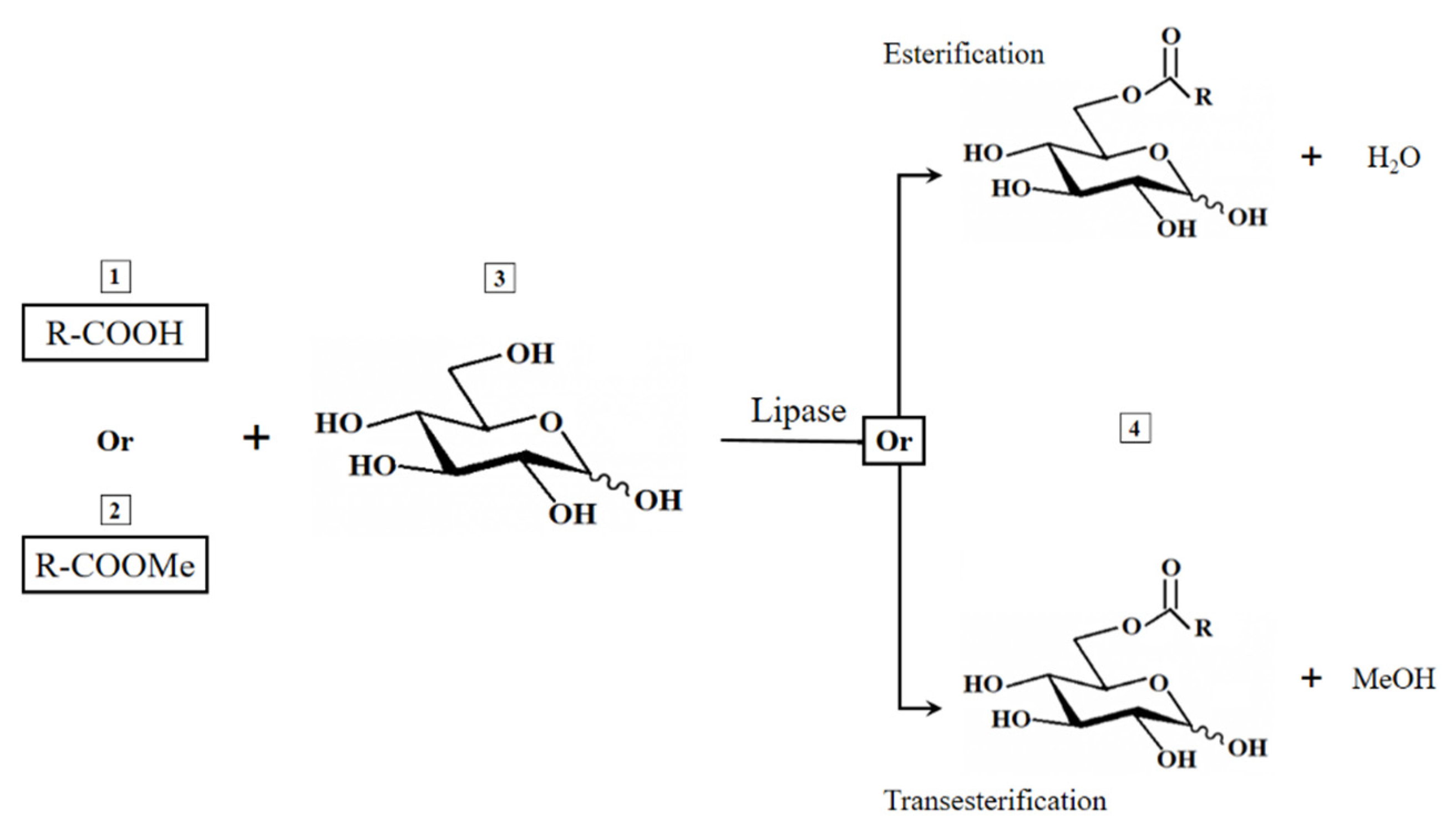
Figure 1. Enzyme-catalyzed synthesis of glucose esters (GEs) using free fatty acids (FFAs) or fatty acid methyl esters (FAMEs) as substrates.
Applsci 11 02700 g001
Figure 2. Reaction course over time of olive oil FAME conversion to glucose esters in different olive oil FAME:glucose ratios.
Applsci 11 02700 g002
Figure 3. NMR monitoring of glucose ester synthesis. The signal intensity at 4.17 and 4.41 ppm derived from the CH2OCO group increased simultaneously with a decrease in the signal intensity derived from the glucose protons of C6 at 3.39 and 3.52 ppm.
Applsci 11 02700 g003
Figure 4. Identification of the ester produced as Glc (C-6)-OCOR by 2D (1H-13C heteronuclear multiple bond correlation (HMBC)) NMR (a) and correlation between the protons of C-6 glucose ester and the carbonyl function in olive oil FAMEs (b).
Applsci 11 02700 g004
Figure 5. FTIR of olive oil FAMEs and their glucose esters (for details, see the text in Section 3.3).
Applsci 11 02700 g005
Figure 6. Reaction course over the time of conversion of FAMEs derived from different origins to glucose esters.
Applsci 11 02700 g006
Figure 7. FTIR analysis of eicosapentaenoic acid (EPA) concentrate FAMEs and their glucose esters.
Applsci 11 02700 g007
Figure 8. The larval mortality effect of GEs derived from C. echinulata, EPA concentrate, olive oil, U. isabellina and N. gaditana at different concentrations against Aedes aegypti after continuous exposure for 48 h.
Applsci 11 02700 g008
Figure 9. Effect of FAMEs and GEs on the SKOV-3 cancer cell line: flow cytometry analysis of apoptosis in SKOV-3 cells either untreated or treated with 10 µg/mL of every compound for 48 h. After the treatment period, the cells were stained with Annexin FITC and subsequently analyzed by flow cytometry. Abbreviations: C. e., C. echinulata; U. i., U. isabellina; N. g., N. gaditana; O. o., Olive oil; EPA, EPA concentrate.
Applsci 11 02700 g009
Table 1. Comparison of GE synthesis using two immobilized lipases and olive oil-derived FFAs or FAMEs as substrates at different molar ratio of the reactants.
EntrySubstrateImmobilized EnzymeOlive Oil:
Glucose Ratio
ProductConversion
(%)
1FFAsLipase CA1:1Still contains FFAs50.5 ± 2.5
21:2Still contains FFAs70.7 ± 3.5
31:3Almost 100% ester95.2 ± 4.8
4FAMEs1:1Still contains FAMEs70.1 ± 3.5
51:2Still contains FAMEs96.1 ± 3.5
61:3100% ester100.0 ± 5.0
7FFAsLipase CR1:1Still contains FFAs30 ± 1.5
81:2Still contains FFAs63 ± 3.2
91:3Still contains FFAs89 ± 4.5
10FAMEs1:1Still contains FAMEs64 ± 3.2
111:2Still contains FAMEs87 ± 4.4
121:3Still contains FAMEs92 ± 4.6
Table 2. GE synthesis using olive oil-derived FAMEs (at a FAMEs:Glc ratio of 1:3), utilizing C. antarctica (CA) lipase as catalyst at different concentrations, in a solvent mixture consisting of 80% DMSO and 20% tert-amyl alcohol and for an incubation duration 50 h.
EntryCA Lipase
(g/25 mL)
Conversion
(%)
10.1080.3 ± 4.0
20.1592.1 ± 4.6
30.2096.3 ± 4.8
40.25100.3 ± 5.0
50.30100.2 ± 5.0
Table 3. GE synthesis by CA lipase in a solvent mixture consisting of 80% DMSO and 20% tert-amyl alcohol) utilizing FAMEs of different origins as substrates and for an incubation duration of 50 h.
EntrySource of FAMEsConversion (%)
1Cunninghamella echinulata80.3 ± 4.0
2Umbelopsis isabellina85.1 ± 4.3
3Nannochloropsis gaditana86.3 ± 4.3
4EPA concentrate99.1 ± 5.0
Table 4. GEs’ antimicrobial activity using human pathogens as test organisms. Data represent the mean of three replicates ± SD of the diameter of the inhibition zones.
GEs Synthesized Using FAMEs (at 40 μg/mL) Derived From:
C. echinulataU. isabellinaN. gaditanaOlive OilEPA
Concentrate
Inhibition Zone (mm)
Escherichia coli
(ATCC 25922)
9.4 ± 0.30.010.0 ± 0.411.5 ± 0.411.1 ± 0.0
Klebsiella pneumoniae
(ATCC 700603)
10.5 ± 0.50.011.1 ± 0.410.3 ± 0.614.2 ± 0.1
Salmonella typhimurium
(ATCC 14028)
5.6 ± 0.55.8 ± 0.76.1 ± 0.310.1 ± 0.715.0 ± 0.0
Pseudomonas aeruginosa
(ATCC 15442)
6.8 ± 0.85.5 ± 0.810.5 ± 0.510.1 ± 0.413.2 ± 0.0
Bacillus subtilis
(ATCC 6633)
14.1 ± 0.50.09.0 ± 0.09.8 ± 0.717.0 ± 0.5
MRSA Staphylococcus aureus
(ATCC 4330)
11.2 ± 0.15.8 ± 1.08.5 ± 0.510.1 ± 0.216.0 ± 0.1
Staphylococcus aureus
(ATCC 25923)
12.3 ± 0.20.08.6 ± 0.312.0 ± 0.617.0 ± 0.2
Candida albicans
(ATCC 10231)
14.3 ± 0.013.3 ± 0.014.0 ± 0.015.0 ± 0.320.0 ± 0.1
Table 5. Minimum inhibitory concentration (MIC, µg/mL) and minimum bactericidal concentration (MBC, µg/mL) of the synthesized GEs against pathogens.
Source of FAMEs
Test organismsC. echinulataN. gaditanaOlive OilEPA Concentrate
MICMBCMICMBCMICMBCMICMBC
Klebsiella pneumoniae
(ATCC 700603)
25.0 ± 0.0100.0 ± 0.050.0 ± 0.0100.0 ± 0.066.7 ± 28.883.3 ± 28.833.3 ± 14.4100.0 ± 0.0
Pseudomonas aeruginosa
(ATCC 15442)
41.7 ± 14.4100.0 ± 0.050.0 ± 0.0100.0 ± 0.025.0 ± 0.083.3 ± 28.820.8 ± 7.241.7 ± 14.4
Bacillus subtilis
(ATCC 6633)
16.7 ± 7.350.0 ± 0.020.8 ± 7.3100.0 ± 0.033.3 ± 14.4100.0 ± 0.010.4 ± 3.641.7 ± 14.4
Staphylococcus aureus
(ATCC 25923)
12.5 ± 0.050.0 ± 0.050.0 ± 0.083.3 ± 28.883.3 ± 28.7100.0 ± 0.08.3 ± 3.633.3 ± 14.4
Table 6. Susceptibility of Aedes aegypti larvae to GEs under laboratory conditions by using dipping methods. Data represent the mean values of six replicates.
Source of GEsLC50
(mg/L)
Lower
Limit
Upper
Limit
RR
Cunninghamellaechinulata0.540.470.641.00
Umbelopsis isabellina39.6233.7546.1073.23
Nannochloropsis gaditana16.9212.6025.8231.27
Olive oil12.8810.0817.5423.81
EPA concentrate10.247.8414.0018.93
Publisher’s Note: MDPI stays neutral with regard to jurisdictional claims in published maps and institutional affiliations.