Article

Radiation Dose Assessment of the Fog Lead Acrylic Shields during Coronary Angiography: A Phantom Study

1
Department of Medical Imaging and Radiological Sciences, Chang Gung University, Taoyuan 33302, Taiwan
2
Department of Nuclear Medicine, Keelung Chang Gung Memorial Hospital, Keelung 20401, Taiwan
3
Department of Medical Imaging and Radiological Technology, Yuanpei University of Medical Technology, Hsinchu 30015, Taiwan
4
Division of Cardiology, Chi Mei Medical Center, Tainan 71004, Taiwan
5
Department of Medical Imaging and Radiology, Shu-Zen Junior College of Medicine and Management, Kaohsiung 82144, Taiwan
6
Department of Diagnostic Radiology, National Cheng Kung University Hospital, Tainan 70403, Taiwan
*
Author to whom correspondence should be addressed.
Appl. Sci. 2021, 11(22), 10743; https://doi.org/10.3390/app112210743
Submission received: 1 October 2021 / Revised: 12 November 2021 / Accepted: 12 November 2021 / Published: 14 November 2021

Abstract

:
This study aimed to evaluate the effects of fogging on the effectiveness of a lead glass shield in protecting an operator from radiation exposure during conventional coronary angiography (CAG). Optically stimulated luminescence dosimeters (OSLDs) were used to measure the effects of fogged lead glass shields (FLSs) and clear lead glass shields (CLSs) on the radiation doses of a cardiac catheterization surgeon. We simulated the scatter radiation incident on the operator with five angiographic projections with 10-s exposures. Experiments were conducted with a field of view of 25 cm, maximum of 100 cm between the X-ray tube and image intensifier, and 80 cm between the image intensifier and operator. Lead glass fogging had no significant effect at any angiographic projection. The average dose at the lens of the eye, thyroid glands, and gonads did not differ significantly between FLS and CLS. Although most surgeons view ceiling-suspended shields as hindrances during surgical procedures, the radiation dose at the operator’s eyes and thyroid glands increased by 13 and 10 times without the shield. The fogging of the shield is probably caused by post-surgery UV decontamination or detergents. An operator has no cause for concern regarding the radiation protection afforded by an FLS during CAG procedures.

1. Introduction

Coronary angiography (CAG) and percutaneous coronary intervention (PCI) are safe and effective surgical procedures, with an overall complication rate of only 2% [1,2]. As PCI is a form of microsurgery, it holds distinct advantages over coronary angiography. However, owing to the growing number of cardiac catheter devices and the increasing complexity of surgical procedures, cardiologists have been subjected to increasingly high radiation doses compared with ordinary radiologists [3]. Furthermore, a clinical examination can be prolonged owing to variations in patient condition, which could lead to high radiation doses in the lenses of the operators’ eyes [4].
Personal radiation protection equipment, such as lead aprons, thyroid shields and lead glasses, have become standard equipment in interventional fluoroscopy. Radiation protection accessories, such as ceiling-suspended lead shielding, table lead shielding and shielding placed on patients are also used in clinical settings. Operators often use ceiling-suspended lead acrylic shields, as there are a number of materials that must be taken in and out during cardiac catheterization, including wires, balloon catheters and stents. However, an operator may choose to remove the lead acrylic shield or avoid using certain X-ray irradiation angles during procedures that require a high level of precision, such as the insertion of a catheter through a blood vessel and patient image evaluation. This occurs most frequently when transradial artery access procedures are performed by a short operator.
Several studies have been conducted on the radiation exposure of operators (doctors, radiologists and nurses) and patients in cardiac catheterization [5,6,7,8]. According to Miller et al., the mean radiation exposure of a physician during cardiac catheterization at the eyes, thyroid glands, chest (inside the apron) and gonads is 20 mR, 2 mR–16 mR, 50 mR and 10 mR, respectively. Furthermore, eye and thyroid exposure are significantly higher during right coronary angiography than in all other diagnostic procedures [9,10]. To elucidate differences in radiation exposure between different types of cardiac catheterization, Dash et al. compared CAG to percutaneous transluminal coronary angioplasty (PTCA) in terms of completion time and radiation dosages. They found that operator radiation exposure was approximately twice as high in PTCA than in CAG; although both of these procedures have similar cineangiographic times, fluoroscopy time is more than twice as long in the former than the latter. Finci et al. compared the operator radiation exposures of CAG, single-, and double-vessel PTCA, and found that irradiation outside the lead protectors was highest in double-vessel PTCA, then single-vessel PTCA and lastly, CAG. However, no significant differences were noted between these procedures inside the lead protectors [11]. Madder et al. showed that the use of lead protectors reduces the radiation exposure of workers, operators, nurses and technical staff by approximately 66%, thus highlighting the importance of accessory lead shields [12].
Air kerma (AK), that is, the kinetic energy released per unit mass when an X-ray beam is traveling through air, is a measure of skin dose in radiology. Since the severity of the deterministic effect is directly proportional to radiation dosage, this effect may be assessed either by AK or radiation dose measurements. According to the linear no-threshold model, the probabilistic effect and risk of tissue damage increase linearly with radiation exposure. Hence, the probabilistic effect can be estimated using the dose-area product (DAP) [13,14].
The 2011 report of the International Committee on Radiological Protection (ICRP) suggested that special attention should be paid to occupational exposure in medical staff [15,16]. The report also specifically raised concerns about the deterministic and stochastic effects of radiation in personnel working in cardiac catheterization laboratories. Venneri et al. found a positive correlation between lifetime attributable risk of cancer and radiation exposure in cardiac catheterization personnel [17]. The ICRP suggested that the threshold for the deterministic effect in the lens of the eyes should be reduced to 0.5 Gy, and that the annual equivalent dose limit for occupational exposure should be reduced by a factor close to 10, i.e., 20 mSv/year [18]. In addition, the ICRP-suggested threshold implies a significantly higher degree of radiation sensitivity in the eye than previous studies [19].
OSLDs’ potential in medical dosimetry was recognized due to their excellent reproducibility, negligible angular dependence, and small size suitable for point measurements. OSLDs are increasingly used for the evaluation of radiation doses in diagnostic radiology [20,21], such as eye lens doses of medical staff during fluoroscopic examination [22] and occupational radiation dose during fluoroscopy-guided interventional procedures [23].
In this work, OSLDs were used to evaluate radiation doses in the lens of the eyes, thyroid glands, and gonads, and to compare radiation doses with fogged and clean lead glass shielding (FLS and CLS). In this way, we evaluated the effects of lead glass fogging on the radiation exposure of surgical personnel during cardiac catheterization.

2. Material and Methods

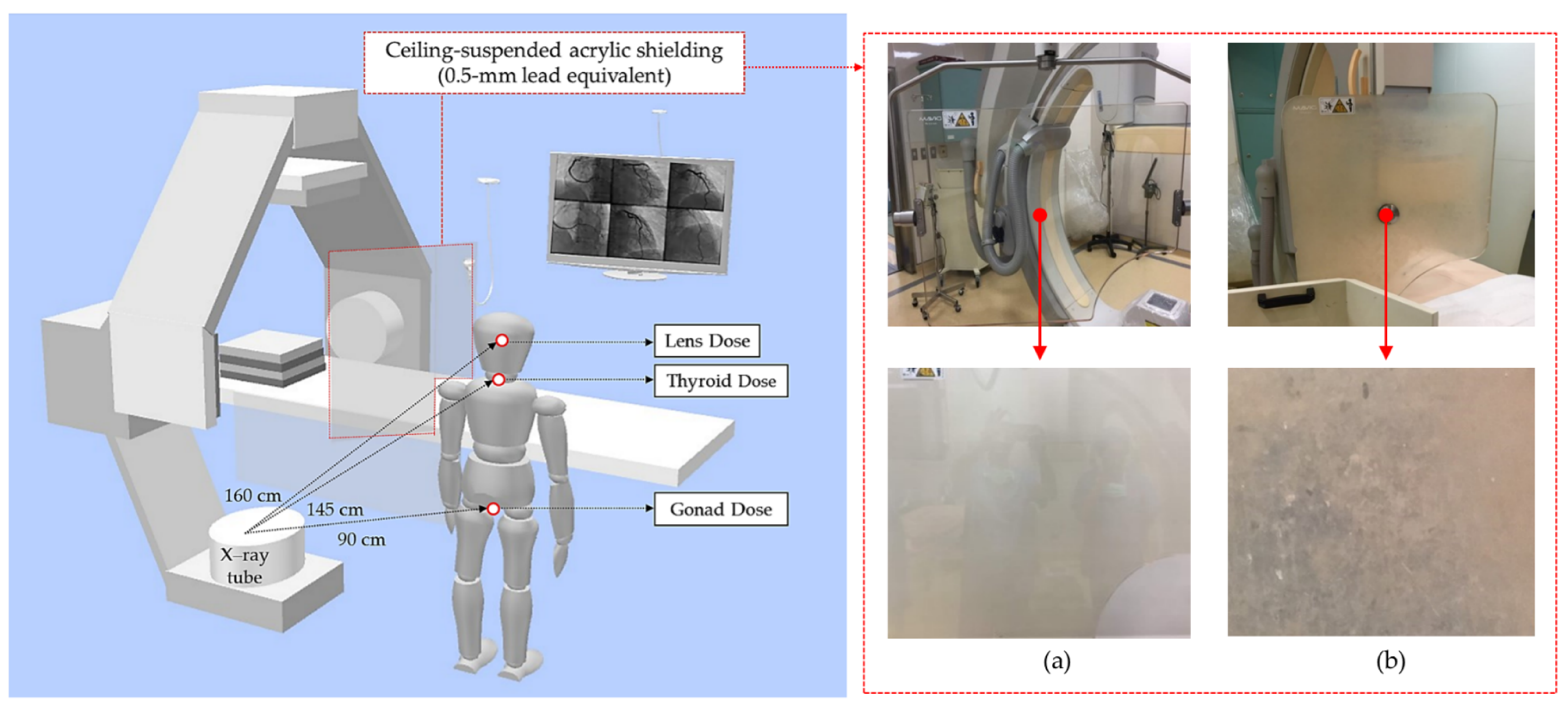
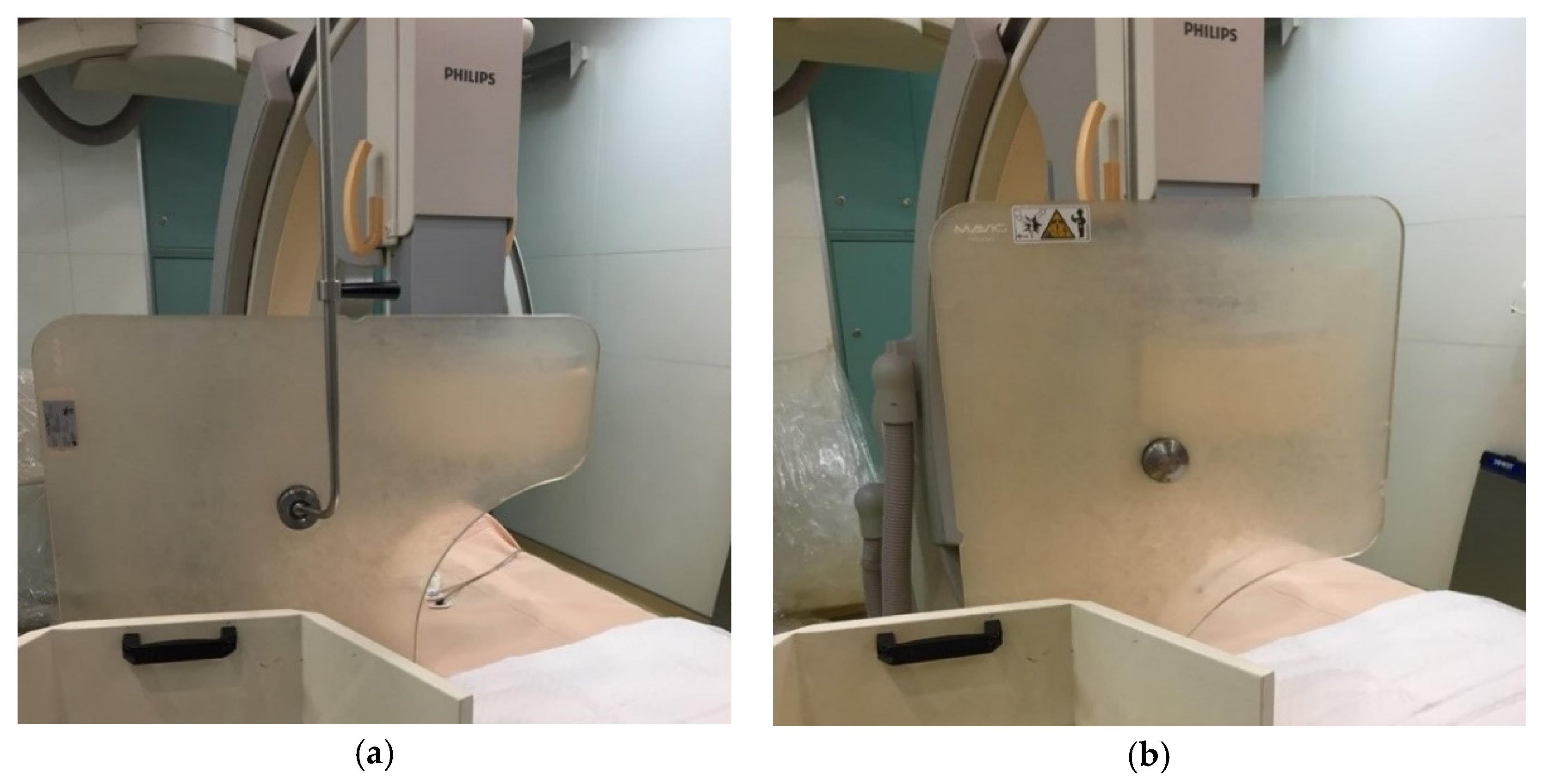
2.1. Study Design and Equipment

All of the experiments were carried out in accordance with approved guidelines. The experimental protocol of this study was approved by the Chi-Mei Medical Center Institutional Review Board without informed consent, as it only involved the use of a phantom. The X-ray system used in this study was a Philips Allura Xper FD10 (Philips Medical Systems, Best, The Netherlands) in the cardiac catheterization laboratory of Chi-Mei Medical Center, which has two X-ray fluoroscopy systems. Fluoroscopy was performed at a rate of 15 frames per second with automatic exposure control (AEC) and a full field of view, using an X-ray tube voltage of 76.50 ± 0.68 kV, tube current of 697.3 ± 21.78 mAs and a 1 mm Al + 0.1 mm Cu filter.
Five commonly used CAG projections under static conditions were used in this experiment, including: (1) caudal (CAU) 30°; (2) right anterior oblique (RAO) 10° + cranial (CRA) 30°; (3) left anterior oblique (LAO) 40° + CAU 30°; (4) LAO 60°; (5) LAO 10° + CRA 30°. The exposure time was 50 s, with each projection angle being held for 10 s. Three trials were performed. The radiation dose of each angle was recorded in terms of AK (in units of mGy) and DAP (in units of mGy∙cm2). In an X-ray system, DAP is the product between the field of view and dose of the incident radiation, and it can be used to monitor the radiation output of an X-ray system.

2.2. Radiation Dose Measurement

An acrylic phantom was placed at the examination table and made to lean against the isocenter of the C-arm X-ray machine for simulating the left chest of patients, as shown in Figure 1. The maximum distance between the two X-ray tubes and the image intensifier was 100 cm. During the image collection process, the image intensifier was placed as close as possible to the phantom. The operator measurement point was located to the right of the patient table, to simulate transradial cardiac catheterization. The linear distance between the image intensifier and operator was 80 cm. The operator radiation dose measurements were performed by measuring radiation exposure at the lens of the eyes, thyroid glands, and gonads, with and without a ceiling-suspended lead glass shield (30 × 50 cm, lead equivalent 0.5 mm, Mavig, Germany), and an FLS or CLS. The distance between the X-ray tubes and lens of the eyes, thyroid glands, and gonads was 160 cm, 145 cm, and 90 cm, respectively. A nanodot OSLD was placed at the location corresponding to each of the aforementioned organs to measure their radiation doses. Three measurements were performed at each organ, and the average radiation dose of each radiation angle was obtained by averaging the readings from three CAG procedures.
Repeated radiation dose measurements were taken with the ceiling-suspended lead glass shield being positioned in four different placements (near or far from the image intensifier, and horizontal or vertical). Regardless of whether the lead glass shield was near or far from the image intensifier, it was always placed close to the patient, at an angle of 90°. The distance between the lead glass shield and image intensifier was set to 20 cm or 70 cm, to realistically simulate a clinical setting. In the former case, the lead glass shield was close to the patient and placed in the horizontal position (see Figure 2a); in the latter, the lead glass shield was close to the operator, and the shield was either placed in the horizontal or vertical position, as shown in Figure 2b. The experimental conditions of geometrical configuration for radiation dose measurements were summarized in Table 1.

2.3. OSLDs

The dosimeters used in this experiment were the nanoDot optically stimulated luminescence (OSL) dosimeters and InLightTM MicroStar reader manufactured by Landauer, Inc., USA (Glenwood, IL, USA). The nanoDot OSL dosimeter was made of 5 mm diameter and 0.2 mm-thick plastic disks infused with Al2O3:C. The disks are encased in 10 × 10 × 2 mm3 light-tight plastic holders with a density of 1.03 g/cm3. Prior to the radiation dose measurements, the OSLDs were calibrated to minimize measurement errors. Dosimetry calibration was performed using a photon energy of 80 kV.
A MicroStar OSLD reader was used for radiation dose measurements. This device uses an LED for dose readings, which allows it to be used for repeated readouts. It also has an integrated calibration source, which is used to construct dose calibration parameters prior to usage. The reader and nanodot OSLDs are shown in Figure 3.
In this study, radiation doses were not calculated based on cumulative OSLD doses. To this end, the OSLDs were optically bleached after each measurement to clear the previous measurement. As it was shown that an incandescent light source is more effective in bleaching OSLDs than florescent lights [24], the OSLDs were illuminated for 24 h by an incandescent light to remove all residual signals. The un-radiated OSLDs were then read to obtain their baseline values.

2.4. Statistical Analysis
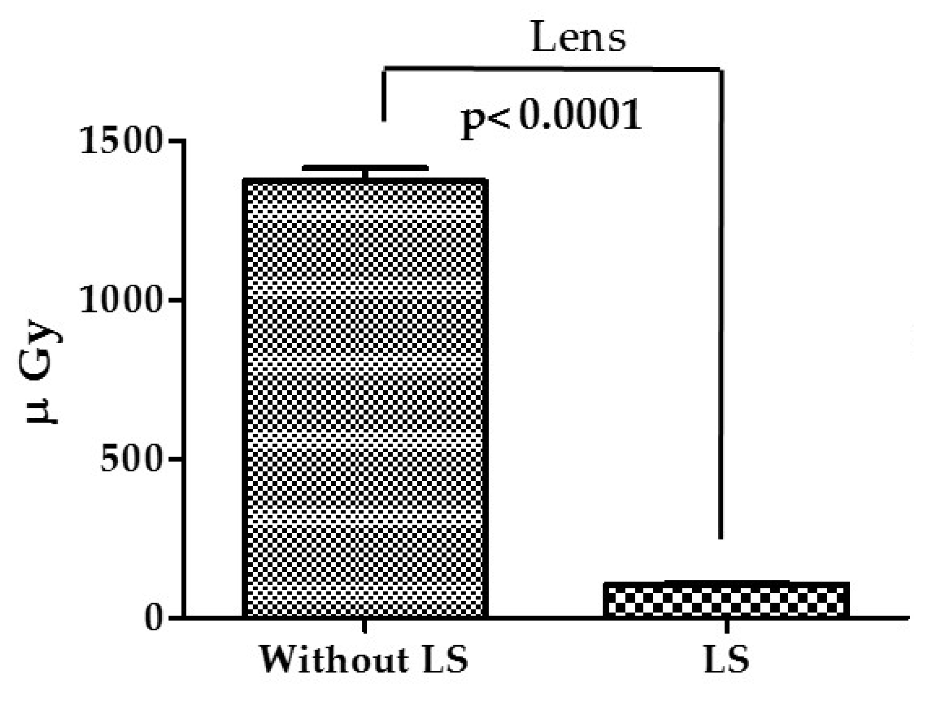
The results of this study were analyzed using paired sample t-tests in SPSS 18.0 (SPSS, Inc., Chicago, IL, USA) to compare the OSLD measurements. The confidence interval (CI) was set to 95%, and a p-value lower than 0.05 was deemed to be indicative of a significant difference between a pair of dose measurements.

3. Results

The radiation dose measurements that were taken with a FLS or CLS placed 70 cm from the image intensifier are shown in Table 2. At the lens of the eye, the average dose was 106.7 ± 5.84 μGy with a horizontal FLS and 103.0 ± 4.77 μGy with a horizontal CLS (p = 0.63). The average doses with a vertical FLS or CLS were 109.3 ± 4.45 μGy and 104.7 ± 3.76 μGy, respectively (p = 0.43). Therefore, neither placement (vertical or horizontal) nor the type of lead glass shield (FLS or CLS) had a significant effect on the reduction of radiation doses at the lens of the eye. The average radiation doses of the thyroid gland with a horizontal FLS or CLS were 91.33 ± 3.02 μGy and 91.00 ± 3.30 μGy (p = 0.94), respectively, and 97.00 ± 2.45 μGy and 95.00 ± 2.61 μGy with a vertical FLS or CLS (p = 0.78). These measurements show that the placement or type of the lead glass shield do not significantly affect the reduction of radiation doses at the thyroid gland. At the gonads, the average radiation dose was 134.7 ± 4.08 μGy and 132.7 ± 3.16 μGy with a horizontal FLS or CLS (p = 0.61), and 135.30 ± 3.42 μGy and 133.3 ± 2.73 μGy with a vertical FLS or CLS (p = 0.65). Likewise, the radiation doses at the gonads was not significantly affected by the type of lead glass shielding or its placement.
The radiation dose measurements that were taken with ceiling-mounted FLS or CLS placed 20 cm from the image intensifier are shown in Table 3. The average radiation dose at the lens of the eye was 106.7 ± 5.83 μGy and 104.0 ± 4.64 μGy with a horizontal FLS or CLS (p = 0.72). The average radiation dose measurements at the thyroid gland with a horizontal FLS or CLS were 89.67 ± 2.93 μGy and 88.0 ± 4.35 μGy (p = 0.75). The average radiation doses of the gonads were 134.7 ± 4.08 μGy and 132.3 ± 3.09 μGy (p = 0.61) with a horizontal FLS or CLS. As before, the type of lead shielding had no significant effect on the ability to reduce radiation doses at the lens of the eye, thyroid glands, and gonads. On the whole, the radiation doses at the gonads are largest, followed by eye lens and thyroid glands during a CAG procedure. The main reason is that the used fluoroscopy system has two X-ray tubes: one of the X-ray tubes is under the operating table and another is in the diagonal top of the operating table for the CAG projections. Such a geometrical setup makes the operators’ lower extremities and heads exposed to higher radiation doses.
The average radiation doses of an operator with and without lead-glass shielding were 1373.0 ± 41.37 μGy and 103.0 ± 4.77 μGy, which are significantly different (p < 0.001). Compared to having a lead shield, not having a lead shield increased the radiation dose at the lens of the eye by approximately 13 times. Furthermore, the p-value was less than 0.001, which is indicative of a highly significant difference (see Figure 4).
AK and DAP values that were measured with different angiographic projections were reported in Table 4. DAP represents the radiation dose at the output of the X-ray tube, while AK represents the skin dose. Between the angiographic projections, it was found that LAO 40 CAU 30 led to the highest radiation dose (DAP = 652.1 ± 3.31 cGy·cm2 and AK = 117.30 ± 0.58 mGy). This is mainly because of the large area of irradiation associated with this angle. The radiation dose measured with LAO 60 is close to that of the LAO 40 CAU 30 angle, with its DAP and AK being 633.6 ± 5.62 cGy·cm2 and 114.1 ± 1.02 mGy, respectively. The angle associated with the lowest radiation dose was LAO 10 CRA 30; since this angle has a small angle of rotation, the irradiated area was also small, which resulted in a low radiation dose.

4. Discussion

As per ICRP guidelines, the occupational exposure limits of radiology personnel are meant to be “as low as reasonably achievable”, so that personnel at risk of high radiation exposure will take special care when using radiation. The standard radiation protectors used in interventional radiology include 0.5 mm-thick lead aprons and thyroid collars. Radiation shields are also used whenever there is any potential for exposure to radiation. In a clinical setting, it may be difficult to determine the optimal placement for a conventional ceiling-suspended protector to maximize radiation protection. Furthermore, the sterile drapes that are necessary for surgical procedures also obstruct vision, and there are many procedures where tube angle problems can only be resolved by performing a series of complex operations. When the lead glass shield is place closed to the operator, it can provide adequate protection to the operator during a CAG procedure; as compared with not having the lead glass shield, this reduces the radiation exposure of the operator by 13 times. However, the optimal placement for a ceiling-suspended lead glass shield remains unclear. Fetterly et al. suggested that the ceiling-mounted shield should be placed far from the radiation source, so as to maximize protection during AP projection [25]. Koukorava et al. on the other hand, suggested that the shield should be placed close to the patient to maximize the size of the “radiation shadow” [26]. Although the lead glass shields are an important form of radiation protection, there are operational limits that prevent operators from using these shields frequently or effectively. Previous studies have shown that operators who do not use lead shields will reach their annual dose limits very quickly, especially medical personnel who are involved in a large number of surgical procedures.
The radiation dose measurements that were obtained in this study provide detailed information about the spatial distribution of radiation exposures on the body of an operator, with a variety of angiographic projections. Cardiac catheterization surgeons should be aware of the substantial differences in radiation exposure that may arise from changes in projection, for their own safety. Therefore, the development of an optimal shielding strategy to minimize occupational radiation exposure is a matter of utmost importance [27]. In this work, a phantom was used as the analogue of an operator, so as to obtain the baseline radiation exposure of the operator at each angiographic projection. We found that lead glass shielding always significantly reduces radiation exposure at the eyes, regardless of its placement (vertical or horizontal, near or far from the operator). Between the five commonly used projection angles that were explored in this study, the CAU 30° and LAO 40° + CAU 30° projections produced the highest radiation doses. Therefore, the operator must use a lead shield when employing these projections or use a different projection to avoid unnecessary radiation exposure. In a clinical setting, the operator may move towards or away from the radiation source, depending on the position they are using and the rotation of the X-ray arm. As long as the ceiling-suspended lead glass shield is carefully kept between the operator and the radiation source, the changes in radiation exposure due to changes in operator position will then become negligible. Therefore, lead glass shields may be expected to provide substantial protection from radiation exposure.
It is well-known that radiation dosage and the spatial distribution of radiation can depend on body type. Therefore, additional studies regarding clinical processes may afford useful insights. As for lens cataracts caused by acute radiation exposure, recent studies based on long-term tracing have shown that the threshold for radiation cataractogenesis is 0.5 Gy. Although the careful placement of radiation protectors can provide adequate protection against radiation during CAG procedures, many doctors focus on problems arising from the procedure itself, especially in the case of complex coronary artery disease or chronic total occlusion. Evidently, eventual radiation damage is difficult to detect initially. Therefore, radiation protection equipment must be enhanced, and the radiation exposure of operators as well as patients must be minimized.

5. Conclusions

Although most operators view ceiling-suspended lead shields as hindrances during surgery, radiation exposure at the operator’s eyes decrease by 13 times with the use of a lead shield (1373.0 ± 41.37 μGy vs. 103.0 ± 4.77 μGy). Moreover, the fogging of lead shields possibly from post-surgery UV decontamination or detergents could cause subjective doubts of the shielding effect. Our results show no statistically significant difference for reducing the radiation dose of the operator using FLS and CLS. This finding supports that the operator does not need to be concerned with the effectiveness of an FLS in blocking radiation during CAG procedures. To further reduce the radiation dose of the operator, we will investigate alternative materials of the shielding such as bismuth lead borate glass in future work.

Author Contributions

Conceptualization, H.-H.L. and C.-Y.T.; methodology, L.-H.L. and C.-Y.T.; validation, K.-T.T.; formal analysis, L.-H.L.; data curation, K.-T.T. and C.-S.L.; writing—original draft preparation, H.-H.L. and L.-H.L.; writing—review and editing, C.-Y.T. All authors have read and agreed to the published version of the manuscript.

Funding

The work was financed by Ministry of Science and Technology of Taiwan (Project No. 110-2314-B-182-032-). HHL was supported by Chang Gung Memorial Hospital (Project No. CMRPD1K0442, BMRP14). We would like to thank the Particle Physics and Beam Delivery Core Laboratory of the Institute for Radiological Research, Chang Gung University/Chang Gung Memorial Hospital, Linkou for their assistance.

Institutional Review Board Statement

Not applicable.

Informed Consent Statement

Not applicable for studies not involving humans.

Data Availability Statement

No new data were created or analyzed in this study. Data sharing is not applicable to this article.

Conflicts of Interest

The authors declare no conflict of interest.

References

  1. Heer, T.; Hochadel, M.; Schmidt, K.; Mehilli, J.; Zahn, R.; Kuck, K.H.; Zeymer, U. Sex differences in percutaneous coronary intervention—insights from the coronary angiography and PCI registry of the German Society of Cardiology. J. Am. Heart Assoc. 2017, 6, e004972. [Google Scholar] [CrossRef]
  2. Navarese, E.P.; Schulze, V.; Andreotti, F.; Kowalewski, M.; Kołodziejczak, M.; Kandzari, D.E.; Valgimigli, M. Comprehensive meta-analysis of safety and efficacy of bivalirudin versus heparin with or without routine glycoprotein IIb/IIIa inhibitors in patients with acute coronary syndrome. JACC: Cardiovasc. Interv. 2015, 8, 201–213. [Google Scholar] [CrossRef]
  3. Dowling, A.; Gallagher, A.; O’connor, U.; Larkin, A.; Gorman, D.; Gray, L.; Malone, J. Acceptance testing and QA of interventional cardiology systems. Radiat. Prot. Dosim. 2008, 129, 291–294. [Google Scholar] [CrossRef]
  4. Vano, E.; Ubeda, C.; Miranda, P.; Leyton, F.; Durán, A.; Nader, A. Radiation protection in pediatric interventional cardiology: An IAEA PILOT program in Latin America. Health Phys. 2011, 101, 233–237. [Google Scholar] [CrossRef]
  5. Andreassi, M.G.; Piccaluga, E.; Guagliumi, G.; Del Greco, M.; Gaita, F.; Picano, E. Occupational health risks in cardiac catheterization laboratory workers. Circ. Cardiovasc. Interv. 2016, 9, e003273. [Google Scholar] [CrossRef] [Green Version]
  6. Khaleghi Fard, A.; Alian, A.H.M.; Pourafkari, L.; Ghojazadeh, M.; Tarighatnia, A.; Farajollahi, A. Impact of pelvic and rad-board lead shields on operator and patient radiation dose in trans-radial coronary procedures. Radiat. Prot. Dosim. 2019, 187, 108–114. [Google Scholar] [CrossRef] [PubMed]
  7. Chida, K.; Kaga, Y.; Haga, Y.; Kataoka, N.; Kumasaka, E.; Meguro, T.; Zuguchi, M. Occupational dose in interventional radiology procedures. Am. J. Roentgenol. 2013, 200, 138–141. [Google Scholar] [CrossRef]
  8. Kim, K.P.; Miller, D.L.; Balter, S.; Kleinerman, R.A.; Linet, M.S.; Kwon, D.; Simon, S.L. Occupational radiation doses to operators performing cardiac catheterization procedures. Health Phys. 2008, 94, 211–227. [Google Scholar] [CrossRef]
  9. Miller, S.W.; Castronovo, F.P., Jr. Radiation exposure and protection in cardiac catheterization laboratories. Am. J. Cardiol. 1985, 55, 171–176. [Google Scholar] [CrossRef]
  10. Dash, H.; Leaman, D.M. Operator radiation exposure during percutaneous transluminal coronary angioplasty. J. Am. Coll. Cardiol. 1984, 4, 725–728. [Google Scholar] [CrossRef] [Green Version]
  11. Finci, L.; Meier, B.; Steffenino, G.; Roy, P.; Rutishauser, W. Radiation exposure during diagnostic catheterization and single-and double-vessel percutaneous transluminal coronary angioplasty. Am. J. Cardiol. 1987, 60, 1401–1403. [Google Scholar] [CrossRef]
  12. Madder, R.D.; LaCombe, A.; VanOosterhout, S.; Mulder, A.; Elmore, M.; Parker, J.L.; Wohns, D. Radiation exposure among scrub technologists and nurse circulators during cardiac catheterization: The impact of accessory lead shields. JACC Cardiovasc. Interv. 2018, 11, 206–212. [Google Scholar]
  13. Patel, C.; Grossman, M.; Shabanova, V.; Asnes, J. Reducing radiation exposure in cardiac Catheterizations for congenital heart disease. Pediatric Cardiol. 2019, 40, 638–649. [Google Scholar] [CrossRef]
  14. Hirshfeld, J.W.; Balter, S.; Brinker, J.A.; Kern, M.J.; Klein, L.W.; Lindsay, B.D.; Weitz, H.H. ACCF/AHA/HRS/SCAI clinical competence statement on physician knowledge to optimize patient safety and image quality in fluoroscopically guided invasive cardiovascular procedures: A report of the American College of Cardiology Foundation/American Heart Association/American College of Physicians Task Force on Clinical Competence and Training. J. Am. Coll. Cardiol. 2004, 44, 2259–2282. [Google Scholar] [PubMed] [Green Version]
  15. Stewart, F.A.; Akleyev, A.V.; Hauer-Jensen, M.; Hendry, J.H.; Kleiman, N.J.; Macvittie, T.J.; Wallace, W.H. ICRP publication 118: ICRP statement on tissue reactions and early and late effects of radiation in normal tissues and organs–threshold doses for tissue reactions in a radiation protection context. Ann. ICRP 2012, 41, 1–322. [Google Scholar] [CrossRef]
  16. Pantelić, G.; Čeliković, I.; Živanović, M.; Vukanac, I.; Nikolić, J.K.; Cinelli, G.; Gruber, V. Qualitative overview of indoor radon surveys in Europe. J. Environ. Radioact. 2019, 204, 163–174. [Google Scholar] [CrossRef]
  17. Venneri, L.; Rossi, F.; Botto, N.; Andreassi, M.G.; Salcone, N.; Emad, A.; Picano, E. Cancer risk from professional exposure in staff working in cardiac catheterization laboratory: Insights from the National Research Council’s Biological Effects of Ionizing Radiation VII Report. Am. Heart J. 2009, 157, 118–124. [Google Scholar] [CrossRef] [PubMed] [Green Version]
  18. UNSCEAR (United Nation Scientific Committee on the Effects of Atomic Radiation Report). Sources and Effects of Ionizing Radiation, Report to the General Assembly; United Nations: New York, NY, USA, 2008. [Google Scholar]
  19. Rajabi, A.B.; Noohi, F.; Hashemi, H.; Haghjoo, M.; Miraftab, M.; Yaghoobi, N.; Khabazkhoob, M. Ionizing radiation-induced cataract in interventional cardiology staff. Res. Cardiovasc. Med. 2015, 4, e25148. [Google Scholar]
  20. Kry, S.F.; Alvarez, P.; Cygler, J.E.; DeWerd, L.A.; Howell, R.M.; Meeks, S.; Mihailidis, D. AAPM TG 191: Clinical use of luminescent dosimeters: TLDs and OSLDs. Med. Phys. 2020, 47, 19–51. [Google Scholar] [CrossRef] [PubMed] [Green Version]
  21. Al-Senan, R.M.; Hatab, M.R. Characteristics of an OSLD in the diagnostic energy range. Med. Phys. 2011, 38, 4396–4405. [Google Scholar]
  22. Matsubara, K.; Yoshida, S.; Hirosawa, A.; Chusin, T.; Furukawa, Y. Characterization of Small Dosimeters Used for Measurement of Eye Lens Dose for Medical Staff during Fluoroscopic Examination. Diagnostics 2021, 11, 150. [Google Scholar] [CrossRef] [PubMed]
  23. Bohari, A.; Hashim, S.; Mustafa, S.N.M. Occupational radiation dose during fluoroscopy guided interventional procedures at Institut Kanser Negara. J. Phys. Conf. Ser. 2019, 1248, 012052. [Google Scholar] [CrossRef]
  24. Jursinic, P.A. Characterization of optically stimulated luminescent dosimeters, OSLDs, for clinical dosimetric measurements. Med. Phys. 2007, 34, 4594–4604. [Google Scholar] [CrossRef] [PubMed]
  25. Fetterly, K.A.; Magnuson, D.J.; Tannahill, G.M.; Hindal, M.D.; Mathew, V. Effective use of radiation shields to minimize operator dose during invasive cardiology procedures. JACC Cardiovasc. Interv. 2011, 4, 1133–1139. [Google Scholar] [CrossRef] [PubMed]
  26. Koukorava, C.; Farah, J.; Struelens, L.; Clairand, I.; Donadille, L.; Vanhavere, F.; Dimitriou, P. Efficiency of radiation protection equipment in interventional radiology: A systematic Monte Carlo study of eye lens and whole body doses. J. Radiol. Prot. 2014, 34, 509–528. [Google Scholar] [CrossRef] [PubMed]
  27. Jia, Q.; Chen, Z.; Jiang, X.; Zhao, Z.; Huang, M.; Li, J.; Zhuang, J.; Liu, X.; Hu, T.; Liang, W. Operator radiation and the efficacy of ceiling-suspended lead screen shielding during coronary angiography: An anthropomorphic phantom study using real-time dosimeters. Sci. Rep. 2017, 7, 1–8. [Google Scholar] [CrossRef]
Figure 1. The geometry of measurement of the coronary angiography for cardiology: (a) clean lead glass shielding (CLS); (b) fogged lead glass shielding (FLS).
Applsci 11 10743 g001
Figure 2. Placement of the ceiling-mounted lead glass shield: (a) horizontal placement; (b) vertical placement.
Applsci 11 10743 g002
Figure 3. OSLD dosimetry system: (A) nanodot OSLDs; (B) microStar reader.
Applsci 11 10743 g003
Figure 4. Radiation doses at the lens of the eye with and without lead shielding.
Applsci 11 10743 g004
Table 1. Summary of geometrical configuration for radiation dose evaluation.
PositionShieldsCAG Projections
TypesOrientation
70 cmCLSHorizontal/VerticalCAU 30°/RAO 10° + CRA 30°/LAO 40° + CAU 30°/LAO 60°/LAO 10° + CRA 30°
FLS
20 cmCLSHorizontalCAU 30°/RAO 10° + CRA 30°/LAO 40° + CAU 30°/LAO 60°/LAO 10° + CRA 30°
FLS
Note: The position represents the distance between lead shields and image intensifier.
Table 2. Organ doses of an operator with the shields placed at 70 cm away from image intensifier.
OrgansHorizontal Placementp-Value
FLS (μGy)CLS (μGy)
Lens106.7 ± 5.84103.00 ± 4.770.63
Thyroid91.33 ± 3.0291.00 ± 3.300.94
Gonad134.7 ± 4.08132.7 ± 3.160.61
OrgansVertical Placementp-Value
FLS (μGy)CLS (μGy)
Lens109.30 ± 4.45104.7 ± 3.760.43
Thyroid97.00 ± 2.4595.00 ± 2.610.78
Gonad135.30 ± 3.42133.3 ± 2.730.65
FLS: fogged lead glass shielding; CLS: clean lead glass shielding.
Table 3. Organ doses of an operator with the shields placed at 20 cm away from image intensifier.
OrgansHorizontal Placementp-Value
FLS (μGy)CLS (μGy)
Lens106.7± 5.83104.0 ± 4.640.72
Thyroid89.67 ± 2.9388.0 ± 4.350.75
Gonad133.2 ± 2.03132.3 ± 3.090.65
FLS: fogged lead glass shielding; CLS: clean lead glass shielding.
Table 4. Dose-area product (DAP) and air kerma (AK) measurements for each angle of projection.
RotationAngulationDAP (cGy·cm2)AK (mGy)
LAO 60° 633.6 ± 5.62114.1 ± 1.02
LAO 10°CRA 30°299.6 ± 1.8541.96 ± 0.26
CAU 30°490.0 ± 0.8278.44 ± 0.13
RAO 10°CRA 30°402.7 ± 9.0165.81 ± 0.71
LAO 40°CAU 30°652.1 ± 3.31117.30 ± 0.58
Publisher’s Note: MDPI stays neutral with regard to jurisdictional claims in published maps and institutional affiliations.

