Review

Carbon Nanotubes-Based Hydrogels for Bacterial Eradiation and Wound-Healing Applications

1
Department of Biosystems Engineering, Institute of Forest Science, Kangwon National University, Chuncheon 24341, Korea
2
Interdisciplinary Program in Smart Agriculture, Kangwon National University, Chuncheon 24341, Korea
3
Department of Microbiology & Biotechnology, Banglore University, Jnana Bharathi Campus, Banglore 560056, India
*
Author to whom correspondence should be addressed.
Appl. Sci. 2021, 11(20), 9550; https://doi.org/10.3390/app11209550
Submission received: 17 September 2021 / Revised: 5 October 2021 / Accepted: 6 October 2021 / Published: 14 October 2021
(This article belongs to the Special Issue Advanced Applications of Carbon Nanotubes)

Abstract

:
Biocompatible nanomaterials have attracted enormous interest for biomedical applications. Carbonaceous materials, including carbon nanotubes (CNTs), have been widely explored in wound healing and other applications because of their superior physicochemical and potential biomedical properties to the nanoscale level. CNTs-based hydrogels are widely used for wound-healing and antibacterial applications. CNTs-based materials exhibited improved antimicrobial, antibacterial, adhesive, antioxidants, and mechanical properties, which are beneficial for the wound-healing process. This review concisely discussed the preparation of CNTs-based hydrogels and their antibacterial and wound-healing applications. The conductive potential of CNTs and their derivatives is discussed. It has been observed that the conductivity of CNTs is profoundly affected by their structure, temperature, and functionalization. CNTs properties can be easily modified by surface functionalization. CNTs-based composite hydrogels demonstrated superior antibacterial potential to corresponding pure polymer hydrogels. The accelerated wound healing was observed with CNTs-based hydrogels.

1. Introduction

The annual wound care costs are estimated to be several billion dollars, which constitute approximately 2–3% total expenditure on health [1]. Wound healing is one of the most complex and essential treatments in the human body. Skin injury also occurs in daily life, where the skin loses its protective action, leading to the formation of a wound in the skin [2]. Multiple cell types are required within the damaged skin layers to accelerate the healing process, such as hemostasis/inflammation, angiogenesis, proliferation, and remodeling. Several therapies are available for the wound-healing process [3]. These therapies include both conventional and modern treatments. Some examples of modern and currently used treatments are stem cell therapy [4], oxygen therapy [5], nitric oxide therapy [6], artificial dressing [7], and growth factor therapy [8]. Comparatively, conventional treatments include using natural substances such as plant extracts, honey, propolis, larvae, etc. [9]. The treatments are effective when the healing materials are fabricated with excellent wound-healing potential. Different materials are developed for this process; still, few reports are available on more effective therapies [2,10]. The incorporation of nanomaterials with biocompatible polymers has been an emerging area for wound-healing application. Commonly, two types of approaches are used where nanomaterials either act as drug or vehicle to deliver the drug. Materials such as silver, gold, zinc, gold copper, titanium, and terbium are used in the form of nanoparticles to act as a drug. Whereas, in another approach, nanomaterials are used to deliver antibiotics, growth factors, nucleic acids, and antioxidants [11]. The design of effective materials for rapid wound healing is emerging, and nanomaterials-based hydrogels exhibited an improved healing process [12,13]. The utilization of inorganic nanomaterials for biomedical applications is an emerging research field. This research topic received significant attention from the researcher community to develop nanocarrier devices for medical applications. Nanomaterials (NM) are classified into four classes: carbon-based NM, inorganic-based NM, organic-based NM, and composite-based NM [14]. Several studies have investigated the development of inorganic conductive materials in combination with non-conductive polymers to support the proliferation and electro-stimuli responsive cell activities [2]. These materials are mainly carbon-based (CNTs and graphene oxide) and metals, which acquired the potential properties for biomedical applications. Since the last decade, CNTs-based nanomaterials have received significant interest from the scientific community and have been widely studied for broad research topics due to their excellent electrical, electronic, and physicochemical properties [15] for electrode materials, biomedical applications, biosensors, bio imaging, drug delivery, tissue engineering, wound healing, sorption materials, and catalysis [16]. Various CNTs composites and their biological applications are listed in Table 1. CNTs-based materials also exhibited antibacterial properties. Antibacterial materials can kill or suppress the growth of bacteria. CNTs are an allotrope of carbon and have high electrical and thermal conductivity and superior mechanical properties [17].
CNTs-based materials can be produced at a large scale that could be an additional advantage in developing industrial-scale biomedical devices [17,18]. Several studies have been conducted to explore the potential of CNTs-based materials in biomedical applications, drug delivery, tissue engineering, cancer therapies, implantable devices such as nanosensors, nanorobotics, antibacterial, and wound dressing [17,19,20]. However, there is broad scope for the utilization of CNTs-based materials in other clinical applications. In the wound-healing process, electro-conductive composites of CNTs and non-conductive polymeric materials have been efficiently utilized. The combination of intelligent hydrogels and CNTs was very effective in wound dressing and antibacterial treatment [21,22].
Table 1. List of CNTs-based composites and their biological applications.
CNT CompositeHighlightBiological ApplicationReference
CP@CNTExamine CNT as a nanocarrier of drug CPCyclophosphamide, anticancer drug delivery, reduced side effects[23]
CNT-Alg-Ch-FAPenetration of functionalized CNT through cell membraneDoxorubicin hydrochloride delivery for cancer treatment[24]
CNT-UHMWPECNT incorporation showed high mechanical and tribological propertiesSustained release (up to 429 h) of gentamicin [25]
CNT-3H2PO4Binding energy of drug to CNT increased with more H2PO4moeitiesDelivery for anti-osteoporosis zolendronate and risedronate drugs[26]
PLGA-CNT-PDA-lamPDA modified scaffold can adhere laminin for longer time and promote neurite outgrowthEnhancement of PC12 cells for nerve tissue engineering[27]
Rh-CNTAnalysis of gases of lung cancer (C6H6 and C6H7N)Biosensor for prediagnosis of lung cancer[28]
CP-cyclophosphamide; Alg-alginate; Ch-chitosan; FA-folic acid; UHMWPE-ultra-high molecular weight polyethylene; PLGA-polylactide-co-glycolide; PDA-polydopamine; lam-laminin; PC12-adrenal phaeochromocytoma cell line; Rh-rhodium.
Hydrogels belong to the group of biomaterials composed of a cross-linked polymer network to form 3D structures that can hold large proportions of water and retain their primary system even after swelling of the wound [21,29,30]. Nanomaterials-based hydrogels are considered an attractive platform for wound-healing applications [13]. These hydrogels demonstrated superior mechanical strength, antioxidant, antibacterial, electrical, and tissue regeneration potentials [31,32,33]. The physicochemical properties of nanomaterials-based hydrogels are profoundly affected by the surface functionalizations [22]. The appealing properties of multifunctional hydrogel make it a promising candidate for wound healing, antibacterial treatment, and other biomedical applications.
This review describes the preparation of CNTs-based hydrogels and their potential applications for skin dressing and antibacterial. We briefly discussed the functionalizations and conductivity of CNTs. The perspective for the possible future directions in developing CNTs-based hydrogels for wound-healing treatment is also summarized. Figure 1 shows the overview of the review article, which includes types of CNT, properties of CNT hydrogels, and their applications.

2. Conductive Properties of CNTs

CNTs are an allotropic form of carbon, as mentioned by Sumiolijima in 1991 [38], which have similar properties as graphene. Structurally, CNTs are cylindrical structures composed by the rolling of graphene sheets with sp2 hybridization. Arc discharge, laser ablation, and chemical vapor deposition (CVD) are commonly utilized to prepare CNTs [39]. In arc discharge and laser ablation methods, the carbon sourced was treated at 3000–4000 °C to generate cylindrical CNTs, whereas the CVD technique involves the pyrolysis of carbon source at a temperature range of 600–1100 °C. The physicochemical properties of the obtained CNTs are widely influenced by synthetic methods [40]. CNTs exhibit remarkable thermal properties due to their structural form and technique of synthesis. The conductivity range of CNTs can vary from 6000 to 0.1 W/mK depending upon the single-walled structure and multi-walled structure, respectively [41,42]. The thermal conductivity is due to the collective vibration of atoms, including phonon and electron transfer [43,44]. The length of CNTs also affects the conductivity [44,45]. Therefore, the optimization of synthetic parameters is required to obtain a certain level of thermal conductivity [46]. Berber et al., studied molecular dynamics simulation to determine the thermal conductivity (k = 6600 W/mK) of CNTs based on Tersoff–Brenner potential, which is similar to a hypothetical isolated graphene monolayer [47]. In comparison, Osman et al., studied the relationship between the physical parameters of CNTs and their thermal conductivity. They examined that the thermal conductivity of single-walled carbon nanotubes (SWCNTs) changes with the temperature. A decrease in the thermal conductivity of armchair (10,10) configured SWCNTs was observed above 400 K, similar to monolayered graphene. The CNTs with similar diameters but different chirality show maximum conductivity at 300 K, and the armchair CNTs have a comparatively sharper peak than zigzag CNTs [48]. Table 2 shows various properties of single-walled carbon nanotubes (SWCNT) and multi-walled carbon nanotubes (MWCNT).

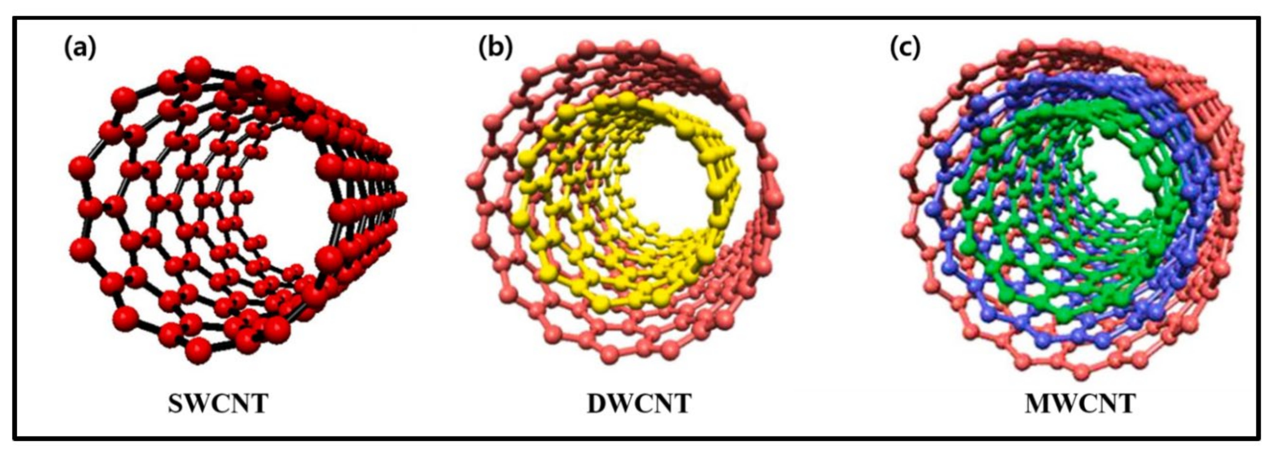
2.1. Single-Walled and Multi-Walled Carbon Nanotubes

Based on the number of inner layers, CNTs are categorized into single-walled CNTs (SWCNTs) and multi-walled CNTs (MWCNTs) [50,51]. CNTs properties are varied with the number of inner layers. Different types of CNTs are presented in Figure 2. SWCNTs are composed of a single layer of a graphene sheet and have 1–2 nm diameters. The controlled catalytic conditions are required to obtain the high quality and purity of SWCNTs. Doubled-walled CNTs (DWCNTs) are generated from two distinct CNTs of inner and outer tubes. The diameter of the outer and inner tubes is approximately 2–4 nm and 1–3 nm, respectively. DWCNTs have similar properties to SWCNTs, including narrow diameter, electrical, and mechanical properties. MWCNTs are comprised of multilayer graphene sheets rolled with a diameter of 2–50 nm; the outer layer dimeters can reach up to 100 nm, and the inner layer diameter is below 1 nm. SWCNTs are classified into structural models such as the Russian doll and Parchment model, depending on the arrangement of the graphene sheet in the concentric cylinder [52]. MWCNTs have defects in their structure, which favor the surface functionalizations, and these defects are increased with increasing the number of graphene sheets rolled up in MWCNTs. MWCNTs are considered potential materials for biomedical applications because of their excellent chemical, thermal, mechanical, electrical, and variable structural properties and functionalities. Additionally, CNTs show semiconducting and superconducting electron transport properties. The biocompatibility, antimicrobial, and antibacterial properties of CNTs are moderately attractive for their use in wound dressing and antibacterial treatment [13].

2.2. Functionalization of CNTs

Nanoparticles and their structure with intrinsic chemical compositions would enhance the surface properties such as variable charge, dispersion, dissolutions, hydrophilicity, hydrophobicity, etc. Many studies and techniques are available for the functionalization of CNTs materials [53,54,55]. The functionalization of CNTs can be categorized as covalentand non-covalent strategies [51]. The covalent modification adds chemical bonds on CNTs by chemical reactions and conjugates hydrophilic molecules or polymers, which increases the solubility of CNTs. Whereas, in non-covalent modification, the hydrophobic behavior of CNTs is used for coating amphiphilic molecules. Thus, the desirable functionalization of CNTs is not a difficult task, which is beneficial for desirable biological or biomedicine feasibility [17,50]. The covalent functionalization generates more hydroxyl and carboxylic groups on the surface of CNTs. The non-covalent functionalization has been performed through the adsorptions of desired moieties, including polymers, proteins, DNA, carbohydrates, and hydrogels with CNTs [20]. Most of these surface modification techniques are helpful for the utilization of functionalized CNTs material in biomedical applications.
The functionalized moieties can be precisely fabricated on CNTs’ surface cell receptors that can go through their internalization. Despite the vast advantage of CNTs, some concerns are to be addressed before biomedical applications. These concerns are (i) the purity of CNTs and their structural reproducibility in large scale synthesis, (ii) the physicochemical and structural properties of CNTs are varied significantly vary with synthesis, purification, and functionalization methods, (iii) the co-operation between the biological environment and CNTs are not always predictable and very complex, (iv) the toxicity of CNTs is a most critical issue, which depends on the synthesis methods and modifications, (iv) the design and synthesis of desired CNTs-based material on a large scale, and (v) hydrophobicity [17]. In addition, they suggested functionalization strategies that can help in minimizing systemic toxicity and inflammation. Usually, CNT materials have been utilized to combine with hydrogels (non-conductive polymers) to form electroactive composite dressings for wound-healing treatment [12]. The combination of CNT and hydrogel renders excellent electro-conductive properties in the resultant composites, which can effectively transmit electrical stimulation to the wounded tissue. In addition, these CNTs–hydrogel composites exhibit an antibacterial phenomenon. Hence, this promising combination could effectively heal wounds by activating local cell proliferation and migration.

3. Development of CNT-Based Conductive Hydrogels

Hydrogels comprise a 3D cross-linked polymeric network structure, holding a large amount of water and maintaining their form even after swelling. Hydrogels are similar to the extracellular matrix and can mimic the microstructure of native cellular environments and provide a moist environment [21]. It can adsorb the fluid excreting from the wound through the porous structure and prevent the growth of anaerobic bacteria by the gaseous exchange phenomenon. It can act as a barrier to prevent bacterial infections and improve epithelization and cell migration into the wound [22,56,57,58,59,60]. Hence, hydrogel materials have been broadly used for wound dressing.Conductive hydrogel has received significant attention for various biomedical applications, including wound dressings, drug delivery, biosensors, bio-imaging, and tissue engineering. The hydrogel conductivity is originated due to the presence of conductive ions and electrons. Naturallyderived biopolymers, such as chitosan, peptides, gelatin, and polyamines, are frequently explored to prepare hydrogels due to their excellent biocompatibility [29]. Recently, Zaho et al., developed injectable antibacterial hydrogels for enhanced skin generation. The injectable hydrogel has certain advantages, such as wound site filling with irregular space, wound adherence, and feasibility toward the insitu encapsulation of bioactive molecules and cells [30]. These hydrogels can be designed with advanced features such as bilayer types to control the infiltration of microbes and moistures and additional antibacterial and antimicrobial properties to protect wound sites from infections and inflammations. Different stimuli-responsive properties can be generated in hydrogel to deliver therapeutic molecules, inhibit bacterial infections, and promote cellular proliferation. Multifunctional hydrogels are an emerging approach. It is the composition of hydrogel constituent materials with or without modification and the incorporation of nanostructures in the hydrogels. These construction strategies improve properties (electrical, mechanical, surface, biocompatibility, and biodegradability) for wound-healing applications [61,62,63,64,65].
Zhang et al., summarized the literature to fabricate different types of hydrogel material with antibacterial agents for wound-healing applications [29]. They included the types of hydrogels and their specific methods of incorporations such as physical combination (incorporation, swelling diffusion, encapsulated with carrier), chemical combinations (chemical bonding, hydrogel with biomedicine activity), and incorporation of photo-assisted antibacterial hydrogels. The physical combination of antimicrobial methods has been considered a straightforward and effective method, in which hydrogel was physically incorporated with antibiotics, biological extracts, antimicrobial peptides, and inorganic nanoparticles. These materials are utilized for wound healing and other biomedical treatments. In chemical combinations, the hydrogel is combined with an antibacterial and antimicrobial agent by chemical treatments. However, these hydrogel derivatives should go through further purification steps before their utilizations. The chemical combination method has been commonly used to synthesize CNT-based hydrogels, and the most common hydrogels are chitosan-based, cationic group-based, polypeptide-based, and halogen-based hydrogels. These hydrogels exhibit strong antibacterial and antimicrobial properties, which are necessary to heal the wound [59] effectively. However, several researchers are developing new hydrogel materials to fulfill their demand in biomedical applications [29,66,67,68,69]. The following sections highlight some recent studies and developments of CNT hydrogels for wound healing and antibacterial property.
As discussed before, CNT functionalization with hydrogel is considered a potential hybrid candidate material for multiple applications in biomedical fields. CNT can be incorporated with hydrogels through their covalent and non-covalent functionalization using different treatment (chemical and mechanical) techniques [70]. Recently, Vashist et al., have nicely summarized the studies on developing CNT-based hybrid hydrogel for wound-healing applications [13]. In addition, they addressed most of the design and synthesis strategies for CNT hydrogel mixed materials and their diverse applications in the biomedical field.
CNT hydrogels are associated with most of the properties of hydrogel polymeric networks. Generally, five polymerization techniques such as (i) covalent cross-linking (insitupolymerization), (ii) exsitu polymerization, (iii) physical cross-linking, (iv) polymer grafting, and (v) smart devices enable techniques that are exploited for the synthesis of CNT hydrogel hybrid. In both insitu and exsitu polymerizations (chemical treatment), CNT hydrogel outfits with excellent mechanical strength, a variety of shapes and surfaces, excellent yields, and it is easy to control the initial and final composition of hybrid gels into the hybrid [71,72]. These techniques involve the introduction of nanofiller during reactions. In physical cross-linking techniques, hydrogels are physically cross-linked with CNT, which showed a high level of biocompatibility but low mechanical strength. These methods have been employed for the synthesis of CNT-based gelatin hydrogels [73]. The schematic representation for the synthesis of CNTs-based hydrogels is shown in Figure 3.
Polymer grafting is employed for the synthesis of CNTs grafted hydrogels. This technique provides the functionalization of the CNTs surface with a polymeric chain, which results in the shielding of the CNT surface [13,76]. This method offers a good dispersion of nanofillers along with their stronger interaction with polymers. Generally, protein and polypeptide-based CNT hydrogels have been synthesized through grafting techniques. Grafting-to and grafting-from polymerization for CNT polymers are two different grafting strategies involved in fabricating CNT hydrogels. In both approaches, the functional groups of the polymer chains are reacted with CNTs, leading to the formation of bioactive materials for biomedical applications. In addition, the intelligent device enable technique is an exciting technique that was applied to develop responsive hydrogel nanocomposites-based smart devices [77]. This method has been widely applied for the synthesis of hydrogel materials that are pH and ionic responsive. Significant research on the development of CNTs-based hydrogels has been done to obtain the desired functionality, shape, and size for specific applications.

4. CNT-Based Antibacterial Applications

It is well known that the wounded areas are more susceptible to bacterial infections. Depending upon the etiology and severity of the microbial invasion, the infections can cause minor to major damage to human life [78]. In the early stage of infection, the Gram-positive bacteria such as Staphylococcus aureus and Streptococcus pyogenes are more populated, whereas the Gram-negative bacteria, such as Escherichia coli (E. coli) and Pseudomonas aeruginosa (P. aeruginosa), are populated later nearby wounded areas [79]. Usually, infection is avoided by activating the immune system for abolishing the invading pathogens. In this process, macrophages initiate the migration to the wound site and subsequently perform phagocytosis of the pathogens (destroyed in a phagolysosome or by nitric acid production). In a later stage of infection, the immune response is performed by activating T-helper lymphocytes, which secrete interferon-γ and CD40 ligand to coordinate the immune adaptive and humoral response to kill and remove the invading bacteria [80]. In the passive stage of the immune system, infection occurs and causes the deterioration of granulation tissue, growth factors, and extracellular matrix components (collagen, elastin, and fibrin) and alters the normal wound-healing process [81,82]. Therefore, it is necessary to develop wound dressing materials to prevent bacterial penetration into the wound or reduce the microorganism’s growth [83].

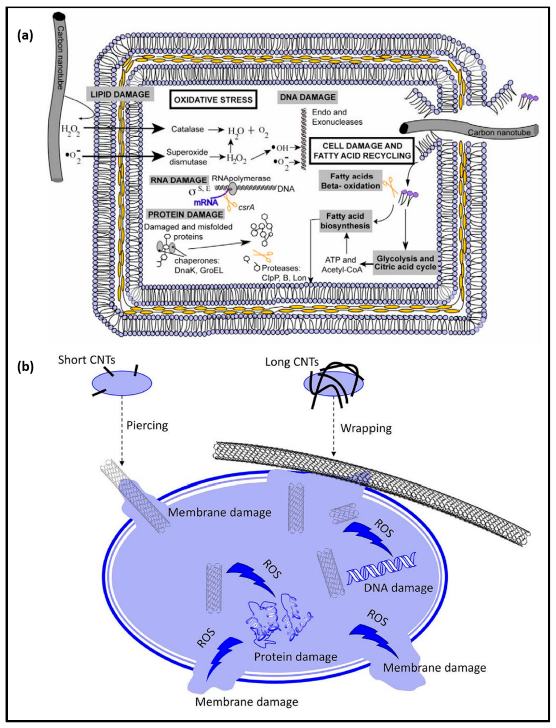
4.1. Antibacterial Activity of Pristine CNT

The antibacterial activity of CNTs has been studied widely against numerous bacteria, such as E. coli, Staphylococcus aureus, Staphylococcus emipermidis, and Bacillus subtilis. Microbes can rapidly increase their population in the wounded areas and adversely affect the wound-healing process [84]. Hence, antibacterial wound-healing patches are desirable and favorable for protecting the wound from infections and accelerating the healing process. Kang et al., demonstrated the antibacterial effect of SWCNTs against E. coli. SWCNTs showed strong antibacterial potential and damaged the bacterial cell membrane [85,86]. CNTs can directly damage the bacterial cell membrane or increase the reactive oxygen species (ROS) production, causing decrease cell viability [87]. Different conditions, such as pH temperature, retention time, and solute and solvent compositions, also affect bacterial eradication [88]. Dong et al., investigated the antibacterial properties of SWCNTs dispersed in different surfactant solutions, including sodium cholate, sodium dodecyl benzenesulfonate, and sodium dodecyl sulfate against Salmonella enterica (S. enterica), E. coli, and Enterococcus faecium. SWCNTs concentrations played a significant role in bacterial cell viability [89]. The probable mechanisms for CNTs induced are the inhibition of bacterial growth by impairing the respiratory chain; inhibition of energy metabolism; physical interaction with the cell membrane, formation of cell–CNTs aggregates, and induction of cell membrane disruption. SWCNTs have also shown excellent antimicrobial properties [90,91,92]. The size of CNTs contributes a considerable role in the deactivation of microorganisms. The smaller-sized CNTs have a larger surface-to-volume ratio, which helps to make strong bonds with the cell wall or membrane of bacteria, showing better antibacterial potential [93].
SWCNTs primarily aggregate with the cell wall, which is followed by induction of cell membrane rupture, hindering DNA replication [94]. It has also been mentioned that the surface charge of CNTs has a significant role in the inactivation of bacteria by cell membrane interruption [95]. SWCNTs have a specific surface area of approximately 407 m2/g, which can remove 3.18 × 10¹² CFU/mL [96]. Bing et al., assessed the effect of CNTs’ surface charge on bacterial death and found that positive and negative charge dots had antibacterial activity. Reactive oxygen species, such as hydroxyl radicals, are generated by the interaction of CNTs and the cell membrane, which kills the bacteria [97]. Yang et al., assessed that longer (≈5 μm) SWCNTs make better aggregation and show more robust antimicrobial activity. The different activity was observed in the solid and liquid media. In solid media, shorted (<1 μm) CNTs showed effective antimicrobial activity than the longer ones [98]. The MWCNTs with 50 μm length wrap on all sides of a microbial cell and cause osmotic lysis. Whereas, in liquid media, longer CNT are more effective in bacterial cell damage. The aggregation or interaction between CNTs and the bacterial cell membrane is unavoidable because of their particular structure and strong van der Waals forces [99]. The tube diameter of CNT also affects the antimicrobial activity. Smaller diameters entail better interactions with the cell wall, mediating determinants to the cell [88]. CNTs with a 1.5 nm diameter act as needles connected to the membrane from one side, and in diameters of 15–30 nm, CNTs are connected to the sidewalls [100]. Chen et al., demonstrated that CNTs have lower activity against Bacilli than Cocci [101]. The mechanism of these bacteriostatic properties is related to their diameter-dependent penetration and length-dependent wrapping on the disruption of cell walls and membranes of bacteria and intracellular substances such as DNA and RNA. In addition, they announced that bacterial survival duration after the direct connection with CNTs enhanced with the increasing length-to-diameter ratios with a linear coefficient >0.79 for all examined doses. Moreover, they reported that the shape, in addition to the size, of a particle can impact on the particle phagocytosis by macrophages.
The nanoscale size, shape, specific surface area, chemical composition, and surface structure of CNTs are the essential factors influencing its toxicity. It has been established that the larger the surface area, the higher the contact area with microbes, increasing the antimicrobial interaction and enhancing the killing mechanism. The dispersed CNTs exhibited significantly more toxic effects on bacterial cells than the aggregates. The inactivation of S. aureus, E. coli, and B. subtilis was increased from 30–50% to 90–100% by adding the functional groups to the surface to improve the dispersibility of the nanomaterials [102]. Additionally, the geometry of CNTs can also affect antimicrobial activity. It has been observed that SWCNTs are more practical to kill E. coli compared to MWCNTs. According to the authors, SWCNTs have better penetration capability into cell walls than MWCNTs due to their smaller diameter and high surface area, which helps for better interaction with cell surface [103]. SWCNTs with sharp diameters can easily penetrate the bacterial cell wall than MWCNTs [88]. Bacteria generate more stress-related gene products with small diameter CNTs [103]. Kang et al., performed a gene expression experiment that revealed the pathways involving membrane damaging, repairing, and lipid recycling, such as fatty acid beta-oxidation and glycolysis [103]. The fatty acid biosynthesis pathways were highly expressed in the presence of SWCNTs than MWCNTs exposure to bacteria. More lipids from the membrane were released to the media due to membrane damage/cell death in the presence of SWCNTs. In the fatty acid biosynthesis pathway, the upregulation of the synthesis of new lipid molecules occurred and was incorporated into the cells for cell growth or repair of the broken membrane. The high expression of glycolysis with SWCNTs indicates that the cells are required to produce more energy to survive the stress-related conditions. The by-products of Acetyl-CoA pathways are typically used to synthesize fatty acids to repair the membranes. Figure 4a represents a schematic summary of E. coli K21 gene expression stress response under exposure of CNT.
The generation of ROS such as O2 and OH with CNTs is also responsible for killing the bacteria [105,106]. The reactive species oxidize the fatty acids in the cell membrane and damage the cell permeability, adversely affecting the cell functions [105,107]. A schematic mechanism of antibacterial activity by ROS generation is presented in Figure 4b. The greater expression of superoxide dismutase and catalase was observed in E. coli in the presence of CNTs. The DNA damage pathways and repair systems were activated with CNTs. A similar gene expression pattern has been previously observed in E. coli cells exposed to hydrogen peroxide and superoxide. Physical membrane damage and oxidative stress are the fundamental mechanisms for the toxicity of all carbon-based nanomaterials [88,103,107,108]. Cell contact time with CNT also influences antimicrobial activity [109]. Enhanced toxicity was observed in Gram-positive and Gram-negative bacteria in the presence of SWCNTs with increasing the contact time from 1 to 3 h [110]. Significant antimicrobial effects were observed in some Gram-positive bacteria after long exposure times (36h). The antimicrobial effects of CNTs also depend on bacterial cell wall characteristics. The Gram-positive bacteria have more resistance to CNTs because of the thicker peptidoglycan cell wall than Gram-negative bacteria [111]. However, more studies are needed better to understand the different tolerances of diverse bacteria to CNTs.

4.2. Antibacterial Properties of CNT Composites

CNTs combined with nanoparticles exhibited an improved antibacterial effect. Several studies have been reported to check the antibacterial effect of CNTs composites with chitosan and silver NP [112]. Dong et al., examined that CNTs conjugated with AgNPs exhibited superior antibacterial properties against E. coli [113]. This was attributed to the more significant disruption of the cell membrane in the presence of CNTs conjugated with AgNPs. Yuan et al., observed that the deposition of AgNPs on MWCNTs with poly(amidoamine) induced bacteriostatic effects against S. aureus, E. coli, and Pseudomonas aeruginosa [114].
Castle et al., prepared the MWCNTs/AgNPs composites and evaluated their biocompatibility and antibacterial potential [115]. Enhanced biocompatibility and antibacterial efficacy were observed in the developed composites. The antibacterial property of CNTs is explored to establish nanocomposite films. CNTs/poly(L-lysine) or poly(L-glutamic acid) composite films were synthesized and their antibacterial potential against E. coli and Staphylococcus epidermidis was evaluated. The composite films showed 90% of bacterial killing efficacy [116]. Gelatin/MWCNTs composite films demonstrated improved bacterial killing properties against Gram-negative and Gram-positive bacteria [117]. An improvement in physicochemical, mechanical, and electrical properties was observed in CNTs incorporated in gelatin. Lu et al., synthesized the poly(lactic acid) (PLA)/AgNPs/CNTs composites and examined their antibacterial potential against Staphylococcus haemolyticus [118]. The developed composite showed superior antibacterial properties against Staphylococcus haemolyticus. They observed that the combination of two nano-fillers demonstrated synergistic features to the polymeric matrix. The nanocomposite also exhibited remarkable thermo-mechanical and antibacterial properties. Benigno et al., developed the low-density poly(ethylene) (LDPE)/MWCNTs nanocomposites and monitored antimicrobial activity against DH5α E. coli [119]. A significant enhancement in the antibacterial potential was observed in 1% MWCNTs-added LDPE film. They also evaluated the effects of the developed nanocomposites on biofilms. They reported that the biofilms boosted the ability of E. coli to adhere to the surface of the materials, thereby attenuating the antimicrobial properties of MWCNTs. Aslan et al., prepared the composites with a combination of SWCNTs and poly(lactic-co-glycolic acid) (PLGA), which effectively declined the viability of both E. coli and S. epidermidis [111]. Table 3 summarizes the antimicrobial effect of CNT and its composite materials.

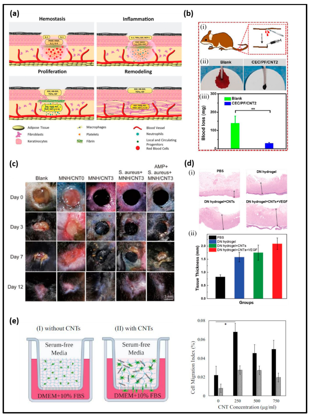
5. CNT-Based Nanocomposites for Wound Healing

CNTs enhance the biological activity of wound-healing materials due to their unique interaction pattern with biomolecules, cells, and nearby tissues [129,130]. As discussed above, wound healing is a natural physiological process that responds to any skin injury. This complicated mechanism involves sophisticated interactions between cell types, coagulation factors, connective tissue, cytokines, growth factors, and the vascular system [2,131]. The schematic wound-healing process is described in Figure 5a, where four stages of wound healing are demonstrated. These stages are hemostasis, inflammatory, proliferative, and maturation phase.
The hemostasis phase is a narrowing of the blood vessels to stop bleeding immediately after an injury to the skin tissue [132]. The blood-clotting system is activated at this stage, and a barrier is formed to prevent excessive bleeding from the injury site. In this process, platelets are attached with collagen, which results in the activation of aggregates. Inflammation is the next stage in the wound-healing process, which is initiated by releasing inflammatory mediators (prostaglandins and histamine) to prepare the wound site for growing new tissue [2,132,133]. In this stage, neutrophils (white blood cells) are introduced into the wound to kill the bacteria and remove the inflammatory mediators. Usually, one to two days are required for complete localization of these cells in the injury, which results in continuous removal of residues. The movement of protein and other factors development by these cells results in immune cells’ attention toward the wound for facilitating the repair of tissue. The next stage is the proliferative phase, where the wound site constructs a new connective tissue with extracellular matrix (ECM) and collagen [133,134]. This construction follows the contraction of wound edges, which significantly promotes wound healing.
Furthermore, the granulation process produces a new wound matrix, in which the ground substance provides a scaffold-like structure for the development of fresh blood capillaries and connective tissues. Re-epithelization is the last stage of proliferation, where the epithelial cells are transferred around the wound. This process is fast in the presence of a moist wound environment [135]. The maturation or remodeling phase is the final phase to the healing of the wound; it can last somewhere between 21 days and 2 years; the maturation period depends on the wound type and condition. In this phase, newly grown tissue becomes strong and flexible, and wound sites free from the blood capillaries, macrophages, and fibroblasts through apoptosis or undefined mechanisms [131,133]. However, this dynamic wound-healing process can be affected due to the local (infection, humidity, maceration) and systemic (age, nutritional status, body status) factors.
Generally, a wound starts with a minor injury, but it can extend without healing for longer durations, leading to a chronic wound condition. The typical chronic wounds are diabetic ulcers, venous and arterial ulcers, pressure ulcers, hypertrophic scars, fibrosis, and pressure sores. No doubt, normal wounds can turn into chronic severe injuries if some healing steps are impaired with specific features. In addition, a few factors such as diabetes, obesity, malnutrition, medication, and lifestyle habits (excessive smoking and excessive alcohol) can be a strong reason for impaired wound healing. Hence, this process is considered more dynamic than other biomedical treatment processes, which could appeal to the development of advanced devices/materials to accelerate this healing process more effectively [136].
He et al., fabricated non-antibiotic but CNT-based hydrogel for photothermal therapy of bacteria-infected skin wounds [137]. The composite was fabricated by N-carboxyethyl chitosan (CEC) benzaldehyde-terminated Pluronic F127/carbon nanotubes (PF127/CNT). The presence of CNT endowed the hydrogel with in vitro and in vivo photothermal antimicrobial activity and good conductivity. Figure 5b shows the hemostatic capability of CEC/PF/CNT2 hydrogel in the mouse tail amputation model. In a recent study, Xu et al., developed a multifunctional conductive microporous nanocomposite hydrogel (MNHs) by air-in-water emulsion [138]. The emulsion was stabilized by colloidal hybrids of CNT and gelatin methacrylate (GelMA). The hydrogels demonstrated tunable pore size, electrical conductivity, and mechanical properties with varying concentrations of CNT. Decreased pore size and increased electrical conductivity were observed with 3 wt% of CNT compared to 0 and 1% CNT added samples. The hydrogel demonstrated remarkable antimicrobial and wound-healing performance on a murine dorsal skin model. The conductivity graph and in vivo wound treatment of MNH/CNT0 and MNH/CNT3 samples is shown in Figure 5c. The presence of CNT promoted the stability of emulsion and enhanced the conductivity and mechanical property of hydrogel. Zhao et al., developed injectable antibacterial and conductive nanocomposite cryogel with similar components (CNT and GelMA) with the addition of functionalized quaternerized chitosan [30]. Their results showed that 4 mg/mL CNT composition was outstanding for hemostatic application compared to gauze. The wound-healing performance was also compared with TegadermTM film, and the fabricated cryogels showed better results in the mouse-liver injury model. In another work, CNT was incorporated with chitosan and poly(acrylamide) to synthesize a multifunctional hydrogel. The film was prepared to deliver vascular endothelial growth factor (VEGF) and check the release pattern in the presence and absence of fluorescein isothiocyanate-tagged bovine serum albumin (FITC-BSA). The presence of CNT enhanced the interaction among cells and promoted cell proliferation. Figure 5d represents the hematoxylin and eosin (H&E) staining of regenerated granulation tissues from wound sites in different groups and their statistical analysis of tissue thickness [139].
Figure 5. (a) Schematic representation of wound-healing phases in cutaneous wound—hemostasis, inflammation, proliferation, and remodeling [2], (b) (i) Scheme representation of mouse-tail amputation model, (ii) bloodstain photograph of mouse tail in blank and hydrogel group, (iii) quantitative data of blood loss (n = 5), ** p < 0.01 [137], (c) In vivo wound-healing model evaluation of MNH hydrogels and observation of treatment in presence of S. aureus infection for 12 days [138], (d) (i) H&E staining of different groups showing the progress in thickness of skin, (ii) respective statistical analysis of average connective tissue thickness in different groups [139], (e) Schematic of chemoattractant-induced cell migration assay using a modified Boyden Chamber experiment. The addition of CNT increased the porosity of the hydrogel and cell migration (left). The average cell migration index for the samples with varying concentration of CNT with (solid pattern) and without (diagonal pattern) chemoattractant. * p < 0.05 corresponds to a significant difference (right) [35].
Applsci 11 09550 g005
He et al., developed a nitrogen-doped CNT encapsulating cobalt (N-CNTs@Co) nanoparticles for antibacterial wound-healing application [140]. The nanocomposite showed oxidase-mimicking activity, which could produce a large number of ROS in an acidic environment. The generation of ROS exhibited good antibacterial activity against Gram-positive Staphylococcus and Gram-negative Escherichia coli. To kill the bacteria, the ROS damaged the bacterial membrane and degraded its DNA. After the treatment of the N-CNTs@Co nanocomposite, the bacteria-infected wound shrank faster than the control group. The H&E analysis confirmed the healing process by showing the high speed of hair follicle recovery, and finally, the skin surface was smooth.
Similarly, Kittana et al., investigated and compared the effect of chitosan complexed SWCNT and MWCNT hydrogels for the wound-healing process [141]. Fibroblasts were viable in the presence of the complexes. The fabricated complexes showed effective organization and contraction of the extracellular matrix. The in vivo data demonstrated the re-epithelialization of the cells in wounded areas and an increase in fibrosis by both the complexes. The MWCNT–chitosan complex showed an enhanced effect in fibrosis formation and collagen deposition compared to the SWCNT–chitosan hybrid material. The inflammatory signs confirmed the wound-healing associated impact. Inflammation plays an essential role in the primary process of wound healing. Here, the activated neutrophils secrete more lysosomal enzymes that facilitate the cleavage and eliminate damaged structural proteins of the extracellular matrix [142]. In addition, the inflammatory process enhances the number of macrophages at the wound site, which secretes more inflammatory mediators and cleanses out the wound area.
Furthermore, it allows the migration of activated fibroblasts to the wound site [143]. Moreover, the activated fibroblasts gain greater capacity to contract the wound tissue and deposit more significant amounts of glycosaminoglycans and collagen, which are essential for tissue repair [144]. In the secondary stage, activated fibroblasts compensate for the damaged extracellular matrix by depositing large amounts of collagen (fibrosis), forming a scar. The next step includes healing, where the bulk of collagen in the scar is subjected to a long-lasting remodeling process over a year or more, which is facilitated by metalloproteinases secreted by fibroblasts and other cells to strengthen the repaired tissue. However, the fibrosis is never entirely resolved [145]. Hence, an increase in the inflammatory signs and collagen deposition can be indicated as an improvement in the wound-healing process, which is also associated with the improved re-epithelialization [141]. The application of CNT composites for cell proliferation and angiogenesis is included in Table 4.
Cell migration is usually determined based on the distance traveled by the cells or the number of migrated cells [155]. Ravanbakhsh et al., studied the cell migration of fibroblasts in the presence of carboxylated MWCNT [35]. To analyze the chemoattractant-induced cell migration of CNT–glycol chitosan hydrogels, a modified Boyden chamber was used. The results showed that even small (250 μL/mL) concentrations of CNT can remarkably increase the cell migration in hydrogels. However, high concentrations did not induce the cell migration further. The schematic representation of cell migration assay and their results for the average cell migration index is shown in Figure 5e. Gelatin-grafted-dopamine (GT-DA) and poly(dopamine) coated CNT (CNT-PDA) hydrogels with chitosan were engineered to present multifunctional injectable hydrogel for antibacterial wound healing [156]. The hydrogel also demonstrated sustained delivery of doxycycline. Poly(dopamine)-coated CNT was incorporated due to its conductive, photothermal, and antibacterial properties. CNT is well known for its excellent electrical, mechanical, and thermal properties.

6. Conclusions and Future Perspective

The application of carbon nanotubes and metal nanoparticles could be considered a promising approach for disinfection purposes. In this review, we summarized the studies related to the design and current strategies for developing CNT hydrogel wound-healing applications. CNT hydrogel composites with conductive and antibacterial properties have been considered as a promising candidate in wound healing and antibacterial treatment due to their excellent electrical and mechanical properties. Advances in the research and development toward the development of multifunctional hydrogels composites using different strategies have been contributing to establish more effective devices for wound-healing applications and enable bringing out effective skin regenerations. However, few obstacles, including low drug resistance, low antibacterial properties, toxicity, and low physical strength, are associated with the hydrogel composites. Thus, CNT hydrogels with antibacterial agents and hydrogel matricesare adopted to solve these issues. However, the diversity of wound healing requires hydrogel composite materials with multifunction to resist infections in or around the wound, enabling an effective and fast healing process. In this contest, CNT hydrogel with multifunctionality (pH-responsive, self-healing hydrogel, conductive hydrogel) needs to be developed to meet the requirements of different wound environments and types to heal it efficiently. Hence, future research should explore more advanced strategies to design efficient hydrogel composite materials with null toxicity.CNTmultifunctional hydrogels will be more attractive wound-healing materials. These composites should possess certain advantages such as easy synthesis procedures, large-scale compatibility, economic viability, degradability, and ready availability. These sustainable aspects should be considered for the further development of CNT-based hydrogels.
In addition to CNT hydrogel’s potential efficiency in wound healing and other biomedical applications, a few necessary research fields need to be addressed in the near future, such as (i) significant studies to understand the toxicity issues related to CNT hydrogel and developing the strategies to overcome these issues, (ii) the development of a multifunctional hydrogel hybrid composite as well as devices (patches in the form of nasal, ocular, oral, intrathecal) for an efficient recovery, (iii) research on the implementation and working mechanism of CNT hydrogel for the complete wound-healing process, (iv) sensing or detection of interacted hydrogel materials to confirm the progress of treatment, and (v) studies related to the hydrogel effect on inflammation and wound site recovery. More importantly, significant studies should be conducted on the applicability of CNT hydrogel materials to check their potential for other innovative biomedical applications such as tissue engineering, organ replacement, biosensing devices, DNA identifications, micro chemotaxis devices, cardiac construction, etc.

Author Contributions

Conceptualization, T.V.P., writing-original draft, T.V.P., S.D.D., K.G., A.R., writing-review and editing, D.K.P., Supervision, K.-T.L., funding acquisition, K.-T.L. All authors have read and agreed to the published version of the manuscript.

Funding

This research was supported by the Basic Research Program through the National Research Foundation of Korea (NRF) funded by the Ministry of Education (NRF-2018R1A6A1A03025582), and the National Research Foundation of Korea (NRF-2019R1D1A3A03103828).

Institutional Review Board Statement

Not applicable.

Informed Consent Statement

Not applicable.

Data Availability Statement

Not applicable.

Acknowledgments

This study has been worked with the support of a research grant of Kangwon National University in 2021.

Conflicts of Interest

The authors declare no competing financial interests.

References

  1. Khalil, H.; Cullen, M.; Chambers, H.; Carroll, M.; Walker, J. Reduction in wound healing times, cost of consumables and number of visits treated through the implementation of an electronic wound care system in rural Australia. Int. Wound J. 2016, 13, 945–950. [Google Scholar] [CrossRef] [PubMed]
  2. Nethi, S.K.; Das, S.; Patra, C.R.; Mukherjee, S. Recent advances in inorganic nanomaterials for wound-healing applications. Biomater. Sci. 2019, 7, 2652–2674. [Google Scholar] [CrossRef] [PubMed]
  3. Feily, A.; Moeineddin, F.; Mehraban, S. Physical Modalities in the Management of Wound (s). In Wound Healing-New Insights into Ancient Challenges; IntechOpen: London, UK, 2016. [Google Scholar]
  4. Kosaric, N.; Kiwanuka, H.; Gurtner, G.C. Stem cell therapies for wound healing. Expert Opin. Biol. Ther. 2019, 19, 575–585. [Google Scholar] [CrossRef]
  5. Gottrup, F.; Dissemond, J.; Baines, C.; Frykberg, R.; Jensen, P.Ø.; Kot, J.; Kröger, K.; Longobardi, P. Use of oxygen therapies in wound healing. J. Wound Care 2017, 26, S1–S43. [Google Scholar] [CrossRef] [PubMed]
  6. Malone-Povolny, M.J.; Maloney, S.E.; Schoenfisch, M.H. Nitric oxide therapy for diabetic wound healing. Adv. Healthc. Mater. 2019, 8, 1801210. [Google Scholar] [CrossRef] [PubMed]
  7. Parani, M.; Lokhande, G.; Singh, A.; Gaharwar, A.K. Engineered nanomaterials for infection control and healing acute and chronic wounds. ACS Appl. Mater. Interfaces 2016, 8, 10049–10069. [Google Scholar] [CrossRef] [PubMed]
  8. Borena, B.M.; Martens, A.; Broeckx, S.Y.; Meyer, E.; Chiers, K.; Duchateau, L.; Spaas, J.H. Regenerative skin wound healing in mammals: State-of-the-art on growth factor and stem cell based treatments. Cell. Physiol. Biochem. 2015, 36, 1–23. [Google Scholar] [CrossRef] [PubMed]
  9. Pereira, R.F.; Bártolo, P.J. Traditional therapies for skin wound healing. Adv. Wound Care 2014, 5, 208–229. [Google Scholar] [CrossRef] [Green Version]
  10. Vieira, S.; Castelli, S.; Falconi, M.; Takarada, J.; Fiorillo, G.; Buzzetti, F.; Lombardi, P.; Desideri, A. Role of 13-(di)phenylalkyl berberine derivatives in the modulation of the activity of human topoisomerase IB. Int. J. Biol. Macromol. 2015, 77, 68–75. [Google Scholar] [CrossRef]
  11. Hamdan, S.; Pastar, I.; Drakulich, S.; Dikici, E.; Tomic-Canic, M.; Deo, S.; Daunert, S. Nanotechnology-driven therapeutic interventions in wound healing: Potential uses and applications. ACS Cent. Sci. 2017, 3, 163–175. [Google Scholar] [CrossRef]
  12. Korupalli, C.; Li, H.; Nguyen, N.; Mi, F.-L.; Chang, Y.; Lin, Y.-J.; Sung, H.-W. Conductive materials for healing wounds: Their incorporation in electroactive wound dressings, characterization, and perspectives. Adv. Healthc. Mater. 2021, 10, 2001384. [Google Scholar] [CrossRef]
  13. Vashist, A.; Kaushik, A.; Vashist, A.; Sagar, V.; Ghosal, A.; Gupta, Y.K.; Ahmad, S.; Nair, M. Advances in carbon nanotubes-hydrogel hybrids in nanomedicine for therapeutics. Adv. Healthc. Mater. 2018, 7, e1701213. [Google Scholar] [CrossRef]
  14. Jeevanandam, J.; Barhoum, A.; Chan, Y.S.; Dufresne, A.; Danquah, M.K. Review on nanoparticles and nanostructured materials: History, sources, toxicity and regulations. Beilstein J. Nanotechnol. 2018, 9, 1050–1074. [Google Scholar] [CrossRef] [Green Version]
  15. Xiang, C.; Zhang, Y.; Guo, W.; Liang, X.-J. Biomimetic carbon nanotubes for neurological disease therapeutics as inherent medication. Acta Pharm. Sin. B 2020, 10, 239–248. [Google Scholar] [CrossRef]
  16. Saito, N.; Usui, Y.; Aoki, K.; Narita, N.; Shimizu, M.; Hara, K.; Ogiwara, N.; Nakamura, K.; Ishigaki, N.; Kato, H.; et al. Carbon nanotubes: Biomaterial applications. Chem. Soc. Rev. 2009, 38, 1897–1903. [Google Scholar] [CrossRef]
  17. Alshehri, R.; Ilyas, A.M.; Hasan, A.; Arnaout, A.; Ahmed, F.; Memic, A. Carbon nanotubes in biomedical applications: Factors, mechanisms, and remedies of toxicity. J. Med. Chem. 2016, 59, 8149–8167. [Google Scholar] [CrossRef]
  18. Liu, Y.; Zhou, J.; Zhang, X.; Liu, Z.; Wan, X.; Tian, J.; Wang, T.; Chen, Y. Synthesis, characterization and optical limiting property of covalently oligothiophene-functionalized graphene material. Carbon 2009, 47, 3113–3121. [Google Scholar] [CrossRef]
  19. Sinha, N.; Yeow, J.T. Carbon nanotubes for biomedical applications. IEEE Trans. NanoBiosci. 2005, 4, 180–195. [Google Scholar] [CrossRef] [Green Version]
  20. Prajapati, S.K.; Malaiya, A.; Kesharwani, P.; Soni, D.; Jain, A. Biomedical applications and toxicities of carbon nanotubes. Drug Chem. Toxicol. 2020, 1–16. [Google Scholar] [CrossRef]
  21. Adorinni, S.; Rozhin, P.; Marchesan, S. Smart hydrogels meet carbon nanomaterials for new frontiers in medicine. Biomedicines 2021, 9, 570. [Google Scholar] [CrossRef]
  22. Asadi, N.; Pazoki-Toroudi, H.; Del Bakhshayesh, A.R.; Akbarzadeh, A.; Davaran, S.; Annabi, N. Multifunctional hydrogels for wound healing: Special focus on biomacromolecular based hydrogels. Int. J. Biol. Macromol. 2021, 170, 728–750. [Google Scholar] [CrossRef]
  23. Kakaei, A.; Mirzaei, M. Cyclophosphamide@ CNT: In silico exploration of nano drug delivery system. Lab-in-Silico 2021, 2, 9–14. [Google Scholar]
  24. Sohrabi, N.; Alihosseini, A.; Pirouzfar, V.; Pedram, M.Z. Analysis of dynamics targeting CNT-based drug delivery through lung cancer cells: Design, simulation, and computational approach. Membranes 2020, 10, 283. [Google Scholar] [CrossRef]
  25. Manoj Kumar, R.; Rajesh, K.; Haldar, S.; Gupta, P.; Murali, K.; Roy, P.; Lahiri, D. Surface modification of CNT reinforced UHMWPE composite for sustained drug delivery. J. Drug Deliv. Sci. Technol. 2019, 52, 748–759. [Google Scholar] [CrossRef]
  26. Nikfar, Z.; Shariatinia, Z. DFT computational study on the phosphate functionalized SWCNTs as efficient drug delivery systems for anti-osteoporosis zolendronate and risedronate drugs. Phys. E Low-Dimens. Syst. Nanostruct. 2017, 91, 41–59. [Google Scholar] [CrossRef]
  27. Nazeri, N.; Karimi, R.; Ghanbari, H. The effect of surface modification of poly-lactide-co-glycolide/carbon nanotube nanofibrous scaffolds by laminin protein on nerve tissue engineering. J. Biomed. Mater. Res. Part. A 2021, 109, 159–169. [Google Scholar] [CrossRef]
  28. Wan, Q.; Xu, Y.; Chen, X.; Xiao, H. Exhaled gas detection by a novel Rh-doped CNT biosensor for prediagnosis of lung cancer: A DFT study. Mol. Phys. 2018, 116, 2205–2212. [Google Scholar] [CrossRef]
  29. Zhang, X.; Qin, M.; Xu, M.; Miao, F.; Merzougui, C.; Zhang, X.; Wei, Y.; Chen, W.; Huang, D. The fabrication of antibacterial hydrogels for wound healing. Eur. Polym. J. 2021, 146, 110268. [Google Scholar] [CrossRef]
  30. Zhao, X.; Guo, B.; Wu, H.; Liang, Y.; Ma, P.X. Injectable antibacterial conductive nanocomposite cryogels with rapid shape recovery for noncompressible hemorrhage and wound healing. Nat. Commun. 2018, 9, 2784. [Google Scholar] [CrossRef] [Green Version]
  31. Sousa, M.P.; Neto, A.I.; Correia, T.R.; Miguel, S.P.; Matsusaki, M.; Correia, I.J.; Mano, J.F. Bioinspired multilayer membranes as potential adhesive patches for skin wound healing. Biomater. Sci. 2018, 6, 1962–1975. [Google Scholar] [CrossRef] [Green Version]
  32. Zhao, X.; Sun, X.; Yildirimer, L.; Lang, Q.; Lin, Z.Y.; Zheng, R.; Zhang, Y.; Cui, W.; Annabi, N.; Khademhosseini, A. Cell infiltrative hydrogel fibrous scaffolds for accelerated wound healing. Acta Biomater. 2017, 49, 66–77. [Google Scholar] [CrossRef] [PubMed] [Green Version]
  33. Tavakoli, J.; Mirzaei, S.; Tang, Y. Cost-effective double-layer hydrogel composites for wound dressing applications. Polymers 2018, 10, 305. [Google Scholar] [CrossRef] [PubMed] [Green Version]
  34. Sun, H.; Mou, Y.; Li, Y.; Li, X.; Chen, Z.; Duval, K.; Huang, Z.; Dai, R.; Tang, L.; Tian, F. Carbon nanotube-based substrates promote cardiogenesis in brown adipose-derived stem cells via β1-integrin-dependent TGF-β1 signaling pathway. Int. J. Nanomed. 2016, 11, 4381. [Google Scholar]
  35. Ravanbakhsh, H.; Bao, G.; Mongeau, L. Carbon nanotubes promote cell migration in hydrogels. Sci. Rep. 2020, 10, 2543. [Google Scholar] [CrossRef]
  36. Shi, H.; Liu, H.; Luan, S.; Shi, D.; Yan, S.; Liu, C.; Li, R.K.Y.; Yin, J. Effect of polyethylene glycol on the antibacterial properties of polyurethane/carbon nanotube electrospun nanofibers. RSC Adv. 2016, 6, 19238–19244. [Google Scholar] [CrossRef]
  37. Nosrati, H.; Aramideh Khouy, R.; Nosrati, A.; Khodaei, M.; Banitalebi-Dehkordi, M.; Ashrafi-Dehkordi, K.; Sanami, S.; Alizadeh, Z. Nanocomposite scaffolds for accelerating chronic wound healing by enhancing angiogenesis. J. Nanobiotechnol. 2021, 19, 1. [Google Scholar] [CrossRef]
  38. Iijima, S. Helical microtubules of graphitic carbon. Nature 1991, 354, 56–58. [Google Scholar] [CrossRef]
  39. Ahmad, M.; Silva, S.R.P. Low temperature growth of carbon nanotubes—A review. Carbon 2020, 158, 24–44. [Google Scholar] [CrossRef]
  40. Gupta, N.; Gupta, S.M.; Sharma, S.K. Carbon nanotubes: Synthesis, properties and engineering applications. Carbon Lett. 2019, 29, 419–447. [Google Scholar] [CrossRef]
  41. Faraji, S.; Stano, K.L.; Yildiz, O.; Li, A.; Zhu, Y.; Bradford, P.D. Ultralight anisotropic foams from layered aligned carbon nanotube sheets. Nanoscale 2015, 7, 17038–17047. [Google Scholar] [CrossRef]
  42. Lee, E.; Salgado, R.A.; Lee, B.; Sumant, A.V.; Rajh, T.; Johnson, C.; Balandin, A.A.; Shevchenko, E.V. Design of lithium cobalt oxide electrodes with high thermal conductivity and electrochemical performance using carbon nanotubes and diamond particles. Carbon 2018, 129, 702–710. [Google Scholar] [CrossRef]
  43. Marconnet, A.M.; Panzer, M.A.; Goodson, K.E. Thermal conduction phenomena in carbon nanotubes and related nanostructured materials. Rev. Mod. Phys. 2013, 85, 1295–1326. [Google Scholar] [CrossRef] [Green Version]
  44. Han, Z.; Fina, A. Thermal conductivity of carbon nanotubes and their polymer nanocomposites: A review. Prog. Polym. Sci. 2011, 36, 914–944. [Google Scholar] [CrossRef] [Green Version]
  45. Maruyama, S. A molecular dynamics simulation of heat conduction of a finite length single-walled carbon nanotube. Microscale Thermophys. Eng. 2003, 7, 41–50. [Google Scholar] [CrossRef]
  46. Kumanek, B.; Janas, D. Thermal conductivity of carbon nanotube networks: A review. J. Mater. Sci. 2019, 54, 7397–7427. [Google Scholar] [CrossRef] [Green Version]
  47. Berber, S.; Kwon, Y.-K.; Tománek, D. Unusually high thermal conductivity of carbon nanotubes. Phys. Rev. Lett. 2000, 84, 4613–4616. [Google Scholar] [CrossRef] [Green Version]
  48. Osman, M.A.; Srivastava, D. Temperature dependence of the thermal conductivity of single-wall carbon nanotubes. Nanotechnology 2001, 12, 21. [Google Scholar] [CrossRef]
  49. Cao, Q.; Yu, Q.; Connell, D.W.; Yu, G. Titania/carbon nanotube composite (TiO2/CNT) and its application for removal of organic pollutants. Clean Technol. Environ. Policy 2013, 15, 871–880. [Google Scholar] [CrossRef]
  50. Simon, J.; Flahaut, E.; Golzio, M. Overview of carbon nanotubes for biomedical applications. Materials 2019, 12, 624. [Google Scholar] [CrossRef] [Green Version]
  51. Rathinavel, S.; Priyadharshini, K.; Panda, D. A review on carbon nanotube: An overview of synthesis, properties, functionalization, characterization, and the application. Mater. Sci. Eng. B 2021, 268, 115095. [Google Scholar] [CrossRef]
  52. Vander Wal, R.L.; Berger, G.M.; Ticich, T.M. Carbon nanotube synthesis in a flame using laser ablation for in situ catalyst generation. Appl. Phys. A 2003, 77, 885–889. [Google Scholar] [CrossRef]
  53. Shah, K.A.; Tali, B.A. Synthesis of carbon nanotubes by catalytic chemical vapour deposition: A review on carbon sources, catalysts and substrates. Mater. Sci. Semicond. Process. 2016, 41, 67–82. [Google Scholar] [CrossRef]
  54. Arora, N.; Sharma, N.N. Arc discharge synthesis of carbon nanotubes: Comprehensive review. Diam. Relat. Mater. 2014, 50, 135–150. [Google Scholar] [CrossRef]
  55. Anzar, N.; Hasan, R.; Tyagi, M.; Yadav, N.; Narang, J. Carbon nanotube—A review on synthesis, properties and plethora of applications in the field of biomedical science. Sens. Int. 2020, 1, 100003. [Google Scholar] [CrossRef]
  56. Qu, J.; Zhao, X.; Liang, Y.; Zhang, T.; Ma, P.X.; Guo, B. Antibacterial adhesive injectable hydrogels with rapid self-healing, extensibility and compressibility as wound dressing for joints skin wound healing. Biomaterials 2018, 183, 185–199. [Google Scholar] [CrossRef]
  57. Zhang, M.; Chen, S.; Zhong, L.; Wang, B.; Wang, H.; Hong, F. Zn2+-loaded TOBC nanofiber-reinforced biomimetic calcium alginate hydrogel for antibacterial wound dressing. Int. J. Biol. Macromol. 2020, 143, 235–242. [Google Scholar] [CrossRef]
  58. Liang, Y.; Zhao, X.; Hu, T.; Chen, B.; Yin, Z.; Ma, P.X.; Guo, B. Adhesive hemostatic conducting injectable composite hydrogels with sustained drug release and photothermal antibacterial activity to promote full-thickness skin regeneration during wound healing. Small 2019, 15, 1900046. [Google Scholar] [CrossRef] [PubMed]
  59. Ghobril, C.; Grinstaff, M.W. The chemistry and engineering of polymeric hydrogel adhesives for wound closure: A tutorial. Chem. Soc. Rev. 2015, 44, 1820–1835. [Google Scholar] [CrossRef] [PubMed]
  60. Gaharwar, A.K.; Peppas, N.A.; Khademhosseini, A. Nanocomposite hydrogels for biomedical applications. Biotechnol. Bioeng. 2014, 111, 441–453. [Google Scholar] [CrossRef] [PubMed] [Green Version]
  61. Guan, Q.; Lin, G.; Gong, Y.; Wang, J.; Tan, W.; Bao, D.; Liu, Y.; You, Z.; Sun, X.; Wen, Z.; et al. Highly efficient self-healable and dual responsive hydrogel-based deformable triboelectric nanogenerators for wearable electronics. J. Mater. Chem. A 2019, 7, 13948–13955. [Google Scholar] [CrossRef]
  62. Lei, Z.; Wu, P. A highly transparent and ultra-stretchable conductor with stable conductivity during large deformation. Nat. Commun. 2019, 10, 3429. [Google Scholar] [CrossRef]
  63. Jin, X.; Shang, Y.; Zou, Y.; Xiao, M.; Huang, H.; Zhu, S.; Liu, N.; Li, J.; Wang, W.; Zhu, P. Injectable hypoxia-induced conductive hydrogel to promote diabetic wound healing. ACS Appl. Mater. Interfaces 2020, 12, 56681–56691. [Google Scholar] [CrossRef]
  64. Chen, Y.; Wu, W.; Yu, J.; Wang, Y.; Zhu, J.; Hu, Z. Mechanical strong stretchable conductive multi-stimuli-responsive nanocomposite double network hydrogel as biosensor and actuator. J. Biomater. Sci. Polym. Ed. 2020, 31, 1770–1792. [Google Scholar] [CrossRef]
  65. Chen, Z.; Chen, Y.; Hedenqvist, M.S.; Chen, C.; Cai, C.; Li, H.; Liu, H.; Fu, J. Multifunctional conductive hydrogels and their applications as smart wearable devices. J. Mater. Chem. B 2021, 9, 2561–2583. [Google Scholar] [CrossRef]
  66. Muthuswamy, S.; Viswanathan, A.; Yegappan, R.; Selvaprithiviraj, V.; Vasudevan, A.K.; Biswas, R.; Jayakumar, R. Antistaphylococcal and neutrophil chemotactic injectable κ-carrageenan hydrogel for infectious wound healing. ACS Appl. Bio Mater. 2019, 2, 378–387. [Google Scholar] [CrossRef]
  67. Han, W.; Zhou, B.; Yang, K.; Xiong, X.; Luan, S.; Wang, Y.; Xu, Z.; Lei, P.; Luo, Z.; Gao, J.; et al. Biofilm-inspired adhesive and antibacterial hydrogel with tough tissue integration performance for sealing hemostasis and wound healing. Bioact. Mater. 2020, 5, 768–778. [Google Scholar] [CrossRef]
  68. Annabi, N.; Rana, D.; Shirzaei Sani, E.; Portillo-Lara, R.; Gifford, J.L.; Fares, M.M.; Mithieux, S.M.; Weiss, A.S. Engineering a sprayable and elastic hydrogel adhesive with antimicrobial properties for wound healing. Biomaterials 2017, 139, 229–243. [Google Scholar] [CrossRef] [Green Version]
  69. Tantiwatcharothai, S.; Prachayawarakorn, J. Property improvement of antibacterial wound dressing from basil seed (O. basilicum L.) mucilage- ZnO nanocomposite by borax cross-linking. Carbohydr. Polym. 2020, 227, 115360. [Google Scholar] [CrossRef]
  70. Meng, L.; Fu, C.; Lu, Q. Advanced technology for functionalization of carbon nanotubes. Prog. Nat. Sci. 2009, 19, 801–810. [Google Scholar] [CrossRef]
  71. Wu, Y.; Xia, M.; Fan, Q.; Zhu, M. Designable synthesis of nanocomposite hydrogels with excellent mechanical properties based on chemical cross-linked interactions. Chem. Commun. 2010, 46, 7790–7792. [Google Scholar] [CrossRef]
  72. Yang, B.X.; Pramoda, K.P.; Xu, G.Q.; Goh, S.H. Mechanical reinforcement of polyethylene using polyethylene-grafted multiwalled carbon nanotubes. Adv. Funct. Mater. 2007, 17, 2062–2069. [Google Scholar] [CrossRef]
  73. De France, K.J.; Chan, K.J.W.; Cranston, E.D.; Hoare, T. Enhanced mechanical properties in cellulose nanocrystal–poly(oligoethylene glycol methacrylate) injectable nanocomposite hydrogels through control of physical and chemical cross-linking. Biomacromolecules 2016, 17, 649–660. [Google Scholar] [CrossRef] [PubMed]
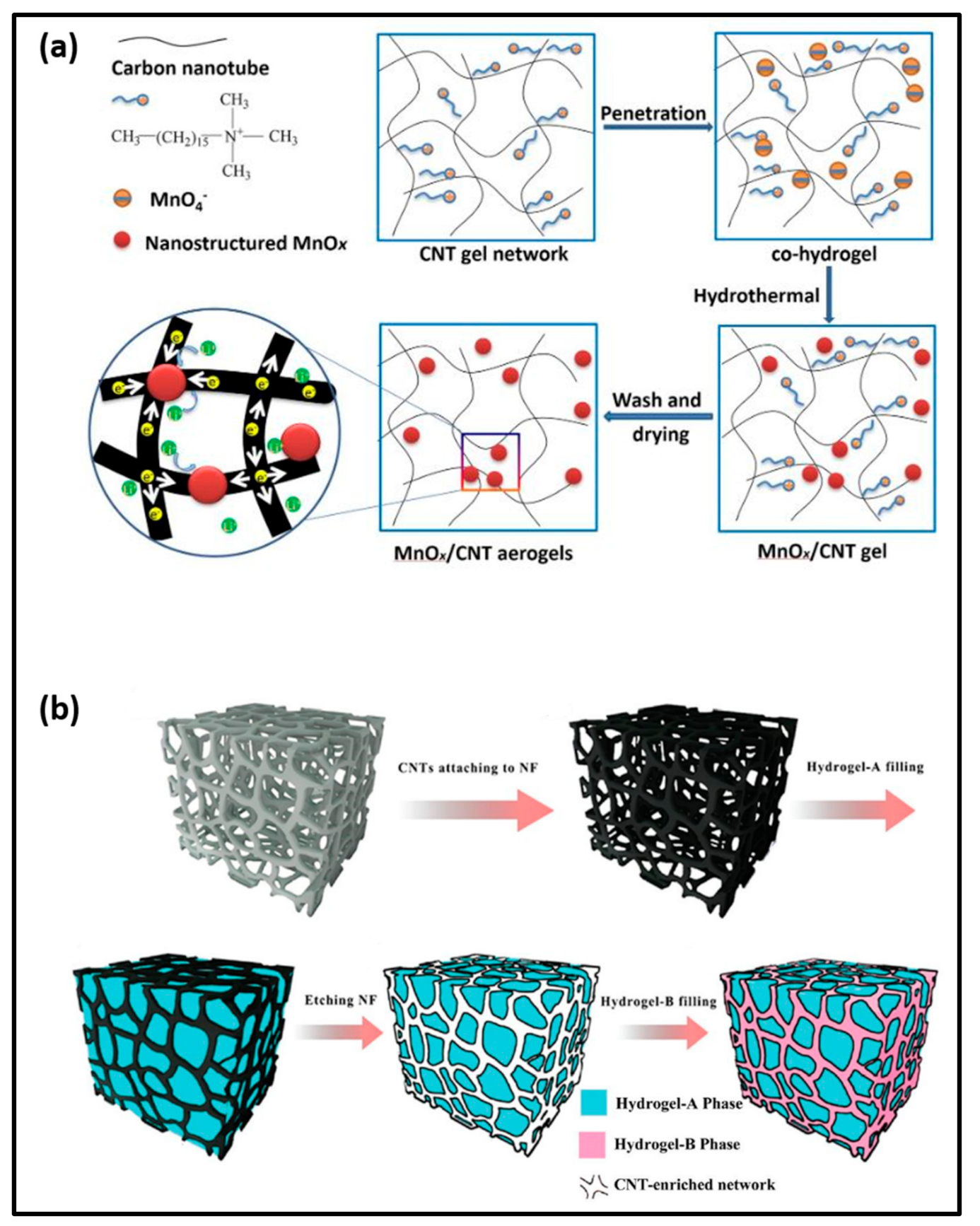
  74. Liu, Y.; Liu, M.; Zheng, P.; Ge, D.; Yang, L. Controllable hydrogel-thermal synthesis of Mn2O3/CNT aerogels: Shape evolution, growth mechanism and electrochemical properties. Mater. Des. 2019, 182, 108022. [Google Scholar] [CrossRef]
  75. Ying, Z.; Wang, Q.; Xie, J.; Li, B.; Lin, X.; Hui, S. Novel electrically-conductive electro-responsive hydrogels for smart actuators with a carbon-nanotube-enriched three-dimensional conductive network and a physical-phase-type three-dimensional interpenetrating network. J. Mater. Chem. C 2020, 8, 4192–4205. [Google Scholar] [CrossRef]
  76. Roy, S.; Das, T.; Ming, Y.; Chen, X.; Yue, C.Y.; Hu, X. Specific functionalization and polymer grafting on multi-walled carbon nanotubes to fabricate advanced nylon 12 composites. J. Mater. Chem. A 2014, 2, 3961–3970. [Google Scholar] [CrossRef]
  77. Liu, S.; Gao, G.; Xiao, Y.; Fu, J. Tough and responsive oppositely charged nanocomposite hydrogels for use as bilayer actuators assembled through interfacial electrostatic attraction. J. Mater. Chem. B 2016, 4, 3239–3246. [Google Scholar] [CrossRef]
  78. Ki, V.; Rotstein, C. Bacterial skin and soft tissue infections in adults: A review of their epidemiology, pathogenesis, diagnosis, treatment and site of care. Can. J. Infect. Dis. Med. Microbiol. 2008, 19, 173–184. [Google Scholar] [CrossRef] [Green Version]
  79. Cardona, A.F.; Wilson, S.E. Skin and soft-tissue infections: A critical review and the role of telavancin in their treatment. Clin. Infect. Dis. Off. Publ. Infect. Dis. Soc. Am. 2015, 61 (Suppl. 2), S69–S78. [Google Scholar] [CrossRef] [Green Version]
  80. Andonova, M.; Urumova, V. Immune surveillance mechanisms of the skin against the stealth infection strategy of Pseudomonas aeruginosa—Review. Comp. Immunol. Microbiol. Infect. Dis. 2013, 36, 433–448. [Google Scholar] [CrossRef]
  81. Han, G.; Zhang, W. Improved biclique cryptanalysis of the lightweight block cipher piccolo. Secur. Commun. Netw. 2017, 2017, 7589306. [Google Scholar] [CrossRef] [Green Version]
  82. Siddiqui, A.R.; Bernstein, J.M. Chronic wound infection: Facts and controversies. Clin. Dermatol. 2010, 28, 519–526. [Google Scholar] [CrossRef]
  83. Vig, K.; Chaudhari, A.; Tripathi, S.; Dixit, S.; Sahu, R.; Pillai, S.; Dennis, V.A.; Singh, S.R. Advances in skin regeneration using tissue engineering. Int. J. Mol. Sci. 2017, 18, 789. [Google Scholar] [CrossRef]
  84. Unalan, I.; Endlein, S.J.; Slavik, B.; Buettner, A.; Goldmann, W.H.; Detsch, R.; Boccaccini, A.R. Evaluation of electrospun poly(ε-caprolactone)/gelatin nanofiber mats containing clove essential oil for antibacterial wound dressing. Pharmaceutics 2019, 11, 570. [Google Scholar] [CrossRef] [Green Version]
  85. Kang, S.; Pinault, M.; Pfefferle, L.D.; Elimelech, M. Single-walled carbon nanotubes exhibit strong antimicrobial activity. Langmuir ACS J. Surf. Colloids 2007, 23, 8670–8673. [Google Scholar] [CrossRef]
  86. Maleki Dizaj, S.; Mennati, A.; Jafari, S.; Khezri, K.; Adibkia, K. Antimicrobial activity of carbon-based nanoparticles. Adv. Pharm. Bull. 2015, 5, 19–23. [Google Scholar] [CrossRef]
  87. Mallaiah, D. Antibacterial activity by functionalized carbon nanotubes. In Nanostructures for Antimicrobial and Antibiofilm Applications; Prasad, R., Siddhardha, B., Dyavaiah, M., Eds.; Springer International Publishing: Cham, Switzeralnd, 2020; pp. 63–77. [Google Scholar]
  88. Smith, S.C.; Rodrigues, D.F. Carbon-based nanomaterials for removal of chemical and biological contaminants from water: A review of mechanisms and applications. Carbon 2015, 91, 122–143. [Google Scholar] [CrossRef]
  89. Dong, L.; Henderson, A.; Field, C. Antimicrobial activity of single-walled carbon nanotubes suspended in different surfactants. J. Nanotechnol. 2012, 2012, 928924. [Google Scholar] [CrossRef] [Green Version]
  90. Assali, M.; Zaid, A.N.; Abdallah, F.; Almasri, M.; Khayyat, R. Single-walled carbon nanotubes-ciprofloxacin nanoantibiotic: Strategy to improve ciprofloxacin antibacterial activity. Int. J. Nanomed. 2017, 12, 6647–6659. [Google Scholar] [CrossRef] [Green Version]
  91. Sapkota, K.P.; Hassan, M.M.; Shrestha, S.; Hanif, M.A.; Islam, M.A.; Akter, J.; Abbas, H.G.; Hahn, J.R. Heterojunction formation between copper(II) oxide nanoparticles and single-walled carbon nanotubes to enhance antibacterial performance. Int. J. Pharm. 2020, 590, 119937. [Google Scholar] [CrossRef]
  92. Masoumeh, A.M.; Nejad, H.R.; Pardis, S.; Ali, K.N.R. Improving antibacterial activity of methicillin by conjugation to functionalized single-wall carbon nanotubes against S. aureus. Res. Sq. 2021. [Google Scholar] [CrossRef]
  93. Khan, A.A.P.; Khan, A.; Rahman, M.M.; Asiri, A.M.; Oves, M. Lead sensors development and antimicrobial activities based on graphene oxide/carbon nanotube/poly(O-toluidine) nanocomposite. Int. J. Biol. Macromol. 2016, 89, 198–205. [Google Scholar] [CrossRef]
  94. Ding, L.; Wang, H.; Liu, D.; Zeng, X.-A.; Mao, Y. Bacteria capture and inactivation with functionalized multi-walled carbon nanotubes (MWCNTs). J. Nanosci. Nanotechnol. 2020, 20, 2055–2062. [Google Scholar] [CrossRef]
  95. Li, P.; Poon, Y.F.; Li, W.; Zhu, H.-Y.; Yeap, S.H.; Cao, Y.; Qi, X.; Zhou, C.; Lamrani, M.; Beuerman, R.W.; et al. A polycationic antimicrobial and biocompatible hydrogel with microbe membrane suctioning ability. Nat. Mater. 2011, 10, 149–156. [Google Scholar] [CrossRef]
  96. Brady-Estévez, A.S.; Kang, S.; Elimelech, M. A single-walled-carbon-nanotube filter for removal of viral and bacterial pathogens. Small 2008, 4, 481–484. [Google Scholar] [CrossRef]
  97. Dwyer, D.J.; Camacho, D.M.; Kohanski, M.A.; Callura, J.M.; Collins, J.J. Antibiotic-induced bacterial cell death exhibits physiological and biochemical hallmarks of apoptosis. Mol. Cell 2012, 46, 561–572. [Google Scholar] [CrossRef] [Green Version]
  98. Yang, C.; Mamouni, J.; Tang, Y.; Yang, L. Antimicrobial activity of single-walled carbon nanotubes: Length effect. Langmuir ACS J. Surf. Colloids 2010, 26, 16013–16019. [Google Scholar] [CrossRef]
  99. Johnston, H.J.; Hutchison, G.R.; Christensen, F.M.; Peters, S.; Hankin, S.; Aschberger, K.; Stone, V. A critical review of the biological mechanisms underlying the in vivo and in vitro toxicity of carbon nanotubes: The contribution of physico-chemical characteristics. Nanotoxicology 2010, 4, 207–246. [Google Scholar] [CrossRef]
  100. Kim, K.-I.; Kim, D.-A.; Patel, K.D.; Shin, U.S.; Kim, H.-W.; Lee, J.-H.; Lee, H.-H. Carbon nanotube incorporation in PMMA to prevent microbial adhesion. Sci. Rep. 2019, 9, 4921. [Google Scholar] [CrossRef] [PubMed]
  101. Chen, H.; Wang, B.; Gao, D.; Guan, M.; Zheng, L.; Ouyang, H.; Chai, Z.; Zhao, Y.; Feng, W. Broad-spectrum antibacterial activity of carbon nanotubes to human gut bacteria. Small 2013, 9, 2735–2746. [Google Scholar] [CrossRef] [PubMed]
  102. Murugan, E.; Vimala, G. Effective functionalization of multi-walled carbon nanotube with amphiphilic poly(propyleneimine) dendrimer carrying silver nanoparticles for better dispersability and antimicrobial activity. J. Colloid Interface Sci. 2011, 357, 354–365. [Google Scholar] [CrossRef] [PubMed]
  103. Kang, S.; Herzberg, M.; Rodrigues, D.F.; Elimelech, M. Antibacterial effects of carbon nanotubes: Size does matter! Langmuir ACS J. Surf. Colloids 2008, 24, 6409–6413. [Google Scholar] [CrossRef]
  104. Song, B.; Xu, P.; Zeng, G.; Gong, J.; Zhang, P.; Feng, H.; Liu, Y.; Ren, X. Carbon nanotube-based environmental technologies: The adopted properties, primary mechanisms, and challenges. Rev. Environ. Sci. Bio/Technol. 2018, 17, 571–590. [Google Scholar] [CrossRef]
  105. Kang, S.; Mauter, M.S.; Elimelech, M. Physicochemical determinants of multiwalled carbon nanotube bacterial cytotoxicity. Environ. Sci. Technol. 2008, 42, 7528–7534. [Google Scholar] [CrossRef]
  106. Akhavan, O.; Ghaderi, E. Toxicity of graphene and graphene oxide nanowalls against bacteria. ACS Nano 2010, 4, 5731–5736. [Google Scholar] [CrossRef]
  107. Boczkowski, J.; Lanone, S. Respiratory toxicities of nanomaterials—A focus on carbon nanotubes. Adv. Drug Deliv. Rev. 2012, 64, 1694–1699. [Google Scholar] [CrossRef]
  108. Vecitis, C.D.; Zodrow, K.R.; Kang, S.; Elimelech, M. Electronic-structure-dependent bacterial cytotoxicity of single-walled carbon nanotubes. ACS Nano 2010, 4, 5471–5479. [Google Scholar] [CrossRef]
  109. Du, J.; Wang, S.; You, H.; Zhao, X. Understanding the toxicity of carbon nanotubes in the environment is crucial to the control of nanomaterials in producing and processing and the assessment of health risk for human: A review. Environ. Toxicol. Pharmacol. 2013, 36, 451–462. [Google Scholar] [CrossRef]
  110. Kang, S.; Mauter, M.S.; Elimelech, M. Microbial cytotoxicity of carbon-based nanomaterials: Implications for river water and wastewater effluent. Environ. Sci. Technol. 2009, 43, 2648–2653. [Google Scholar] [CrossRef]
  111. Aslan, S.; Loebick, C.Z.; Kang, S.; Elimelech, M.; Pfefferle, L.D.; Van Tassel, P.R. Antimicrobial biomaterials based on carbon nanotubes dispersed in poly(lactic-co-glycolic acid). Nanoscale 2010, 2, 1789–1794. [Google Scholar] [CrossRef]
  112. Azizi-Lalabadi, M.; Hashemi, H.; Feng, J.; Jafari, S.M. Carbon nanomaterials against pathogens; the antimicrobial activity of carbon nanotubes, graphene/graphene oxide, fullerenes, and their nanocomposites. Adv. Colloid Interface Sci. 2020, 284, 102250. [Google Scholar] [CrossRef]
  113. Dong, X.; Koo, Y.; Tang, Y.; Yun, Y.; Yang, L. Superior antibacterial activity of photochemical synthesized Ag-CNT composites and their synergistic effects in combination with other antimicrobial agents. Nanomed. Nanotechnol. 2015, 6, 279. [Google Scholar]
  114. Yuan, W.; Jiang, G.; Che, J.; Qi, X.; Xu, R.; Chang, M.W.; Chen, Y.; Lim, S.Y.; Dai, J.; Chan-Park, M.B. Deposition of silver nanoparticles on multi-walled carbon nanotubes grafted with hyperbranched poly(amidoamine) and their antimicrobial effects. J. Phys. Chem. C 2008, 112, 18754–18759. [Google Scholar] [CrossRef]
  115. Castle, A.B.; Gracia-Espino, E.; Nieto-Delgado, C.; Terrones, H.; Terrones, M.; Hussain, S. Hydroxyl-functionalized and N-doped multiwalled carbon nanotubes decorated with silver nanoparticles preserve cellular function. ACS Nano 2011, 5, 2458–2466. [Google Scholar] [CrossRef]
  116. Aslan, S.; Deneufchatel, M.; Hashmi, S.; Li, N.; Pfefferle, L.D.; Elimelech, M.; Pauthe, E.; Van Tassel, P.R. Carbon nanotube-based antimicrobial biomaterials formed via layer-by-layer assembly with polypeptides. J. Colloid Interface Sci. 2012, 388, 268–273. [Google Scholar] [CrossRef]
  117. Kavoosi, G.; Dadfar, S.M.M.; Dadfar, S.M.A.; Ahmadi, F.; Niakosari, M. Investigation of gelatin/multi-walled carbon nanotube nanocomposite films as packaging materials. Food Sci. Nutr. 2014, 2, 65–73. [Google Scholar] [CrossRef]
  118. Gan, L.; Geng, A.; Jin, L.; Zhong, Q.; Wang, L.; Xu, L.; Mei, C. Antibacterial nanocomposite based on carbon nanotubes–silver nanoparticles-co-doped polylactic acid. Polym. Bull. 2020, 77, 793–804. [Google Scholar] [CrossRef]
  119. Benigno, E.; Lorente, M.A.; Olmos, D.; González-Gaitano, G.; González-Benito, J. Nanocomposites based on low density polyethylene filled with carbon nanotubes prepared by high energy ball milling and their potential antibacterial activity. Polym. Int. 2019, 68, 1155–1163. [Google Scholar] [CrossRef]
  120. Wang, W.; Zhu, L.; Shan, B.; Xie, C.; Liu, C.; Cui, F.; Li, G. Preparation and characterization of SLS-CNT/PES ultrafiltration membrane with antifouling and antibacterial properties. J. Membr. Sci. 2018, 548, 459–469. [Google Scholar] [CrossRef]
  121. Pandiyan, R.; Mahalingam, S.; Ahn, Y.-H. Antibacterial and photocatalytic activity of hydrothermally synthesized SnO2 doped GO and CNT under visible light irradiation. J. Photochem. Photobiol. B Biol. 2019, 191, 18–25. [Google Scholar] [CrossRef]
  122. Lakourj, M.M.; Norouzian, R.-S.; Esfandyar, M. Conducting nanocomposites of polypyrrole-co-polyindole doped with carboxylated CNT: Synthesis approach and anticorrosion/antibacterial/antioxidation property. Mater. Sci. Eng. B 2020, 261, 114673. [Google Scholar] [CrossRef]
  123. Shaham, G.; Veisi, H.; Hekmati, M. Silver nanoparticle-decorated multi-walled carbon nanotube/pramipexole nanocomposite: Synthesis, characterization and application as an antibacterial agent. Appl. Organomet. Chem. 2017, 31, e3737. [Google Scholar] [CrossRef]
  124. Baek, S.; Joo, S.H.; Su, C.; Toborek, M. Antibacterial effects of graphene-and carbon-nanotube-based nanohybrids on Escherichia coli: Implications for treating multidrug-resistant bacteria. J. Environ. Manag. 2019, 247, 214–223. [Google Scholar] [CrossRef] [PubMed]
  125. Xu, Y.; Liu, J.; Xie, M.; Jing, L.; Xu, H.; She, X.; Li, H.; Xie, J. Construction of novel CNT/LaVO4 nanostructures for efficient antibiotic photodegradation. Chem. Eng. J. 2019, 357, 487–497. [Google Scholar] [CrossRef]
  126. Palza, H.; Saldias, N.; Arriagada, P.; Palma, P.; Sanchez, J. Antibacterial carbon nanotubes by impregnation with copper nanostructures. JOM 2017, 69, 1319–1324. [Google Scholar] [CrossRef]
  127. Joghataeian, M.; Bahari, A.; Qavami, A.; Raeisi, M.J. An antibacterial study of a new magnetic carbon nanotube/core-shell nanohybrids. J. Environ. Chem. Eng. 2020, 8, 104150. [Google Scholar] [CrossRef]
  128. Sivaraj, D.; Vijayalakshmi, K. Preferential killing of bacterial cells by hybrid carbon nanotube-MnO2 nanocomposite synthesized by novel microwave assisted processing. Mater. Sci. Eng. C 2017, 81, 469–477. [Google Scholar] [CrossRef]
  129. Ku, S.H.; Lee, M.; Park, C.B. Carbon-based nanomaterials for tissue engineering. Adv. Healthc. Mater. 2013, 2, 244–260. [Google Scholar] [CrossRef]
  130. Goenka, S.; Sant, V.; Sant, S. Graphene-based nanomaterials for drug delivery and tissue engineering. J. Control. Release 2014, 173, 75–88. [Google Scholar] [CrossRef]
  131. Gonzalez, A.C.; Costa, T.F.; Andrade, Z.A.; Medrado, A.R. Wound healing—A literature review. An. Bras. Dermatol. 2016, 91, 614–620. [Google Scholar] [CrossRef] [Green Version]
  132. Robson, M.C.; Steed, D.L.; Franz, M.G. Wound healing: Biologic features and approaches to maximize healing trajectories. Curr. Probl. Surg. 2001, 38, 72–140. [Google Scholar] [CrossRef]
  133. Velnar, T.; Bailey, T.; Smrkolj, V. The wound healing process: An overview of the cellular and molecular mechanisms. J. Int. Med. Res. 2009, 37, 1528–1542. [Google Scholar] [CrossRef]
  134. Natarajan, S.; Williamson, D.; Stiltz, A.J.; Harding, K. Advances in wound care and healing technology. Am. J. Clin. Dermatol. 2000, 1, 269–275. [Google Scholar] [CrossRef]
  135. Junker, J.P.; Kamel, R.A.; Caterson, E.J.; Eriksson, E. Clinical impact upon wound healing and inflammation in moist, wet, and dry environments. Adv. Wound Care 2013, 2, 348–356. [Google Scholar] [CrossRef] [Green Version]
  136. Zhao, R.; Liang, H.; Clarke, E.; Jackson, C.; Xue, M. Inflammation in chronic wounds. Int. J. Mol. Sci. 2016, 17, 2085. [Google Scholar] [CrossRef]
  137. He, J.; Shi, M.; Liang, Y.; Guo, B. Conductive adhesive self-healing nanocomposite hydrogel wound dressing for photothermal therapy of infected full-thickness skin wounds. Chem. Eng. J. 2020, 394, 124888. [Google Scholar] [CrossRef]
  138. Xu, M.; Li, Q.; Fang, Z.; Jin, M.; Zeng, Q.; Huang, G.; Jia, Y.-G.; Wang, L.; Chen, Y. Conductive and antimicrobial macroporous nanocomposite hydrogels generated from air-in-water Pickering emulsions for neural stem cell differentiation and skin wound healing. Biomater. Sci. 2020, 8, 6957–6968. [Google Scholar] [CrossRef]
  139. Liang, N.; Wu, L.; Yang, Y. Multifunctional double network hydrogel film for skin wound healing. Mater. Express 2021, 11, 1084–1091. [Google Scholar] [CrossRef]
  140. He, S.; Huang, J.; Zhang, Q.; Zhao, W.; Xu, Z.; Zhang, W. Bamboo-like nanozyme based on nitrogen-doped carbon nanotubes encapsulating cobalt nanoparticles for wound antibac-terial applications. Adv. Funct. Mater. 2021, 31, 2105198. [Google Scholar] [CrossRef]
  141. Kittana, N.; Assali, M.; Abu-Rass, H.; Lutz, S.; Hindawi, R.; Ghannam, L.; Zakarneh, M.; Mousa, A. Enhancement of wound healing by single-wall/multi-wall carbon nanotubes complexed with chitosan. Int. J. Nanomed. 2018, 13, 7195–7206. [Google Scholar] [CrossRef] [Green Version]
  142. Simpson, D.M.; Ross, R. The neutrophilic leukocyte in wound repair a study with antineutrophil serum. J. Clin. Investig. 1972, 51, 2009–2023. [Google Scholar] [CrossRef] [Green Version]
  143. Diegelmann, R.F.; Evans, M.C. Wound healing: An overview of acute, fibrotic and delayed healing. Front. Biosci. J. Virtual Libr. 2004, 9, 283–289. [Google Scholar] [CrossRef] [PubMed]
  144. Tocco, I.; Zavan, B.; Bassetto, F.; Vindigni, V. Nanotechnology-based therapies for skin wound regeneration. J. Nanomater. 2012, 2012, 714134. [Google Scholar] [CrossRef] [Green Version]
  145. Gurtner, G.C.; Werner, S.; Barrandon, Y.; Longaker, M.T. Wound repair and regeneration. Nature 2008, 453, 314–321. [Google Scholar] [CrossRef] [PubMed]
  146. Wang, L.; Hu, S.; Ullah, M.W.; Li, X.; Shi, Z.; Yang, G. Enhanced cell proliferation by electrical stimulation based on electroactive regenerated bacterial cellulose hydrogels. Carbohydr. Polym. 2020, 249, 116829. [Google Scholar] [CrossRef]
  147. Omrani, M.M.; Ansari, M.; Kordestani, S.S.; Kiaie, N.; Salati, A. Enhanced bone marrow stem cell attachment and differentiation on PCL/CNT substrate. Inorg. Nano-Metal. Chem. 2019, 49, 136–142. [Google Scholar] [CrossRef]
  148. Wang, S.; Li, Y.; Zhao, R.; Jin, T.; Zhang, L.; Li, X. Chitosan surface modified electrospun poly (ε-caprolactone)/carbon nanotube composite fibers with enhanced mechanical, cell proliferation and antibacterial properties. Int. J. Biol. Macromol. 2017, 104, 708–715. [Google Scholar] [CrossRef]
  149. Di Leo, N.; Battaglini, M.; Berger, L.; Giannaccini, M.; Dente, L.; Hampel, S.; Vittorio, O.; Cirillo, G.; Raffa, V. A catechin nanoformulation inhibits WM266 melanoma cell proliferation, migration and associated neo-angiogenesis. Eur. J. Pharm. Biopharm. 2017, 114, 1–10. [Google Scholar] [CrossRef]
  150. Madhusoodan, A.; Das, K.; Mili, B.; Kumar, K.; Kumar, A.; Saxena, A.; Singh, P.; Dutt, T.; Bag, S. In vitro proliferation and differentiation of canine bone marrow derived mesenchymal stem cells over hydroxyl functionalized CNT substrates. Biotechnol. Rep. 2019, 24, e00387. [Google Scholar] [CrossRef]
  151. Mombini, S.; Mohammadnejad, J.; Bakhshandeh, B.; Narmani, A.; Nourmohammadi, J.; Vahdat, S.; Zirak, S. Chitosan-PVA-CNT nanofibers as electrically conductive scaffolds for cardiovascular tissue engineering. Int. J. Biol. Macromol. 2019, 140, 278–287. [Google Scholar] [CrossRef]
  152. Khan, A.; Hussain, A.; Sidra, L.; Sarfraz, Z.; Khalid, H.; Khan, M.; Manzoor, F.; Shahzadi, L.; Yar, M.; Rehman, I. Fabrication and in vivo evaluation of hydroxyapatite/carbon nanotube electrospun fibers for biomedical/dental application. Mater. Sci. Eng. C 2017, 80, 387–396. [Google Scholar] [CrossRef]
  153. Lu, X.; Zhu, Y.; Bai, R.; Wu, Z.; Qian, W.; Yang, L.; Cai, R.; Yan, H.; Li, T.; Pandey, V. Long-term pulmonary exposure to multi-walled carbon nanotubes promotes breast cancer metastatic cascades. Nat. Nanotechnol. 2019, 14, 719–727. [Google Scholar] [CrossRef]
  154. Shokraei, N.; Asadpour, S.; Shokraei, S.; Nasrollahzadeh Sabet, M.; Faridi-Majidi, R.; Ghanbari, H. Development of electrically conductive hybrid nanofibers based on CNT-polyurethane nanocomposite for cardiac tissue engineering. Microsc. Res. Tech. 2019, 82, 1316–1325. [Google Scholar] [CrossRef]
  155. Kramer, N.; Walzl, A.; Unger, C.; Rosner, M.; Krupitza, G.; Hengstschläger, M.; Dolznig, H. In vitro cell migration and invasion assays. Mutat. Res. 2013, 752, 10–24. [Google Scholar] [CrossRef]
  156. Liang, Y.; Zhao, X.; Hu, T.; Han, Y.; Guo, B. Mussel-inspired, antibacterial, conductive, antioxidant, injectable composite hydrogel wound dressing to promote the regeneration of infected skin. J. Colloid Interface Sci. 2019, 556, 514–528. [Google Scholar] [CrossRef]
Figure 1. A scheme showing the types of CNT, properties of CNT hydrogel patches, and their application in cell proliferation [34], cell migration [35], antibacterial [36], and wound healing [37].
Applsci 11 09550 g001
Figure 2. Surface and internal view of (a) single-walled CNTs, (b) double-walled CNTs, and (c) multi-walled CNTs [51].
Applsci 11 09550 g002
Figure 3. Scheme showing the synthesis of CNT-based hydrogels: (a) MnOx/CNT aerogels [74], (b) general process of the double network hydrogel [75].
Applsci 11 09550 g003
Figure 4. (a) Scheme showing antibacterial activity through cell membrane rupture in E. coli K12 bacteria [103], (b) Schematic diagram of bacteria killing through ROS mechanism in presence of long and short CNT [104].
Applsci 11 09550 g004
Table 2. Various properties of SWCNT and MWCNT [49].
PropertiesUnitsSWCNTMWCNT
Specific gravity (bulk)g/cm30.8–1.31.8–2.6
Specific aream2/g400–900200–400
Young’s modulusPa≈1000≈1000
Tensile strengthPa3.1010–5.10111.1010–15.1010
Thermal conductivityW/m.K3000–60002000–3000
Electrical conductivityS/cm102–106103–105
Thermal stability temperature in air°C550–650550–650
Table 3. Application and aspect of CNT-based composites for antibacterial activity on various bacteria.
CNT CompositeTreatmentOrganismEffectReference
SLS-CNT/PESUltrafiltration Escherichia coli (E. coli)Antifouling and antibacterial[120]
SCNT-SnO2 Wastewater treatmentEscherichia coli and Pseudomonas graminisPhotolytic degradation and Bactericidal activity [121]
PPy-co-PIn@CNTAnticorrosion coating to prevent the surface of metals from being damagedS. aureus and B. subtilis, E. coli, and P. aeruginosa.Antibacterial and anticorrosion activity[122]
(CNT/pra/Ag)Increased inhibitory activity toward Gram-positive bacteriaStaphylococcus aureus, Pseudomonasaeruginosa, and E. coliAntibacterial activity[123]
GO/ZnO–CNTTreatment of multidrug-resistant bacteriaE. coli (DH5α)Damage the E. coli cell membranes[124]
CNT/LaVO4Antibiotic
photodegradation
E. coliLow toxicity, improve water quality[125]
Cu/CNTEnhance antimicrobial activityS. aureus and E. coliAntibacterial activity[126]
Co0.7Zn0.3 Fe2O4/PET/Ag/CNTWater-dispersible antibacterial activityE. coli,
Pseudomonas aeruginosa, Bacillus subtilis, Staphylococcus aureus
Improved antibacterial activity[127]
CNT/MnO2Enhanced antibacterial activity of Gram-negative bacteriaProteus mirabilis and Escherichia coliInhibit bacterial growth.[128]
SLS-sodium lignosulfonate, PES-poly(ethersulfone), SCNT-SnO2-sonicated CNT-SnO2, PPy-co-Pin-pyrrole indole copolymer, pra-pramipexole, Ag-silver, ZnO-zinc oxide, GO-graphene oxide, LaVO4-lanthanum vanadate, Cu-copper, PET-poly(ethylene terephthalate), MnO2-manganese dioxide.
Table 4. Application and aspect of CNT hybrids for cell proliferation and angiogenesis.
CNT CompositeTreatmentCell LineEffectReference
rBC/PPy/CNTWound healing The mouse embryo fibroblast (NIH3T3) cell lineGood biocompatibility, enhanced cell proliferation, excellent
thermal stability, mechanical properties, recoverability, and good water-absorbing capability.
[146]
PCL/CNTneuron differentiation
and regeneration
human bone-derived mesenchymal stem cellsImproved cell adhesion and differentiation.[147]
Ch-PCL/CNTAntibacterial activity and other biomedical applicationsL929 fibroblast cellsImproved cell attachment and proliferation, antibacterial activity against Gram-positive and Gram-negative bacteria. [148]
CNT: gel-CTCancer treatmentsWM266-4 human melanoma cell lineInduce cell growth inhibition and cell death.[149]
(OH)- CNTsStem cell-based regenerative medicines.Canine bone marrow-derived
mesenchymal stem cells
Positive influence on osteogenic and chondrogenic differentiation as well as neuronal differentiation.[150]
PVA-Ch-CNTCardiac differentiationUndifferentiated mesenchymal stem cells
(MSCs)
Upregulation of cardiomyocytes differentiation. Increased conductivity, chemical stability, and enhanced adherence.[151]
HA/CNTDental application Induce angiogenesis and exhibited biocompatibility. Improved mechanical properties[152]
MWCNTInfluence breast cancer
metastatic cascades
BALB/c-derived 4T1Enhances tumor angiogenesis and metastatic processes.[153]
CNT/PUImproved cytocompatibility for cardiomyoblasts.H9c2 cells and human umbilical vein endothelial cells (HUVECs)Appropriate attachment, high viability, and proliferation of cardiomyoblasts.[154]
rBC-regenerated bacterial cellulose, PPy-polypyrrole, PCL-poly(caprolactone), Ch-chitosan, gel-gelatin, CT-catechin, PVA-poly(vinyl alcohol), HA-hydroxyapatite, PU-polyurethane.
Publisher’s Note: MDPI stays neutral with regard to jurisdictional claims in published maps and institutional affiliations.

