Communication

Cytochrome cM Is Probably a Membrane Protein Similar to the C Subunit of the Bacterial Nitric Oxide Reductase

by
Tomás Rodríguez-Gil
1,2,
Alejandro Torrado
1,2,
Macarena Iniesta-Pallarés
1,2,
Consolación Álvarez
1,
Vicente Mariscal
1 and
Fernando P. Molina-Heredia
1,2,*
1
Instituto de Bioquímica Vegetal y Fotosíntesis, Universidad de Sevilla and CSIC, C. Américo Vespucio 49, 41092 Seville, Spain
2
Departamento de Bioquímica Vegetal y Biología Molecular, Facultad de Biología, Universidad de Sevilla, Avda. Reina Mercedes s/n, 41012 Seville, Spain
*
Author to whom correspondence should be addressed.
Appl. Sci. 2021, 11(20), 9396; https://doi.org/10.3390/app11209396
Submission received: 9 September 2021 / Revised: 29 September 2021 / Accepted: 6 October 2021 / Published: 10 October 2021
(This article belongs to the Special Issue Electron Transfer in Photosynthesis)

Abstract

:
Cytochrome cM was first described in 1994 and its sequence has been found in the genome of manifold cyanobacterial species ever since. Numerous studies have been carried out with the purpose of determining its function, but none of them has given place to conclusive results so far. Many of these studies are based on the assumption that cytochrome cM is a soluble protein located in the thylakoid lumen of cyanobacteria. In this work, we have reevaluated the sequence of cytochrome cM, with our results showing that its most probable 3D structure is strongly similar to that of the C subunit of the bacterial nitric oxide reductase. The potential presence of an α-helix tail, which could locate this protein in the thylakoid membrane, further supports this hypothesis, thus providing a new, unexpected role for this redox protein.

1. Introduction

Electron transport chains play a fundamental role in the metabolism of all living beings. They consist of a series of redox reactions, spatially separated, in which electrons are transferred from a donor molecule to an acceptor one. In phototrophic eukaryotes, the respiratory and photosynthetic electron transport chains are spatially separated into specialized organelles (mitochondria and chloroplasts). However, in cyanobacteria, both electron transport chains are located in the thylakoid membranes and share some components, such as plastoquinone (PQ), plastocyanin (Pc) or cytochrome (Cyt) c6 and the Cyt b6-f complex.
In cyanobacteria, two c-type cytochromes involved in the electron transport chains have been described: Cyt c6 and Cyt c550. While Cyt c6 is a soluble electron carrier involved both in photosynthesis and in respiration, Cyt c550 is structurally bound to photosystem (PS) II and is not directly involved in electron transport [1]. One further c-type cytochrome has been described in cyanobacteria: the so-called Cyt cM [2]. The gene encoding for Cyt cM is conserved in nearly every sequenced cyanobacterium [3,4], and it has been consistently studied in the unicellular organism Synechocystis sp. PCC 6803 [5,6]. In Synechocystis, the annotated gene contains two methionine codons (Figure 1), of which the distal one was proposed to be the actual start codon as it was preceded by a GA-rich motif, i.e., a probable ribosome binding site [2,6]. The sequence further comprised a hydrophobic amino-terminal region that was assumed to constitute either a membrane anchor or a signal peptide. However, due to its close similarity to the signal peptide of Cyt c6 from Anabaena sp. PCC 7119, the latter was regarded as the most probable option [6].
Cho et al. [5] were the first to successfully detect the protein in cyanobacteria through Western blot analysis. Nevertheless, their results were not clear enough to conclude whether the protein featured the N-terminal hydrophobic domain in vivo or not, since the molecular weight of the protein detected by Western blotting did not match either of the two possible options [6]. As for the protein location, even though Bernroitner et al. [3] claimed to have determined that the thylakoid and plasma membranes are the exact places where Cyt cM is located in the cell, these results have later been questioned [7] given the high likelihood that cross-contamination between membranes occurred. Ever since, no other author has declared to have successfully established the cellular region where the protein is placed, probably due to its extremely scarce expression levels under normal conditions [8].
Based on the hypothesis that the actual Cyt cM protein sequence starts with the second methionine and that it contains a signal peptide to the thylakoid lumen—which would be subsequently released thereby generating a soluble globular protein—several authors have tried to determine its physiological function. As an alleged electron carrier similar to Cyt c6, speculations about the biological role of Cyt cM have been mainly linked it to a hypothetical role in respiratory and photosynthetic electron transport chains. The fact that deletion mutants were able to carry out both processes without any significant phenotype [2] dismissed the possibility of Cyt cM being a main component of any of these systems. However, there was a chance that Cyt cM participated in respiration or photosynthesis as a secondary element, i.e., as a backup piece that is only expressed under certain conditions to substitute or complement a major constituent. Interestingly, the expression levels of Cyt cM sharply increase in stress conditions such as low temperature and high-intensity light whereas those of the two main electron carriers, plastocyanin (Pc) and Cyt c6, decrease [9]. Despite some further evidence indicating a “spare component” role for Cyt cM in photosynthesis [10,11], we ruled this out by showing that the surface electrostatic potential of Cyt cM is too different from those of Pc and Cyt c6 and by proving that the kinetic interaction constant between Cyt cM and PS I, to which Pc and Cyt c6 donate electrons, is negligible [6]. Moreover, the low redox potential of Cyt cM (+150 mV) makes it thermodynamically unlikely that it can be reduced by Cyt f (with a redox potential of +320 mV), as is the case with Pc and Cyt c6 [6,12]. Consequently, only respiration remained as a real possibility. Initially, it was suggested that Cyt cM could reduce respiratory terminal oxidases (COX) since it was kinetically plausible [3] and it was impossible to create a ΔcytM/PSI double mutant [10]. However, the hypothesis of Cyt cM working as an electron donor in respiration was dismissed once it was observed that ΔcytM mutants have in fact a higher respiration rate under dark heterotrophic conditions [13], with the lack of COX not affecting this whatsoever [8].
In conclusion, none of the hypotheses formulated to this day regarding the physiological role or the cellular location of Cyt cM has proved solid, and this matter remains altogether an open debate. Since most attempts to determine any of these have been based on the assumption that it is a small soluble protein located in the lumen of the thylakoid, we have reevaluated the sequence and structure of Cyt cM in the present work. Our results suggest that, contrarily to what was previously thought, Cyt cM could be a membrane protein with a structure similar to that of the C subunit of bacterial nitric oxide reductase.

2. Materials and Methods

For the sequence collection, we searched for protein sequences corresponding to the cytM gene appearing in either of the two main repositories of annotated cyanobacterial genes: CYORF [14] (http://cyano.genome.jp/, accessed on 11 May 2021) and CyanoBase [15] (http://genome.microbedb.jp/cyanobase/, accessed on 11 May 2021).
Secondary and 3D protein structure prediction was made with the tools Phyre2 [16] (http://www.sbg.bio.ic.ac.uk/phyre2/html/page.cgi?id=index, accessed on 11 May 2021), I-TASSER [17] and AlphaFold [18].
The free software UCSF Chimera [19] (https://www.cgl.ucsf.edu/chimera/, version 1.15, accessed on 11 May 2021) was used to visualize protein 3D structures and to determine hydrophobicity and electrostatic potential, as well as for distance measurements and structure matching. For the electrostatic potential, the “Coulombic Surface Coloring” option was chosen. For comparison with NorC, we used the structure of nitric oxide reductase of Pseudomonas aeruginosa [20], which we obtained from the Protein Data Bank (https://www.rcsb.org/; PDB entry: 3O0R, accessed on 14 May 2021).

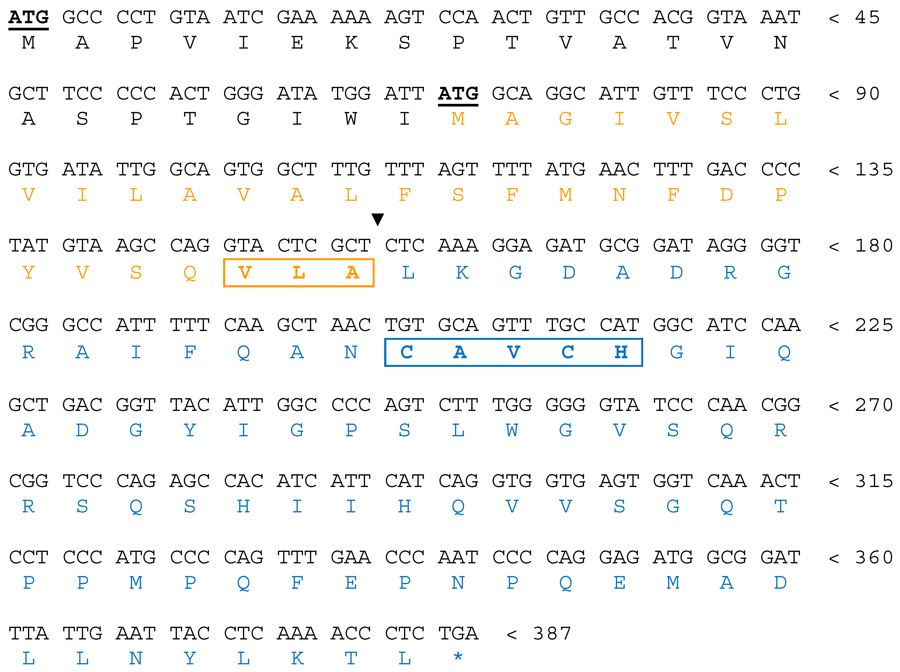
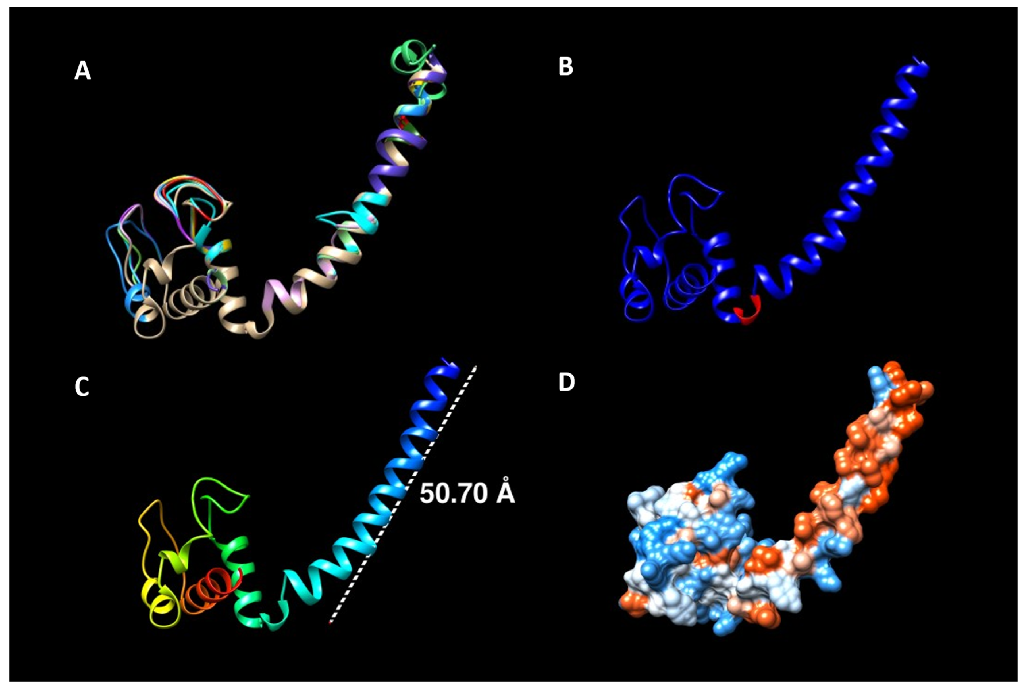
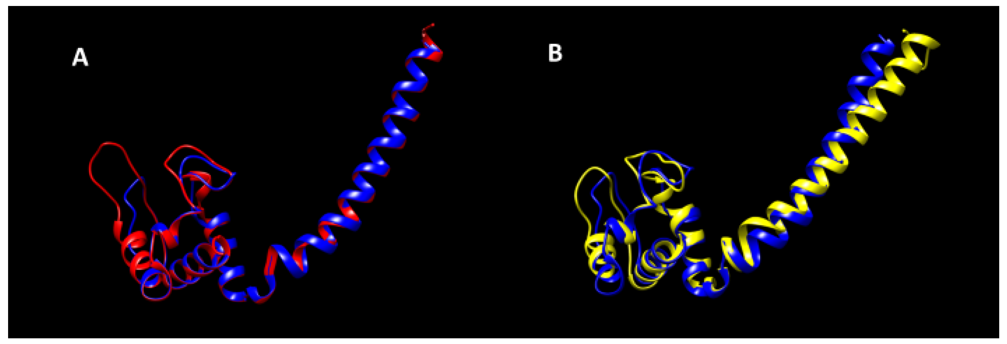
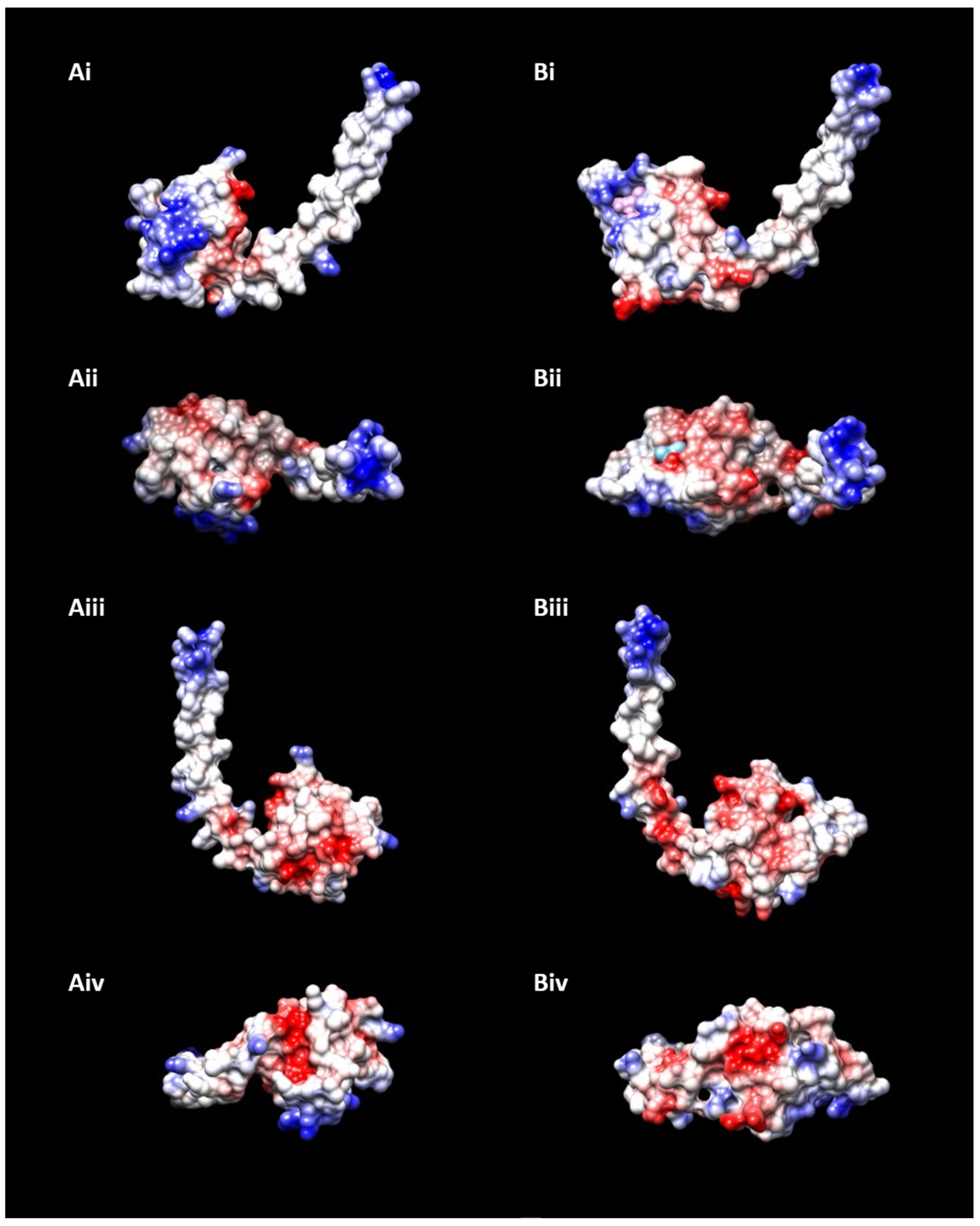
3. Results and Discussion

With the purpose of determining whether the N terminal part of the protein encodes for a signal peptide or a transmembrane helix, we retrieved all the protein sequences corresponding to the cytM gene that appear in either of the two main repositories of annotated cyanobacterial genes: CYORF and CyanoBase. A total of 18 sequences was obtained (Table 1), and they were then subjected to a structural prediction through Phyre2, which uses the homology recognition (threading) approach.
The first striking result was that 15 out of 18 sequences produced almost identical structures with a long, protruding α-helix (or “tail”) corresponding to the initial section of the protein (Figure 2A). This was the precise same region whose nature—signal peptide or transmembrane domain—has not been clearly established to this day. In fact, the VLA motif, repeatedly alleged as the cleavage site of the signal peptide, is located exactly at the junction of the tail with the “body” of the protein in the newly proposed structure (Figure 2B). The length of this helix motif of 5.1 nm (the average membrane being between 5 and 10 nm wide; Figure 2C) and the remarkably hydrophobic nature of this section (Figure 2D) agree with a membrane anchoring function.
At this point, it is important to bring up the fact that the signal peptides of Cyt c6 and Cyt c6-like proteins from the cyanobacterium Anabaena sp. PCC 7119, having a processing site (AXA) downstream from the N-terminal end, are recognized by E. coli which results in transport of the proteins to periplasmic space, a subcellular compartment equivalent to thylakoid lumen, where the signal peptide is correctly processed [21,22,23]. However, the hydrophobic tail of the Synechocystis Cyt cM is not recognized by E. coli as a signal peptide. For this reason, it has only been possible to express the soluble region of Synechocystis Cyt cM in E. coli by substituting its hydrophobic tail for the signal peptide of Anabaena Cyt c6 [6].
The template protein used by Phyre2 for calculating those 15 structures happened to be the c subunit of nitric oxide reductase (NorC) from Pseudomonas aeruginosa. Thus, the similarity between the returned models and the original template were compared. Surprisingly, the overlap between both proteins was clear and almost total (Figure 3A). To confirm this, a second, independent prediction for the same 15 sequences was performed using I-TASSER, a different structure prediction software that incorporates the ab initio approach in its algorithm. In this new test, the structures obtained were, once again, almost identical to that of NorC (Figure 3B). We obtained similar results once more using AlphaFold, a novel deep learning algorithm that incorporates physical and biological knowledge about protein structure and that leverages multi-sequence alignments [18].
Crucially, NorC is a subunit of nitric oxide reductase that has never been found in cyanobacteria, while the core subunit NorB is present in several cyanobacterial species [24] and its expression decreases in ΔcytM mutants [7]. Furthermore, Cyt cM sharply increases its expression in oxidative stress conditions [9], a condition in which nitric oxide reductase is highly active [25].
In order to determine similarities in charges between Cyt cM and NorC, we subsequently evaluated their surface electrostatic potential (Figure 4).
Also in this aspect, we found similarities between Cyt cM and NorC that might be indicative of a similar function, with Cyt cM acting as a functional part of a nitric oxide reductase and therefore being involved in the oxidative stress response. These included a positively charged tip at the end of the tail, a negatively charged region at the body closest to the tail, another positively charged region on the opposite side of the body and yet one more negatively charged pocket on the bottom. On the other hand, there are some clear differences too, such as a more extensive positive zone in the case of NorC, but the resemblances between both structures here are again undeniable. Moreover, the midpoint redox potential of Cyt cM and NorC, a fundamental parameter to establish the function of redox proteins, are relatively close with +150 mV and +183 mV, respectively [6,26]. This is interestingly opposed to the major divergences found previously between the surface electrostatic potential and midpoint redox potential of Cyt cM and the ones of Cyt c6 and Pc [6].

4. Conclusions

Here we show simple yet solid evidence that Cyt cM most probably has a transmembrane domain, and that it might well be a protein very similar to—or simply the equivalent of—the C subunit of nitric oxide reductase in cyanobacteria. If this last was the case, it would explain why it has been so difficult to assign any clear function to the protein so far, since all previous studies have focused on the soluble part of it when in fact it could hardly carry out any task without the rest of the protein it belongs to.

Author Contributions

Conceptualization, F.P.M.-H.; methodology, T.R.-G. and F.P.M.-H.; writing—original draft preparation, T.R.-G. and F.P.M.-H.; writing—review and editing, F.P.M.-H., T.R.-G., A.T., M.I.-P., C.Á. and V.M.; funding acquisition, F.P.M.-H. All authors have read and agreed to the published version of the manuscript.

Funding

This research was funded through the Fundación de Investigación de la Universidad de Sevilla (Spain) (grant FIUS05710000).

Institutional Review Board Statement

Not applicable.

Informed Consent Statement

Not applicable.

Data Availability Statement

The data presented in this study are available upon reasonable request from the corresponding author.

Conflicts of Interest

The authors declare no conflict of interest.

References

  1. Roncel, M.; Kirilovsky, D.; Guerrero, F.; Serrano, A.; Ortega, J.M. Photosynthetic cytochrome c550. Biochim. Biophys. Acta 2012, 1817, 1152–1163. [Google Scholar] [CrossRef] [PubMed] [Green Version]
  2. Malakhov, M.P.; Wada, H.; Los, D.A.; Semenenko, V.E.; Murata, N. A New Type of Cytochrome c from Synechocystis PCC6803. J. Plant Physiol. 1994, 144, 259–264. [Google Scholar] [CrossRef]
  3. Bernroitner, M.; Tangl, D.; Lucini, C.; Furtmüller, P.G.; Peschek, G.A.; Obinger, C. Cyanobacterial cytochrome cM: Probing its role as electron donor for CuA of cytochrome c oxidase. Biochim. Biophys. Acta Bioenerg. 2009, 1787, 135–143. [Google Scholar] [CrossRef] [PubMed] [Green Version]
  4. Bialek, W.; Szczepaniak, A.; Kolesinski, P.; Kallas, T. Cryptic c6-like and cM cytochromes of cyanobacteria. In Cytochrome Complexes: Evolution, Structures, Energy Transduction, and Signaling; Kramer, W.A., T. Kallas, T., Eds.; Springer: Dordrecht, The Netherlamnds, 2016; pp. 713–734. [Google Scholar]
  5. Cho, Y.S.; Pakrasi, H.B.; Whitmarsh, J. Cytochrome cM from Synechocystis 6803. Detection in cells, expression in Escherichia coli, purification and physical characterization. Eur. J. Biochem. 2000, 267, 1068–1074. [Google Scholar] [CrossRef]
  6. Molina-Heredia, F.P.; Balme, A.; Hervás, M.; Navarro, J.A.; De la Rosa, M.A. A comparative structural and functional analysis of cytochrome cM, cytochrome c6 and plastocyanin from the cyanobacterium Synechocystis sp. PCC 6803. FEBS Lett. 2002, 517, 50–54. [Google Scholar] [CrossRef]
  7. Solymosi, D.; Nikkanen, L.; Muth-Pawlak, D.; Fitzpatrick, D.; Vasudevan, R.; Howe, C.J.; Lea-Smith, D.J.; Allahverdiyeva, Y. Cytochrome cM decreases photosynthesis under photomixotrophy in Synechocystis sp. PCC 6803. Plant Physiol. 2020, 183, 700–716. [Google Scholar] [CrossRef] [Green Version]
  8. Baers, L.L.; Breckels, L.M.; Mills, L.A.; Gatto, L.; Deery, M.J.; Stevens, T.J.; Howe, C.J.; Lilley, K.S.; Lea-Smith, D.J. Proteome mapping of a cyanobacterium reveals distinct compartment organization and cell-dispersed metabolism. Plant Physiol. 2019, 181, 1721–1738. [Google Scholar] [CrossRef] [Green Version]
  9. Malakhov, M.P.; Malakhova, O.A.; Murata, N. Balanced regulation of expression of the gene for cytochrome c(M) and that of genes for plastocyanin and cytochrome c6 in Synechocystis. FEBS Lett. 1999, 444, 281–284. [Google Scholar] [CrossRef] [Green Version]
  10. Pils, D.; Gregor, W.; Schmetterer, G. Evidence for in vivo activity of three distinct respiratory terminal oxidases in the cyanobacterium Synechocystis sp. strain PCC6803. FEMS Microbiol. Lett. 1997, 152, 83–88. [Google Scholar] [CrossRef]
  11. Manna, P.; Vermaas, W. Lumenal proteins involved in respiratory electron transport in the cyanobacterium Synechocystis sp. PCC6803. Plant. Mol. Biol 1997, 35, 407–416. [Google Scholar] [CrossRef]
  12. Shuvalov, V.A.; Allakhverdiev, A.I.; Sakamoto, A.; Malakhov, M.; Murata, N. Optical study of cytochrome cM formation in Synechocystis. IUBMB Life 2001, 51, 93–97. [Google Scholar] [CrossRef]
  13. Hiraide, Y.; Oshima, K.; Fujisawa, T.; Uesaka, K.; Hirose, Y.; Tsujimoto, R.; Yamamoto, H.; Okamoto, S.; Nakamura, Y.; Terauchi, K.; et al. Loss of cytochrome cM stimulates cyanobacterial heterotrophic growth in the dark. Plant Cell Physiol. 2015, 56, 334–345. [Google Scholar] [CrossRef]
  14. Furumichi, M.; Sato, Y.; Omata, T.; Ikeuchi, M.; Kanehisa, M. CYORF: Community Annotation of Cyanobacteria Genes. Genome Inf. 2002, 13, 402–403. [Google Scholar]
  15. Nakao, M.; Okamoto, S.; Kohara, M.; Fujishiro, T.; Fujisawa, T.; Sato, S.; Tabata, S.; Kaneko, T.; Nakamura, Y. CyanoBase: The cyanobacteria genome database update 2010. Nucleic Acids Res. 2009, 38, D379–D381. [Google Scholar] [CrossRef] [Green Version]
  16. Kelley, L.A.; Mezulis, S.; Yates, C.M.; Wass, M.N.; Sternberg, M.J. The Phyre2 web portal for protein modeling, prediction and analysis. Nat. Protoc. 2015, 10, 845–858. [Google Scholar] [CrossRef] [PubMed] [Green Version]
  17. Zhang, Y. I-TASSER server for protein 3D structure prediction. BMC Bioinf. 2008, 9, 1–8. [Google Scholar] [CrossRef] [Green Version]
  18. Jumper, J.; Evans, R.; Pritzel, A.; Green, T.; Figurnov, M.; Ronneberger, O.; Tunyasuvunakool, K.; Bates, R.; Žídek, A.; Potapenko, A.; et al. Highly accurate protein structure prediction with AlphaFold. Nature 2021, 596, 583–589. [Google Scholar] [CrossRef] [PubMed]
  19. Pettersen, E.F.; Goddard, T.D.; Huang, C.C.; Couch, G.S.; Greenblatt, D.M.; Meng, E.C.; Ferrin, T.E. UCSF Chimera—A visualization system for exploratory research and analysis. J. Comput. Chem. 2004, 25, 1605–1612. [Google Scholar] [CrossRef] [PubMed] [Green Version]
  20. Hino, T.; Matsumoto, Y.; Nagano, S.; Sugimoto, H.; Fukumori, Y.; Murata, T.; Iwata, S.; Shiro, Y. Structural basis of biological N2O generation by bacterial nitric oxide reductase. Science 2010, 330, 1666–1670. [Google Scholar] [CrossRef] [PubMed]
  21. Molina-Heredia, F.P.; Hervás, M.; Navarro, J.A.; De la Rosa, M.A. Cloning and correct expression in Escherichia coli of the petE and petJ genes respectively encoding plastocyanin and cytochrome c6 from the cyanobacterium Anabaena sp. PCC 7119. Biochem. Biophys. Res. Commun. 1998, 243, 302–306. [Google Scholar] [CrossRef] [PubMed]
  22. Reyes-Sosa, F.M.; Gil-Martínez, J.; Molina-Heredia, F.P. Cytochrome c6-like protein as a putative donor of electrons to photosystem I in the cyanobacterium Nostoc sp. PCC 7119. Photosynth. Res. 2011, 110, 61–72. [Google Scholar] [CrossRef] [PubMed]
  23. Torrado, A.; Valladares, A.; Puerto-Galán, L.; Hervás, M.; Navarro, J.A.; Molina-Heredia, F.P. Cyt c6-3: A New Isoform of Photosynthetic Cyt c6 Exclusive to Heterocyst-Forming Cyanobacteria. Plant Cell Physiol. 2017, 58, 256–265. [Google Scholar] [PubMed] [Green Version]
  24. Büsch, A.; Friedrich, B.; Cramm, R. Characterization of the norB gene, encoding nitric oxide reductase, in the nondenitrifying cyanobacterium Synechocystis sp. strain PCC6803. Appl. Environ. Microbiol. 2002, 68, 668–672. [Google Scholar] [CrossRef] [Green Version]
  25. Murphy, M.P. Nitric oxide and cell death. Biochim. Biophys. Acta Bioenerg. 1999, 1411, 401–414. [Google Scholar] [CrossRef] [Green Version]
  26. Oubrie, A.; Gemeinhardt, S.; Field, S.; Marritt, S.; Thomson, A.J.; Saraste, M.; Richardson, D.J. Properties of a soluble domain of subunit c of a bacterial nitric oxide reductase. Biochemistry 2002, 41, 10858–10865. [Google Scholar] [CrossRef] [PubMed]
Figure 1. Cyt cM protein and gene sequences from Synechocystis sp. PCC 6803. The two potential translation initiation sites are underlined. The black triangle separates the hydrophobic region (orange) from the hydrophilic one (blue) and marks the point at which the signal peptide was presumed to finish, i.e., right after the VLA motif (boxed). The characteristic CXXCH heme binding motif (boxed) can also be observed.
Applsci 11 09396 g001
Figure 2. Visualization of the predicted 3D structures for Cyt cM. (A) shows the superposition of all the 15 predicted structures that coincided, each one displayed in the ribbon representation and bearing a different color. Among all the sequences that gave rise to the same structure, that of Nostoc sp. PCC 7120 was used as consensus model for (BD), since its prediction had both the sequence with the highest coverage (92%) and the structure with the highest confidence index (99.8%). (B) displays in red the place where the VLA motif would be located, whereas (C) shows the length of the tail in a rainbow-colored model. (D) represents the hydrophobicity surface of the model with an orange-hydrophobic/blue-hydrophilic palette. Note the considerably more hydrophobic nature of the tail.
Applsci 11 09396 g002
Figure 3. Comparative of the predicted structures of Cyt cM and NorC. (A) shows the predicted structure for Cyt cM by Phyre2 (blue) vs. NorC (red) while (B) shows the same prediction (blue) vs. the one by I-TASSER (yellow). In both panels, the structure predicted by Phyre2 is the one for Cyt cM of Nostoc sp. PCC 7120, as in Figure 2.
Applsci 11 09396 g003
Figure 4. Predicted surface electrostatic potential of Cyt cM and NorC. Cyt cM (the same as in Figure 2 and Figure 3) is represented in (A), whereas NorC (the same as in Figure 3) is represented in (B). The displayed POVs are front (i), top (ii), back (iii) and bottom (iv). The color code is as customary, with a blue-positive/red-negative/white-neutral palette.
Applsci 11 09396 g004
Table 1. Cyt cM protein sequences retrieved from CyanoBase and/or CYORF.
OrganismStrainPhyre2 Structure MatchingDB of Retrieval
Gloeobacter violaceusPCC7421YesCyanoBase
Prochlorococcus marinusMIT9211YesCyanoBase
Synechococcus elongatusPCC6301No *CyanoBase & CYORF
Synechococcus sp.WH7803No *CyanoBase & CYORF
Synechocystis sp.PCC6803YesCyanoBase & CYORF
Nostoc sp.PCC7120YesCyanoBase & CYORF
Prochlorococcus marinusNATL1AYesCyanoBase & CYORF
Prochlorococcus marinus subsp. pastorisCCMP1986YesCyanoBase & CYORF
Synechococcus elongatusPCC7942No **CyanoBase
Prochlorococcus marinusMIT9303YesCyanoBase & CYORF
Synechococcus sp.RCC307YesCyanoBase & CYORF
Synechococcus sp.WH8102YesCyanoBase & CYORF
Acaryochloris marinaMBIC11017YesCyanoBase & CYORF
Cyanothece sp.ATCC51142YesCyanoBase & CYORF
Synechococcus sp.PCC7002YesCyanoBase & CYORF
Thermosynechococcus elongatusBP-1YesCyanoBase & CYORF
Microcystis aeruginosaNIES-843YesCYORF
Prochlorococcus marinusMIT9313YesCYORF
The “Phyre2” column states whether the structure predicted for that specific sequence by the tool Phyre2 matched most of the structures predicted for the other sequences or was different from the rest. * First model proposed by Phyre2 did not match the rest, but the second one (the next most probable according to the software’s algorithm) did. ** None of the models proposed for this sequence by Phyre2 matched the rest.
Publisher’s Note: MDPI stays neutral with regard to jurisdictional claims in published maps and institutional affiliations.

