Article

Puerarin Improves Dexamethasone-Impaired Wound Healing In Vitro and In Vivo by Enhancing Keratinocyte Proliferation and Migration

1
Department of Physiology, College of Korean Medicine, Dongguk University, Gyeongju 38066, Korea
2
Department of Anatomy, College of Korean Medicine, Semyung University, Jecheon 27136, Korea
*
Authors to whom correspondence should be addressed.
Appl. Sci. 2021, 11(19), 9343; https://doi.org/10.3390/app11199343
Submission received: 2 September 2021 / Revised: 1 October 2021 / Accepted: 4 October 2021 / Published: 8 October 2021
(This article belongs to the Special Issue Advances in Natural Bioactive Compounds and Biological Effects)

Abstract

:
The delayed and impaired wound healing caused by dexamethasone (DEX) is commonly reported. Puerarin, the major isoflavone found in Pueraria montana var. lobata (Willd.) Sanjappa & Pradeep promoted the wound healing process in diabetic rats. However, the effects and underlying mechanisms of puerarin on DEX-impaired wound healing have not been investigated. This study examined the potential uses of puerarin in upregulating keratinocyte proliferation and migration in dexamethasone (DEX)-suppressed wound healing model. The effects of puerarin on wound healing in vivo were investigated by taking full-thickness 5 mm punch biopsies from the dorsal skin of BALB/c mice and then treating them topically with 0.1% DEX. For the in vitro study, DEX-treated HaCaT cells were used to examine the effects of puerarin on DEX-induced keratinocyte proliferation and migration and the mechanisms of its action. Puerarin, when applied topically, accelerated the wound closure rate, increased the density of the capillaries, and upregulated the level of collagen fibers and TGF-β in the wound sites compared to the DEX-treated mice. Puerarin promoted the proliferation and migration of keratinocytes by activating the ERK and Akt signaling pathways in DEX-treated HaCaT cells. In conclusion, puerarin could be effective in reversing delayed and disrupted wound healing associated with DEX treatments.

1. Introduction

Dexamethasone (DEX) is a synthetic glucocorticoid commonly used as an anti-inflammatory and immunosuppressant reagent to treat atopic dermatitis and psoriasis [1]. Despite these beneficial effects of dermatitis, adverse effects, such as skin atrophy and impaired wound healing, are reported [2].
Wound healing is a dynamic and complex process that requires the involvement of various cell types, including fibroblasts, keratinocytes, endothelial cells, and macrophages [3]. Keratinocytes play an important role in re-epithelialization and contribute to skin barrier formation. Keratinocytes are activated to help repair cutaneous wounds. Activated keratinocytes secrete various cytokines which are involved in the proliferation and re-epithelialization phase of the wound healing process [4]. In general, DEX, when applied topically, impairs or delays the process of skin wound healing by inhibiting the proliferation and migration of keratinocytes [5]. Hence, enhancing the migration and proliferation rate of keratinocytes may promote wound healing in the DEX-impaired wound healing process.
Pueraria montana var. lobata (Willd.) Sanjappa & Pradeep is traditionally used to treat fever, dysentery, cardiovascular diseases, and diabetes mellitus in many countries in Asia [6]. P. montana extract shows the beneficial effects on skin aging by suppressing intracellular reactive oxygen species production and increasing collagen formation in UVB-irradiated human dermal fibroblasts [6]. Moreover, the main compositions of P. montana are isoflavonoids, which are suggested to possess skin regeneration and wound healing activity by enhancing proliferation, migration, and collagen production [7,8,9,10]. Previous studies indicated that puerarin is the major isoflavone found in the root extract of P. montana [11,12]. Puerarin has attracted considerable attention in recent years, especially for its pharmacological effects such as anti-atopic dermatitis, pro-angiogenesis, and antioxidant effects [13,14,15]. In particular, the oral administration of puerarin promoted the formation of new vessels and supported the wound healing process in diabetic rats [16]. On the other hand, its potential for improving impaired wound healing is not fully elucidated. Therefore, this study examines the effects of puerarin on keratinocytes migration and proliferation in DEX-treated cells in vitro and assesses the medicinal value of puerarin in surgically induced skin wounds in mice.

2. Materials and Methods

2.1. Animals and Treatment

Male BALB/c (5-week-old) mice were procured from Koatech Lab Animal Inc. (Seoul, Korea). The Institutional Animal Care and Use Committee of Dongguk University approved all animal experiments (Approval no. IACUC-2019-7). All mice were fed with the commercial pellet diet (5L79, PMI Nutrition, St Louis, MO, USA) and water ad libitum. The animal room was maintained at 22 ± 2 °C, humidity 65 ± 2%, and 12 h light/dark cycle (light on/off, 8:00 a.m./8:00 p.m.). The mice were assigned into five groups (n = 6 per group): NC group (normal control group), DEX group (dexamethasone (DEX) treated group), DEX+puerarin 0.1% group, DEX+puerarin 1% group, and DEX+Centella asiatica group. DEX (D4902) was purchased from Sigma-Aldrich (St. Louis, MO, USA). Puerarin (CFN99169, purity 98%) was obtained from Chemfaces (Hubei, China). The powdered Centella asiatica extract (#1297770) was obtained from United States pharmacopeia (Rockville, MD, USA). Centella asiatica extract containing asiaticoside as a main component was known to promote wound healing [17,18,19,20]. Therefore, Centella asiatica extract was used as a positive control drug to assess test validity and compare with the wound healing efficacy of puerarin. After shaving the backs of the mice, excision wounds were produced using 5 mm biopsy punches (Kai Industries Co., Ltd., Saki, Japan), as described previously [21]. The wounds were treated daily with DEX (0.1% in acetone) for 14 days. For therapy, puerarin (0.1%, 1%) and Centella asiatica extract (1%) in 50 μg Vaseline were applied topically to the wounds, daily for 14 days. The wound sizes were measured on 0, 7, and 14 days after wounding. The mice were sacrificed on day 14 using isoflurane (Piramal Critical Care Inc., Bethlehem, PA, USA), and the wounds were collected using 6 mm biopsy punches for further analysis.

2.2. Histological and Immunohistochemical Examinations

Skin samples of all wounds were collected and fixed in 4% paraformaldehyde, then embedded in paraffin, and cut into 5 µm thick sections. The skin sections were stained with hematoxylin and eosin (H&E) for evaluation of wound morphology. Masson trichrome (M/T) and phloxine-tartrazine (P/T) staining were performed to examine the fibrotic changes to the dermis and the angiogenesis changes to the epidermis, respectively, according to previous studies [22,23]. Immunohistochemical staining was conducted to evaluate the changes in the fibrotic scarring response related to TGF-β. After the proteolytic process, the skin sections were treated with proteinase K (20 μg/mL, Dako, Carpinteria, CA, USA) and incubated for 1 h in a blocking solution containing 10% goat serum (Vector Lab, USA) and 1% bovine serum (Sigma-Aldrich, St. Louis, MO, USA). The samples were then incubated with anti-TGF-β antibody (sc-130348, 1:100, Santa Cruz Biotechnology, Dallas, TX, USA) at 4 °C for 72 h in a humidified chamber and then with biotinylated goat anti-mouse IgG (ab6788, 1:100, Abcam, Cambridge, MA, USA) for 24 h at room temperature. The sections were processed using an avidin-biotin complex kit (PK-4000, Vector Lab, Burlingame, CA, USA) for 1 h at room temperature, then incubated with a 0.05 M Tris-HCl buffer solution (pH 7.4) and counter-stained with hematoxylin. The images were captured using a Lionheart FX Automated imaging system and Gen5 software (Biotek Instruments Inc., Winooski, VT, USA).

2.3. Cell Culture

HaCaT cells, an immortalized human keratinocyte cell line, were provided by Korea Institute of Oriental Medicine (KIOM) (Daegu, Korea) and cultured in Eagle’s Minimum Essential Medium (EMEM) (#06-174G, Lonza, Walkersville, MD, USA) at a low Ca2+ concentration (0.03 mM) supplemented with 10% fetal bovine serum (FBS) (#TMS-013-BKR, EMD Millipore, Billerica, MA, USA) and 1% penicillin-streptomycin (#15140-122, Life Technologies, Grand Island, NY, USA), at 37 °C in a humidified atmosphere containing 5% CO2.

2.4. Cell Viability

The cell viability was determined using an XTT assay. HaCaT cells were treated with puerarin (1 or 10 μM) for 24 h, then incubated with 50 μL of an XTT reagent (#11465015001, Roche Diagnostics GmbH, Mannheim, Germany) for 4 h. The optical densities were assessed at 450 nm (with a reference wavelength at 650 nm) using an absorbance reader (Tecan, Männedorf, Switzerland).

2.5. Scratch Wound Healing Assay

HaCaT cells were seeded at a density of 4 × 10⁵ cells/well on 12-well plates and cultured in EMEM medium with 10% FBS to achieve 100% confluence. The cells were then incubated with mitomycin C (Sigma-Aldrich, St. Louis, MO, USA) for 1 h to inhibit cell proliferation, based on a previous study [24]. They were then scratched and treated with dexamethasone (DEX, 100 nM) and puerarin (1, 10 µM) or Centella asiatica extract (a positive control, 100 µg/mL). In the experiment using inhibitors, the cells were pretreated with U0126 (an ERK inhibitor, 10 µM) or wortmannin (an Akt inhibitor, 1 µM) for 1 h before scratching instead of mitomycin C. Images of the scratch gap width for each time-point were captured using a Lionheart FX Automated imaging system and measured using Gen5 software (Biotek Instruments Inc., Winooski, VT, USA).

2.6. Cell Proliferation Assay

The proliferation of HaCaT cells was evaluated using a BrdU proliferation assay kit (CBA-251, Cell Biolabs, San Diego, CA, USA). The HaCaT cells were pretreated with U0126 (an ERK inhibitor, 10 µM) or wortmannin (an Akt inhibitor, 1 µM) for 1 h, and incubated with DEX (100 nM) and puerarin (1 µM) for 48 h. After treatment, 10 µL of a BrdU-labeling solution was added and incubated for 4 h. The optical density was measured at 450 nm using an absorbance reader (Tecan, Männedorf, Switzerland).

2.7. Cell Cycle Analysis

The effects of puerarin on the cell cycle were determined using a Muse Cell Cycle kit (MCH100106, Luminex Corporation, Austin, TX, USA). HaCaT cells were seeded at a density of 5 × 10⁵ cells/well on six-well plates. After 24 h, the cells were incubated with DEX (100 nM) and either puerarin (1, 10 µM) or Centella asiatica extract (100 µg/mL) for 48 h. The cells were detached using trypsin-EDTA, collected by centrifuging, and fixed with 70% ethanol (1 mL/sample) for 3 h at −20 °C. The cells were then incubated with Muse Cell Cycle reagent (200 μL) for 30 min at room temperature, protected from the light. The proportion of cells arrested in the G0/G1, S, and G2/M phases was evaluated using a Muse Cell Analyzer (Merck KGaA, Darmstadt, Germany).

2.8. Ki67 Proliferation Assay

A Ki67 proliferation assay was performed using a Muse Ki67 Proliferation kit (MCH100114, Luminex Corporation, Austin, TX, USA) according to the manufacturer’s protocol. HaCaT cells were seeded at a density of 5 × 10⁵ cells/well on six-well plates. After 24 h, the cells were incubated with DEX (100 nM) and puerarin (1, 10 µM) or Centella asiatica extract (100 µg/mL) for 48 h. The cells were then harvested, fixed, permeabilized, and incubated with Muse Human Ki67-PE and IgG1-PE antibodies. The cells were then analyzed and the percentage of Ki67(+) cells was detected using a Muse Cell Analyzer (Merck KGaA, Darmstadt, Germany).

2.9. Western Blotting

HaCaT cells were homogenized using RIPA lysis buffer (containing 1% protease and phosphatase inhibitors) (WSE-7420, Atto, Tokyo, Japan) on ice. The obtained cell lysates were centrifuged at 8000× g for 15 min and the supernatants were collected. The amounts of proteins were calculated using a Bradford assay reagent (#5000006, BioRad, Hercules, CA, USA). Next, 25–50 µg of proteins were separated using 10% SDS-PAGE electrophoresis and then transferred onto PVDF membranes (Merck Millipore, Carrigtwohill, Ireland). All membranes were then incubated with a blocking buffer containing 5% skimmed milk in 1X PBS for 2 h at room temperature. After washing, all membranes were incubated with the primary antibodies at 4 °C overnight, followed by the horseradish peroxidase-conjugated anti-IgG secondary antibody. The primary antibodies for phosphorylated Akt (p-Akt, #9271), Akt (#9272), phosphorylated extracellular signal-regulated kinase (p-ERK, #4370), and ERK (#9102) were procured from Cell Signaling (Danvers, MA, USA). The antibodies against keratin 16 (K16, ab53117) and K17 (ab53707) were obtained from Abcam (Cambridge, MA, USA). The antibody for β-actin (A1978) was purchased from Sigma-Aldrich (St. Louis, MO, USA). HRP-conjugated secondary antibodies, including goat anti-rabbit IgG (#31460) and goat anti-mouse IgG (#32430), were purchased from Invitrogen Inc. (Carlsbad, CA, USA). The blots were detected using ECL solution (#1705061, BioRad, Hercules, CA, USA). Quantification of protein band intensities was conducted using GelPro V3.1 software (Media Cybernetics, Rockville, MD, USA).

2.10. Statistical Analysis

All experiments were conducted independently, at least in triplicate. Statistical analysis was performed using GraphPad Prism V5.0 (GraphPad Software, San Diego, CA, USA). The data are presented as means ± standard deviations (SDs). p-values < 0.05 were considered statistically significant using one-way analysis of variance (ANOVA) with post hoc Tukey’s test.

3. Results

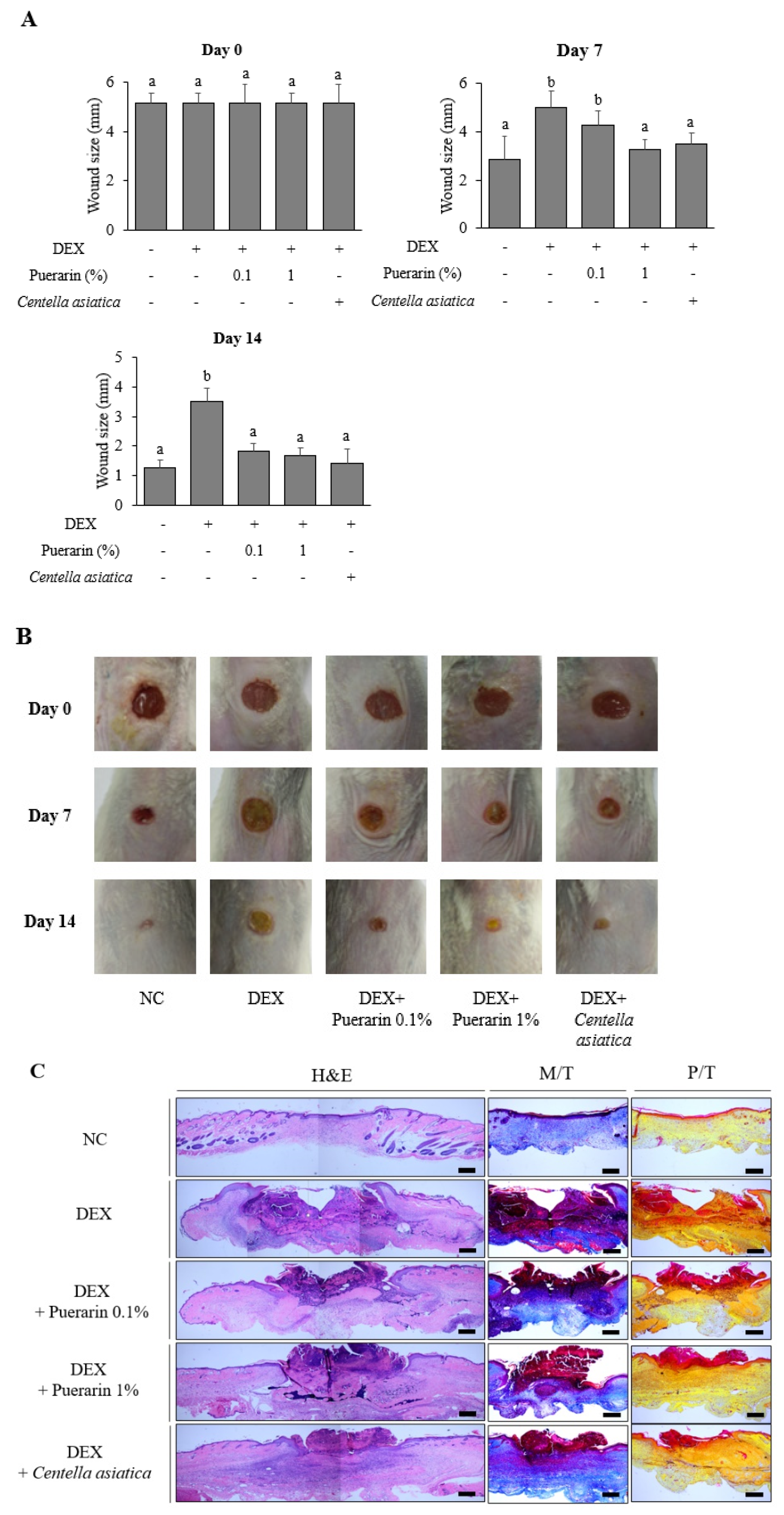
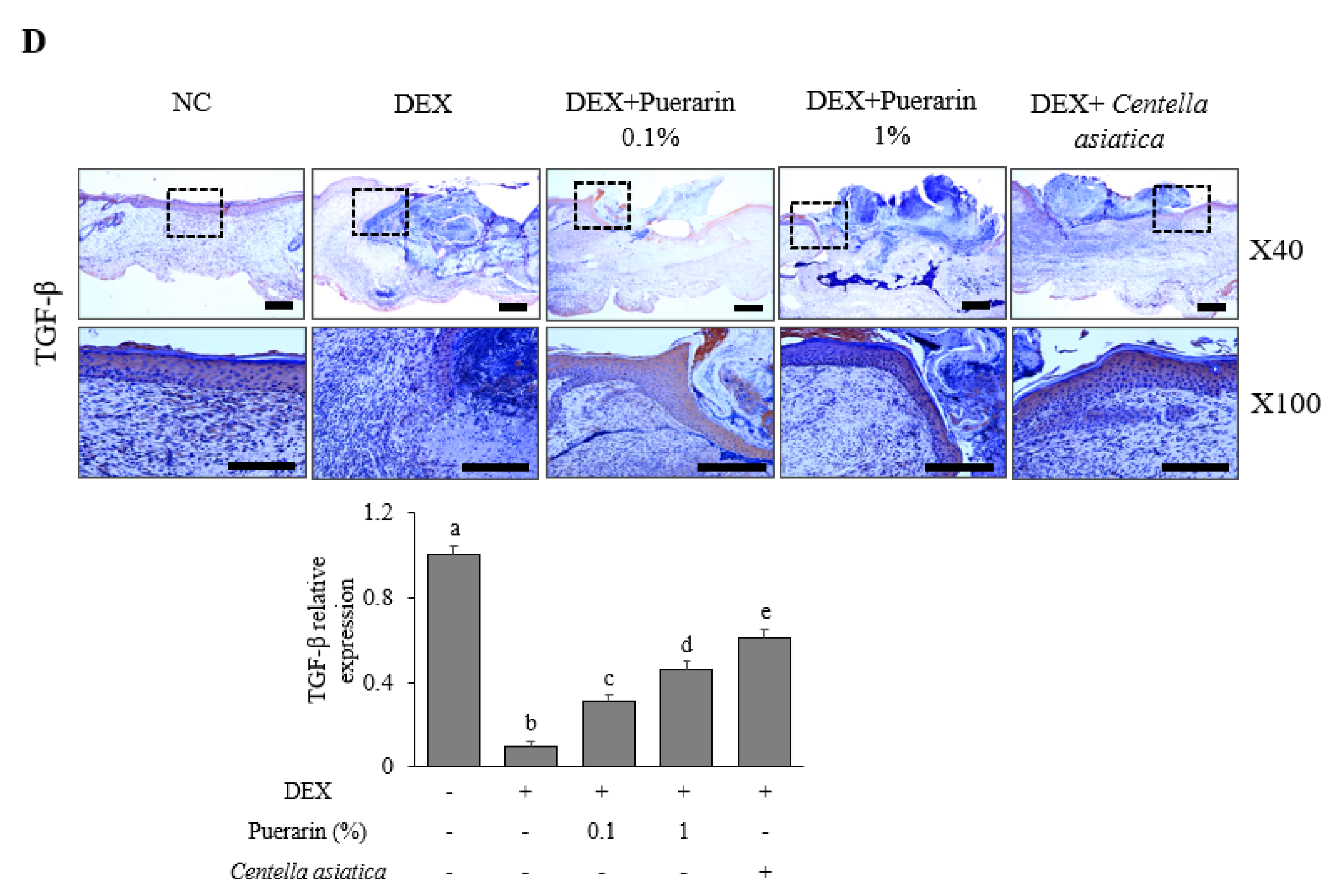
3.1. Effects of Puerarin on Cutaneous Wound Healing in DEX-Treated Mice

In the excision wound model, DEX significantly inhibited a wound contraction compared to the normal control group (day 7, day 14) (Figure 1A,B). In contrast, the treatment with puerarin promoted wound healing by decreasing the wound size compared to the DEX group (Figure 1A,B). Moreover, on Day 7 and Day 14 the wound size in the puerarin 1% group was comparable to that of the Centella asiatica treated group, which was known to promote DEX-suppressed wound healing [20]. Figure 1C presents the results of histological analysis of wound areas. H&E staining showed that the scar size in the DEX group was larger than the NC group, but this was decreased by a puerarin treatment (Figure 1C). M/T staining showed that the DEX group exhibited a lower level of collagen fiber (blue color) than the NC group, which was recovered by puerarin application (Figure 1C). P/T staining showed that the DEX group had fewer capillaries (yellow color under the epidermis) than the NC group, which was improved in the puerarin treated groups (Figure 1C). An immunohistochemical analysis (day 14) showed that the decreased TGF-β expression by DEX stimulation was increased significantly by the puerarin treatment (Figure 1D).

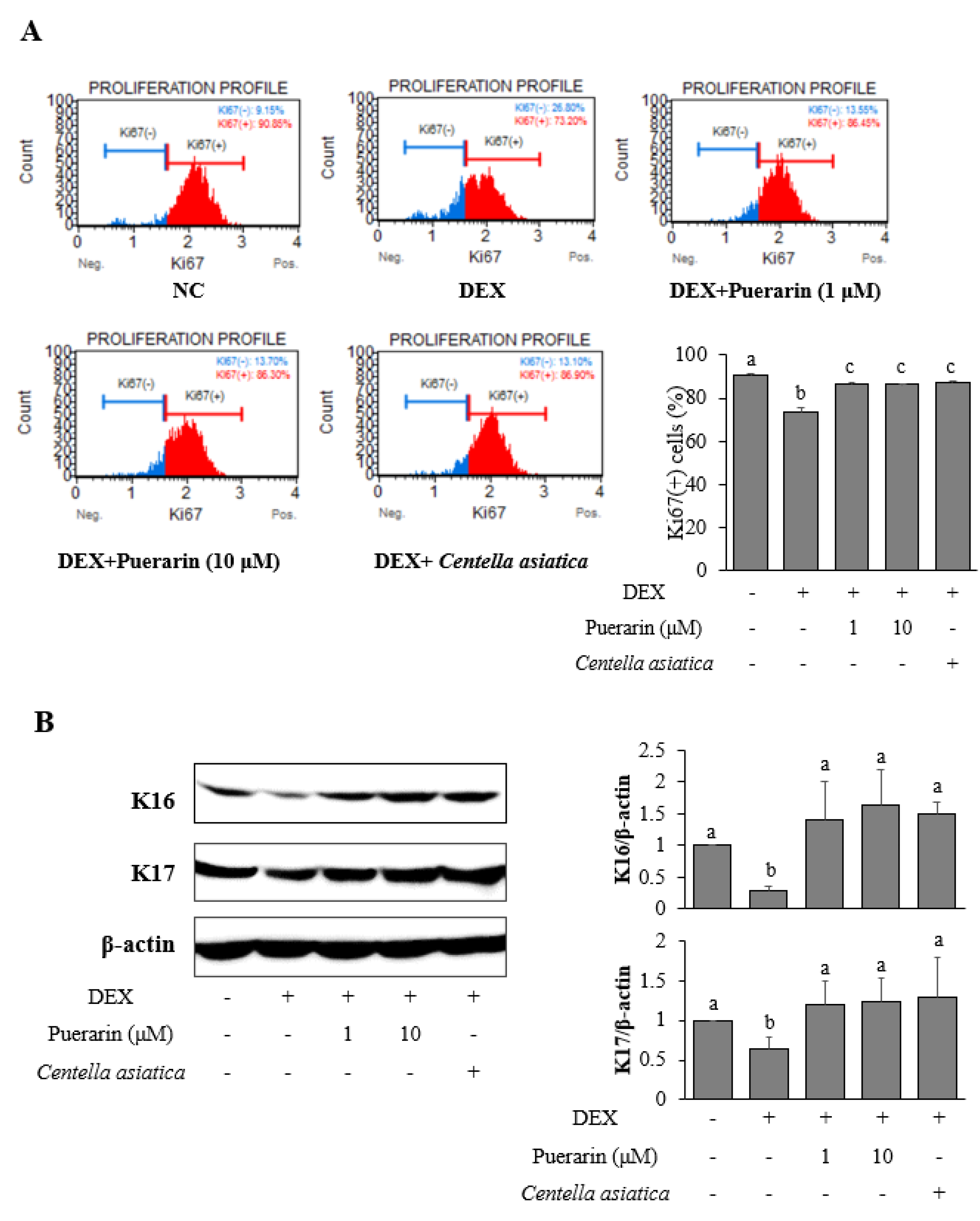
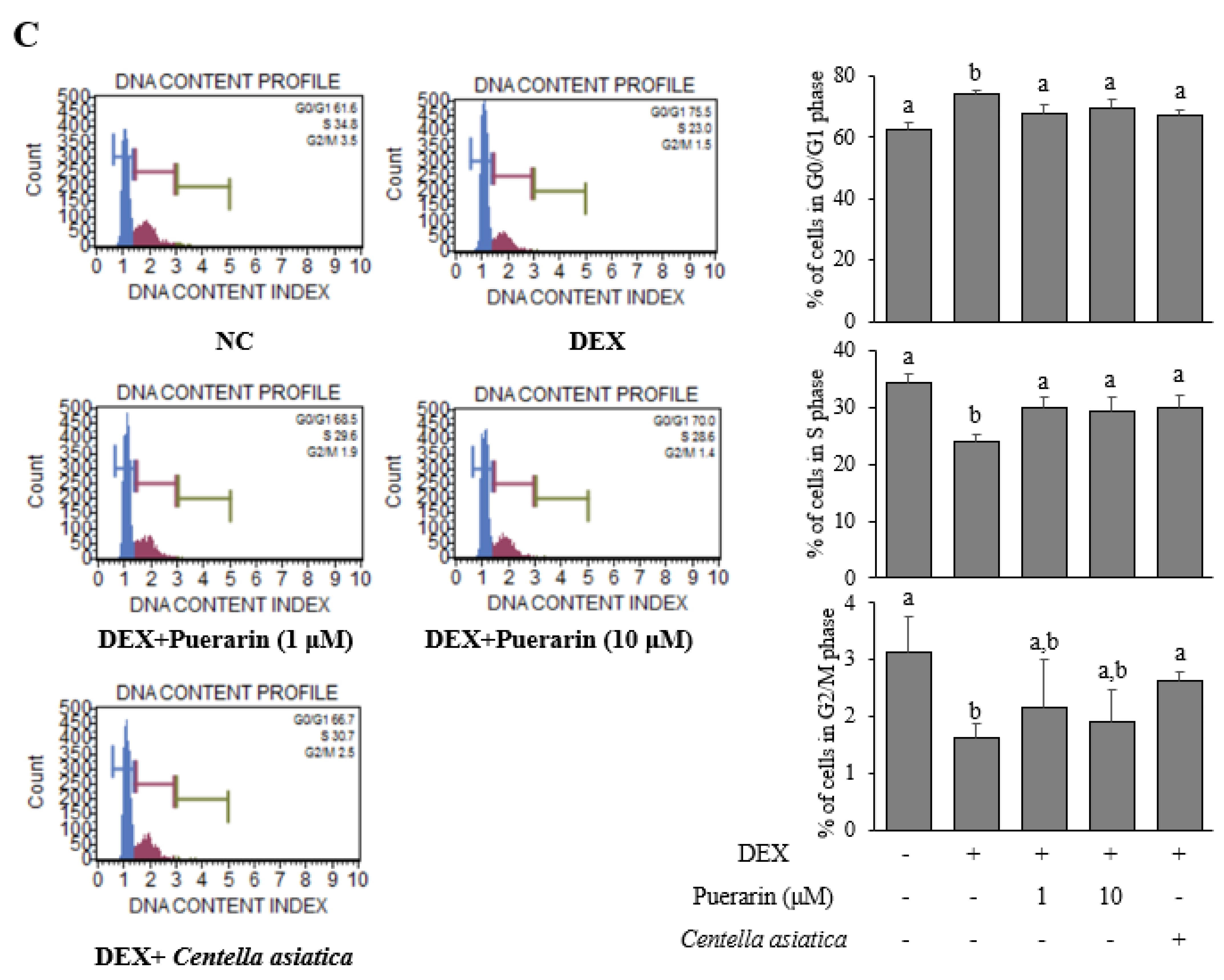
3.2. Effects of Puerarin on the Proliferation in DEX-Treated HaCaT Cells

A BrdU proliferation assay was performed to examine the effects of puerarin on the proliferation in DEX-treated HaCaT cells. The effects of puerarin on proliferation were evaluated using a Muse Ki67 assay, which showed that DEX remarkably decreased the number of Ki67(+) cells, which were recovered by treatment with puerarin (Figure 2A). Moreover, puerarin upregulated the expression of the proliferation markers, K16 and K17, in DEX-stimulated HaCaT cells (Figure 2B).
A flow cytometry analysis was conducted to examine if the cell cycle was involved in regulating cell proliferation in DEX-stimulated HaCaT cells. DEX significantly increased the proportion of cells arrested in the G0/G1 phase and decreased the proportion of cells arrested in the S and G2/M phase, whereas the treatment with puerarin had the opposite effects (Figure 2C).

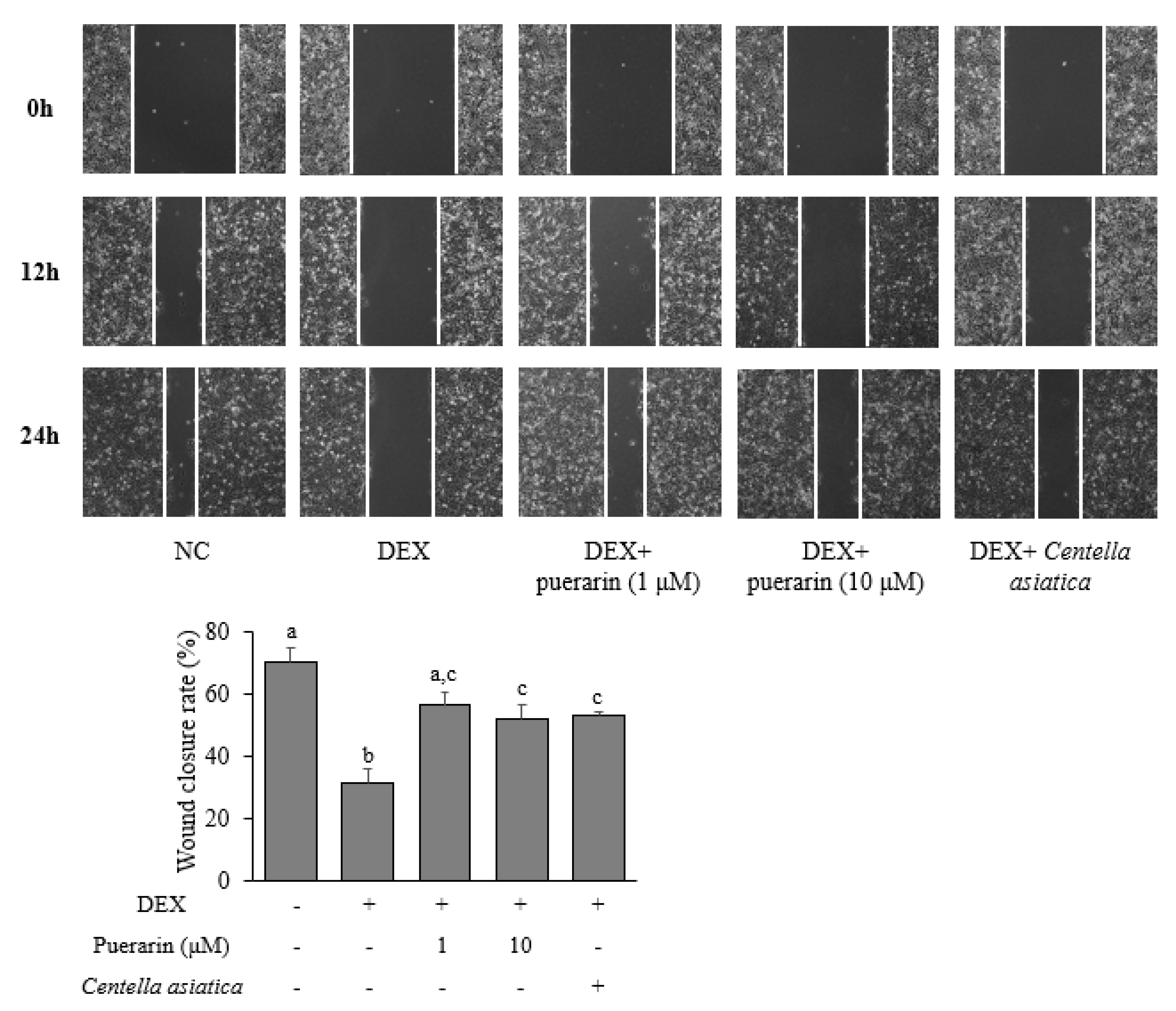
3.3. Effects of Puerarin on the Migration in DEX-Treated HaCaT Cells

The scratch wound healing assay was used to determine the effect of puerarin on cell migration. Figure 3 showed that DEX inhibited cell migration after 24 h. On the other hand, the treatment with puerarin increased the wound closure rate, in comparison with the DEX-only treated group.

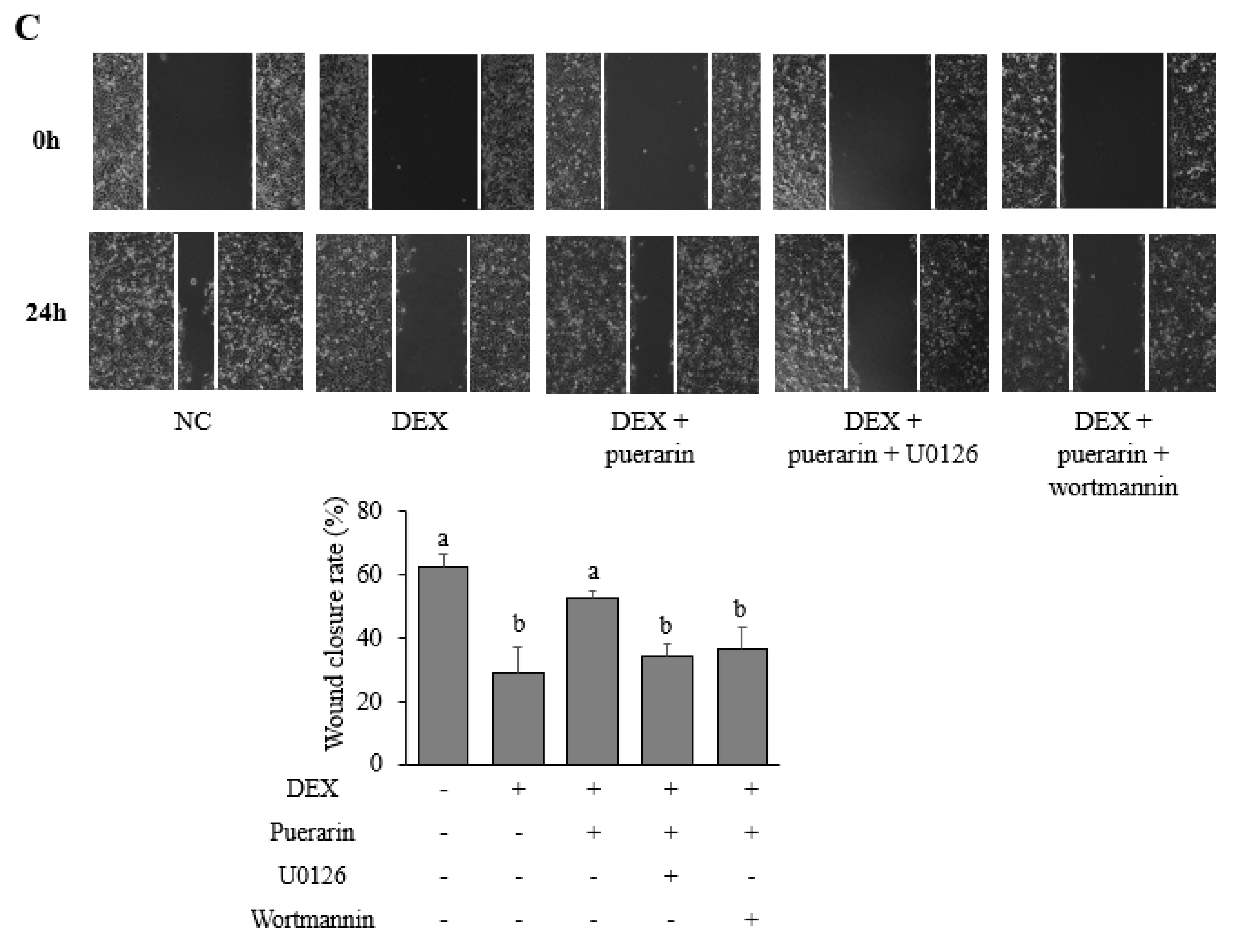
3.4. Effects of Puerarin on Signaling Pathways in DEX-Treated HaCaT Cells

Next, the effects of puerarin on the proliferation- and migration-related signaling pathways in DEX-stimulated HaCaT cells were evaluated. DEX inhibited the phosphorylation of ERK and Akt significantly, but the treatment with puerarin significantly increased the levels of p-ERK and p-Akt (Figure 4A). The role of ERK and Akt in mediating the effects of puerarin on proliferation and migration was confirmed using the inhibitors. As shown in Figure 4B, the treatment with U0126 (an ERK inhibitor) and wortmannin (an Akt inhibitor) prevented the effects of puerarin on cell proliferation in DEX-stimulated HaCaT. Moreover, U0126 and wortmannin abrogated the effects of puerarin on cell migration in DEX-stimulated HaCaT cells (Figure 4C).

4. Discussion

Wound healing is a complex process involving four overlapping phases: hemostasis, inflammation, proliferation, and remodeling [3]. The dysregulation of these stages leads to delayed wound healing. DEX is one of the most common corticosteroids used to treat inflammatory diseases but has adverse effects on wound healing by delaying the inflammation phase and inhibiting the proliferation phase of this process [5]. In this study, DEX impaired the wound healing process. A treatment with puerarin recovered the impaired wound healing process in both the in vivo and in vitro models.
Puerarin is a major isoflavonoid from the root of Pueraria montana var. lobata (Willd.) Sanjappa & Pradeep [6]. Several studies suggest that isoflavonoids have beneficial effects on the wound healing process [25,26]. An extract from the flower of Pueraria thunbergiana Benth (synonymous with Pueraria montana var. lobata (Willd.) Sanjappa & Pradeep), which contains isoflavonoids and essential oil components, has the potential for skin regeneration by stimulating the migration, proliferation, and collagen synthesis of epidermal keratinocytes [10]. A previous study reported that the oral administration of puerarin promoted wound healing in a diabetic rat model by enhancing the connective tissue and new vessel formation [16]. In the current study, the topical treatment of puerarin for 14 days also promoted the wound closure process in DEX-treated mice. Centella asiatica is a well-known wound healing agent in both clinical and experimental studies [18,27]. Centella asiatica used as the positive control drug is known to improve DEX-impaired wound healing ability [20]. In our experiments, the topical treatment of Centella asiatica restored the delayed wound closure by DEX application. The efficacy of Centella asiatica to improve wound healing is related to promoting collagen synthesis, angiogenesis, and keratinocyte migration [17,28]. Therefore, we investigated whether puerarin might be related to these mechanisms.
Collagen is the essential component of the dermal extracellular matrix (ECM), and collagen deposition in the dermis plays an important role in the wound healing process. Collagen is involved in the hemostasis phase by interacting with platelets in the wound areas [29]. The recruited platelets can release various growth factors, including the transforming growth factor-beta (TGF-β), platelet-derived growth factor (PDGF), and vascular endothelial growth factor (VEGF) [30]. TGF-β can induce fibroblast differentiation and collagen synthesis to promote wound closure and scarring [31]. VEGF is a crucial factor in the wound angiogenesis process as it promotes the proliferation and migration of endothelial cells [32]. Previous studies reported that glucocorticoids impaired wound healing by inhibiting collagen synthesis and angiogenesis in vivo [33,34]. In this study, DEX delayed the healing process and decreased the level of collagen fibers and the density of capillaries in a mouse excisional wound model. Moreover, the expression of TGF-β in the wound sites was also reduced in the DEX-treated mice. Puerarin treatment improved these phenomena, suggesting that puerarin might accelerate wound healing by promoting collagen deposition and angiogenesis.
Keratinocytes, the most predominant component of the epidermis, play an essential role in the wound healing process, particularly in the proliferation phase [35]. The proliferation phase includes granulation tissue and re-epithelialization. Granulation tissue formation consists of collagen synthesis by fibroblasts and neovascularization by endothelial cells, whereas re-epithelialization is characterized by the proliferation and migration of keratinocytes [36]. In this study, DEX inhibited wound healing significantly in the mouse model (day 7, day 14) and suppressed the proliferation and migration in HaCaT keratinocytes. The puerarin treatment recovered these impairments. Puerarin promoted keratinocyte proliferation by increasing Ki67, K16, and K17 expressions and switching the cell cycle from the G1 to the S phase in DEX-treated HaCaT cells. This result was consistent with a previous study showing that puerarin enhanced the proliferation and differentiation of osteoblasts to stimulate bone regeneration [37]. Puerarin also recovered DEX-impaired migration in HaCaT cells in the scratching sound healing assay. In contrast to the inhibitory effects of puerarin on the proliferation and migration in cancer studies [38,39], this report was the first indicating that puerarin could promote proliferation and migration in keratinocytes, improving the wound healing process in various DEX-treated models.
The underlying mechanisms mediating the effects of puerarin were investigated. ERK and Akt were critical signaling pathways that regulated a range of cellular processes, such as cell growth, proliferation, differentiation, apoptosis, and mobility [40,41]. The roles of ERK and Akt in keratinocyte proliferation and migration were demonstrated [42]. In the current study, DEX suppressed ERK and Akt phosphorylation, which were restored by a puerarin treatment. This result was consistent with a previous study indicating that icariin, a flavonoid from Herba Epimedii (the aerial parts from several Epimedium species), enhanced the migration and proliferation of keratinocytes via the ERK and Akt pathways [43]. Furthermore, ERK and Akt inhibitors repressed puerarin-induced proliferation and migration in DEX-treated HaCaT cells, highlighting the crucial roles of the ERK and Akt signaling pathways in mediating the effects of puerarin.
This study had some limitations. Wound healing is a complicated process with the involvement of various cellular components and overlapping events. This study only demonstrated the effects of puerarin on keratinocyte proliferation and migration. On the other hand, puerarin may affect other cell types, such as fibroblasts and endothelial cells. Therefore, further studies will be needed to prove the beneficial effects of puerarin on wound healing and to investigate other mechanisms mediating these effects.

5. Conclusions

Puerarin rescued DEX-impaired wound closure in an excision wound in a mouse model. This effect can be explained by the enhanced proliferation and migration of keratinocytes via the activation of ERK and Akt signaling pathways. These findings suggest that puerarin may be a potential candidate for treating DEX-suppressed wound healing.

Author Contributions

Conceptualization, I.-J.Y. and H.-M.S.; methodology, I.-J.Y. and S.-H.A.; formal analysis, L.T.H.N., S.-H.A. and M.-J.C.; writing—original draft preparation, L.T.H.N. and M.-J.C.; writing—review and editing, I.-J.Y. and H.-M.S. All authors have read and agreed to the published version of the manuscript.

Funding

This work was supported by the Dongguk University Research Fund (2020) and the National Research Foundation of Korea (NRF) funded by the Ministry of Education (grant no. NRF-2019R1F1A1059856).

Institutional Review Board Statement

The study was conducted according to the guidelines of the Declaration of Helsinki, and approved by the Institutional Review Board (or Ethics Committee) of Institutional Animal Care and Use Committee of Dongguk University approved all animal experimental procedures (No. IACUC-2019-7, 1 May 2019).

Informed Consent Statement

Not applicable.

Data Availability Statement

The data presented in this study are available in this article.

Conflicts of Interest

The authors declare no conflict of interest.

References

  1. Mizuno, K.; Morizane, S.; Takiguchi, T.; Iwatsuki, K. Dexamethasone but not tacrolimus suppresses TNF-alpha-induced thymic stromal lymphopoietin expression in lesional keratinocytes of atopic dermatitis model. J. Dermatol. Sci. 2015, 80, 45–53. [Google Scholar] [CrossRef] [PubMed] [Green Version]
  2. Hengge, U.R.; Ruzicka, T.; Schwartz, R.A.; Cork, M.J. Adverse effects of topical glucocorticosteroids. J. Am. Acad. Dermatol. 2006, 54, 1–15. [Google Scholar] [CrossRef] [PubMed]
  3. Velnar, T.; Bailey, T.; Smrkoli, V. The Wound Healing Process: An Overview of the Cellular and Molecular Mechanisms. J. Int. Med. Res. 2009, 37, 1528–1542. [Google Scholar] [CrossRef] [PubMed]
  4. Santoro, M.M.; Gaudino, G. Cellular and molecular facets of keratinocyte reepithelization during wound healing. Exp. Cell. Res. 2005, 304, 274–286. [Google Scholar] [CrossRef] [PubMed]
  5. Slominski, A.T.; Zmijewski, M.A. Glucocorticoids Inhibit Wound Healing: Novel Mechanism of Action. J. Investig. Dermatol. 2017, 137, 1012–1014. [Google Scholar] [CrossRef] [PubMed] [Green Version]
  6. Heo, H.S.; Han, G.E.; Won, J.; Cho, Y.; Woo, H.; Lee, J.H. Pueraria montana var. lobata root extract inhibits photoaging on skin through Nrf2 pathway. J. Microbiol. Biotechnol. 2019, 29, 518–526. [Google Scholar] [CrossRef]
  7. Tungmunnithum, D.; Intharuksa, A.; Sasaki, Y. A Promising View of Kudzu Plant, Pueraria montana var. lobata (Willd.) Sanjappa & Pradeep: Flavonoid Phytochemical Compounds, Taxonomic Data, Traditional Uses and Potential Biological Activities for Future Cosmetic Application. Cosmetics 2020, 7, 12. [Google Scholar]
  8. Carvalho, M.T.B.; Araújo-Filho, H.G.; Barreto, A.S.; Quintans-Júnior, L.J.; Quintans, J.S.S.; Barreto, R.S.S. Wound healing properties of flavonoids: A systematic review highlighting the mechanisms of action. Phytomedicine 2021, 90, 153636. [Google Scholar] [CrossRef]
  9. Zhang, S.; Ou, Q.; Xin, P.; Yuan, Q.; Wang, Y.; Wu, J. Polydopamine/puerarin nanoparticle-incorporated hybrid hydrogels for enhanced wound healing. Biomater. Sci. 2019, 7, 4230–4236. [Google Scholar] [CrossRef]
  10. Kim, D.Y.; Won, K.J.; Hwang, D.I.; Yoon, S.W.; Lee, S.J.; Park, J.H.; Yoon, M.S.; Kim, B.; Lee, H.M. Potential Skin Regeneration Activity and Chemical Composition of Absolute from Pueraria thunbergiana Flower. Nat. Prod. Commun. 2015, 10, 2009–2012. [Google Scholar] [CrossRef]
  11. Reppert, A.; Yousef, G.G.; Rogers, R.B.; Lila, M.A. Isolation of radiolabeled isoflavones from kudzu (Pueraria lobata) root cultures. J. Agric. Food Chem. 2008, 56, 7860–7865. [Google Scholar] [CrossRef] [Green Version]
  12. Jung, H.W.; Kang, A.N.; Kang, S.Y.; Park, Y.-K.; Song, M.Y. The root extract of Pueraria lobata and its main compound, puerarin, prevent obesity by increasing the energy metabolism in skeletal muscle. Nutrients 2017, 9, 33. [Google Scholar] [CrossRef] [PubMed]
  13. Lee, J.H.; Jeon, Y.D.; Lee, Y.M.; Kim, D.K. The suppressive effect of puerarin on atopic dermatitis-like skin lesions through regulation of inflammatory mediators in vitro and in vivo. Biochem. Biophys. Res. Commun. 2018, 498, 707–714. [Google Scholar] [CrossRef]
  14. Zhang, S.; Chen, S.; Shen, Y.; Yang, D.; Liu, X.; Sun-Chi, A.C.; Xu, H. Puerarin induces angiogenesis in myocardium of rat with myocardial infarction. Biol. Pharm. Bull. 2006, 29, 945–950. [Google Scholar] [CrossRef] [Green Version]
  15. Jin, S.E.; Son, Y.K.; Min, B.S.; Jung, H.A.; Choi, J.S. Anti-inflammatory and antioxidant activities of constituents isolated from Pueraria lobata roots. Arch. Pharm. Res. 2012, 35, 823–837. [Google Scholar] [CrossRef] [PubMed]
  16. Turer, A.; Onger, M.E. The effect of oral puerarin administration on wound healing in diabetic rat model. Ann. Med. Res. 2018, 25, 536–539. [Google Scholar] [CrossRef]
  17. Somboonwong, J.; Kankaisre, M.; Tantisira, B.; Tantisira, M.H. Wound healing activities of different extracts of Centella asiatica in incision and burn wound models: An experimental animal study. BMC Complement. Altern. Med. 2012, 12, 103. [Google Scholar] [CrossRef] [Green Version]
  18. Azis, H.; Taher, M.; Ahmed, A.; Sulaiman, W.; Susanti, D.; Chowdhury, S.; Zakaria, Z. In vitro and In vivo wound healing studies of methanolic fraction of Centella asiatica extract. S. Afr. J. Bot. 2017, 108, 163–174. [Google Scholar] [CrossRef]
  19. Ruksiriwanich, W.; Khantham, C.; Sringarm, K.; Sommano, S.; Jantrawut, P. Depigmented Centella asiatica Extraction by Pretreated with Supercritical Carbon Dioxide Fluid for Wound Healing Application. Processes 2020, 8, 277. [Google Scholar] [CrossRef] [Green Version]
  20. Shetty, B.S.; Udupa, S.L.; Udupa, A.L.; Somayaji, S.N. Effect of Centella asiatica L (Umbelliferae) on normal and dexamethasone-suppressed wound healing in Wistar Albino rats. Int. J. Low. Extrem. Wounds 2006, 5, 137–143. [Google Scholar] [CrossRef] [PubMed]
  21. Moreira, C.F.; Cassini-Vieira, P.; da Silva, M.F.; Barcelos, L. Skin wound healing model-excisional wounding and assessment of lesion area. Bio-Protocol 2015, 5, e1661. [Google Scholar] [CrossRef]
  22. Suvik, A.; Effendy, A. The use of modified Masson’s trichrome staining in collagen evaluation in wound healing study. Mal. J. Vet. Res. 2012, 3, 39–47. [Google Scholar]
  23. Cho, A.-R.; Hong, S.-U.; Yoon, Y.-J. Gagamyeonryunggobon-dan (Jiājiǎnyánlínggùběn-dān) induces hair regrowth effect from activating hair follicle. J. Korean Orient. Otolaryngol. Dermatol. 2016, 29, 65–80. [Google Scholar]
  24. Su, L.; Fu, L.; Li, X.; Zhang, Y.; Li, Z.; Wu, X.; Li, Y.; Bai, X.; Hu, D. Loss of CAR promotes migration and proliferation of HaCaT cells and accelerates wound healing in rats via Src-p38 MAPK pathway. Sci. Rep. 2016, 6, 19735. [Google Scholar] [CrossRef] [Green Version]
  25. Aslam, M.S.; Ahmad, M.S.; Riaz, H.; Raza, S.A.; Hussain, S.; Qureshi, O.S.; Maria, P.; Hamzah, Z.; Javed, O. Role of Flavonoids as Wound Healing Agent. In Phytochemicals-Source of Antioxidants and Role in Disease Prevention; Asao, T., Asaduzzaman, M., Eds.; IntechOpen: London, UK, 2018; pp. 95–102. [Google Scholar]
  26. Öz, B.E.; İşcan, G.S.; Akkol, E.K.; Süntar, İ.; Acıkara, Ö.B. Isoflavonoids as wound healing agents from Ononidis Radix. J. Ethnopharmacol. 2018, 211, 384–393. [Google Scholar]
  27. Bylka, W.; Znajdek-Awiżeń, P.; Studzińska-Sroka, E.; Dańczak-Pazdrowska, A.; Brzezińska, M. Centella asiatica in dermatology: An overview. Phytother. Res. 2014, 28, 1117–1124. [Google Scholar] [CrossRef]
  28. Singkhorn, S.; Tantisira, M.H.; Tanasawet, S.; Hutamekalin, P.; Wongtawatchai, T.; Sukketsiri, W. Induction of keratinocyte migration by ECa 233 is mediated through FAK/Akt, ERK, and p38 MAPK signaling. Phytother. Res. 2018, 32, 1397–1403. [Google Scholar] [CrossRef]
  29. Davison-Kotler, E.; Marshall, W.S.; Garcia-Gareta, E. Sources of Collagen for Biomaterials in Skin Wound Healing. Bioengineering 2019, 6, 56. [Google Scholar] [CrossRef] [Green Version]
  30. Chicharro-Alcantara, D.; Rubio-Zaragoza, M.; Damia-Gimenez, E.; Carrillo-Poveda, J.M.; Cuervo-Serrato, B.; Pelaez-Gorrea, P.; Sopena-Juncosa, J.J. Platelet Rich Plasma: New Insights for Cutaneous Wound Healing Management. J. Funct. Biomater. 2018, 9, 10. [Google Scholar] [CrossRef] [PubMed] [Green Version]
  31. Penn, J.W.; Grobbelaar, A.O.; Rolfe, K.J. The role of the TGF-beta family in wound healing, burns and scarring: A review. Int. J. Burns Trauma 2012, 2, 18–28. [Google Scholar]
  32. Johnson, K.E.; Wilgus, T.A. Vascular Endothelial Growth Factor and Angiogenesis in the Regulation of Cutaneous Wound Repair. Adv. Wound Care 2014, 3, 647–661. [Google Scholar] [CrossRef] [Green Version]
  33. Wicke, C.; Halliday, B.; Allen, D.; Roche, N.S.; Scheuenstuhl, H.; Spencer, M.M.; Roberts, A.B.; Hunt, T.K. Effects of steroids and retinoids on wound healing. Arch. Surg. 2000, 135, 1265–1270. [Google Scholar] [CrossRef] [Green Version]
  34. Morgan, R.; Keen, J.; Halligan, D.; O’Callaghan, A.; Andrew, R.; Livingstone, D.; Abernethie, A.; Maltese, G.; Walker, B.; Hadoke, P. Species-specific regulation of angiogenesis by glucocorticoids reveals contrasting effects on inflammatory and angiogenic pathways. PLoS ONE 2018, 13, e0192746. [Google Scholar] [CrossRef] [Green Version]
  35. Pastar, I.; Stojadinovic, O.; Tomic-Canic, M. Role of keratinocytes in healing of chronic wounds. Surg. Technol. Int. 2008, 17, 105–112. [Google Scholar]
  36. Hopf, H.W.; Kelly, M.; Shapshak, D. Oxygen and the basic mechanisms of wound healing. In Physiology and Medicine of Hyperbaric Oxygen Therapy; Neuman, T.S., Thom, S.R., Eds.; Saunders: Philadelphia, PA, USA, 2008; pp. 203–228. [Google Scholar]
  37. Zeng, X.; Feng, Q.; Zhao, F.; Sun, C.; Zhou, T.; Yang, J.; Zhan, X. Puerarin inhibits TRPM3/miR-204 to promote MC3T3-E1 cells proliferation, differentiation and mineralization. Phytother. Res. 2018, 32, 996–1003. [Google Scholar] [CrossRef] [PubMed]
  38. Jia, L.; Hu, Y.; Yang, G.; Li, P. Puerarin suppresses cell growth and migration in HPV-positive cervical cancer cells by inhibiting the PI3K/mTOR signaling pathway. Exp. Ther. Med. 2019, 18, 543–549. [Google Scholar] [CrossRef] [Green Version]
  39. Pan, L.M.; Wu, M.; Yuan, S.F.; Geng, J.H.; Kong, X.D.; Shen, J.L. Effects of puerarin on proliferation, apoptosis, migration and invasion of hormone-independent prostate cancer cells. Tumor 2018, 38, 680–688. [Google Scholar]
  40. Manning, B.D.; Toker, A. AKT/PKB signaling: Navigating the network. Cell 2017, 169, 381–405. [Google Scholar] [CrossRef] [Green Version]
  41. McCubrey, J.A.; Steelman, L.S.; Chappell, W.H.; Abrams, S.L.; Wong, E.W.; Chang, F.; Lehmann, B.; Terrian, D.M.; Milella, M.; Tafuri, A. Roles of the Raf/MEK/ERK pathway in cell growth, malignant transformation and drug resistance. Biochim. Biophys. Acta Mol. Cell Res. 2007, 1773, 1263–1284. [Google Scholar] [CrossRef] [PubMed] [Green Version]
  42. Zhou, T.; Yang, Z.; Chen, Y.; Chen, Y.; Huang, Z.; You, B.; Peng, Y.; Chen, J. Estrogen accelerates cutaneous wound healing by promoting proliferation of epidermal keratinocytes via Erk/Akt signaling pathway. Cell. Physiol. Biochem. 2016, 38, 959–968. [Google Scholar] [CrossRef] [PubMed]
  43. Mi, B.; Liu, J.; Liu, G.; Zhou, W.; Liu, Y.; Hu, L.; Xiong, L.; Ye, S.; Wu, Y. Icariin promotes wound healing by enhancing the migration and proliferation of keratinocytes via the AKT and ERK signaling pathway. Int. J. Mol. Med. 2018, 42, 831–838. [Google Scholar] [CrossRef] [PubMed]
Figure 1. Effects of puerarin on cutaneous wound healing in mice treated with DEX. (A) Wound sizes were measured on day 0, day 7, and day 14 of the experiment. (B) Representative images of wounds from five groups. (C) Hematoxylin and eosin (H&E), Masson trichrome (M/T), and phloxine-tartrazine (P/T) stained sections of wound samples from the five groups. Scale bar: 50 μm. (D) Immunohistochemical analysis (day 14) of TGF-β in the wound areas. The data are presented as the means ± SDs (n = 6 per experiment). Bars labeled by the same letter on the graph are not significantly different from each other (p < 0.05) by one-way ANOVA followed by post hoc Tukey’s test.
Applsci 11 09343 g001aApplsci 11 09343 g001b
Figure 2. Effects of puerarin on the proliferation of HaCaT cells stimulated with DEX. HaCaT cells were incubated with DEX (100 nM) and puerarin (1, 10 µM) or Centella asiatica extract (100 µg/mL) for 48 h. (A) Ki67 expression was assessed using a Muse Ki67 Proliferation kit. (B) Protein levels of K16 and K17 were determined by Western blotting. (C) Cell cycle analysis was performed using a Muse Cell Cycle kit. The results are presented as the means ± SDs of three independent experiments. Bars labeled by the same letter on the graph are not significantly different from each other (p < 0.05) by one-way ANOVA followed by post hoc Tukey’s test.
Applsci 11 09343 g002aApplsci 11 09343 g002b
Figure 3. Effects of puerarin on cell migration in HaCaT cells stimulated with DEX. The HaCaT cells were incubated with DEX (100 nM) and puerarin (1, 10 µM) or Centella asiatica extract (100 µg/mL). Before the scratch wound was made, the cells were treated with mitomycin C (10 µg/mL) for 1 h to inhibit cell proliferation. The images of the scratch gap width were captured at 0, 12, and 24 h post-scratching. The wound closure rates were defined as the difference between the wound width at 0 and 24 h. The results are presented as the means ± SDs of three independent experiments. Bars labeled by the same letter on the graph are not significantly different from each other (p < 0.05) by one-way ANOVA followed by post hoc Tukey’s test.
Applsci 11 09343 g003
Figure 4. Effects of puerarin on the ERK and Akt signaling pathways in HaCaT cells stimulated with DEX. (A) HaCaT cells were incubated with DEX (100 nM) and puerarin (1, 10 µM) or Centella asiatica extract (100 µg/mL) for 30 min. Protein expression of p-ERK, p-Akt, ERK, Akt was determined by Western blotting. (B) HaCaT cells were pretreated with U0126 (10 µM) or wortmannin (1 µM) for 1 h and then treated with DEX (100 nM) and puerarin (1 µM) for 48 h. The proliferation rate was assessed using BrdU assays. (C) HaCaT cells were pretreated with U0126 (10 µM) or wortmannin (1 µM) for 1 h and scratched and treated with dexamethasone (DEX, 100 nM) and puerarin (1 µM). Images of the scratch gap width were captured at 0 and 24 h post-scratching. The wound closure rates were defined as the difference between the wound width at 0 and 24 h. The results are presented as the means ± SDs (n = 3 per experiment). Bars labeled by the same letter on the graph are not significantly different from each other (p < 0.05) by one-way ANOVA followed by post hoc Tukey’s test.
Applsci 11 09343 g004aApplsci 11 09343 g004b
Publisher’s Note: MDPI stays neutral with regard to jurisdictional claims in published maps and institutional affiliations.

