Article

Increased Expression of TGF-β1 by 4-hexylresorcinol Is Mediated by Endoplasmic Reticulum and Mitochondrial Stress in Human Umbilical Endothelial Vein Cells

1
Hallym University Kangnam Sacred Heart Hospital, Hallym University Medical Center, 1, Singil-ro, Yeongdeungpo-gu, Seoul 07441, Korea
2
Department of Oral Biochemistry, College of Dentistry, Gangneung-Wonju National University, Gangneung 28644, Korea
3
Institution of Hydrogen Magnetic Reaction Gene Regulation, Daejeon 34140, Korea
4
Department of Biochemistry and Cell Biology, Cell and Matrix Research Institute, Skeletal Diseases Analysis Center, Korea Mouse Phenotyping Center (KMPC), School of Medicine, Kyungpook National University, Daegu 41944, Korea
5
Department of Oral and Maxillofacial Surgery, College of Dentistry, Gangneung-Wonju National University, Gangneung 28644, Korea
6
Biomedical, Surgical and Oral Sciences Department, Maxillofacial and Dental Unit, School of Dentistry, University of Milan, 20122 Milan, Italy
*
Author to whom correspondence should be addressed.
Appl. Sci. 2021, 11(19), 9128; https://doi.org/10.3390/app11199128
Submission received: 1 September 2021 / Revised: 27 September 2021 / Accepted: 28 September 2021 / Published: 30 September 2021

Abstract

:

Featured Application

For uneventful wound healing, endothelial cell differentiation is a vital component, and transforming growth factor-β1 (TGF-β1) orchestrates this process. The administration of 4-hexylresorcinol increased transforming growth factor-β1 expression in endothelial cells via endoplasmic reticulum (ER) and mitochondrial stress combined with its histone deacetylase inhibitor activity.

Abstract

In our previous study, 4-hexylresorcinol (4HR) increased the expression level of vascular endothelial growth factor in human umbilical vein endothelial cells (HUVECs) via the transforming growth factor-β1 (TGF-β1)-mediated pathway. Endoplasmic reticulum (ER) and mitochondrial stress is a positive regulator of cellular differentiation. As TGF-β1 is a master regulator for cellular differentiation, 4HR treatment may increase TGF-β1 expression via ER stress. In this study, HUVECs were treated using 4HR (1–100 μM) for 24 h. The 4HR treatment increased ER stress-associated markers and mitochondrial stress. Increased TGF-β1 expression by 4HR administration was alleviated by tauroursodeoxycholate (ER stress inhibitor) treatment. Combining these activities with the elevated acetylation level of histone 3 (H3) by 4HR treatment, TGF-β1 expression was increased in HUVECs. Overall, 4HR increased TGF-β1 expression through upregulation of the stress response of ER as well as H3 acetylation in HUVECs.

1. Introduction

Some phenolic compounds such as resorcinolic lipids can bind to proteins and change their conformation [1]. The administration of resorcinolic lipids suppresses microbial growth and induces dormancy [2]. Although it is hard to define dormancy as a biochemical aspect, a life in dormancy shows decreased metabolism with resistance to environmental stress [2,3]. Therefore, resorcinolic lipids can be considered chemical chaperones. Resorcinolic lipids can be produced by cells or synthesized chemically [1]. When a chemical chaperone is applied from outside, most active proliferating microorganisms will die [3]. Thus, chemical chaperones have two conflicting aspects: one protects them from environmental stress [2], while the other kills actively proliferating competitors to save the available energy resources [3]. Translating the activity of a chemical chaperone from a microorganism to multi-cellular life, such as mammals, is a logical leap, and many variables should be considered. However, the application of chemical chaperones may have therapeutic benefits. Application of some phenolic compounds having chaperone-like properties induces apoptosis or differentiates cancer cells [4,5]. Endoplasmic reticulum (ER) and mitochondria are the main intracellular organelles that control this response [6,7]. Chemical chaperones may stimulate ER and mitochondrial production of proteins inducing cellular differentiation or apoptosis. If resorcinolic lipids are used in a controlled manner, their administration may differentiate cells via ER and mitochondrial stress, which could be helpful for wound healing.
4-Hexylresorcinol (4HR) is a resorcinolic lipid and has been studied for angiogenesis and wound healing [ref]. In our previous study, application of 4HR on human umbilical vein endothelial cells (HUVECs) increased vascular endothelial growth factor (VEGF) via the hypoxia-inducible factor (HIF)-independent pathway [8]. 4HR-induced apoptotic stress increased the expression of transforming growth factor-β1 (TGF-β1) [9]. Treatment of TGF-β1 siRNA decreased VEGF expression in HUVECs, and TGF-β1-mediated angiogenesis increased capillary formation in a diabetic animal model [9]. The application of TGF-β1 supplemented with VEGF resulted in early wound closure and improved angiogenesis and vasculogenesis [10]. The application of TGF-β1 on stem cells induced cellular differentiation into functional pericyte-like cells [11]. However, the mechanism of 4HR-induced TGF-β1 is unclear.
Cellular stress is mainly transmitted to mitochondria and ER, inducing physiological or pathological events [7]. Stressed mitochondria and ER perform important functions in cellular differentiation and apoptosis [7]. If mitochondria and the ER fail to maintain cellular homeostasis, apoptosis can be activated. A successful adaptive response may introduce cellular growth and differentiation. As ER stress increases the expression level of TGF-β1 [12], an elevated level of TGF-β1 after 4HR administration may be induced by ER stress. Tauroursodeoxycholate (TUDCA) has been used as an ER stress reliever [13]. If 4HR increases TGF-β1 expression via an ER stress-mediated pathway, the application of TUDCA will alleviate TGF-β1 expression after 4HR administration. Accordingly, the association between 4HR administration and ER stress can be evaluated by investigating the change in TGF-β1 expression with or without TUDCA addition.
Many proteins are involved in the adaptive response of the ER. Sirtuins (SIRTs) are class III histone deacetylases (HDACs) and have seven distinct isoforms (SIRT1-7) [14]. SIRTs are mainly localized in the nucleus and mitochondria [15]. The expression level of SIRTs increases in response to cellular stress [15]. SIRTs help cells to maintain cellular homeostasis in stressful situations [12,15]. 4HR induced several antioxidant proteins in RAW 264.7 cells [16] and HUVECs [17] and has similar antioxidant activity to resveratrol in HUVECs [8] and lymphocytes [18]. As SIRTs induce antioxidant enzymes, 4HR may have a role as an SIRT activator. These responses may be associated with reducing mitochondrial and ER stress. Inhibition of specificity protein 1 (Sp1) disrupts ER homeostasis [19]. Sp1 is a transcription factor for TGF-β1 [20]. Interestingly, 4HR also increases Sp1 expression in oral cancer cells and SCC-9 cells [21]. In addition, 4HR is a class I histone deacetylase inhibitor (HDACi) [22]. It is known that hyper-acetylated histone 3 (H3) is important for TGF-β1 expression [23]. The inhibition of class I HDACs by 4HR might induce the hyper-acetylation of H3 and consequent TGF-β1 expression.
The aim of this study was to clarify the cellular mechanism of TGF-β1 expression after 4HR administration in HUVECs. To answer this question, the relationship between 4HR application and ER and mitochondrial stress was studied. To clarify ER stress, changes in the expression levels of ER stress marker proteins were observed after 4HR administration. The ATP level in the mitochondria, mitochondrial membrane potential (MMP), and cytochrome c level were checked for the evaluation of mitochondrial stress. Additionally, the level of acetylated H3 and Sp1 activity after 4HR application was also clarified.

2. Materials and Methods

2.1. HUVEC Culture

HUVECs (Lonza, Walkersville, MD, USA) were cultured as described in our previous publications [15,24]. The medium was endothelial cell growth medium-2 (Clonetics®, Lonza). Cells were cultured in a CO2 incubator. To prevent mycoplasma contamination, tests were performed on a regular basis. All cellular experiments were performed at least in triplicate.

2.2. Western Blot and Sp1 Transcription Factor Assay

When HUVECs were grown (approximately 70% confluent), cells were treated with 1, 10, and 100 µM 4HR for 2, 8, or 24 h; control cells were treated with 0.1% dimethyl sulfoxide in culture medium. TUDCA, a known ER stress inhibitor, was purchased from Sigma-Aldrich (CAT#: T0266, St. Lois, MO, USA). TUDCA inhibits ER stress with an EC50 value of 100 μM [25]. Accordingly, 100 μM TUDCA was applied. Cultured cells were harvested with protein lysis buffer (PRO-PREPTM, iNtRON Biotechnology INC, Sungnam, Korea) and underwent Western blotting for TGF-β1, VEGF-A, VEGF-C, Sp1, p-Sp1, GRP78, PERK, p-PERK, eIF2, p-eIF2, ATF4, ATF6α, Ac-lys, H3, and Ac-H3. Mithramycin is an Sp1 inhibitor and was purchased from Sigma-Aldrich (CAT#: M6891). Mithramycin inhibits Sp1 with an EC50 value of 25 μM. Additional Western blot analyses for SIRT1, SIRT3, and SIRT6 were also carried out. Antibodies against TGF-β1, VEGF-A, VEGF-C, Sp1, p-Sp1, GRP78, PERK, p-PERK, eIF2, p-eIF2, ATF4, ATF6α, Ac-lys, H3, and Ac-H3 were purchased from Santa Cruz Biotechnology (Santa Cruz, CA, USA). Antibodies against SIRTs were purchased from Abcam (Cambridge, UK). Quantification of the proteins was performed as described previously [16,17]. For the assessment of apoptosis by 4HR administration, a cytochrome c apoptosis antibody cocktail (CAT: ab110415, Abcam) was used. The final concentration of the antibody cocktail was 3.6 μg/mL. The cell collection was separated into mitochondrial and cytoplasmic fractions. Each fraction underwent Western blot analysis using an antibody cocktail.
Sp1 activation detection was performed using an Sp1 transcription factor assay kit (CAT: ab207226, Abcam). To detect GC box-bound Sp1, an oligonucleotide with an Sp1 consensus binding site coated plate was used. To assess the effect of 4HR administration on Sp1 activation levels, 1, 10, and 100 μM 4HR were applied, and nuclear extracts were collected after 2, 8, and 24 h. The subsequent procedure was in accordance with the manufacturer’s protocol. Briefly, samples were added to appropriate wells and the plate was incubated for 1 h at room temperature. Primary antibody was then added to the wells, and the plate was incubated again for 1 h at room temperature. After the washing process, HRP-conjugated secondary antibody was applied, and the plate was incubated for 1 h at room temperature. After another washing process, a developing solution was added, and we waited until the medium color turned dark blue. Then, a stop solution was added, and the absorbance was measured at 450 nm.

2.3. ATP, Mitochondrial Membrane Potential (MMP), and Oxygen Consumption Assay

ATP detection was performed using a luminescent ATP detection assay kit (CAT: ab113849, Abcam). HUVECs were seeded into 6 well plate. The number of seeded cells was 2 × 10⁵ cells per well. To assess the effect of 4HR administration on ATP levels, 1, 10, and 100 μM 4HR were applied, and cellular lysates were collected after 8 and 24 h. The subsequent procedure was in accordance with the manufacturer’s protocol. Briefly, ATP standard was added into standard wells, and medium was added into control wells. The detergent solution was added and incubated for 5 min. Then, the substrate solution was added and incubated for 5 min. The plate was stored in a dark room for 10 min. The luminescence was measured using a plate reader.
MMP was measured using a commercially available kit (CAT: MAK159, Sigma-Aldrich). Cells were prepared on a 96-well plate. The number of seeded cells was 5 × 10⁴ cells per well. The JC-10 Dye Loading Solution was prepared by adding 50 μL of 100× JC-10 to 5 mL of Assay Buffer A. Cells were treated with 10 μL of 10× test compounds to induce apoptosis. In parallel, we set up negative (vehicle only) and 4HR-treated samples. The cells were incubated for 24 h. JC-10 Dye Loading Solution (50 μL) was added to each well. Cells were protected from light and incubated in a 5% CO2 incubator at 37 °C for 30 min. Assay Buffer B was added at 50 mL to each well. The fluorescence intensity was measured at 490 and 540 nm. The ratio of red/green intensity was used to determine MMP.
Oxygen consumption assay was performed using an oxygen consumption assay kit (CAT:ab197243, Abcam). Briefly, HUVECs (4.0 × 10⁴ cells/well) were plated on 96-well cell culture plates and incubated overnight. After removing the media from all wells, they were replaced with 150 μL of fresh culture media. Reconstituted extracellular O2 consumption reagent (10 μL) was added to each sample well. For the blank control wells, 10 μL of fresh culture media was added. Each well was sealed by adding 100 μL of pre-warmed high sensitivity mineral oil. For the measurement, the prepared plate was inserted into a fluorescence plate reader pre-set to the measurement temperature (37 °C).

2.4. Confocal Microscopic Exam

When HUVECs were grown on chamber slides at approximately 70% confluence, they were treated with 1, 10, and 100 µM 4HR for 24 h. After fixation, the slides were washed with phosphate-buffered saline with Tween20. Then, protein blocking was performed with a blocking reagent (DAKO, Glosturp, Denmark) for 30 min. The cytochrome c apoptosis ICC antibody kit (CAT: ab110417, Abcam) was composed of cytochrome c monoclonal antibody and ATP synthase V subunit alpha monoclonal antibody. Cytochrome c and ATP synthase V are localized in mitochondria, and only cytochrome c is released from mitochondria during apoptosis. As cytochrome c was conjugated with FITC and ATP synthase V with TXRD, the cells with cytochrome c leakage showed isolated green fluorescence. They were applied to slides and incubated in a humidified dark chamber for 1 h. After washing, each slide was mounted. The mounted slide was examined with Stellaris 5 (Leica Microsystems, Wetzlar, Germany) at the Center for Scientific Instruments, Gangneung-Wonju National University.

2.5. Immunoprecipitation High-Performance Liquid Chromatography (IP-HPLC)

IP-HPLC detects every modified target protein containing epitopes against specific polyclonal or monoclonal antisera, while Western blot shows only the precursor protein migrated to the expected molecular size. In this study, IP-HPLC was performed to detect the expressional changes of antioxidant and survival-related proteins to know the activities of target proteins undergoing modification. While 70–80% confluent HUVECs were treated with 10 μg/mL 4HR for 8, 16, and 24 h, control cells were treated with 100 μL of normal saline. Cultured cells were harvested with protein lysis buffer (PRO-PREPTM, iNtRON Biotechnology, Daejeon, Korea) on ice, and the protein samples were immunoprecipitated using antisera of NRF2, HO-1, GSTO1, SOD-1, NOS-1, HSP-70, HSP-90, Sp1, and Sp3, followed by HPLC analysis [16,23]. Proportional data (%) were plotted on a line graph and a star plot. The expression of housekeeping proteins, i.e., β-actin, α-tubulin, and glyceraldehyde 3-phosphate dehydrogenase (GAPDH), was compared to that of the non-responsive control (≤5%) at 8, 16, or 24 h after 4HR treatment.

2.6. Statistical Analysis

Numerical data are shown as average ± standard deviation. Independent samples t-tests were performed for the comparison of two independent groups. For the comparison of more than three independent groups, analysis of variance was used. For post hoc tests, Bonferroni’s method was used. The statistical significance level was set as p < 0.05.

3. Results

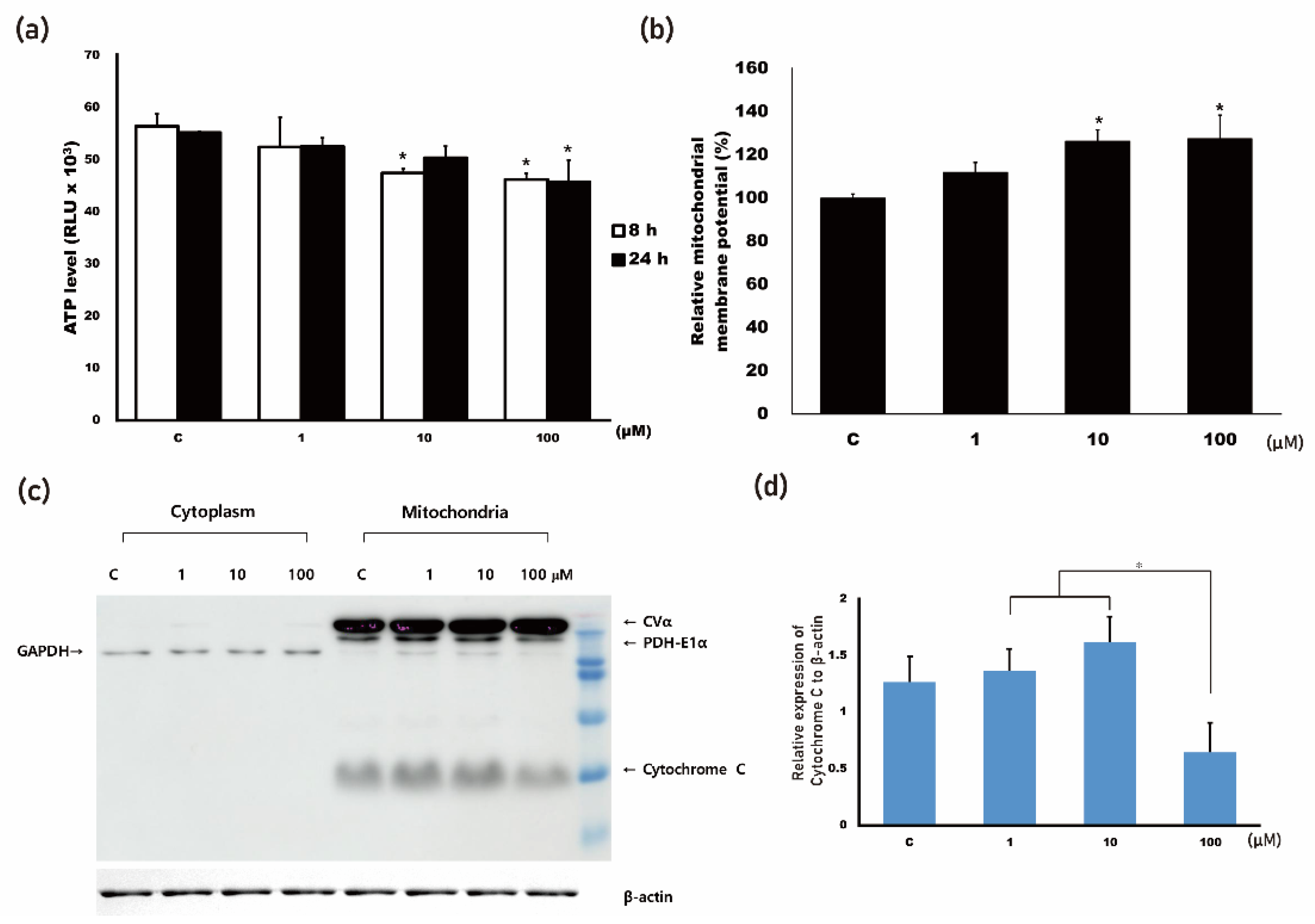
3.1. 4HR Increased Mitochondrial Stress

To clarify the effect of 4HR administration on mitochondrial stress, ATP level and MMP were measured. Mitochondria are the main cellular power factories for the production of ATP, a cellular energy source. Oxygen consumption decreased after 4HR administration (Supplementary Figure S1), dropping to 94.7% of that of the untreated control when HUVECs were treated with 100 μM 4HR (p < 0.001). Thereafter, the production of ATP was decreased after 4HR administration in HUVECs (Figure 1a). MMP increased by 20% after 100 μM 4HR administration (Figure 1b). Increased MMP following cellular stress can protect cells from apoptosis [26].
Mitochondrial stress would decrease the cytochrome c level in mitochondria. The administration of 4HR decreased cytochrome c levels at 100 μM but not at other concentrations in the mitochondrial fraction (Figure 1c). The level of PDH-E1α was also decreased following 100 μM 4HR administration, which might have caused the decreased ATP level and oxygen consumption (Figure 1c and Supplementary Figure S2). The administration of 100 μM 4HR significantly decreased the cytochrome c level of mitochondria when compared to the 1 and 10 μM 4HR administration groups (p = 0.029 and 0.005, respectively, Figure 1d). Decreased cytochrome c following 100 μM 4HR administration was not detected in the cytoplasmic fraction. This may be due to a negligible amount of cytochrome c leakage. Interestingly, there was no significant difference in cytochrome c level between the 100 μM 4HR administration group and the untreated control group (p > 0.05). This finding was confirmed through subsequent confocal microscopy observations (Supplementary Figure S3).

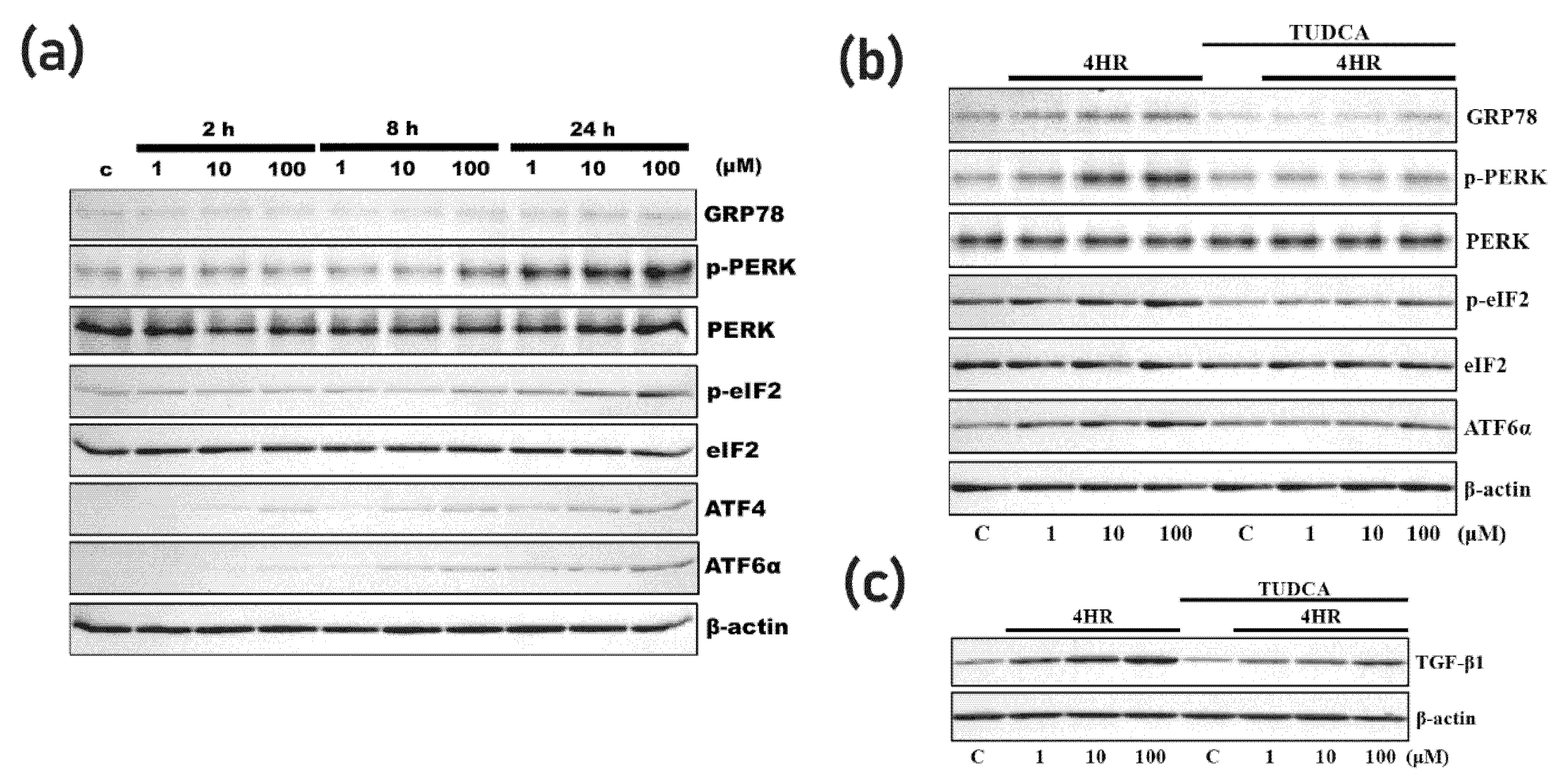
3.2. 4HR Increased ER Stress

To clarify the effect of 4HR administration on ER stress, the expression levels of ER stress markers were evaluated. The administration of 4HR increased ER stress markers such as phosphorylated protein kinase R-like endoplasmic reticulum kinase (p–PERK), phosphorylated eukaryotic initiation factor 2 (p-eIF2), activating transcription factor 4 (ATF4), and ATF6α (Figure 2a and Supplementary Figure S4). The results of quantification are shown in Supplementary Figure S3. TUDCA is an inhibitor of ER stress [13], the administration of which decreased the expression of ER stress markers which were increased by 4HR administration (Figure 2b and Supplementary Figure S5). TGF-β1 increases VEGF expression in HUVECs and helps differentiate them, and the administration of TUDCA decreased 4HR-induced TGF-β1 expression (Figure 2c and Supplementary Figure S6).
The phosphorylation level of Sp1 is increased by mitochondrial and ER stress [27]. Dissociation of p-Sp1 from HDAC increases the expression level of TGF-β1 [20]. The administration of 4HR increased Sp1 phosphorylation and TGF-β1 expression (Figure 3a). Mithramycin is an Sp1 inhibitor [28], the administration of which decreased 4HR-induced TGF-β1 expression (Figure 3b). The transcription level of Sp1 was significantly increased following 100 μM 4HR administration (p < 0.05; Figure 3c). Hyper-acetylated H3 binds to TGF-β1 promoter and increases the expression level of TGF-β1 [23]. The administration of 4HR to HUVECs increased the expression level of acetylated proteins (Supplementary Figure S7). The administration of 4HR increased H3 acetylation, too (Figure 3d). The data for the quantification of Western blot are shown in Supplementary Figure S7.

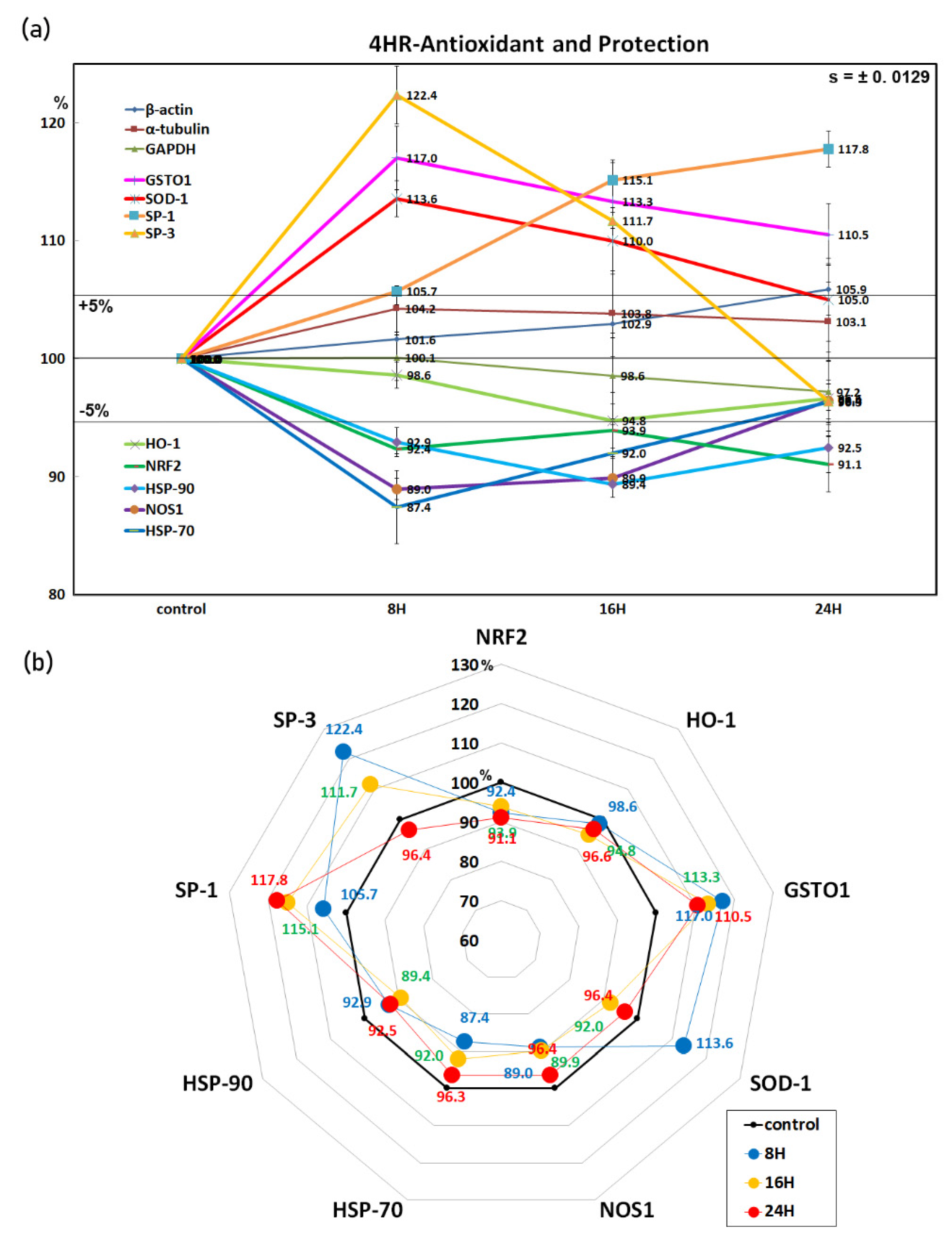
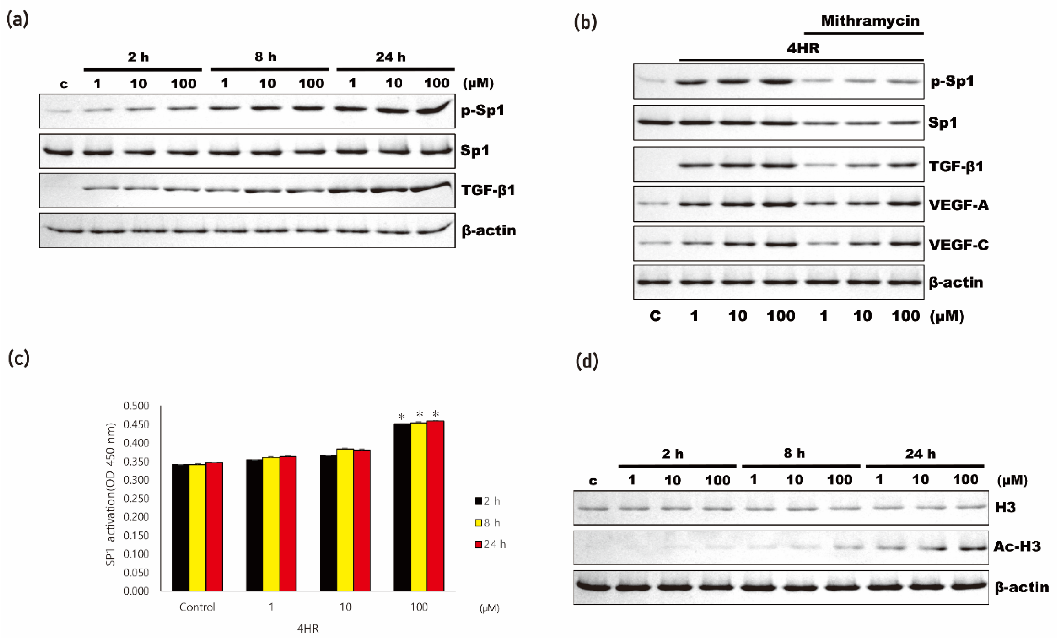
3.3. 4HR Increased Antioxidant Proteins and Sp1/Sp3 Ratio

Immunoprecipitation high-performance liquid chromatography (IP-HPLC) was conducted to evaluate the cellular survival mechanism against mitochondrial and ER stress. The IP-HPLC results also demonstrated that 4HR increased the expression of the antioxidant proteins glutathione s-transferase omega-1 (GSTO1, by 17% at 8 h) and superoxide dismutase-1 (SOD-1, 13.6% at 8 h) while decreasing the expression of the protective proteins heme oxygenase-1 (HO-1, by 5.2% at 12 h), nitric oxide synthase-1 (NOS-1, 11% at 12 h), heat shock protein-70 (HSP-70, 12.6% at 8 h), and HSP-90 (10.6% at 12 h).
A transcription factor regulating antioxidant proteins, nuclear factor erythroid-2-related factor 2 (NRF2), was downregulated by 4HR by 8.9% at 24 h, but the survival transcription factor Sp1/Sp3 ratio gradually increased at 8 (86.3%), 16 (103%), and 24 h (122.2%) after 4HR administration (Figure 4). These data indicate that 4HR showed an antioxidant effect by upregulation of GSTO1 and SOD-1 and downregulation of NOS-1 and also enhanced cell survival by Sp1/Sp3 ratio increase.

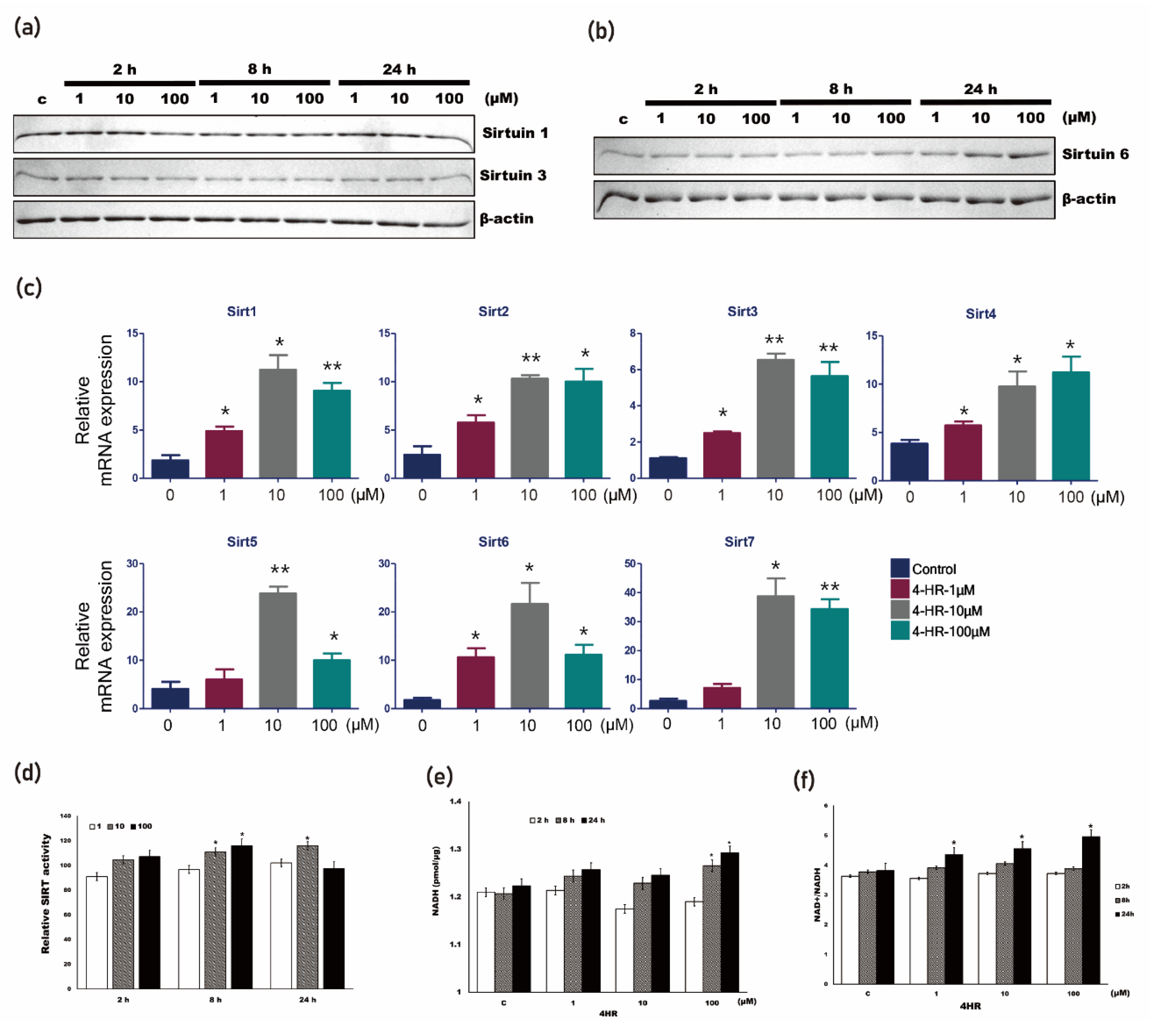
3.4. 4HR Increased SIRT Activity
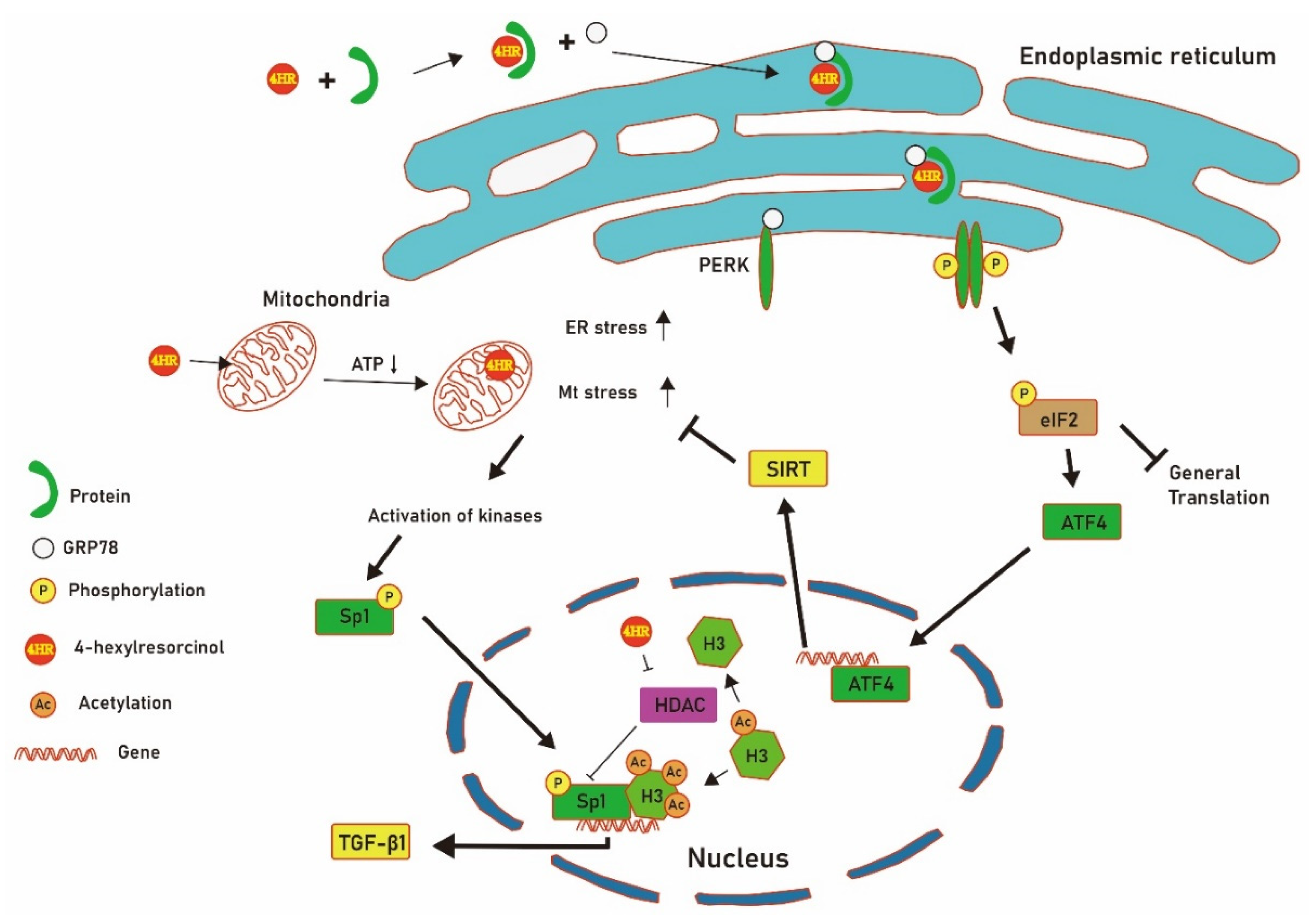
To clarify the cellular survival mechanism against mitochondrial and ER stress after 4HR administration, the expression level of SIRTs and SIRT activity were evaluated. The administration of 4HR did not change the expression of SIRT1 and SIRT3 in HUVECs (Figure 5a). Interestingly, the administration of 4HR did increase the expression of SIRT6 in HUVECs (Figure 5b). Quantitative RT-PCR results also demonstrated that the SIRTs mRNA expression level was increased at 24 h after 4HR administration (Figure 5c). A pan-SIRTs activity assay kit can detect the activity of all SIRT enzymes (SIRT1–7). Pan-SIRTs enzyme activity increased significantly with 10 and 100 μM 4HR after 8 h and with 10 μM 4HR after 24 h (Figure 5d, p < 0.05). The data for the quantification of Western blot are shown in Supplementary Figure S9.
Nicotinamide adenine dinucleotide+ (NAD+) is a substrate for SIRTs. NAD+ and the NAD+/NADH ratio were increased after 4HR administration (Figure 5e,f). NADH levels were increased significantly upon the administration of 100 μM 4HR after 8 and 24 h (Supplementary Figure S10, p < 0.05).

4. Discussion

In this study, we found that 4HR increased TGF-β1 expression through upregulation of stress responses in both mitochondria and the ER in HUVECs. ER stress-associated markers such as GRP78, ATF4, ATF6α, p-eIF2, and p-PERK were increased after 4HR administration (Figure 2a). The administration of TUDCA, an ER stress inhibitor, attenuated 4HR-induced TGF-β1 expression (Figure 2c). The mitochondrial-stress-inducing chemical carbonyl cyanide m-chlorophenylhydrazone increases TGF-β1 expression [29]. Decreased mitochondrial activity following 4HR administration reduced both ATP production (Figure 1a) and oxygen consumption (Supplementary Figure S1).
As noted in our previous study, 4HR is a class I HDACi [22]. HDACis induce TGF-β1 expression via activation of Sp1 [20]. In this study, 4HR increased the expression level of p-Sp1. Hyper-acetylated H3 is also required for the expression of TGF-β1, and this response is enhanced by HDACi [23]. In this study, H3 was hyper-acetylated by 4HR administration (Figure 3d). 4HR exhibited a wide variety of functions, such as anti-cancer effects [21], pro-angiogenic effects [8,17], anti-inflammatory effects [30], and increased bone formation [31]. These divergent effects are likely to be involved in the broad-spectrum regulation of cellular events [24,32]. In this study, 4HR increased TGF-β1 expression via hyper-acetylated H3 and p-Sp1 (Figure 5).
Sp1 is a transcription factor for TGF-β1, and its expression is increased by histone deacetylase inhibitors [20]. Interestingly, 4HR also increases Sp1 expression in oral cancer cells and SCC-9 cells [21]. Therefore, increased TGF-β1 expression as a result of 4HR administration might be associated with its HDACi activity and the subsequent promotion of Sp1 expression (Figure 3). In fact, 4HR is a class I HDACi [22]. Hyper-acetylated H3 is also required for the expression of TGF-β1, and this response is enhanced by HDACi [23]. Therefore, 4HR administration might increase acetylated H3. In this study, the Sp1/Sp3 ratio gradually increased at 8, 16, and 24 h after 4HR administration (Figure 4). In addition, 4HR administration increased p-Sp1 and TGF-β1 expression (Figure 3a). The administration of mithramycin inhibited 4HR-induced p-Sp1 and TGF-β1 expression (Figure 3b). Sp1 transcription activity and (Figure 3c) acetylated H3 were also increased by 4HR administration (Figure 3d).
When cells face ER stress, the mechanism for restoring ER homeostasis is activated [8]. As active protein synthesis increases ER loading, protein translation activity is decreased [33]. To neutralize ER stress, the expression of chaperones is increased [34]. Organic chemicals which may induce ER stress can bind to intracellular decoy proteins and change the protein conformation. This process may increase the production of misfolded proteins. ATF6 is activated by ER stress [35], which leads to the removal of misfolded proteins [36]. In this study, the expression level of ATF6α was increased by 4HR administration (Figure 2a). Increases in p–PERK, p-eIF2, and ATF4 reduce protein translation activity [37]. The expression levels of p–PERK, p-eIF2, and ATF4 were increased by 4HR administration (Figure 2a). TUDCA is an inhibitor of ER stress [13], the administration of which decreased the expression of ER stress markers such as ATF6α, p–PERK, p-eIF2, and ATF4, which were increased by 4HR administration (Figure 2b). TGF-β1 increases VEGF expression in HUVECs and helps differentiate HUVECs [9]. The administration of TUDCA decreased 4HR-induced TGF-β1 expression (Figure 2c).
The expression levels of SIRT1 and SIRT3 were not dramatically changed by 4HR administration (Figure 5a). Interestingly, the expression level of SIRT6 was increased by 4HR (Figure 5b). SIRTs gene expression was increased by 4HR administration (Figure 5c). A discrepancy between the quantity of SIRT1 and -3 proteins and their mRNA molecules was observed. This type of discrepancy has been frequently observed [38]. Though the detailed mechanism remains elusive, interaction with microRNA has been suggested as a reason for this discrepancy [39]. Interestingly, many types of microRNAs interact with SIRT1 [40]. Pan-SIRTs activity was increased by 4HR administration (Figure 5d). To increase SIRT activity, NAD+ (which is the substrate of SIRT) should be increased [41]. NADH and the NAD+/NADH ratio were significantly increased by 4HR administration (p < 0.05, Figure 5e,f). The NAD+/NADH ratio has been reported as 2–10, and it is different to the cellular type and metabolic status [42,43]. In this study, the NAD+/NADH ratio was 3–5 and increased following 4HR administration (Figure 5f). In addition, NAD+ and NADH distribute differently to cellular compartments [44]. In the case of hepatocytes, the NAD+/NADH ratio in cytoplasm is 725, and that in mitochondria is 8 [45]. When cells undergo stress, maintaining the mitochondrial NAD+ level is important for cell survival, despite its drop in the nucleus and cytoplasm [46]. Thus, the change in NAD+ level following 4HR administration in each cellular compartment could be an interesting topic for future study. The increased apoptotic stress by ER and mitochondria seemed to be ameliorated by SIRT activation. 4HR and resveratrol (10–100 μM) show antioxidant activity after hydrogen peroxide treatment in lymphocytes [18]. Resveratrol activates SIRT and ameliorates mitochondrial stress [47]. The increased apoptotic stress induced by 4HR seemed to be ameliorated by SIRT activation as with resveratrol, which was also induced by 4HR as a delayed response.
In this study, the administration of 4HR on HUVECs induced ER stress (Figure 2a). As 4HR has a long alky group, it can bind to any hydrophobic domain of proteins with different ranges of affinity [1]. Some proteins show high affinity to 4HR, which has been confirmed by chromatography with 4HR-bound beads [16]. The protein conformation would be changed after binding to 4HR and could be considered as misfolded protein. GRP78 binds to misfolded proteins and is transported to the ER [47]. This protein complex phosphorylates PERK and subsequently activates ATF4 (Figure 6). As a consequence, apoptotic stress increases. In this study, ATP synthesis in mitochondria was decreased (Figure 1a), and NAD+ level was increased by 4HR administration (Figure 5e). Reduced ATP synthesis and increased NAD+ level increase SIRT transcription [48]. The activation of several kinases by mitochondrial and ER stress phosphorylates Sp1 [27]. By increasing phosphorylation, Sp1 is dissociated from HDACs, and gene promoter activity is increased [49]. 4HR could bind to class I HDACs and inhibits their enzyme activity [22]. Accordingly, Ac-H3 was increased by 4HR administration (Figure 3d). When 4HR binds to HDACs, they dissociate from p-Sp1 via conformation change. In fact, HDACi binding to HDACs induces HDAC conformation change and dissociation from Sp1 [20]. HDACis such as trichostatin A and suberoylanilide hydroxamic acid increase TGF-β1 expression through this mechanism [20]. These events activated the transcription of TGF-β1 (Figure 6). Topical application of 4HR accelerated capillary regeneration in a diabetic animal model [9]. Besides the above results of different ER and mitochondrial stress, the expression levels of antioxidant and protection-related proteins through IP-HPLC showed consistent upregulation of GSTO1 and SOD-1 and downregulation of NOS-1 (an enzyme responsible for the synthesis of NO) and NRF2 (a transcription factor regulating antioxidant proteins to protect against oxidative damage). On the other hand, the expression levels of the protection-related proteins HO-1, HSP-70, and HSP-90 were simultaneously downregulated. Therefore, it is suggested that 4HR has a potent antioxidant effect in HUVECs, and its local administration should be safe given the lack of oxidative cellular damage.
4HR is known as an antiseptic and kills actively proliferating microorganisms [50]. Therefore, 4HR should be considered a serious stress on proliferating microorganisms. However, some microorganisms reduce their metabolic rate and enter a state of dormancy after 4HR administration [51]. In this case, 4HR may play a role as a chemical chaperone, and it has an amphiphilic structure like other chemical chaperones [1]. Microorganisms in a state of dormancy are more resistant to environmental stress [51]. Based on these observations, 4HR in eukaryotes may mimic its action in prokaryotes. However, there may be differences in the details, and this should be clarified in future studies. TUDCA has an amphiphilic structure similar to 4HR and is considered a chemical chaperone [13]. However, TUDCA alleviates ER stress [13]. The difference in chemical structure between the two chemicals may result in different patterns of cellular reaction after their administration.
The limitation of the current study was that the data on mitochondrial stress were not sufficient. Particularly, the relation between mitochondrial stress and kinase activation was unclear. Kinases activated after 4HR administration should be clarified in a further study. In addition, we assumed that SIRT activation might ameliorate mitochondrial stress induced by 4HR administration. This could be clarified by the suppression of SIRT activity. As SIRT activity is associated with longevity [42,46], increased SIRT activity after 4HR administration could be an interesting topic for further studies.

5. Conclusions

4HR induced mitochondrial and ER stress in HUVECs. 4HR-induced ER stress seemed to be ameliorated by an increase in the expression of SIRTs and SIRT activity. Therefore, administration of 1–100 μM 4HR did not lead to apoptosis induction. The mild increase in mitochondrial and ER stress by 4HR administration increased p-Sp1 and Ac-H3. Both the ER stress inhibitor (TUDCA) and the Sp1 inhibitor (mithramycin) reduced the expression level of TGF-β1, which was increased by 4HR administration.

Supplementary Materials

The following are available online at https://www.mdpi.com/article/10.3390/app11199128/s1, Figure S1: Oxygen consumption assay, Figure S2: Quantification of PDH-E1α in Figure 1c, Figure S3: Confocal microscopic findings after 4HR administration, Figure S4: Quantification of Western blot of Figure 3a, Figure S5: Quantification of Western blot of Figure 3b, Figure S6: Quantification of Western blot of Figure 3c, Figure S7: Protein acetylation was increased by 4HR administration, Figure S8: Quantification of Western blot of Figure 4, Figure S9: Quantification of Western blot of Figure 5, Figure S10: NADH level after 4HR administration.

Author Contributions

Conceptualization, J.-Y.K. and S.-G.K.; methodology, D.-W.K., X.C. and S.-G.K.; validation, D.-W.K. and J.-Y.C.; formal analysis, J.-Y.K.; investigation, D.-W.K. and X.C.; data curation, X.C. and J.-Y.C.; writing—original draft preparation, S.-G.K.; writing—review and editing, J.-Y.K., U.G. and J.-Y.C.; supervision, S.K.L., U.G. and J.-Y.C.; funding acquisition, S.-G.K. All authors have read and agreed to the published version of the manuscript.

Funding

This study was carried out with the support of the “Cooperative Research Program for Agriculture Science and Technology Development (Project no. PJ01562601)” Rural Development Administration, Korea.

Institutional Review Board Statement

HUVECs are commercially available and frequently used in many labs. Accordingly, ethics approval was not sought for the present study.

Informed Consent Statement

Not applicable.

Data Availability Statement

There was no publicly archived datasets in this study.

Conflicts of Interest

The authors declare no conflict of interest.

References

  1. Kozubek, A.; Tyman, J.H.P. Resorcinolic lipids, the natural non-isoprenoid phenolic amphiphiles and their biological activity. Chem. Rev. 1999, 99, 1–26. [Google Scholar] [CrossRef]
  2. Muliukin, A.L.; Demkina, E.V.; Kozlova, A.N.; Soina, V.S.; El’-Registan, G.I. Synthesis of anabiosis autoinducers in non-spore-forming bacteria as a mechanism regulating their activity in soil and subsoil sedimentary rocks. Mikrobiologiia 2001, 70, 620–628. [Google Scholar] [PubMed]
  3. Nikolaev, Y.A.; Tutel’yan, A.V.; Loiko, N.G.; Buck, J.; Sidorenko, S.V.; Lazareva, I.; Gostev, V.; Manzen’yuk, O.Y.; Shemyakin, I.G.; Abramovich, R.A.; et al. The use of 4-Hexylresorcinol as antibiotic adjuvant. PLoS ONE 2020, 15, e0239147. [Google Scholar] [CrossRef] [PubMed]
  4. Hosseinzadeh, E.; Hassanzadeh, A.; Marofi, F.; Alivand, M.R.; Solali, S. Flavonoid-based cancer therapy: An updated review. Anticancer Agents Med. Chem. 2020, 20, 1398–1414. [Google Scholar] [CrossRef] [PubMed]
  5. Tamaddoni, A.; Mohammadi, E.; Sedaghat, F.; Qujeq, D.; As’Habi, A. The anticancer effects of curcumin via targeting the mammalian target of rapamycin complex 1 (mTORC1) signaling pathway. Pharmacol. Res. 2020, 156, 104798. [Google Scholar] [CrossRef]
  6. Havalová, H.; Ondrovičová, G.; Keresztesová, B.; Bauer, J.A.; Pevala, V.; Kutejová, E.; Kunová, N. Mitochondrial HSP70 chaperone system-The influence of post-translational modifications and involvement in human diseases. Int. J. Mol. Sci. 2021, 22, 8077. [Google Scholar] [CrossRef]
  7. Healy, S.J.M.; Verfaillie, T.; Jäger, R.; Agostinis, P.; Samali, A. Biology of the Endoplasmic Reticulum. In Endoplasmic Reticulum Stress in Health and Disease; Agostinis, P., Samali, A., Eds.; Springer: Berlin/Heidelberg, Germany, 2012; pp. 3–22. ISBN 978-94-007-4350-2. [Google Scholar]
  8. Jo, Y.Y.; Kim, D.W.; Choi, J.Y.; Kim, S.G. 4-Hexylresorcinol and silk sericin increase the expression of vascular endothelial growth factor via different pathways. Sci. Rep. 2019, 9, 3448. [Google Scholar] [CrossRef] [Green Version]
  9. Kim, D.W.; Jo, Y.Y.; Garagiola, U.; Choi, J.Y.; Kang, Y.J.; Oh, J.H.; Kim, S.G. Increased level of vascular endothelial growth factors by 4-hexylresorcinol is mediated by transforming growth factor-β1 and accelerates capillary regeneration in the burns in diabetic animals. Int. J. Mol. Sci. 2020, 21, 3473. [Google Scholar] [CrossRef] [PubMed]
  10. Shukla, A.; Choudhury, S.; Chaudhary, G.; Singh, V.; Prabhu, S.N.; Pandey, S.; Garg, S.K. Chitosan and gelatin biopolymer supplemented with mesenchymal stem cells (Velgraft®) enhanced wound healing in goats (Capra hircus): Involvement of VEGF, TGF and CD31. J. Tissue Viability 2021, 30, 59–66. [Google Scholar] [CrossRef]
  11. Zhang, Y.; Liu, J.; Zou, T.; Qi, Y.; Yi, B.; Dissanayaka, W.L.; Zhang, C. DPSCs treated by TGF-β1 regulate angiogenic sprouting of three-dimensionally co-cultured HUVECs and DPSCs through VEGF-Ang-Tie2 signaling. Stem Cell Res. Ther. 2021, 12, 281. [Google Scholar] [CrossRef]
  12. Zhou, L.; Liu, Z.; Chen, S.; Qiu, J.; Li, Q.; Wang, S.; Zhou, W.; Chen, D.; Yang, G.; Guo, L. Transcription factor EB-mediated autophagy promotes dermal fibroblast differentiation and collagen production by regulating endoplasmic reticulum stress and autophagy-dependent secretion. Int. J. Mol. Med. 2021, 47, 547–560. [Google Scholar] [CrossRef] [PubMed]
  13. Berger, E.; Haller, D. Structure-function analysis of the tertiary bile acid TUDCA for the resolution of endoplasmic reticulum stress in intestinal epithelial cells. Biochem. Biophys. Res. Commun. 2011, 409, 610–615. [Google Scholar] [CrossRef] [PubMed]
  14. Vassilopoulos, A.; Fritz, K.S.; Petersen, D.R.; Gius, D. The human sirtuin family: Evolutionary divergences and functions. Hum. Genom. 2011, 5, 485–496. [Google Scholar] [CrossRef] [PubMed] [Green Version]
  15. Aka, J.A.; Kim, G.W.; Yang, X.J. K-Acetylation and Its Enzymes: Overview and New Developments. In Histone Deactylases: The Biology and Clinical Implication; Yao, T.P., Seto, E., Eds.; Springer: New York, NY, USA, 2011; pp. 1–12. ISBN 978-3-642-21630-5. [Google Scholar]
  16. Kim, M.K.; Yoon, C.S.; Kim, S.G.; Park, Y.W.; Lee, S.K. Effects of 4-hexylresorcinol on protein expressions in RAW 264.7 cells as determined by immunoprecipitation high performance liquid chromatography. Sci. Rep. 2019, 9, 3379. [Google Scholar] [CrossRef]
  17. Kim, M.K.; Kim, S.G.; Lee, S.K. 4-Hexylresorcinol-induced angiogenesis potential in human endothelial cells. Maxillofac. Plast. Reconstr. Surg. 2020, 42, 23. [Google Scholar] [CrossRef]
  18. Yen, G.C.; Duh, P.D.; Lin, C.W. Effects of resveratrol and 4-hexylresorcinol on hydrogen peroxide-induced oxidative DNA damage in human lymphocytes. Free Radic. Res. 2003, 37, 509–514. [Google Scholar] [CrossRef]
  19. Dauer, P.; Gupta, V.K.; McGinn, O.; Nomura, A.; Sharma, N.S.; Arora, N.; Giri, B.; Dudeja, V.; Saluja, A.K.; Banerjee, S. Inhibition of Sp1 prevents ER homeostasis and causes cell death by lysosomal membrane permeabilization in pancreatic cancer. Sci. Rep. 2017, 7, 1564. [Google Scholar] [CrossRef]
  20. Martin-Gallausiaux, C.; Béguet-Crespel, F.; Marinelli, L.; Jamet, A.; Ledue, F.; Blottière, H.M.; Lapaque, N. Butyrate produced by gut commensal bacteria activates TGF-beta1 expression through the transcription factor SP1 in human intestinal epithelial cells. Sci. Rep. 2018, 8, 9742. [Google Scholar] [CrossRef]
  21. Kim, S.G.; Kim, A.S.; Jeong, J.H.; Choi, J.Y.; Kweon, H. 4-hexylresorcinol stimulates the differentiation of SCC-9 cells through the suppression of E2F2, E2F3 and Sp3 expression and the promotion of Sp1 expression. Oncol. Rep. 2012, 28, 677–681. [Google Scholar] [CrossRef] [Green Version]
  22. Kim, J.Y.; Kweon, H.Y.; Kim, D.W.; Choi, J.Y.; Kim, S.G. 4-Hexylresorcinol inhibits class I histone deacetylases in human umbilical cord endothelial cells. Appl. Sci. 2021, 11, 3486. [Google Scholar] [CrossRef]
  23. Deb, D.K.; Chen, Y.; Sun, J.; Wang, Y.; Li, Y.C. ATP-citrate lyase is essential for high glucose-induced histone hyperacetylation and fibrogenic gene upregulation in mesangial cells. Am. J. Physiol. Renal Physiol. 2017, 313, F423–F429. [Google Scholar] [CrossRef] [Green Version]
  24. Kim, Y.S.; Kim, D.W.; Kim, S.G.; Lee, S.K. 4-hexylresorcinol-induced protein expression changes in human umbilical cord vein endothelial cells as determined by immunoprecipitation high-performance liquid chromatography. PLoS ONE 2020, 15, e0243975. [Google Scholar] [CrossRef]
  25. Yoon, Y.M.; Lee, J.H.; Yun, S.P.; Han, Y.S.; Yun, C.W.; Lee, H.J.; Noh, H.; Lee, S.J.; Han, H.J.; Lee, S.H. Tauroursodeoxycholic acid reduces ER stress by regulating of Akt-dependent cellular prion protein. Sci. Rep. 2016, 6, 39838. [Google Scholar] [CrossRef] [PubMed] [Green Version]
  26. Zhou, J.; Yang, Z.; Shen, R.; Zhong, W.; Zheng, H.; Chen, Z.; Tang, J.; Zhu, J. Resveratrol Improves Mitochondrial Biogenesis Function and Activates PGC-1α Pathway in a Preclinical Model of Early Brain Injury Following Subarachnoid Hemorrhage. Front. Mol. Biosci. 2021, 8, 620683. [Google Scholar] [CrossRef] [PubMed]
  27. Chu, S. Transcriptional regulation by post-transcriptional modification—Role of phosphorylation in Sp1 transcriptional activity. Gene 2012, 508, 1–8. [Google Scholar] [CrossRef] [PubMed]
  28. Wang, S.; Li, Y.; Sun, S.; Cai, J.; Cao, J. Sp1 promotes ovarian cancer cell migration through repressing miR-335 expression. Biochem. Biophys. Res. Commun. 2020, 524, 211–216. [Google Scholar] [CrossRef]
  29. Amuthan, G.; Biswas, G.; Ananadatheerthavarada, H.K.; Vijayasarathy, C.; Shephard, H.M.; Avadhani, N.G. Mitochondrial stress-induced calcium signaling, phenotypic changes and invasive behavior in human lung carcinoma A549 cells. Oncogene 2002, 21, 7839–7849. [Google Scholar] [CrossRef] [Green Version]
  30. Ahn, J.H.; Kim, S.G.; Kim, M.K.; Kim, D.W.; Lee, J.H.; Seok, H.; Choi, J.Y. Topical delivery of 4-hexylresorcinol promotes wound healing via tumor necrosis factor-α suppression. Burns 2016, 42, 1534–1541. [Google Scholar] [CrossRef]
  31. Kweon, H.Y.; Kim, S.G.; Choi, J.Y. Inhibition of foreign body giant cell formation by 4-hexylresorcinol through suppression of diacylglycerol kinase delta gene expression. Biomaterials 2014, 35, 8576–8584. [Google Scholar] [CrossRef]
  32. Kim, S.G. Immunomodulation for maxillofacial reconstructive surgery. Maxillofac. Plast. Reconstr. Surg. 2020, 42, 5. [Google Scholar] [CrossRef]
  33. Harding, H.P.; Zhang, Y.; Ron, D. Protein translation and folding are coupled by an endoplasmic-reticulum-resident kinase. Nature 1999, 397, 271–274. [Google Scholar] [CrossRef]
  34. Yamamoto, K.; Sato, T.; Matsui, T.; Sato, M.; Okada, T.; Yoshida, H.; Harada, A.; Mori, K. Transcriptional induction of mammalian ER quality control proteins is mediated by single or combined action of ATF6alpha and XBP1. Dev. Cell 2007, 13, 365–376. [Google Scholar] [CrossRef] [Green Version]
  35. Ye, J.; Rawson, R.B.; Komuro, R.; Chen, X.; Dave, U.P.; Prywes, R.; Brown, M.S.; Goldstein, J.L. ER stress induces cleavage of membrane-bound ATF6 by the same proteases that process SREBPs. Mol. Cell 2000, 6, 1355–1364. [Google Scholar] [CrossRef]
  36. Douglas, P.M.; Summers, D.W.; Cyr, D.M. Molecular chaperones antagonize proteotoxicity by differentially modulating protein aggregation pathways. Prion 2009, 3, 51–58. [Google Scholar] [CrossRef] [PubMed] [Green Version]
  37. Lu, P.D.; Harding, H.P.; Ron, D. Translation reinitiation at alternative open reading frames regulates gene expression in an integrated stress response. J. Cell Biol. 2004, 167, 27–33. [Google Scholar] [CrossRef] [PubMed]
  38. Flory, M.R.; Lee, H.; Bonneau, R.; Mallick, P.; Serikawa, K.; Morris, D.R.; Aebersold, R. Quantitative proteomic analysis of the budding yeast cell cycle using acid-cleavable isotope-coded affinity tag reagents. Proteomics 2006, 6, 6146–6157. [Google Scholar] [CrossRef] [PubMed]
  39. Wang, D. Discrepancy between mRNA and protein abundance: Insight from information retrieval process in computers. Comp. Biol. Chem. 2008, 32, 462–468. [Google Scholar] [CrossRef] [Green Version]
  40. Karbasforooshan, H.; Roohbakhsh, A.; Karimi, G. SIRT1 and microRNAs: The role in breast, lung and prostate cancers. Exp. Cell Res. 2018, 367, 1–6. [Google Scholar] [CrossRef]
  41. Xiao, C.; Kim, H.S.; Lahusen, T.; Wang, R.H.; Xu, X.; Gavrilova, O.; Jou, W.; Gius, D.; Deng, C.X. SIRT6 deficiency results in severe hypoglycemia by enhancing both basal and insulin-stimulated glucose uptake in mice. J. Biol. Chem. 2010, 285, 36776–36784. [Google Scholar] [CrossRef] [PubMed] [Green Version]
  42. Mostoslavsky, R.; Chua, K.F.; Lombard, D.B.; Pang, W.W.; Fischer, M.R.; Gellon, L.; Liu, P.; Mostoslavsky, G.; Franco, S.; Murphy, M.M.; et al. Genomic instability and aging-like phenotype in the absence of mammalian SIRT6. Cell 2006, 124, 315–329. [Google Scholar] [CrossRef] [Green Version]
  43. Easlon, E.; Tsang, F.; Dilova, I.; Wang, C.; Lu, S.P.; Skinner, C.; Lin, S.J. The dihydrolipoamide acetyltransferase is a novel metabolic longevity factor and is required for calorie restriction-mediated life span extension. J. Biol. Chem. 2007, 282, 6161–6171. [Google Scholar] [CrossRef] [PubMed] [Green Version]
  44. Anderson, K.A.; Madsen, A.S.; Olsen, C.A.; Hirschey, M.D. Metabolic control by sirtuins and other enzymes that sense NAD+, NADH, or their ratio. Biochim. Biophys. Acta Bioenerg. 2017, 1858, 991–998. [Google Scholar] [CrossRef]
  45. Williamson, D.H.; Lund, P.; Krebs, H.A. The redox state of free nicotinamide-adenine dinucleotide in cytoplasm and mitochondria of rat liver. Biochem. J. 1967, 103, 514–527. [Google Scholar] [CrossRef] [PubMed]
  46. Yang, H.; Yang, T.; Baur, J.A.; Perez, E.; Matsui, T.; Carmona, J.J.; Lamming, D.W.; Souza-Pinto, N.C.; Bohr, V.A.; Rosenzweig, A.; et al. Nutrient-sensitive mitochondrial NAD+ levels dictate cell survival. Cell 2007, 130, 1095–1107. [Google Scholar] [CrossRef] [PubMed] [Green Version]
  47. Xu, S.; Gao, Y.; Zhang, Q.; Wei, S.; Chen, Z.; Dai, X.; Zeng, Z.; Zhao, K.S. SIRT1/3 activation by resveratrol attenuates acute kidney injury in a septic rat model. Oxid. Med. Cell. Longev. 2016, 2016, 7296092. [Google Scholar] [CrossRef]
  48. Basseri, S.; Austin, R.C. Endoplasmic Reticulum Stress and the Unfolded Protein Response in Lipid Metabolism and Obesity. In Endoplasmic Reticulum Stress in Health and Disease; Agostinis, P., Samali, A., Eds.; Springer: Berlin/Heidelberg, Germany, 2012; pp. 231–256. ISBN 978-94-007-4350-2. [Google Scholar]
  49. Liao, M.; Zhang, Y.; Dufau, M.L. Protein kinase Calpha-induced derepression of the human luteinizing hormone receptor gene transcription through ERK mediated release of HDAC1/Sin3A repressor complex from Sp1 sites. Mol. Endocrinol. 2008, 22, 1449–1463. [Google Scholar] [CrossRef] [Green Version]
  50. Evans, R.T.; Baker, P.J.; Coburn, R.A.; Fischman, S.L.; Genco, R.J. In vitro antiplaque effects of antiseptic phenols. J. Periodontol. 1977, 48, 156–162. [Google Scholar] [CrossRef]
  51. Kushneruk, M.A.; Tugarova, A.V.; Il’chukova, A.V.; Slavkina, E.A.; Starichkova, N.I.; Bogatyrev, V.A.; Antoniuk, L.P. Factors inducing transition from growth to dormancy in rhizobacteria Azospirillum brasilense. Mikrobiologiia 2013, 82, 563–570. [Google Scholar] [CrossRef]
Figure 1. ATP and mitochondrial membrane potential (MMP) assay. (a) Administration of 4HR slightly decreased the ATP levels in HUVECs (* p < 0.05 compared to the control group; RLU, relative light units). (b) MMP was slightly increased by 4HR administration in HUVECs. In the case of 10 and 100 µM 4HR administration, MMP was increased by approximately 20 % compared to the untreated control (* p < 0.05). (c) Administration of 4HR (100 μM) decreased the cytochrome c level in the mitochondrial fraction in HUVECs. However, cytoplasmic leakage of mitochondrial cytochrome c was not clearly detected in the cytoplasmic fraction. (d) Cytochrome c level relative to mitochondrial β-actin was calculated and compared. There was no significant difference in the cytochrome c level of the mitochondria among the untreated control and the 1 and 10 µM 4HR administration groups (p > 0.05). In the case of 100 µM 4HR administration, the cytochrome c level of the mitochondria was decreased significantly compared to the untreated control and the 10 µM 4HR administration group (* p < 0.05).
Applsci 11 09128 g001
Figure 2. The expression of ER stress-associated markers. (a) 4HR administration increased ER stress markers such as p-PERK, p-eIF2, ATF4, and ATF6α. TUDCA is an inhibitor of the ER stress-mediated signaling pathway. (b) The expression of ER stress-associated markers was decreased by the administration of the inhibitor. (c) The administration of TUDCA decreased 4HR-induced TGF-β1 expression. All blots were performed in triplicate, and the statistical analysis of the relative expression level is shown in Supplementary Figures S3–S5.
Applsci 11 09128 g002
Figure 3. Sp1 and H3 acetylation by 4HR administration. (a) 4HR administration increased the expression level of p-Sp1 and TGF-β1. (b) Mithramycin is an inhibitor of Sp1. The expression of Sp1-associated proteins was decreased by the administration of mithramycin. (c) The administration of 4HR increased Sp1 activity (* p < 0.05 compared to control). (d) 4HR administration increased the expression level of Ac-H3. All blots were performed in triplicate, and the statistical analysis of the relative expression level is shown in Supplementary Figure S8.
Applsci 11 09128 g003
Figure 4. IP-HPLC analysis. Expression of antioxidant- and protection-related proteins in 4HR-treated HUVECs. The antioxidant proteins GSTO1 and SOD-1 were upregulated by 4HR compared to the untreated control, while the antioxidant regulating transcription factor NRF2 and the protection-related proteins HO-1, HSP-70, and HSP-90 were downregulated. On the other hand, Sp-1 expression gradually increased by 5.7%, 15.1%, and 17.8% at 8, 16, and 24 h, respectively, while Sp-3 expression increased up to 122.4% at 8 h but gradually decreased to a normal level at 24 h. The line graph (a) shows protein expression (%) at 8, 16, and 24 h after 4HR administration, whereas the star plot (b) shows the differential expression levels (%).
Applsci 11 09128 g004
Figure 5. 4HR administration and SIRT expression. (a) The administration of 4HR did not change the expression of SIRT1 and SIRT3 in HUVECs. (b) The administration of 4HR increased the expression of SIRT6 in HUVECs. (c) Quantitative RT-PCR results showed that the SIRTs mRNA expression level was significantly increased by 4HR administration (* p < 0.05, ** p < 0.01 compared to untreated control). (d) The administration of 4HR (1–100 μM) increased pan-SIRTs enzyme activity (* p < 0.05 compared to the control group). (e) The administration of 4HR increased NAD+ production (* p < 0.05 compared to the control group). (f) 4HR also increased the NAD+/NADH ratio (* p < 0.05 compared to the control group). All blots were performed in triplicate, and the statistical analysis of the relative expression level is shown in Supplementary Figure S8.
Applsci 11 09128 g005
Figure 6. Schematic drawing of signaling pathway. 4HR administration induced both mitochondrial and ER stress. ER stress-induced ATF4 expression may increase SIRT, and SIRT may alleviate mitochondrial stress and kinase activation. Mitochondrial stress induced kinase activation and increased Sp1 phosphorylation. To activate TGF-β1 transcription, not only p-Sp1 but also hyper-acetylated H3 is required. Since 4HR is a class I HDAC inhibitor and reduces the expression levels of HDAC4 and HDAC5 [22], H3 could be hyper-acetylated by 4HR administration.
Applsci 11 09128 g006
Publisher’s Note: MDPI stays neutral with regard to jurisdictional claims in published maps and institutional affiliations.

