
Facing Phototrophic Microorganisms That Colonize Artistic Fountains and Other Wet Stone Surfaces: Identification Keys

 ,
,
Abstract
Featured Application
Abstract
1. Introduction
2. Materials and Methods
2.1. Morphological Identification
2.2. Identification Keys
3. Results
3.1. First Screening
- 1a
- visible vivid green, light green, green or brownish phototrophic growth.....................................2
- 1b
- visible dark green, deep brown, deep grey or black phototrophic growth.....................................3
- 2a
- with a mucilaginous, wet or powdering aspect, it seems not well adhered to the stone surface ...................................................4
- 2b
- with an aspect of patina, pellicle or mat, that seems adhered to the stone surface... ......5
- 3a
- dark green, dark grey or dark brown wet formations, with filamentous or spherical aspect ...................................... see 3.2. Cyanobacteria (blue-green algae) and 3.3. Green Algae
- 3b
- dark patina with intergranular growth on dry surfaces ................... see 3.2. Cyanobacteria
- 4a
- vivid and light green color........................................................................see 3.3. Green Algae
- 4b
- green, yellow-green or light brown color, sometimes with aspect of pustule...................................................................................see 3.3. Green Algae and 3.4. Diatoms
- 5a
- pellicles more or less gelatinous....................... see 3.2. Cyanobacteria and 3.3. Green Algae
- 5b
- aspect of mats and patina, with different thicknesses, dark green or dark blue-green color with many filaments........................................................see mainly 3.2. Cyanobacteria
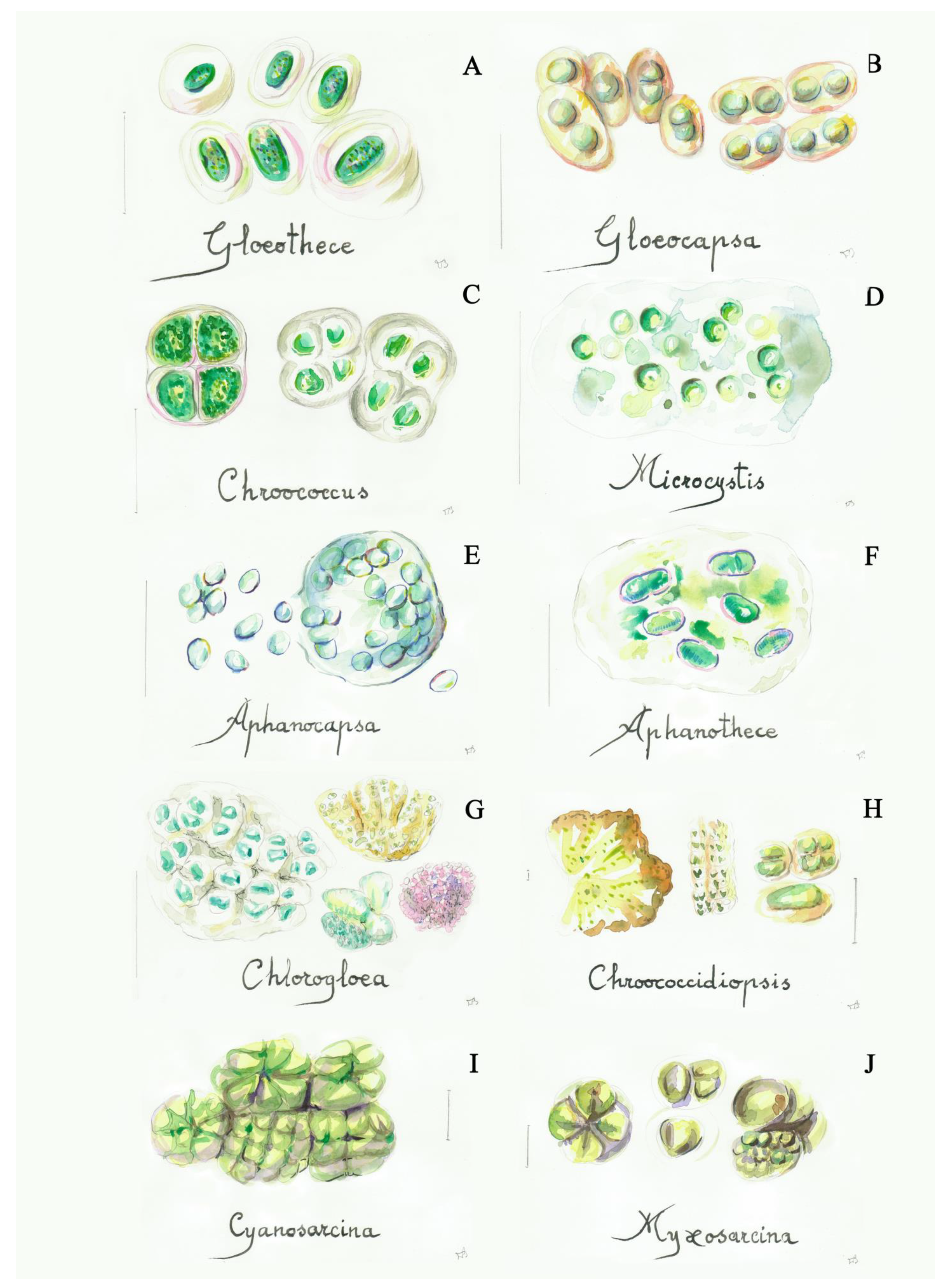
3.2. Cyanobacteria (Blue-Green Algae)
- 1a
- mucilaginous or compact groups of cells ......………………………....................................2
- 1b
- cells lacking mucilage..........................................................................................................10
- 2a
- irregular agglomerations of cells embedded in mucilage....................................................3
- 2b
- agglomerations of cells with a regular packet-like arrangement .....................................13
- 3a
- concentrically layered gelatinous sheaths around the cells (with distinct or indistinct lamellation).................................................................................................................................4
- 3b
- sheaths without clearly concentric lamellation.....................................................................6
- 4a
- ovoid to rod shaped cells, well delimitated and lamelled sheaths......................Gloeothece
- 4b
- spherical to hemispherical cell shape, with colorless or light yellow-brown sheaths.........................................................................................................................................5
- 5a
- sheaths wide and vesiculous………………………………….........………...……Gloeocapsa
- 5b
- sheaths thin and usually colorless, usually small colonies (2–16 cells, with the shape of a half or a quarter of sphere after division)......................................................Chroococcus
- 6a
- irregular colonies forming amorphous masses ....................................................................7
- 6b
- irregular colonies with their cells distributed in the mucilage............................................8
- 7a
- spherical cells............................................................................................................Microcystis
- 7b
- spherical or slightly elongated cells (oval, ovoid).............................................Aphanocapsa
- 8a
- ellipsoidal cells with homogeneous sheaths.......................................................Aphanothece
- 8b
- spherical cells.............................................................................................................................9
- 9a
- cells with an irregular-rounded to polygonal-rounded outline, and with the margin of the colony in a more or less radial row..................................................................Chlorogloea
- 9b
- cells more or less uniform in size in the colony...........................................Chroococcidiopsis
- 10a
- cells ovate to oblong, single or in small colonies.............................................................11
- 10b
- spherical and solitary cells...............................................................................Synechocystis
- 11a
- cells with a dimension of (1.5)3–15(40) × 0.4–3(6) μm ..............................Synechococcus
- 11b
- very small cells, up to 1.5 × 3 μm..................................................................Gloeobacter
- 12a
- agglomerations of cells with a more regular arrangement (cubical)..........Cyanosarcina
- 12b
- agglomerations of cells with a less regular arrangement ............................Myxosarcina
- 1a
- trichomes not clearly developed, compact groups of cells.....………….…....………..…..2
- 1b
- trichomes clearly developed, isolated or forming mats or pellicles.......……………....…3
- 2a
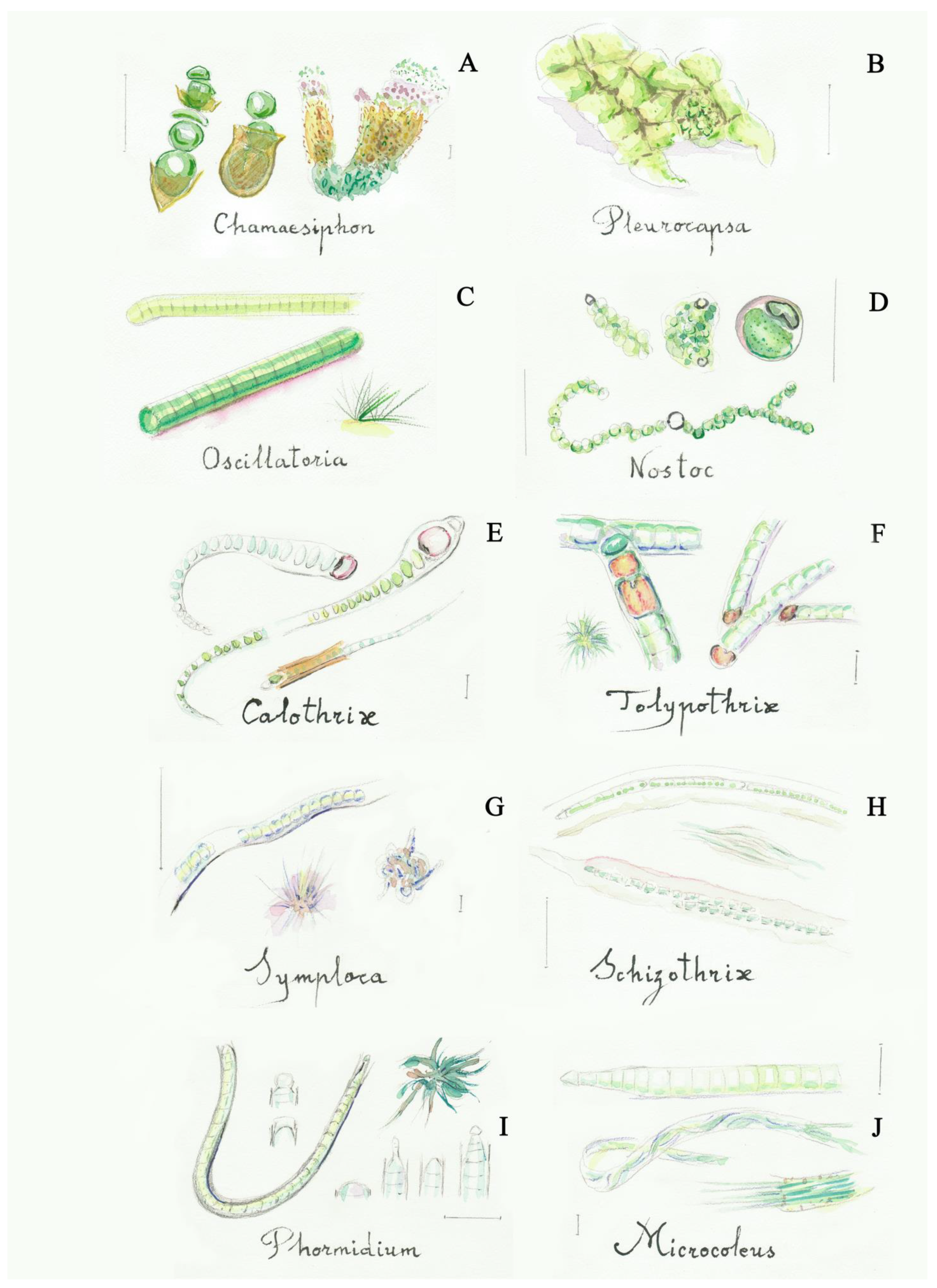
- short apical row of cells easily damaged, basal cells with colored sheath, forming macroscopic brown spots……….......................………........................……….Chamaesiphon
- 2b
- irregular groups of rows—pseudofilaments—built by polygonal cells...........Pleurocapsa
- 3a
- trichomes without sheath or with a very diffluent sheath..........................……………….4
- 3b
- trichomes with a visible sheath……………………….………..………….........…………....6
- 4a
- trichomes very short, up to 8 cells (rarely 16)................................................................Borzia
- 4b
- trichomes with a higher number of cells ................................................................................5
- 5a
- trichomes with very short cells, coin shaped and close together, visible moving filaments under the microscope …...............................................……..............….…Oscillatoria
- 5b
- trichomes with long cells more distant from one to another...……….....…Pseudanabaena
- 6a
- trichomes forming firm colonies with mucilage .........................................................Nostoc
- 6b
- trichomes forming compact clusters with a covering character .........................................7
- 7a
- presence of differentiated cells (heterocytes) in addition to the rest of the cells .............8
- 7b
- all cells with the same morphology, sometimes apical cells are differentiated...............11
- 8a
- heterocytes present at the base of polar filaments, filaments narrow into the shape of a hair...............................................................................................................................................9
- 8b
- heterocytes intercalated along the filaments, filaments not narrow.................................10
- 9a
- filaments with thin sheaths, no branching…............................................................Calothrix
- 9b
- filaments with wide sheaths, false-branching .......................................................Dichothrix
- 10a
- filament usually with rounded cells, no branching…………....……..….………..Nostoc
- 10b
- filaments with rectangular or squared cells and false branching, usually prostrated ……………………………………………………………................….............……Tolypothrix
- 11a
- one trichome within each sheath ......................................................................................12
- 11b
- several trichomes within each sheath ..............................................................................17
- 12a
- consistent sheaths with apical cells not differentiated, filaments can form mats.......13
- 12b
- mucilaginous sheaths with apical cells usually differentiated.....................................15
- 13a
- mats formed exclusively by prostrated filaments...........................................................14
- 13b
- mats formed by a base of prostrated filaments that form peripheral erect bundles with a conical shape, cells lack the calypra...............................................................Symploca
- 14a
- strongly intertwined filaments with false branching and wide sheaths, and sometimes more than one trichoma.................................................................................Schizothrix
- 14b
- very intertwined filaments forming less consistent mats, usually between mineral concretions......................................................................................................................Lyngbya
- 15a
- squared or slightly rectangular cells................................................................................16
- 15b
- cylindrical cells longer than wide, less than 3 µm in width, unbranched or with false branching............................................................................................................Leptolyngbya
- 16a
- filaments with no branching, sheath sometimes difficult to see..................Phormidium
- 16b
- filaments with false branching...............................................................Pseudophormidium
- 17a
- numerous trichomes within each sheath..........................................................Microcoleus
- 17b
- one or few trichomes within each sheath.........................................................................18
- 18a
- few-celled and short trichomes, constricted at cross walls, with a more or less cylindrical shape.....................................................................................................Pseudanabaena
- 18b
- trichomes in vegetative state always within distinct sheaths, filaments with typical “scytonematoid” (double and single) false branching, forming slimy mats.....Plectonema
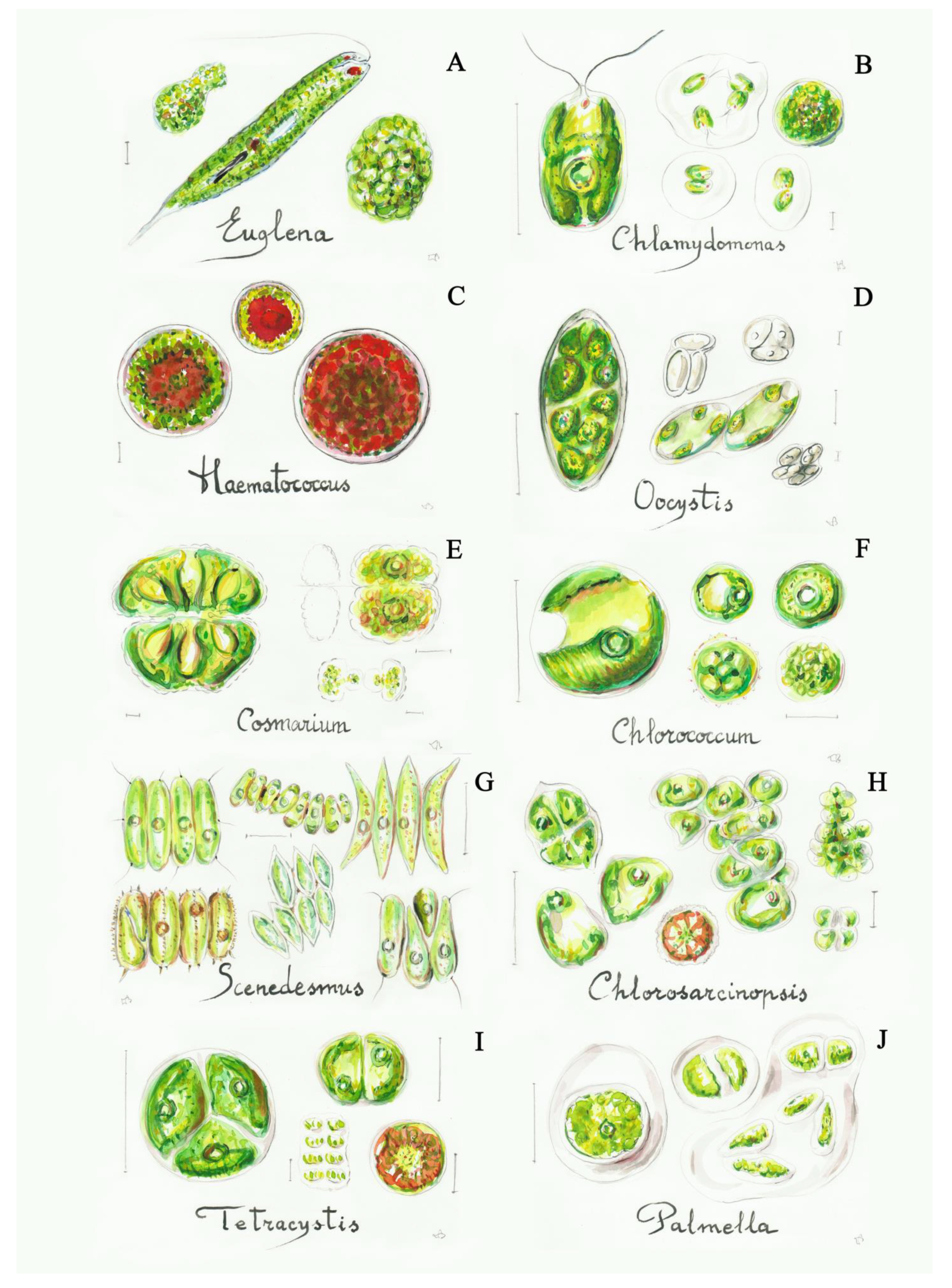
3.3. Green Algae (Chlorophyta)
- 1a
- solitary cells................................................................................................................................2
- 1b
- cells forming colonies..............................................................................................................14
- 2a
- motile cells .................................................................................................................................3
- 2b
- non-motile cells..........................................................................................................................5
- 3a
- cells with two flagella and with a rigid cellular surface.......................................................4
- 3b
- cells with flagella but with elastic (deformable) cellular surface.............................Euglena
- 4a
- solitary cells with an oval or globular shape.................................................Chlamydomonas
- 4b
- cells with a more or less globular shape, with a red spot or totally red-colored in adverse conditions. ………………........................................................................Haematococcus
- 5a
- cells not closely grouped, more or less spherical or globular.............................................6
- 5b
- cells closely and continuously grouped...............................................................................14
- 6a
- elliptical, egg-shaped cells........................................................................................................7
- 6b
- spherical cells, isolated or arranged in unstructured groups ….........................................9
- 7a
- cells clearly elongated, fusiform…………………………………........…......Monoraphidium
- 7b
- cells are ellipsoidal or globular................................................................................................8
- 8a
- cells clearly elliptic, sometimes lemon-shaped, colonies delimitated by a parental wall..................................................................................................................................Oocystis
- 8b
- ovoid cells, almost globular, solitary or in irregular mucilaginous colonies...Coccomyxa
- 9a
- many parietal plastids inside the cell, without pyrenoids................................................10
- 9b
- few plastids inside the cell, usually with pyrenoids………..............................................11
- 10a
- solitary multinucleated large cells, maximum 85 μm in diameter............Bracteococcus
- 10b
- spherical cells with a diameter of 5–18 μm............................................................Muriella
- 11a
- spherical cells with a dimension of 2–25 μm, with a huge star-shaped plastid, usually as a lichen symbiont.............................................................................................Trebouxia
- 11b
- ellipsoidal, globular or sub-spherical cells......................................................................12
- 12a
- cells with a median constriction dividing them into two semi-cells.............Cosmarium
- 12b
- cells without constriction…………………………………………………….....………..13
- 13a
- cells with a cup- or plated-like plastid, maximum diameter of 20 μm..............Chlorella
- 13b
- spherical, sometimes ellipsoidal cells with a variable diameter inside the same population (from 10–15 to 100 μm), with the plastid filling the cell………………………………………...............................................................Chlorococcum
- 14a
- groups of cells that are close together……………...........................................................15
- 14b
- groups of usually separated cells forming mucilaginous structures...........................20
- 15a
- small and bidimensional groups of cells..........................................................................16
- 15b
- large groups of cells, not bidimensional ..........................................................................18
- 16a
- cells are arranged on a single layer, with a two-dimensional structure .....................17
- 16b
- cells are arranged in groups of four (sometimes in a clear gelatinous matrix) or isolated, with strip- or plated-like plastids …………….........…………...…….Pseudochlorella
- 17a
- cells are globular ...................................................................................................Planophila
- 17b
- cells are elliptical, spindle-shaped or pear-shaped, forming groups of 4–8 cells (or multiples of four), arranged linearly or in zig-zag ............................................Scenedesmus
- 18a
- cubical shape of the colony..............................................................................Chlorosarcina
- 18b
- non-cubic colonies……………………………………………...….......………………….19
- 19a
- cells are very compacted, forming angular-shaped colonies.............Chlorosarcinopsis
- 19b
- globular shape of the colony, with its cells isolated or in small groups (two or four cells) usually tetrahedron-like shaped....................................................................Tetracystis
- 20a
- globular or amorphous colonies, cells with a diameter of 10–15 μm.................Palmella
- 20b
- globular, tetrahedron-like or irregular shape of the colony, with a consistent gelatinous matrix and with every cell enclosed by a visible membrane, the diameter varies between 3 and 23 μm ...........................................................................................Gloeocystis
- 1a
- filaments not branched..............................................................................................................2
- 1b
- filaments branched, sometimes forming very compact structures ....................................5
- 2a
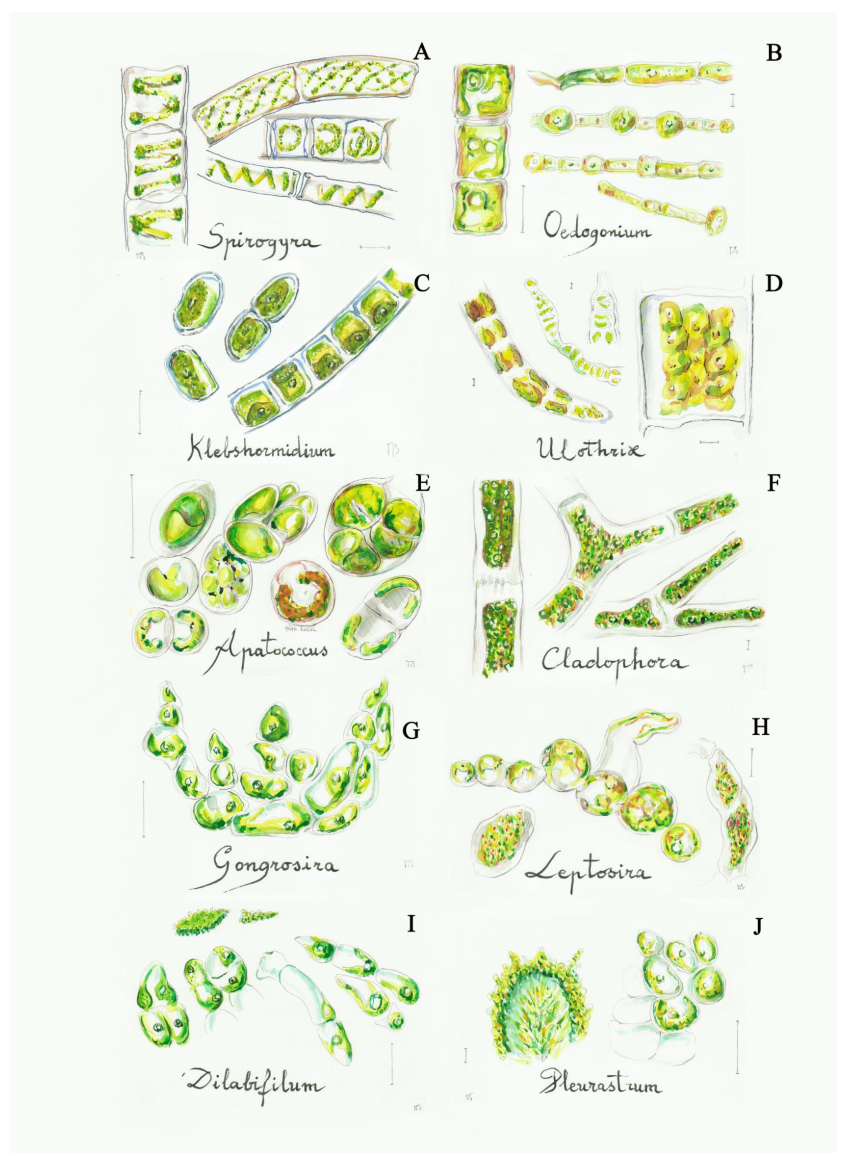
- plastids with the shape of a spiral.............................................................................Spirogyra
- 2b
- plastids with another shape .....................................................................................................3
- 3a
- presence of polar rings and intercalary widened cells (oogonia) ….....…...…Oedogonium
- 3b
- all cells with similar morphology and without polar rings…………....………....……….4
- 4a
- long filaments fixed at their base, with a stable structure, annular plastids….....Ulothrix
- 4b
- filaments that disarticulate easily, usually short, annular or elliptic plastids, usually aerophytic..........................................................................................................Klebshormidium
- 5a
- short filaments forming packet-like colonies, sometimes cubical in the first stages, cells globular or elongated..............................................................................................Apatococcus
- 5b
- filaments clearly uniseriate, linear or branched....................................................................6
- 6a
- long filaments made of long cylindrical cells with laminated wall and reticulate plastids with numerous pyrenoids ……….…………….…….……....…..............…..Cladophora
- 6b
- short filaments or small groups of cells usually with a prostrate base and a small erect part, parietal plastids with few or no pyrenoids………………….…....……......................7
- 7a
- cylindrical or club-shaped cells with a blunt apex, usually tangled filaments, built crustose lime impregnated ......................................................................................Gongrosira
- 7b
- globular or cylindrical cells forming compact groups, sometimes without a clear filament structure............................................................................................................................8
- 8a
- globular cells, parietal plastids without pyrenoids, short filaments without branches or with short branches of 1–2 cells……...........................................................................Leptosira
- 8b
- cylindrical or globular cells, with pyrenoids, from irregular groups to small filaments.9
- 9a
- cylindrical-elongated cells with short branches ...................................................Dilabifilum
- 9b
- cylindrical or subglobular cells, forming both irregular and dense groups and branched filaments .................................................................................................Pleurastrum
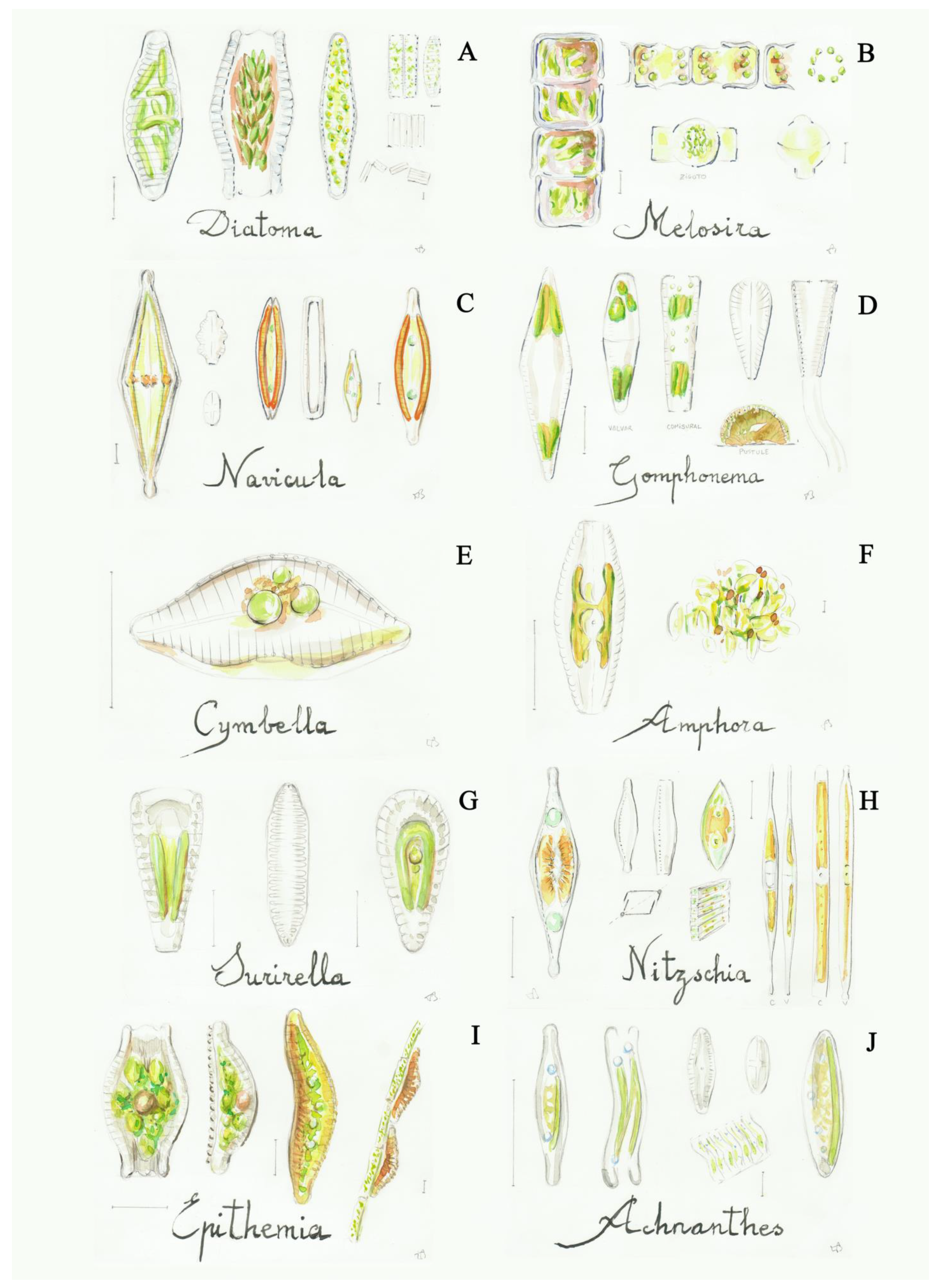
3.4. Diatoms (Bacillariophyta)
- 1a
- cells with a colonial organization, united in filament-like structures, numerous discoidal plastids.......................................................................................................................2
- 1b
- solitary cells, sometimes forming colonies, with valves of different shapes (elliptic, lanceolate, oblong, etc.), but never circular and with no radial symmetry, two lateral plastids........................................................................................................................................3
- 2a
- short cells, the filaments are easily separated in small groups of cells, valves symmetric both on the frontal and lateral faces, septa are lacking in the frustules .................Diatoma
- 2b
- long cells, valves with a circular shape (only visible in valvar view of isolated cells), the cells form filaments often found inside the shady fountains..................................Melosira
- 3a
- both valves with a raphe (structure composed by two slits or fissures), usually with a striation from the raphe to marginal part..............................................................................4
- 3b
- both valves without a raphe..................................................................................................14
- 4a
- the raphe is located in the middle of both valves.................................................................5
- 4b
- the raphe is located in a different part……..........................................................................12
- 5a
- the raphe is located on both valves.........................................................................................6
- 5b
- the raphe is located along the margin of the valves...........................................................11
- 6a
- bi-symmetric valves, central raphe.........................................................................................7
- 6b
- mono-symmetric valve, central or lateral raphe…………………………...........................9
- 7a
- striation narrow or not very wide that is not visible............................................................8
- 7b
- very wide striations, the raphe bent at both ends..................................................Pinnularia
- 8a
- thin striation, visible in dead cells, valve elliptical to boat-shaped.......................Navicula
- 8b
- very thin striations (horizontal and vertical), difficult to see even in dead cells.................................................................................................................................Frustulia
- 9a
- the valves are longitudinally asymmetric, raphe marginal...............................................10
- 9b
- the valves are transversely asymmetric, with the upper part broader than the lower one, with a trapezoidal girdle view…….............................................................Gomphonema
- 10a
- the valve margins are different, one convex and the other straight or slightly curved, raphe parallel to the straight margin and striations do not strongly radiate in the center .............................................................................................................................Cymbella
- 10b
- elliptic or oblong valves vision, intercalary bands and striations are imperceptible....................................................................................................................................Amphora
- 11b
- the raphe is present on two sides of each valve, with thick striation and a big central channel............................................................................................................................Surirella
- 11a
- the raphe is present on one side of each valve …………………..………………...…..13
- 12a
- hyaline valves, usually linear with lateral refractory dots (fibulae).........Nitzschia
- 12b
- the raphe forms a peak in the ventral part of the valve.....................................Epithemia
- 13a
- linear-elliptic valve, with a bent girdle view…………………..........................Achnantes
- 13b
- elliptic and almost circular valve, straight girdle view ………….……….…...Cocconeis
- 14a
- needle-shaped valves with parallel and thin striations, lacking septa (a silica internal sheet, occluding a portion of the frustula)..................................................Synedra (Ulnaria)
- 14b
- the frustules present septa, observed in girdle view, forming filaments........Tabellaria
4. Discussion
4.1. Fountains’ Phototrophic Biodiversity
4.2. Phototrophic Control on the Fountains
5. Conclusions
Supplementary Materials
Author Contributions
Funding
Institutional Review Board Statement
Informed Consent Statement
Acknowledgments
Conflicts of Interest
References
- Juuti, P.S.; Antoniou, G.P.; Dragoni, W.; El-Gohary, F.; De Feo, G.; Katko, T.S.; Rajala, R.P.; Zheng, X.Y.; Drusiani, R.; Angelakis, A.N. Short Global History of Fountains. Water 2015, 7, 2314–2348. [Google Scholar] [CrossRef]
- Tomaselli, L.; Pietrini, A.M. Alghe e cianobatteri. In La Biologia Vegetale per i Beni Culturali. Biodeterioramento e Conservazione; Caneva, G., Nugari, M.P., Salvadori, O., Eds.; Nardini Editore: Firenze, Italy, 2007; Volume 1, pp. 71–77. [Google Scholar]
- Gattuso, C.; Cozza, R.; Gattuso, P.; Villella, F. La Conoscenza Per il Restauro e la Conservazione. Il Ninfeo di Vadue a Carolei e la Fontana Nuova di Lamezia Terme; Franco Angeli S.R.L.: Milano, Italy, 2012; p. 75. [Google Scholar]
- Altieri, A.; Pietrini, A.M.; Ricci, S.; Roccardi, A. Il Controllo del Biodeterioramento. In Il Restauro della Fontana del Fuga nell’Orto Botanico di Roma; Micheli, M.P., Tammeo, G., Eds.; Gangemi Editore Spa: Roma, Italy, 2011; pp. 111–117. [Google Scholar]
- Villalba Corredor, L.S.; Malagón Forero, A. Biodeterioro de la Fuente de Lavapatas, parque arqueológico de San Agustín-Huila. Colombia. Ge-Conserv. 2011, 2, 65–80. [Google Scholar] [CrossRef][Green Version]
- Sánchez-Castillo, P.M.; Bolivar-Galiano, F.C. Characterización de comunidades algales epilìticas en fuentes monumentales y aplicacion a la diagnosis del biodeterioro. Limnetica 1997, 13, 31–46. [Google Scholar]
- Not, R.; Miceli, G.; Terranova, F.; Catalisano, A.; Lo Campo, P. Indagini sulla natura biotica delle alterazioni. In Fontana Pretoria—Studi Per un Progetto di Restauro. Regione Siciliana—Assessorato dei Beni Culturali e Ambientali e della Pubblica Istruzione, Centro Regionale Per la Progettazione e il Restauro e Per le Scienze Naturali ed Applicate ai Beni Culturali, Palermo; Catalisano, A., Di Natale, R., Gallo, E., Vergara, F., Eds.; Arti Grafiche Siciliane: Palermo, Italy, 1996; pp. 51–64. [Google Scholar]
- Hamadeh, S. Splash and Spectacle: The Obsession with Fountains in Eighteenth-Century Istanbul. Muqarnas Online 2002, 19, 123. [Google Scholar] [CrossRef]
- Guillaud, H. Socio-Cultural Sustainability in Vernacular Architecture. Versus: Heritage for Tomorrow; Lotti, L., Ed.; Firenze University Press: Firenze, Italy, 2014; pp. 48–55. [Google Scholar]
- Delgado-Rodrigues, J. Stone patina. A controversial concept of relevant importance in conservation. In Proceedings of International Seminar Theory and Practice in Conservation-A Tribute to Cesare Brandi, Lisbon, Portugal, 4–5 May 2006; Delgado Rodrigues, J., Mimoso, J.-M., Eds.; LNEC Editor: Lisbon, Portugal, 2006; pp. 163–174. [Google Scholar]
- Bolivar-Galiano, F.C.; Sánchez-Castillo, P.M. Claves de identificación de microalgas frecuentes en monumentos. PH: Boletín Del Inst. Andal. Del Patrim. Histórico 1999, 7, 93–98. [Google Scholar] [CrossRef]
- Komárek, J.; Anagnostidis, K. Cyanoprokaryota Chroococcales. In Süsswasserflora von Mitteleuropa; Ettl, H., Gärtner, G., Heynig, H., Mollenhauer, D., Eds.; Gustav Fischer Verlag, JenaStuttgart-Lübeck-Ulm: Jena, Germany, 1998; p. 548. [Google Scholar]
- Biggs, B.J.F.; Kilroy, C. Identification guide to common periphyton in New Zealand streams and rivers. In Stream Periphyton Monitoring Manual, Prepared for The New Zealand Ministry for the Environment; NIWA: Christchurch, New Zealand, 2000; pp. 121–210. [Google Scholar]
- Komárek, J. Coccoid and Colonial Cyanobacteria. In Freshwater Algae of North America: Ecology and Classification; Wehr, J.D., Sheath, R.G., Eds.; Academic Press: Cambridge, MA, USA, 2003; pp. 59–116. [Google Scholar]
- B-Komárek, J.; Komárková, J.; Kling, H. Filamentous cyanobacteria. Freshwater Algae of North America: Ecology and Classification, Wehr , J.D., Sheath, R.G., Eds.; Academic Press: Cambridge, MA, USA, 2003; 117–196. [Google Scholar]
- Komárek, J.; Anagnostidis, K. Cyanoprokaryota. 2. Teil: Oscillatoriales. In Süsswasserflora von Mitteleuropa; Büdel, B., Krienitz, L., Gärtner, G., Schagerl, M., Eds.; Springer Spektrum: Heidelberg, Germany, 2005; pp. 1–759. [Google Scholar]
- Komárek, J. Cyanoprokaryota. Heterocytous genera. In Süswasserflora von Mitteleuropa/Freshwater Flora of Central Europe; Büdel, B., Gärtner, G., Krienitz, L., Schagerl, M., Eds.; Springer Spektrum: Berlin/Heidelberg, Germany, 2013; p. 1130. [Google Scholar]
- Komárek, J.; Johansen, J.R. Filamentous Cyanobacteria. In Freshwater Algae of North America, 2nd ed; Academic Press: Cambridge, MA, USA, 2015; pp. 135–235. [Google Scholar] [CrossRef]
- Komárek, J.; Johansen, J. Coccoid Cyanobacteria. In Freshwater Algae of North America, 2nd ed; Academic Press: Cambridge, MA, USA, 2015; pp. 75–133. [Google Scholar] [CrossRef]
- Bourrelly, P. Diatomophycées. In Les Alguesd’eaudouce. Initiation à la Systématique. Tome II: Les Algues Jaunes et Brunes-Chrysophyycées, Phéophycées, Xanthophycées et Diatomées; Boubée, N., Ed.; Cie: Paris, France, 1968; pp. 259–399. [Google Scholar]
- Bolívar-Galiano, F.; Abad-Ruiz, C.; Sánchez-Castillo, P.; Toscano, M.; Romero-Noguera, J. Frequent Microalgae in the Fountains of the Alhambra and Generalife: Identification and Creation of a Culture Collection. Appl. Sci. 2020, 10, 6603. [Google Scholar] [CrossRef]
- Bolívar-Galiano, F.C.; Abad-Ruiz, C.; Yebra, A.; Romero-Noguera, J.; Sánchez-Castillo, P. Chromatic alterations by microalgae at National Mall fountains in Washington DC (USA). In Proceedings of the 6th International Conference on Heritage and Sustainable Development, Granada, Spain, 12–15 June 2018, Granada, Spain; EUG Green Lines Institute for Sustainable Development: Barcelos, Portugal, 2018; Volume 2, pp. 1211–1218. [Google Scholar]
- Cuzman, O.A.; Ventura, S.; Sili, C.; Mascalchi, C.; Turchetti, T.; D’Acqui, L.P.; Tiano, P. Biodiversity of Phototrophic Biofilms Dwelling on Monumental Fountains. Microb. Ecol. 2010, 60, 81–95. [Google Scholar] [CrossRef]
- Zurita, Y.P.; Cultrone, G.; Castillo, P.S.; Sebastián, E.; Bolívar, F. Microalgae associated with deteriorated stonework of the fountain of Bibatauín in Granada, Spain. Int. Biodeterior. Biodegrad. 2005, 55, 55–61. [Google Scholar] [CrossRef]
- Peraza Zurita, Y. Biodeterioro por Microalgas en Fuentes de Mármol. Ph.D. Thesis, University of Granada Departamento de Pintura, Granada, Spain, 2004. [Google Scholar]
- Bolívar-Galiano, F.C.; Sánchez-Castillo, P.M. Biodeterioro del patrimonio artístico por cianobacterias, algas verdes y diatomeas. Idea PH Boletìn 1998, 24, 52–63. [Google Scholar] [CrossRef]
- De Natale, A.; Mele, B.H.; Cennamo, P.; Del Mondo, A.; Petraretti, M.; Pollio, A. Microbial biofilm community structure and composition on the lithic substrates of Herculaneum Suburban Baths. PLoS ONE 2020, 15, e0232512. [Google Scholar] [CrossRef] [PubMed]
- Pietrini, A.M. La microflora fotosintetica. In Il restauro della Fontana del Fuga nell’Orto Botanico di Roma; Micheli, M.P., Tammeo, G., Eds.; Gangemi Editore Spa: Roma, Italy, 2011; pp. 107–111. [Google Scholar]
- Sarró, M.I.; Garcia, A.M.; Rivalta, V.M.; Moreno, D.A.; Arroyo, I. Biodeterioration of the Lions Fountain at the Alhambra Palace, Granada (Spain). Build. Environ. 2006, 41, 1811–1820. [Google Scholar] [CrossRef]
- Hindáková, A.; Hindác, F. Green algae of five city fountains in Bratislava (Slovakia). Biol. Bratisl. 1998, 53, 481–493. [Google Scholar]
- Nugari, M.P.; Pietrini, A.M. Trevi Fountain: An evaluation of inhibition effect of water-repellents on cyanobacteria and algae. Int. Biodeterior. Biodegrad. 1997, 40, 247–253. [Google Scholar] [CrossRef]
- Gandhi, H.P. Notes on the diatomaceae from Ahmedabad and its environs ? VI. On some diatoms from fountain reservoir of Seth Sarabhai’s garden. Hydrobiology 1967, 30, 248–272. [Google Scholar] [CrossRef]
- Chorus, I.; Cavalieri, M. Cyanobacteria and algae. In Monitoring Bathing Waters—A practical Guide to the Design and Implementation of Assessments and Monitoring Programme; Bartram, J., Rees, G., Eds.; CRC Press: London, UK, 2000; pp. 219–272. Available online: https://www.who.int/water_sanitation_health/bathing/bathwatchap10.pdf (accessed on 28 August 2021).
- Trobajo, R.; Mann, D.G. A rapid cleaning method for diatoms. Diatom Res. 2019, 34, 115–124. [Google Scholar] [CrossRef]
- Franchini, W. The Collecting, Cleaning, and Mounting of Diatoms. Available online: https://www.mccrone.com/mm/the-collecting-cleaning-and-mounting-of-diatoms/ (accessed on 30 August 2021).
- Popović, S.; Nikolić, N.; Jovanović, J.; Predojević, D.; Trbojević, I.; Manić, L.; Subakov Simić, G. Cyanobacterial and algal abundance and biomass in cave biofilms and relation to environmental and biofilm parameters. Int. J. Speleol. 2019, 48, 49–61. [Google Scholar] [CrossRef]
- Bolívar-Galiano, F.C. Diagnosis y Tratamiento del Deterioro por Microalgas en los Palacios Nazaries de la Alhambra. Ph.D. Thesis, Universidad de Granada, Granada, Spain, 1994; pp. 193–218. Available online: https://digibug.ugr.es/ (accessed on 1 September 2021).
- Phyco Key, A List of Helpful Texts and Websites with Excellent Images; Baker, A.L., Ed.; University of Hampshire: Hampshire, UK; Available online: http://cfb.unh.edu/phycokey/Choices/Text_html/groups/genera.htm (accessed on 1 September 2021).
- Mihajlovski, A.; Seyer, D.; Benamara, H.; Bousta, F.; Di Martino, P. An overview of techniques for the characterization and quantification of microbial colonization on stone monuments. Ann. Microbiol. 2015, 65, 1243–1255. [Google Scholar] [CrossRef]
- Piñar, G.; Sterflinger, K. Natural sciences at the service of art and cultural heritage: An interdisciplinary area in development and important challenges. Microb. Biotechnol. 2021, 14, 806–809. [Google Scholar] [CrossRef]
- Pandey, D.C.; Mitra, A.K. On the morphology and life-history of a form of gloeocapsa showing some new stages. Hydrobiologia 1966, 27, 379–384. [Google Scholar] [CrossRef]
- Absalón, I.B.; Muñoz-Martín, M.A.; Montejano, G.; Mateo, P. Differences in the Cyanobacterial Community Composition of Biocrusts from the Drylands of Central Mexico. Are There Endemic Species? Front. Microbiol. 2019, 10, 937. [Google Scholar] [CrossRef]
- Charola, A.E.; Wendler, E. An Overview of the Water-Porous Building Materials Interactions. Restor. Build. Monum. 2015, 21, 55–65. [Google Scholar] [CrossRef]
- Gaylarde, C.; Gaylarde, P.M.; Neilan, B. Endolithic Phototrophs in Built and Natural Stone. Curr. Microbiol. 2012, 65, 183–188. [Google Scholar] [CrossRef]
- Cuzman O., A.; Tiano, P.; Ventura, S.; Frediani, P. Biodiversity on Stone Artifacts. In The Importance of Biological Interactions in the Study of Biodiversity; IntechOpen: Rijeka, Croatia, 2011; pp. 367–390. [Google Scholar]
- Conference on Conservation of Fountains, organized by The National Center for Preservation Technology and Training, in partnership with The Nelson-Atkins Museum of Art and conservator Martin Burke. Kansas City, MO, USA, 10–11 July 2013; Available online: https://www.ncptt.nps.gov/blog/fountain-fundamentals/ (accessed on 10 July 2021).
- Krueger, R. A new approach to maintaining water features and reducing biological growth. In Objects Specialty Group Postprints; Riccardelli, C., Del Re, C., Eds.; The American Institute for Conservation of Historic & Artistic Works: Washington, DC, USA, 2010; Volume 17, pp. 33–39. [Google Scholar]
- Salvadori, O.; Charola, A.E. Methods to Prevent Biocolonization and recolonization: An overview of current research for architectural and archaeological heritage. In Biocolonization of Stone: Control and Preventive Methods Proceedings from the MCI Workshop Series; Charola, A.E., McNamara, C., Koestler, R.J., Eds.; Smithsonian Institution Scholarly Press: Washington, DC, USA, 2009; pp. 37–50. [Google Scholar]
- Lo Schiavo, S.; De Leo, F.; Urzì, C. Present and Future Perspectives for Biocides and Antifouling Products for Stone-Built Cultural Heritage: Ionic Liquids as a Challenging Alternative. Appl. Sci. 2020, 10, 6568. [Google Scholar] [CrossRef]
- Cuzman, O.A.; Camaiti, M.; Sacchi, B.; Tiano, P. Natural antibiofouling agents as new control method for phototrophic biofilms dwelling on monumental stone surfaces. Int. J. Conserv. Sci. 2011, 2, 3–16. [Google Scholar]
- Yebra, D.M.; Kiil, S.; Dam-Johansen, K. Antifouling technology—past, present and future steps towards efficient and environmentally friendly antifouling coatings. Prog. Org. Coat. 2004, 50, 75–104. [Google Scholar] [CrossRef]
- Pandolfi, A.; Capponi, G.; Fazio, G. The restoration of historical fountains. In Restoring in Italy—Art and Technology in the Activities of the Istituto Superiore per la Conservazione ed Il Restauro; Gangemi Editore: Rome, Italy, 2012; pp. 87–94. [Google Scholar]
- Gaylarde, C.; Morton, L.H.G. Deteriogenic biofilms on buildings and their control: A review. Biofouling 1999, 14, 59–74. [Google Scholar] [CrossRef]
- May, E.; Zamarreño, D.; Hotchkiss, S.; Mitchell, J.; Inkpen, R. Bioremediation of Algal Contamination on Stone. In Biocolonization of Stone: Control and Preventive Methods Proceedings from the MCI Workshop Series; Charola, A.E., McNamara, C., Koestler, R.J., Eds.; Smithsonian Institution Scholarly Press: Washington, DC, USA, 2009; pp. 59–70. [Google Scholar]


| Location | Fountains List |
|---|---|
| A = San Agustín-Huila, Colombia [5] | 1 = Fountain of Lavapatas (green area, made of volcanic rock) |
| B = Lamezia Terme, Italy [3] | 2 = New Fountain |
| C = Florence, Italy [23] | 3 = Tacca’s Fountain (urban area, made of marble (pedestal and border of basins) and pietra Serena sandstone (basins)) |
| 4 = Second Fountain of Villa la Pietra (green area, made of concrete) | |
| D = Palermo, Italy [7] | 5 = Pretoria Fountain (urban area, made of marble (decorative elements and border of the basin), red granite and limestone (all the rest)) |
| E = Pompei, Italy [27] | 6 = Vestibulum, Tepidarium and Swimming pool of the Herculaneum Suburban Baths (green area, sampled from plaster (Vestibulum), marble (Tepidarium) and mortar (swimming pool)) |
| F = Rome, Italy [28,31] | 7 = Trevi Fountain (urban area, marble and travertine); the presented genera in the tables were observed on the same type of stone samples immersed in the fountain basin |
| 8 = Fuga’s Fountain from the Botanical Garden of Rome (green area) | |
| G = Gujarat, India [32] | 9 = Fountain Reservoir of Seth Sarabhai’s Garden (green area) |
| H = Bratislava, Slovakia [30] | 10 = Planet of Peace Fountain (urban area) |
| 11 = Hviezdoslav’s Fountain (urban area) | |
| 12 = Ganymede’s Fountain (urban area) | |
| 13 = Friendship Fountain (urban area) | |
| 14 = Veil Fountain (urban area) | |
| I = Granada, Spain [21,23,24,25,26] | 15 = Patio Sultana (Alhambra Complex) (green area, made of Sierra Elvira stone) 16 = Lindaraja Fountain (Alhambra Complex) (green area, made of marble (pedestal) and Sierra Elvira stone (basin)) 17 = Patio Naranjos (Alhambra Complex) (green area) 18 = Patio Reja (Alhambra Complex) (green area) 19 = Lions Fountain (Alhambra Complex) (green area) 20 = Mexuar Fountain (Alhambra Complex) (green area) 21 = North “guitar” Fountain of the Court of the Myrtles (Alhambra Complex) (green area) 22 = South “guitar” fountain of the Court of the Myrtles (Alhambra Complex) (green area) 23 = East channel of the Court of the Myrtles (Alhambra Complex) (green area) 24 = “Guitar” Fountain of the Ladies Tower (Alhambra Complex) (green area) 25 = Jardín de los Adarves east basin (Alhambra Complex) (green area) 26 = Mirador del Partal basin (Alhambra Complex) (green area) 27 = Glorieta del Secano Fountain (Alhambra Complex) (green area) 28 = Jardines Bajos South Fountain (Alhambra Complex) (green area) 29 = Escalera del Agua handrail (Alhambra Complex) (green area) 30 = Fuente del Tomate (Alhambra Complex) (green area) 31 = Ángel Ganivet pool Fountain (Alhambra Complex) (green area) 32 = Carlos V basin (Alhambra Complex) (green area) 33 = Washington Irving basin (Alhambra Complex) (green area) 34 = Puerta de las Granadas basin (Alhambra Complex) (green area) 35 = Right fountain of the 2nd terrace of the Carmen de Bellavista (Alhambra Complex) (green area) 36 = Bibatauín Fountain (urban area) |
| J = Seville, Spain [25] | 37 = Reales Alcázares Fountains (unspecified fountains) (green area) |
| K = Washington, DC, USA [22] | 38 = The outside pool of the National Museum of African American History and Culture, Smithsonian Institution 39 = The pool in the plaza of the Hirshhorn Museum, Smithsonian Institution 40 = Large Acanthus Fountain, Smithsonian Institution 41 = Keith Fountain, Smithsonian Institution 42 = The pool in front of the National Museum of American History, Smithsonian Institution 43 = The fountain on the northwest corner of the National Museum of the American Indian, Smithsonian Institution 44 = The fountain outside the National Gallery, which flows into the cafeteria 45 = The fountain located in front of the west façade of the West Building of the National Gallery 46 = Girl Water Lilies Fountain of the National Gallery |
| Location | Cyanobacteria in the Worldwide Fountains | |||||||||||||||||||||||||||||||||
|---|---|---|---|---|---|---|---|---|---|---|---|---|---|---|---|---|---|---|---|---|---|---|---|---|---|---|---|---|---|---|---|---|---|---|
| Country | CO | IT | ES | USA | ||||||||||||||||||||||||||||||
| City | A | B | C | D | E | F | I | J | K | |||||||||||||||||||||||||
| Fountain (1–46) | 1 | 2 | 3 | 4 | 5 | 6 | 7 | 8 | 15 | 16 | 18 | 19 | 21 | 22 | 23 | 24 | 26 | 27 | 28 | 29 | 30 | 31 | 32 | 34 | 35 | 36 | 37 | 38 | 39 | 40 | 41 | 43 | 44 | 46 |
| Microorganisms | ||||||||||||||||||||||||||||||||||
| Aphanocapsa | x | x | x | |||||||||||||||||||||||||||||||
| Aphanothece | x | x | ||||||||||||||||||||||||||||||||
| Borzia | x | |||||||||||||||||||||||||||||||||
| Calothrix | x | x | x | x | x | x | x | x | x | x | x | x | ||||||||||||||||||||||
| Chamaesiphon | x | x | x | x | x | x | x | x | x | |||||||||||||||||||||||||
| Chlorogloea | x | x | x | x | ||||||||||||||||||||||||||||||
| Chroococcidiopsis | x | x | x | x | x | |||||||||||||||||||||||||||||
| Chroococcopsis | x | x | x | |||||||||||||||||||||||||||||||
| Chroococcus | x | x | x | x | x | x | x | |||||||||||||||||||||||||||
| Cyanosarcina | x | x | ||||||||||||||||||||||||||||||||
| Dichothrix | x | |||||||||||||||||||||||||||||||||
| Gloeobacter | x | |||||||||||||||||||||||||||||||||
| Gloeocapsa | x | x | x | x | x | x | x | |||||||||||||||||||||||||||
| Gloeocapsopsis | x | |||||||||||||||||||||||||||||||||
| Gloeothece | x | x | ||||||||||||||||||||||||||||||||
| Leptolyngbya | x | x | x | x | x | x | x | x | x | x | x | x | x | x | x | x | x | x | x | |||||||||||||||
| Lyngbya | x | x | x | x | ||||||||||||||||||||||||||||||
| Microcoleus | x | |||||||||||||||||||||||||||||||||
| Microcystis | x | x | ||||||||||||||||||||||||||||||||
| Myxosarcina | x | x | x | |||||||||||||||||||||||||||||||
| Nostoc | x | x | x | x | ||||||||||||||||||||||||||||||
| Oscillatoria | x | x | x | x | x | |||||||||||||||||||||||||||||
| Phormidium | x | x | x | x | x | x | x | x | x | x | x | x | x | x | x | x | ||||||||||||||||||
| Plectonema | x | |||||||||||||||||||||||||||||||||
| Pleurocapsa | x | x | x | x | x | x | ||||||||||||||||||||||||||||
| Pseudanabaena | x | x | x | x | ||||||||||||||||||||||||||||||
| Pseudophormidium | x | x | x | x | ||||||||||||||||||||||||||||||
| Rivularia | x | |||||||||||||||||||||||||||||||||
| Schizothrix | x | |||||||||||||||||||||||||||||||||
| Scytonema | x | |||||||||||||||||||||||||||||||||
| Stanieria | x | |||||||||||||||||||||||||||||||||
| Symploca | x | x | x | |||||||||||||||||||||||||||||||
| Synechococcus | x | |||||||||||||||||||||||||||||||||
| Synechocystis | x | |||||||||||||||||||||||||||||||||
| Location | Green Algae in the Worldwide Fountains | |||||||||||||||||||||||||||||||||||||||||||
|---|---|---|---|---|---|---|---|---|---|---|---|---|---|---|---|---|---|---|---|---|---|---|---|---|---|---|---|---|---|---|---|---|---|---|---|---|---|---|---|---|---|---|---|---|
| Country | CO | IT | IN | SK | ES | USA | ||||||||||||||||||||||||||||||||||||||
| City (A–K) | A | B | C | D | E | F | G | H | I | J | K | |||||||||||||||||||||||||||||||||
| Fountain (1–46) | 1 | 2 | 3 | 4 | 5 | 6 | 7 | 8 | 9 | 10 | 11 | 12 | 13 | 14 | 15 | 16 | 17 | 18 | 19 | 20 | 21 | 22 | 23 | 24 | 25 | 26 | 27 | 29 | 30 | 31 | 32 | 33 | 34 | 36 | 37 | 38 | 39 | 40 | 41 | 42 | 43 | 44 | 45 | 46 |
| Microorganisms | ||||||||||||||||||||||||||||||||||||||||||||
| Apatococcus | x | x | x | x | x | x | x | x | x | |||||||||||||||||||||||||||||||||||
| Bracteacoccus | x | x | x | x | x | x | x | x | x | x | x | |||||||||||||||||||||||||||||||||
| Chlamydomonas | x | x | x | x | x | x | x | |||||||||||||||||||||||||||||||||||||
| Chlorella | x | x | x | x | x | x | x | x | x | x | x | x | x | x | x | x | ||||||||||||||||||||||||||||
| Chlorococcum | x | x | x | x | x | x | x | |||||||||||||||||||||||||||||||||||||
| Chlorosarcina | x | x | x | x | ||||||||||||||||||||||||||||||||||||||||
| Chlorosarcinopsis | x | x | x | x | x | x | x | x | x | x | x | x | ||||||||||||||||||||||||||||||||
| Choricystis | x | x | x | x | x | x | ||||||||||||||||||||||||||||||||||||||
| Coccomyxa | x | x | x | x | x | |||||||||||||||||||||||||||||||||||||||
| Cosmarium | x | x | x | x | x | x | x | x | x | |||||||||||||||||||||||||||||||||||
| Dilabifilum | x | |||||||||||||||||||||||||||||||||||||||||||
| Euglena | x | |||||||||||||||||||||||||||||||||||||||||||
| Gloeocystis | x | |||||||||||||||||||||||||||||||||||||||||||
| Gongrosira | x | |||||||||||||||||||||||||||||||||||||||||||
| Haematococcus | x | x | x | |||||||||||||||||||||||||||||||||||||||||
| Klebsormidium | x | x | x | x | ||||||||||||||||||||||||||||||||||||||||
| Leptosira | x | |||||||||||||||||||||||||||||||||||||||||||
| Microthamnion | x | |||||||||||||||||||||||||||||||||||||||||||
| Monoraphidium | x | x | x | x | ||||||||||||||||||||||||||||||||||||||||
| Muriella | x | x | x | |||||||||||||||||||||||||||||||||||||||||
| Oocystella | x | x | x | x | x | |||||||||||||||||||||||||||||||||||||||
| Oocystis | x | x | ||||||||||||||||||||||||||||||||||||||||||
| Palmella | x | |||||||||||||||||||||||||||||||||||||||||||
| Pediastrum | x | x | x | x | x | x | ||||||||||||||||||||||||||||||||||||||
| Planophila | x | x | ||||||||||||||||||||||||||||||||||||||||||
| Pleurastrum | x | x | ||||||||||||||||||||||||||||||||||||||||||
| Pseudochlorella | x | x | x | |||||||||||||||||||||||||||||||||||||||||
| Pseudopleurococcus | x | |||||||||||||||||||||||||||||||||||||||||||
| Selenastrum | x | x | ||||||||||||||||||||||||||||||||||||||||||
| Scenedesmus | x | x | x | x | x | x | x | x | x | x | x | x | x | x | x | x | ||||||||||||||||||||||||||||
| Spirogyra | x | x | x | x | ||||||||||||||||||||||||||||||||||||||||
| Stichococcus | x | x | x | x | x | x | x | x | x | x | ||||||||||||||||||||||||||||||||||
| Tetracystis | x | |||||||||||||||||||||||||||||||||||||||||||
| Trebouxia | x | x | ||||||||||||||||||||||||||||||||||||||||||
| Ulothrix | x | x | x | x | x | |||||||||||||||||||||||||||||||||||||||
| Location | Diatoms in the Worldwide Fountains | |||||||||||||||||||||
|---|---|---|---|---|---|---|---|---|---|---|---|---|---|---|---|---|---|---|---|---|---|---|
| Country | CO | IT | IN | ES | USA | |||||||||||||||||
| City (A–K) | A | B | C | D | F | G | I | J | K | |||||||||||||
| Fountain (1–46) | 1 | 2 | 3 | 4 | 5 | 7 | 8 | 9 | 15 | 16 | 18 | 19 | 20 | 21 | 22 | 23 | 26 | 28 | 36 | 37 | 38 | 43 |
| Microorganisms | ||||||||||||||||||||||
| Achnanthes | x | x | x | x | x | x | x | x | ||||||||||||||
| Amphora | x | x | ||||||||||||||||||||
| Cymbella | x | x | x | x | x | |||||||||||||||||
| Diatoma | x | x | ||||||||||||||||||||
| Epithemia | x | x | x | |||||||||||||||||||
| Frustulia | x | x | ||||||||||||||||||||
| Gomphonema | x | x | x | |||||||||||||||||||
| Melosira | x | x | ||||||||||||||||||||
| Navicula | x | x | x | x | x | x | x | x | x | x | x | x | x | x | x | x | x | x | ||||
| Nitzschia | x | x | x | x | x | x | x | x | x | x | ||||||||||||
| Pinnularia | x | x | x | x | ||||||||||||||||||
| Surirella | x | x | ||||||||||||||||||||
| Synedra | x | x | x | x | x | |||||||||||||||||
| Tabellaria | x | x | ||||||||||||||||||||
Publisher’s Note: MDPI stays neutral with regard to jurisdictional claims in published maps and institutional affiliations. |
© 2021 by the authors. Licensee MDPI, Basel, Switzerland. This article is an open access article distributed under the terms and conditions of the Creative Commons Attribution (CC BY) license (https://creativecommons.org/licenses/by/4.0/).
Share and Cite
Bolivar-Galiano, F.; Cuzman, O.A.; Abad-Ruiz, C.; Sánchez-Castillo, P. Facing Phototrophic Microorganisms That Colonize Artistic Fountains and Other Wet Stone Surfaces: Identification Keys. Appl. Sci. 2021, 11, 8787. https://doi.org/10.3390/app11188787
Bolivar-Galiano F, Cuzman OA, Abad-Ruiz C, Sánchez-Castillo P. Facing Phototrophic Microorganisms That Colonize Artistic Fountains and Other Wet Stone Surfaces: Identification Keys. Applied Sciences. 2021; 11(18):8787. https://doi.org/10.3390/app11188787
Chicago/Turabian StyleBolivar-Galiano, Fernando, Oana Adriana Cuzman, Clara Abad-Ruiz, and Pedro Sánchez-Castillo. 2021. "Facing Phototrophic Microorganisms That Colonize Artistic Fountains and Other Wet Stone Surfaces: Identification Keys" Applied Sciences 11, no. 18: 8787. https://doi.org/10.3390/app11188787
APA StyleBolivar-Galiano, F., Cuzman, O. A., Abad-Ruiz, C., & Sánchez-Castillo, P. (2021). Facing Phototrophic Microorganisms That Colonize Artistic Fountains and Other Wet Stone Surfaces: Identification Keys. Applied Sciences, 11(18), 8787. https://doi.org/10.3390/app11188787