Review

Protective Effects of Tocotrienols in Cerebral and Myocardial Ischemia-Reperfusion Injury: A Systematic Review

by
Fitri Fareez Ramli
1,2,*,
Adli Ali
3,4 and
Nurul ’Izzah Ibrahim
1,2
1
Department of Pharmacology, Faculty of Medicine, Universiti Kebangsaan Malaysia, Cheras, Kuala Lumpur 56000, Malaysia
2
Cardiovascular Health Research Group, Faculty of Medicine, Universiti Kebangsaan Malaysia, Cheras, Kuala Lumpur 56000, Malaysia
3
Department of Pediatrics, Faculty of Medicine, Universiti Kebangsaan Malaysia, Cheras, Kuala Lumpur 56000, Malaysia
4
Infection and Immunology Health and Advanced Medicine Cluster, Universiti Kebangsaan Malaysia, Cheras, Kuala Lumpur 56000, Malaysia
*
Author to whom correspondence should be addressed.
Appl. Sci. 2021, 11(17), 7994; https://doi.org/10.3390/app11177994
Submission received: 26 July 2021 / Revised: 25 August 2021 / Accepted: 27 August 2021 / Published: 29 August 2021

Abstract

:
Although the current treatments for stroke and myocardial infarction contribute to an improvement in mortality rates, the consequences of reperfusion therapy have remained a challenge. Tocotrienols have been shown to exert beneficial effects on the brain and heart. This review aimed to determine the effects of tocotrienols in cerebral and myocardial ischemia-reperfusion (I/R) injury. We retrieved articles from Scopus, MEDLINE and PubMed from inception to June 2021, and included any studies using tocotrienols as a treatment for cerebral or myocardial I/R injury therapy. Observational studies and review articles were excluded, and the risk of bias was conducted using a specific tool for animal study (SYRCLE). The data were analyzed qualitatively. Twelve articles met the eligibility criteria. Tocotrienols significantly improved the structural, functional, and biochemical parameters in both cerebral and myocardial I/R injury models. In contrast, oxidative stress, inflammation, and apoptosis were markedly attenuated by tocotrienol treatment. Limitations to the analysis included marked differences in animal models, disease inductions, forms of tocotrienols, and an unclear risk of bias in certain types of bias. However, tocotrienols have the potential to serve as a supplement for reducing the impact of reperfusion injury.

1. Introduction

Ischemic heart disease (IHD) and stroke are the world’s leading causes of death. The Global Burden of Disease study reported that around 126 million people have been affected with IHD, with nine million deaths in 2017 [1]. Moreover, around 80 million people were affected by stroke in 2016, mainly the ischaemic type (84.4%), and accounted for the deaths of about 5.5 million people worldwide in that year [2]. Although advancements in treatment significantly reduced the mortality rate, IHD became the most common cause of disability-adjusted life years (DALYs) in 2017, with more than two thousand people per hundred thousand being affected [1]. Stroke was the second-greatest contributor to DALYs in 2016, affecting more than a thousand people per hundred thousand [2]. Therefore, exploring potential approaches that promote additional protection against these diseases is essential to reduce debilitating effects after the condition’s onset.
Nutraceuticals in the forms of food or its components that are utilized for the prevention or treatment of diseases have been shown to exert various benefits on cardiovascular and cerebrovascular health [3,4]. Vitamin E is one of these nutraceuticals that can be found in most edible oils [5]. Two forms of vitamin E exist, namely, tocopherols and tocotrienols. The latter is primarily found in annatto, palm, and rice bran oils [5,6]. Tocotrienol differs from tocopherol in that it contains an isoprenoid side chain, making it polyunsaturated in nature [6]. Both isomers can be further classified, based on the amount and position of the methyl group in the chemical structures, into alpha, beta, gamma, and delta groups [7].
A recent meta-analysis reported that vitamin E significantly reduced the risk of ischemic stroke but not hemorrhagic stroke [8]. However, this meta-analysis was limited to α-tocopherol treatment, as the authors retrieved no studies on tocotrienols. Another meta-analysis reported the remarkable effects of vitamin E in reducing myocardial infarction when used alone at a high dose concentration [9]. Again, no clinical data is available concerning tocotrienol supplementation for risk reduction.
Results in pre-clinical studies have reported various beneficial effects of tocotrienols as a cardioprotective agent [10,11,12,13,14,15,16] and neuroprotective agent [17,18,19,20,21]. The effects vary across different types of tocotrienols, and the gamma-type has been observed to produce remarkable effects compared to other types as a cardioprotective agent [10,11]. In contrast, a study using the cerebral ischemia-reperfusion (I/R) injury model reported that α- and γ-tocopherols and α-tocotrienol are neuroprotective. The latter two were found to be more potent than α-tocopherol [18]. The mechanisms of the protective effects of tocotrienols in cerebral and myocardial I/R injuries are attributable to their antioxidative, anti-inflammatory, and anti-apoptotic activities, making the brain and heart more resistant toward I/R injury [10,11,13,14,15,17,19,20,21].
Given their significant positive impact on the brain and heart, tocotrienols have a huge potential to be used as a supplement or incorporated into a diet as part of cerebral and coronary ischemic disease prevention and management. To our knowledge, no systematic review has been conducted that focuses on the effects of tocotrienol on both cerebral and myocardial I/R injury. Therefore, a systematic approach to analyzing the current state of evidence, drawing conclusions on the significance of tocotrienol use and addressing the current limitations for future research, is indispensable. This review aimed to determine the effects of tocotrienols on the structural, functional, biochemical, and other possible signaling mechanisms of tocotrienol-induced neuroprotection and cardioprotection, in cerebral and myocardial I/R injury models.

2. Methods

The study was conducted according to the preferred reporting items for systematic reviews and meta-analyses (PRISMA) [22]; however, we did not register our protocol in any database.

2.1. Eligibility Criteria

We included articles of the interventional studies of the I/R injury models (P) (in/ex vivo or in vitro) using tocotrienol in a pure form or as a component of food (I) in comparison to control (C), assessing the effects on the structural, functional, biochemical, and molecular signaling aspects of the brain or heart (O). We excluded observational studies, animal studies using other than I/R models such as congenital heart disease or cardiomyopathy models, review articles, and articles published in languages other than English.

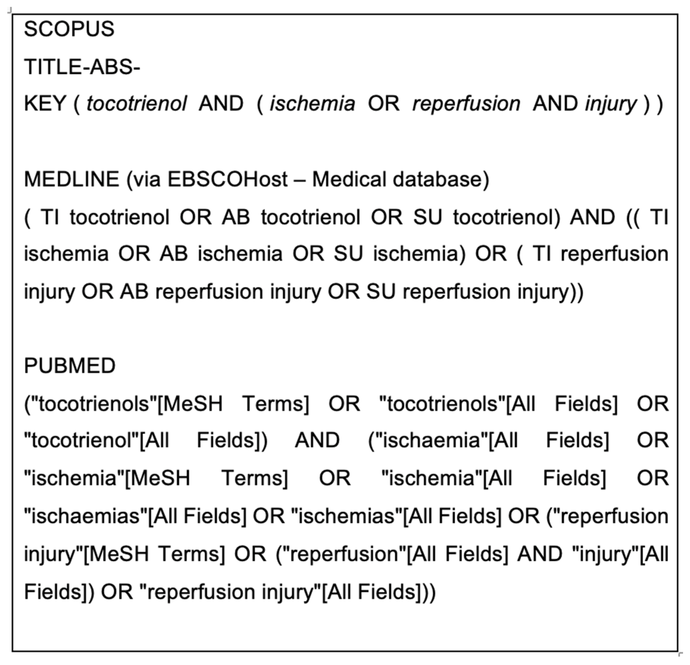
2.2. Search Strategy and Study Selection

We sought articles from Scopus, MEDLINE (via EBSCOhost), and PubMed for studies submitted from inception until June 2021. We used the terms “tocotrienol” AND (“ischemia” OR “reperfusion injury”) in our searching (Figure 1). For reasons of feasibility, the study authors were not contacted for missing data or verification. The screening was performed by two independent persons at each step, first in terms of titles, then abstracts, and finally, full texts based on the eligibility criteria. Any disagreement was resolved by discussion. Our protocol was not registered in any database.

2.3. Data Extraction

The following information was extracted: the first author’s name, year of publication, animal model characteristics (species, gender, age, and disease induction), intervention characteristics (intervention, duration, dose, route of administration), and outcomes. The outcome measures included any structural, functional, and biochemical parameters. Any disagreement was resolved by discussion.

2.4. Quality Assessment

Risk-of-bias assessments were conducted by two investigators independently using the Systematic Review Center for Laboratory Animal Experimentation (SYRCLE) [23].

2.5. Data Analysis

We planned to extract data in terms of mean and standard deviation to calculate the mean differences between the tocotrienol-treated group and control for numerous parameters. The 95% confidence interval would also be calculated. However, given the substantial differences in methodology, including the forms of tocotrienol preparation and the duration of I/R, we decided not to conduct the meta-analysis. The results were reported qualitatively.

3. Results

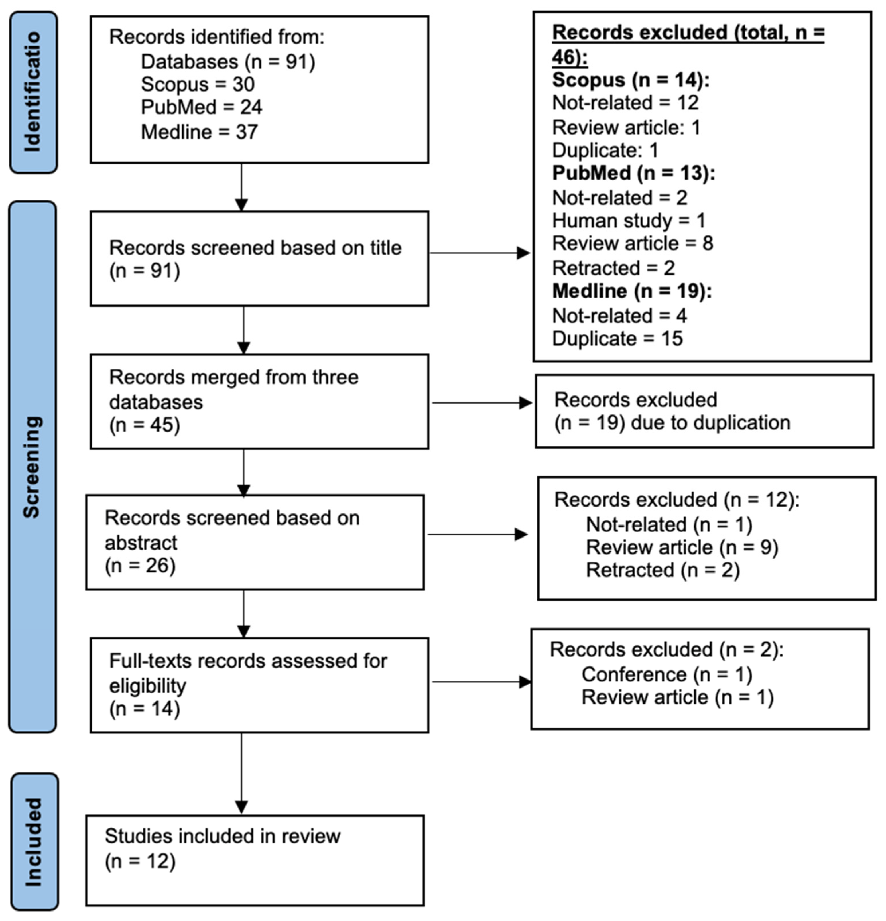
3.1. Description of Article Selection Process

Initially, we identified a total of 91 articles from three databases (Figure 2). Next, we excluded 46 articles for various reasons, including: not being related to tocotrienols (n = 18), review articles (n = 9), duplicates (n = 16), human study (n = 1), and retracted articles (n = 2), based on the title screening. All articles were then merged (n = 45), and 19 articles were eliminated due to duplications. Twenty-six articles were then screened, and 12 were excluded during abstract screening, while two (a conference abstract [24] and a review) were removed during full-text screening, leaving a final total of 12 articles that were included in the qualitative analysis.

3.2. Characteristics of Included Studies

Twelve articles from the years 1992 to 2018 were included in our qualitative analysis (Table 1). Seven articles reported the effects of tocotrienols in myocardial I/R injury [10,11,12,13,14,15,16], and the other five reported their effects in cerebral I/R injury [17,18,19,20,21]. In the cerebral I/R injury studies, all except one (a canine model [20]) utilized various types of mice, including ICR [17,21], C57BL/6 [19], and ddY [18]. The stroke induction was carried out by the occlusion of the middle cerebral artery (MCA) for 60 min, followed by reperfusion for one day [20] or seven days [17,21]. Another study performed 90 min of MCA occlusion followed by two days of reperfusion [19], while Mishima et al. [18] occluded MCA for 4 h, then reperfused for 24 h. In terms of treatment, two studies utilized a Tocovid treatment that contained α-tocotrienol (12.4%), α-tocopherol (11.3%), β-tocotrienol (2.5%), γ-tocotrienol (19.2%), and 𝛿-tocotrienol (6.3%) [17,21], while another study used a combined tocotrienol pill that contained α-tocotrienol (61.52 mg), γ-tocotrienol (112.8 mg), and δ-tocotrienol (25.68 mg) [20]. Furthermore, other studies administered individual tocotrienols [18,19] and tocopherols [18].
In contrast, most of the studies in myocardial I/R injury models utilized various types of rat models, such as male Sprague-Dawley [10,11,14,15,16], male Wistar [13], and male Long-Evans [12]. Moreover, all studies of the myocardial I/R injury had conducted ex vivo studies to induce global ischemia for 30 min [10,14,15], followed by 120 min of reperfusion [10,11,14,15]. However, two studies had induced 25 min of ischemia, followed by 25 min of reperfusion [12,13], while another study subjected the heart to 40 min of global ischemia and 20 min of reperfusion [16]. In terms of treatment, all the studies in the myocardial I/R models used individual tocotrienols in their research [10,11,14,15], except for two that utilized red palm oils [12,13].

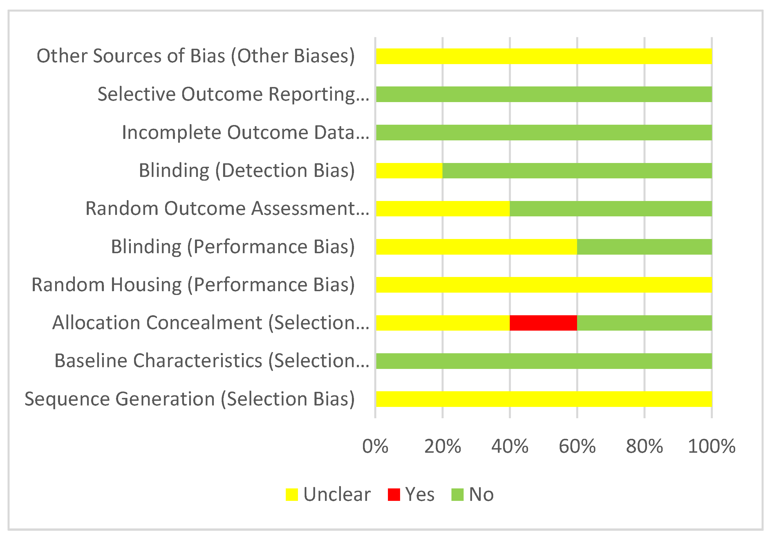
3.3. Risk of Bias Assessment

3.3.1. Cerebral Ischemia-Reperfusion Injury

All the studies (n = 5) reported an unclear risk of bias in sequence generation, random housing, and other sources of bias (Figure 3). However, all studies also reported no risk of bias in the baseline characteristics, incomplete outcome data, and selective outcome reporting. Most studies reported no risk of bias regarding detection bias (blinding and random outcome assessment) but an unclear risk of bias regarding performance bias (blinding). One study reported a high risk of bias for allocation concealment.

3.3.2. Myocardial Ischemia-Reperfusion Injury

All studies (n = 7) reported an unclear risk of bias in sequence generation, allocation concealment, blinding (detection bias), and other sources of bias (Figure 4). In contrast, all studies reported no risk of bias for baseline characteristics, incomplete outcome data, and selective outcome reporting. Most studies had no risk of bias for performance bias (housing and blinding).

3.4. Tocotrienol’s Effects on Cerebral Ischaemia Reperfusion Injury

Table 2 shows how all studies reported that various tocotrienol forms, including α-tocotrienol [18,19], combined tocotrienols [20], and combined tocotrienols and α-tocopherol [17,21], significantly reduced the infarct size in those animals subjected to the MCA occlusion. Reduction in cytotoxic edema was another structural improvement mediated by tocotrienol treatment [20]. Other than that, the improvement of collateral circulation was also evident in the same study that utilized a canine model [20]. Furthermore, various parameters were also reported to be significantly changed, including the attenuation of oxidative stress markers (oxidized glutathione (GSSG) to glutathione (GSH) ratio [21], 4-hydroxy-2-nonenal (4-HNE) [19,21], nitrotyrosine [21], 8-hydroxy-2′-deoxyguanosine (8-OHdG) [21], the receptor for advanced glycation end products (RAGE) [21], N (omega)-(carboxymethyl) arginine (CMA) [21], N (omega)-(carboxymethyl) lysine (CML) [21]), the regulation of antioxidant-related markers (increased NF-E2 p45-related factor 2 (Nrf2) [21]), reductions in inflammation parameters (tumor necrosis factor α (TNF-α) [17], monocyte chemotactic protein 1 (MCP-1), Iba-1, IgG [17], light chain 3 (LC3) II [21]) and a reduction of apoptotic marker (cleaved caspase-3 [21]). In addition, the extracellular matrix was significantly affected, as evidenced by the increase in collagen IV [17] and tissue inhibitor of metalloproteinase 1 (TIMP-1), and the reduction in matrix metalloproteinase (MMP)-9 (not MMP-2) levels [20]. Two transporters that were regulated by tocotrienol treatment were multidrug-resistant protein 1 (MRP1) [19,21] and chloride intracellular channel protein 1 (CLIC1) [20]. Neurobehavioral assessment of the mice demonstrated a significant improvement in Rotarod time but a negligible change in the Bederson score and Corner test [17,21].

3.5. Tocotrienol’s Effects in Myocardial Ischemia-Reperfusion Injury

Differing from the studies of cerebral I/R injury, the early reports regarding myocardial I/R injury focused on the use of palm oils containing both tocotrienols and tocopherols [12,13,16]. The more recent studies used individual tocotrienols, with the primary focus on γ-tocotrienol. Although both α- and γ-tocotrienol isomers have been reported to affect many parameters significantly, the latter isomer has the most prominent cardioprotective effects [10,11], particularly in females [11]. Improvements in structural [11,14,15] and functional parameters have been observed, although functional parameters reported conflicting outcomes (Table 3) [11,12,13,14,15,16].
In addition, tocotrienol treatment remarkably reduced the oxidative stress markers (reactive oxygen species [15], lipid peroxidation product [16]), angiogenic factor (vascular endothelial growth factor [15]), collagen homeostasis regulators (MMP-2, MMP-9, transforming growth factor β (TGF-β) [11]) and the pro-apoptotic marker (cleaved caspase-3 [10]). In contrast, tocotrienols significantly increased the expression of the antioxidative enzyme of superoxide dismutase [11], the anti-apoptotic marker of b-cell lymphoma 2 (bcl-2) [14], mitochondrial-related proteins (ATP synthase, cytochrome-c oxidase 1, 2, 3) [11], and calcium pump-related proteins (calpain, calsequestrin, phospholamban) [11].
Red palm oil was reported to increase cyclic GMP levels after 10 min during the ischemic period [12,13]. However, a negligible effect was found on nitric oxide levels [13]. Furthermore, γ-tocotrienol was reported to increase the phosphorylation of Akt and reduce the phosphorylated mechanistic target of rapamycin (mTOR) [14]. The addition of wortmannin, an inhibitor of phosphatidylinositol-3-kinase (PI3K), reversed the effects of tocotrienols on Akt and mTOR [14]. Lekli et al. [14] also showed an elevated Beclin-1 level and the ratio of LCII to LCI. Other than that, tocotrienol treatment significantly increased the bindings of p38-mitogen-activated protein kinase (MAPK) α and SRC kinase, while reducing the endothelial nitric oxide synthase (eNOS) and heme oxygenase 1 (HO-1) with caveolin-1 [10]. In contrast, the interaction of p38MAPKβ and caveolin-3 was significantly attenuated in rats treated with tocotrienols [10].

4. Discussion

Our review identified numerous beneficial effects of tocotrienols on many parameters regarding both the brain and heart. Tocotrienols, either used as individual isomers or when combined with other tocotrienols and tocopherol isomers, have various positive impacts as neuroprotective and cardioprotective agents.
Mishima et al. [18] reported that both α-tocotrienol and γ-tocopherol were more potent than α-tocopherol in cerebral protection, as evidenced by significant reductions of infarct volume. Similarly, Park et al. [19] found a remarkable reduction in infarct volume in the α-tocotrienol-treated group. The mechanism of neuroprotection might be attributable to the ability of tocotrienols to reverse oxidative stress conditions.
Cerebral I/R injury causes the excessive generation of reactive oxygen species (ROS), primarily from the electron transport chain [25]. ROS interacts with various cellular components, including DNA, proteins, and lipids, as evidenced by the significant increment of oxidative markers of 8–hydroxy-2′-deoxyguanosine (8-OHdG), nitrotyrosine, and 4-hydroxy-2-nonenal (4-HNE), respectively [19,21]. Membrane disruption following the insult causes the leakage of intracellular components, such as lactate dehydrogenase (LDH) [19,21].
The endogenous antioxidant system comprises catalase (CAT), glutathione peroxidase (GPx), glutathione reductase (GR), glutathione S transferase (GST), and superoxide dismutase (SOD), which are distributed unevenly in the different brain regions [26]. SOD plays an essential role in converting superoxide into hydrogen peroxide, while CAT, GPx, and GST act in concert to neutralize hydroperoxides [26]. On the other hand, GR serves as a GSH regenerator, reducing the GSSG by transferring an electron from nicotinamide adenine dinucleotide phosphate (NADPH) to GSSG [26].
Cerebral I/R injury has been shown to affect antioxidant enzymes differently, depending on the region and duration of the insult. For instance, GR activity is reduced during ischemia in the cerebral cortex, cerebellum, and hippocampus but becomes elevated at one hour following reperfusion [26]. In contrast, Candelario-Jalil et al. [27] reported negligible hippocampus changes during ischemia, with a reperfusion period of up to 24 h. The inconsistent results might be attributable to the different durations of ischemia and probably to the different animal models [26,27]. Moreover, during the first six hours of reperfusion, the levels of GSH significantly decreased, along with a notable increase in GSSG levels. GSSG is a toxic metabolite and can be transported away via the multidrug resistance-associated protein 1 (MRP1) [21]. Shang et al. [21] reported that the I/R injury contributed to the increase in GSSG levels and MRP1 expression. In contrast, MRP1-deficient mice had significantly higher GSSG accumulation and infarct volume than non-deficient mice. α-tocotrienol upregulates MRP1, attenuating the elevation of the intracellular GSSG level and, thus, reducing the infarct volume [19,21].
Three included studies utilized a fixed-dose preparation of tocotrienols or in combination with tocopherol [17,20,21]. Rink et al. [20] used a canine model to elucidate the potential mechanism of combined tocotrienols in stroke via collateral circulation and reported significant improvements in cytotoxic edema and infarct volume in the first hour following the initiation of reperfusion. Furthermore, the infarct volume remained significantly lower in the combined tocotrienols group at 24 h [20]. The effects might be attributed to arteriogenesis, a process that takes place in an early phase due to enhanced blood flow in the existing collateral blood vessels [28]. This improvement in collateral circulation might be attributed to the regulation of the extracellular matrix, as evidenced by the increase in TIMP-1 and reductions in both MMP-2 and MMP-9 [17,20]. MMPs, together with plasmin, are essential for the degradation of the external elastic lamina and elastin, forming a space for expanding blood vessels [28]. Excessive MMP activity during the I/R event causes damage to the basal lamina, promoting blood-brain barrier disruption and inflammation [29,30]. TIMP-1, the inhibitor of MMP-9, was significantly elevated in the tocotrienol-treated group [20]. The downregulation of MMP-9 and the elevation of TIMP-1 are essential mechanisms that promote neuroprotection [30].
On the contrary, the level of vascular endothelial growth factor (VEGF) is not significantly changed by tocotrienol-enriched treatment [20]. VEGF is an essential factor for angiogenesis, the process of sprouting of new blood vessels [31]. However, several studies had reported that the induction of angiogenesis in the early phase contributes to the aggravation of stroke, due to disruption of the blood-brain barrier and increased vascular permeability, resulting in worsening vasogenic edema and infarct size [31]. Thus, the downregulation of angiogenic factors, including VEGF by tocotrienols, promotes beneficial outcomes in ischemic stroke.
Another interesting finding is the increase in CLIC1 but not CLIC4 expression in the tocotrienol-enriched group [20]. A previous study by Chalothorn et al. [32] reported that CLIC4 was an essential factor for collateral circulation and demonstrated that mice that were deficient in CLIC4 genes (homozygous Clic4−/−) had a fourfold-larger infarct size compared to the homozygous Clic4+/+. However, they did not extend their study regarding CLIC1, as negligible changes were observed in the collateral density and diameter between the postnatal and adult period in Clic1−/− mice [32]. The exact reason for the contrasting expression of CLIC1 and CLIC4 remains to be elucidated, particularly with tocotrienol treatment.
In terms of myocardial I/R injury, tocotrienols, particularly γ-tocotrienol, have been reported to protect the heart, as evidenced by the significant improvement of various functional parameters, including aortic flow and output recovery, the left ventricular developed pressure, and the first derivative of developed pressure [11,12,13,14,15]. These changes are often accompanied by a reduction in infarct size [11,14,15].
Mechanisms of tocotrienol-induced cardioprotective effects are numerous, including regulation of PI3K/Akt/mTOR and MAPK pathways. The activation of PI3K promotes the phosphorylation of Akt, which in turn leads to the activation of mTOR [33]. Myocardial I/R injury has been shown to reduce the phosphorylation of Akt and mTOR [33]. Lekli et al. [14] had confirmed the role of tocotrienol in modulating the PI3K/Akt/mTOR-signaling pathway since the use of wortmannin, the inhibitor of PI3K, reversed the inhibition of this pathway. Interestingly, they also found the potential role of tocotrienols in modulating autophagy via the PI3K/Akt/mTOR pathway.
Autophagy is an essential process that degrades damaged organelles and proteins and is frequently activated during stress conditions, such as myocardial I/R injury [34]. Tocotrienol has been reported to enhance the Beclin-1 level and LCII/LCI ratio [14]. The former component is required to form phosphatidylinositol 3-kinase class III (PI3K-III), a complex that is required for the initiation of autophagy in the presence of another complex, namely, an autophagy-related 1 (ATG1)/Unc–51 such as autophagy-activating kinase-1 (ULK1) [35]. In its inactive form, Beclin-1 presents in a homodimer configuration, with Bcl-2/Bcl-XL proteins attached to the specific binding site (Bcl-2 homology-3) on each Beclin-1 molecule [35]. Tocotrienols inhibit the PI3K/Akt/mTOR-signaling pathway [14], resulting in increased ULK1 activity due to reduced MTORC1 activity, which is required to inactivate ULK1 [35]. LC3 also plays an essential role in the formation of the autophagosome. LC3II is an autophagosome marker produced by LC3I conversion, via a series of ATG reactions that mediate the incorporation of LC3II into the membrane of the autophagosome [36]. Then, the autophagosome fuses with a lysosome, forming an autolysosome that degrades the accumulated products [35].
Another mechanism that contributes to tocotrienol-induced cardioprotective effects is the regulation of a variety of signaling molecules via caveolin. Caveolin is an integral protein that is associated with caveolae, the lipid rafts in the plasma membrane that forms an invagination in the presence of multiple oligomerized caveolins [37]. Both caveolin-1 and -3 are critical regulators in myocardial I/R injury. Das et al. [10] reported that γ-tocotrienol promotes caveolin-1 interaction with p38α, while reducing the interaction of caveolin-3 with p38β, resulting in the inhibition of the mitogen-activated protein kinase (MAPK) pathway [10]. The former p38 isoform is associated with the pro-apoptotic pathway, while the latter promotes anti-apoptotic pathway [38]. Moreover, tocotrienols attenuate mitochondrial injury, reducing cytochrome c translocation from mitochondria into the cytoplasm, and the activation of caspase-3 that promotes apoptosis [10]. Other than that, tocotrienols also increase the interaction of another pro-apoptotic marker (Src kinase) and caveolin-1 but weaken the binding to anti-apoptotic markers of eNOS and HO-1 [10].
In addition, tocotrienols promote cardioprotection via the regulation of angiogenesis [15]. Mukhopadhyay et al. [15] reported that the microRNA-20b level was significantly elevated in the γ-tocotrienol-treated group. In contrast, both hypoxia-induced factor-1α (HIF-1α) and vascular endothelial growth factor (VEGF) were significantly reduced. In the event of acute myocardial ischemia, HIF-1α expression is elevated, promoting the transcription of various molecules, including VEGF [39]. The exact reason why the tocotrienol modulation of angiogenesis leads to cardioprotective effects is unclear. However, it may be partly related to the attenuation of vascular permeability, reducing the inflammatory reaction.
Interestingly, the dose ranges for both cardioprotective and neuroprotective effects are distinct and broad. The studies involving the myocardial I/R injury model utilized long-term individual tocotrienols (α, γ, 𝛿) with a dose of between 0.02 and 5 mg/kg/day [10,11,14,15]. This dose range is substantially lower than in the studies of the cerebral I/R injury model that used either combined tocotrienols and α-tocopherol (200 mg/kg) [17,21], mixed tocotrienols (400 mg/day) [20], or α-tocotrienol (50 mg/kg/day) [19]. The human equivalent dose (HED) calculated (based on a 60-kg human) [40] for cardioprotective effects ranged from 0.2 to 48 mg/day [9,10,22,30], while the dose range for neuroprotective effects was 240–960 mg/day [17,19,21]. The question of the tolerability of the dose might arise, as the upper limit of the dose required is remarkably high. In an open-label, randomized study, Qureshi et al. [41] reported that 750–1000 mg of tocotrienols given orally were tolerated well and showed a similar pharmacokinetic profile compared to lower doses. Furthermore, no adverse effects were reported [41]. This study indicates that the HED derived from the studies is safe to be consumed.
The reason for the higher dose requirement for neuroprotection is obscure but is probably due to the different pharmacokinetic properties of tocotrienols. The α-tocopherol transfer protein (α-TTP) plays a pivotal role in the incorporation of α-tocopherol into very-low-density lipoprotein (VLDL) before its release into the circulation or other cells [42,43]. Although the liver is the major site of α-TTP, other organs have been shown to express low levels of α-TTP. Hosomi et al. [42] detected the presence of α-TTP mRNA in the brain. This protein has a 100% binding affinity toward α-tocopherol but variable affinity toward other tocopherol isomers and tocotrienols [43]. Lower binding affinity may partly explain why the brain requires a higher dose of tocotrienols to increase their transportation into the brain. For instance, Hansen et al. [44] reported a remarkable accumulation of vitamin E (α-tocopherol, γ-tocotrienol, 𝛿-tocotrienol) in the brains of hens fed with high doses of tocopherol and/or annatto tocotrienols.
Our systematic review has several limitations. The search databases were limited to only three: PubMed, Scopus, and MEDLINE. Other databases such as Ovid, Cochrane, and Google Scholar and the grey literature were not sought but could contribute more potential articles. In terms of the included articles, some data provided were contradictory. For instance, the values given in the text descriptions were not parallel to those stated in the included figures. Other than that, the authors of the articles were not contacted to verify the data, for reasons of feasibility. Moreover, there are a variety of animal models with different durations of ischemia and reperfusion and different forms of tocotrienols. These differences might produce variable effects, contributing to non-homogeneous findings. On the other hand, the exploration of the molecular signaling pathway of tocotrienols in attenuating cerebral and myocardial I/R injury was still limited. Further studies should be conducted to determine other related molecular pathways, such as RhoA/ROCK, Sirtuin-1, and JNK-mediated-nuclear factor-kappa b. Despite the differences in terms of animal models and treatments, all studies demonstrated the positive effects of tocotrienols. In addition, this review addresses the gap of knowledge that can be filled in future studies.

5. Conclusions

Tocotrienols have the potential to serve as a supplement to reduce the impact of reperfusion injury via the regulation of oxidative stress, inflammation, and apoptosis, promoting structural, biochemical, and functional improvements.

Author Contributions

Conceptualization, F.F.R.; methodology, F.F.R.; formal analysis, F.F.R., A.A. and N.’I.I.; writing—original draft preparation, F.F.R., A.A. and N.’I.I.; writing—review and editing, F.F.R., A.A. and N.’I.I.; funding acquisition, F.F.R. All authors have read and agreed to the published version of the manuscript.

Funding

This research was funded by Universiti Kebangsaan Malaysia (UKM), grant number FF-2021-055.

Institutional Review Board Statement

Not applicable.

Informed Consent Statement

Not applicable.

Data Availability Statement

Not applicable.

Conflicts of Interest

The authors confirm that this article has no conflict of interest.

References

  1. Khan, M.A.B.; Hashim, M.J.; Mustafa, H.; Baniyas, M.Y.; Al Suwaidi, S.K.B.M.; AlKatheeri, R.; Alblooshi, F.M.K.; Almatrooshi, M.E.A.H.; Alzaabi, M.E.H.; Al Darmaki, R.S.; et al. Global Epidemiology of Ischemic Heart Disease: Results from the Global Burden of Disease Study. Cureus 2020, 12, e9349. [Google Scholar] [CrossRef]
  2. Johnson, C.O.; Nguyen, M.; Roth, G.A.; Nichols, E.; Alam, T.; Abate, D.; Abd-Allah, F.; Abdelalim, A.; Abraha, H.N.; Abu-Rmeileh, N.M.; et al. Global, regional, and national burden of stroke, 1990–2016: A systematic analysis for the Global Burden of Disease Study 2016. Lancet Neurol. 2019, 18, 439–458. [Google Scholar] [CrossRef] [Green Version]
  3. Chelluboina, B.; Vemuganti, R. Therapeutic potential of nutraceuticals to protect brain after stroke. Neurochem. Int. 2021, 142, 104908. [Google Scholar] [CrossRef] [PubMed]
  4. Sosnowska, B.; Penson, P.; Banach, M. The role of nutraceuticals in the prevention of cardiovascular disease. Cardiovasc. Diagn. Ther. 2017, 7, S21–S31. [Google Scholar] [CrossRef] [PubMed] [Green Version]
  5. Aggarwal, B.B.; Sundaram, C.; Prasad, S.; Kannappan, R. Tocotrienols, the vitamin E of the 21st century: Its potential against cancer and other chronic diseases. Biochem. Pharmacol. 2010, 80, 1613–1631. [Google Scholar] [CrossRef] [PubMed] [Green Version]
  6. Ahsan, H.; Ahad, A.; Siddiqui, W.A. A review of characterization of tocotrienols from plant oils and foods. J. Chem. Biol. 2015, 8, 45–59. [Google Scholar] [CrossRef] [PubMed] [Green Version]
  7. Shahidi, F.; De Camargo, A.C. Tocopherols and tocotrienols in common and emerging dietary sources: Occurrence, applications, and health benefits. Int. J. Mol. Sci. 2016, 17, 1745. [Google Scholar] [CrossRef]
  8. Loh, H.C.; Lim, R.; Lee, K.W.; Ooi, C.Y.; Chuan, D.R.; Looi, I.; Hay, Y.K.; Khan, N.A.K. Effects of vitamin E on stroke: A systematic review with meta-analysis and trial sequential analysis. Stroke Vasc. Neurol. 2021, 6, 109–120. [Google Scholar] [CrossRef] [PubMed]
  9. Loffredo, L.; Perri, L.; Di Castelnuovo, A.; Iacoviello, L.; De Gaetano, G.; Violi, F. Supplementation with vitamin E alone is associated with reduced myocardial infarction: A meta-analysis. Nutr. Metab. Cardiovasc. Dis. 2015, 25, 354–363. [Google Scholar] [CrossRef]
  10. Das, M.; Das, S.; Wang, P.; Powell, S.R.; Das, D.K. Caveolin and proteasome in tocotrienol mediated myocardial protection. Cell. Physiol. Biochem. 2008, 22, 287–294. [Google Scholar] [CrossRef]
  11. Das, S.; Mukherjee, S.; Lekli, I.; Gurusamy, N.; Bardhan, J.; Raychoudhury, U.; Chakravarty, R.; Banerji, S.; Knowlton, A.A.; Das, D.K. Tocotrienols confer resistance to ischemia in hypercholesterolemic hearts: Insight with genomics. Mol. Cell Biochem. 2012, 360, 35–45. [Google Scholar] [CrossRef] [PubMed]
  12. Esterhuyse, A.J.; Du Toit, E.; Van Rooyen, J. Dietary red palm oil supplementation protects against the consequences of global ischemia in the isolated perfused rat heart. Asia Pac. J. Clin. Nutr. 2005, 14, 340–347. [Google Scholar]
  13. Esterhuyse, J.S.; van Rooyen, J.; Strijdom, H.; Bester, D.; du Toit, E.F. Proposed mechanisms for red palm oil induced cardioprotection in a model of hyperlipidaemia in the rat. Prostaglandins Leukot. Essent. Fat. Acids 2006, 75, 375–384. [Google Scholar] [CrossRef] [PubMed]
  14. Lekli, I.; Ray, D.; Mukherjee, S.; Gurusamy, N.; Ahsan, M.K.; Juhasz, B.; Bak, I.; Tosaki, A.; Gherghiceanu, M.; Popescu, L.M.; et al. Co-ordinated autophagy with resveratrol and gamma-tocotrienol confers synergetic cardioprotection. J. Cell. Mol. Med. 2010, 14, 2506–2518. [Google Scholar] [CrossRef] [PubMed] [Green Version]
  15. Mukhopadhyay, P.; Das, S.; Ahsan, M.K.; Otani, H.; Das, D.K. Modulation of microRNA 20b with resveratrol and longevinex is linked with their potent anti-angiogenic action in the ischaemic myocardium and synergestic effects of resveratrol and γ-tocotrienol. J. Cell. Mol. Med. 2012, 16, 2504–2517. [Google Scholar] [CrossRef]
  16. Serbinova, E.; Khwaja, S.; Catudioc, J.; Ericson, J.; Torres, Z.; Gapor, A.; Kagan, V.; Packer, L. Palm oil vitamin E protects against ischemia/reperfusion injury in the isolated perfused langendorff heart. Nutr. Res. 1992, 12, S203–S215. [Google Scholar] [CrossRef]
  17. Jiao, Y.; Shang, J.; Ohta, Y.; Yan, H.; Liu, X.; Li, X.; Morihara, R.; Nakano, Y.; Fukui, Y.; Shi, X.; et al. Neuroprotective Effects of Tocovid Pretreatment in a Mouse Stroke Model. J. Stroke Cerebrovasc. Dis. 2018, 27, 2166–2174. [Google Scholar] [CrossRef]
  18. Mishima, K.; Tanaka, T.; Pu, F.; Egashira, N.; Iwasaki, K.; Hidaka, R.; Matsunaga, K.; Takata, J.; Karube, Y.; Fujiwara, M. Vitamin E isoforms alpha-tocotrienol and gamma-tocopherol prevent cerebral infarction in mice. Neurosci. Lett. 2003, 337, 56–60. [Google Scholar] [CrossRef]
  19. Park, H.A.; Kubicki, N.; Gnyawali, S.; Chan, Y.C.; Roy, S.; Khanna, S.; Sen, C.K. Natural vitamin E α-tocotrienol protects against ischemic stroke by induction of multidrug resistance-associated protein 1. Stroke 2011, 42, 2308–2314. [Google Scholar] [CrossRef] [Green Version]
  20. Rink, C.; Christoforidis, G.; Khanna, S.; Peterson, L.; Patel, Y.; Khanna, S.; Abduljalil, A.; Irfanoglu, O.; Machiraju, R.; Bergdall, V.K.; et al. Tocotrienol vitamin E protects against preclinical canine ischemic stroke by inducing arteriogenesis. J. Cereb. Blood Flow Metab. 2011, 31, 2218–2230. [Google Scholar] [CrossRef] [PubMed] [Green Version]
  21. Shang, J.; Yan, H.; Jiao, Y.; Ohta, Y.; Liu, X.; Li, X.; Morihara, R.; Nakano, Y.; Fukui, Y.; Shi, X.; et al. Therapeutic Effects of Pretreatment with Tocovid on Oxidative Stress in Postischemic Mice Brain. J. Stroke Cerebrovasc. Dis. 2018, 27, 2096–2105. [Google Scholar] [CrossRef] [PubMed]
  22. Moher, D.; Liberati, A.; Tetzlaff, J.; Altman, D.G. Preferred reporting items for systematic reviews and meta-analyses: The PRISMA statement. PLoS Med. 2009, 6, e1000097. [Google Scholar] [CrossRef] [Green Version]
  23. Hooijmans, C.R.; Rovers, M.M.; De Vries, R.B.; Leenaars, M.; Ritskes-Hoitinga, M.; Langendam, M.W. SYRCLE’s risk of bias tool for animal studies. BMC Med. Res. Methodol. 2014, 14, 43. [Google Scholar] [CrossRef] [PubMed] [Green Version]
  24. Das, S.; Tosaki, A.; Juhasz, B.; Lekli, I.; Varadi, J.; Nesaretam, K.; Maulik, N.; Das, D.K. Tocotrienols in cardioprotection: Role of different isomers. FASEB J. 2007, 21, A1112. [Google Scholar] [CrossRef]
  25. Lin, L.; Wang, X.; Yu, Z. Ischemia-reperfusion Injury in the Brain: Mechanisms and Potential Therapeutic Strategies. Biochem. Pharmacol. 2016, 5. [Google Scholar] [CrossRef] [Green Version]
  26. Kumari Naga, K.; Panigrahi, M.; Prakash Babu, P. Changes in endogenous antioxidant enzymes during cerebral ischemia and reperfusion. Neurol. Res. 2007, 29, 877–883. [Google Scholar] [CrossRef]
  27. Candelario-Jalil, E.; Mhadu, N.H.; Al-Dalain, S.M.; Martínez, G.; León, O.S. Time course of oxidative damage in different brain regions following transient cerebral ischemia in gerbils. Neurosci. Res. 2001, 41, 233–241. [Google Scholar] [CrossRef] [Green Version]
  28. Liu, J.; Wang, Y.; Akamatsu, Y.; Lee, C.C.; Stetler, R.A.; Lawton, M.T.; Yang, G.Y. Vascular remodeling after ischemic stroke: Mechanisms and therapeutic potentials. Prog. Neurobiol. 2014, 115, 138–156. [Google Scholar] [CrossRef] [Green Version]
  29. Rosenberg, G.A.; Cunningham, L.A.; Wallace, J.; Alexander, S.; Estrada, E.Y.; Grossetete, M.; Razhagi, A.; Miller, K.; Gearing, A. Immunohistochemistry of matrix metalloproteinases in reperfusion injury to rat brain: Activation of MMP-9 linked to stromelysin-1 and microglia in cell cultures. Brain Res. 2001, 893, 104–112. [Google Scholar] [CrossRef]
  30. Tejima, E.; Guo, S.; Murata, Y.; Arai, K.; Lok, J.; van Leyen, K.; Rosell, A.; Wang, X.; Lo, E.H. Neuroprotective effects of overexpressing tissue inhibitor of metalloproteinase TIMP-1. J. Neurotrauma 2009, 26, 1935–1941. [Google Scholar] [CrossRef] [PubMed]
  31. Adamczak, J.; Hoehn, M. Poststroke angiogenesis, con: Dark side of angiogenesis. Stroke 2015, 46, e103–e104. [Google Scholar] [CrossRef] [PubMed] [Green Version]
  32. Chalothorn, D.; Zhang, H.; Smith, J.E.; Edwards, J.C.; Faber, J.E. Chloride intracellular channel-4 is a determinant of native collateral formation in skeletal muscle and brain. Circ. Res. 2009, 105, 89–98. [Google Scholar] [CrossRef] [PubMed] [Green Version]
  33. Cui, Z.H.; Zhang, X.J.; Shang, H.Q.; Wang, X.; Rong, D. Glutamine protects myocardial ischemia-reperfusion injury in rats through the PI3K/Akt signaling pathway. Eur. Rev. Med. Pharmacol. Sci. 2020, 24, 444–451. [Google Scholar] [CrossRef] [PubMed]
  34. Wu, D.; Zhang, K.; Hu, P. The Role of Autophagy in Acute Myocardial Infarction. Front. Pharmacol. 2019, 10, 551. [Google Scholar] [CrossRef] [Green Version]
  35. Menon, M.B.; Dhamija, S. Beclin 1 Phosphorylation—At the Center of Autophagy Regulation. Front. Cell Dev. Biol. 2018, 6, 137. [Google Scholar] [CrossRef] [PubMed] [Green Version]
  36. Tanida, I.; Ueno, T.; Kominami, E. LC3 conjugation system in mammalian autophagy. Int. J. Biochem. Cell Biol. 2004, 36, 2503–2518. [Google Scholar] [CrossRef] [PubMed]
  37. Yang, Y.; Ma, Z.; Hu, W.; Wang, D.; Jiang, S.; Fan, C.; Di, S.; Liu, D.; Sun, Y.; Yi, W. Caveolin-1/-3: Therapeutic targets for myocardial ischemia/reperfusion injury. Basic Res. Cardiol. 2016, 111, 45. [Google Scholar] [CrossRef] [PubMed]
  38. Romero-Becerra, R.; Santamans, A.M.; Folgueira, C.; Sabio, G. p38 MAPK Pathway in the Heart: New Insights in Health and Disease. Int. J. Mol. Sci. 2020, 21, 7412. [Google Scholar] [CrossRef] [PubMed]
  39. Lee, S.H.; Wolf, P.L.; Escudero, R.; Deutsch, R.; Jamieson, S.W.; Thistlethwaite, P.A. Early expression of angiogenesis factors in acute myocardial ischemia and infarction. N. Engl. J. Med. 2000, 342, 626–633. [Google Scholar] [CrossRef]
  40. FDA. Guidance for Industry, Estimating the Maximum Safe Starting Dose in Initial Clinical Trials for Therapeutics in Adult Healthy Volunteers; Food and Drug Administration: Rockville, MD, USA, 2005. [Google Scholar]
  41. Qureshi, A.A.; Khan, D.A.; Silswal, N.; Saleem, S.; Qureshi, N. Evaluation of Pharmacokinetics, and Bioavailability of Higher Doses of Tocotrienols in Healthy Fed Humans. J. Clin. Exp. Cardiol. 2016, 7, 1–11. [Google Scholar] [CrossRef] [Green Version]
  42. Hosomi, A.; Goto, K.; Kondo, H.; Iwatsubo, T.; Yokota, T.; Ogawa, M.; Arita, M.; Aoki, J.; Arai, H.; Inoue, K. Localization of alpha-tocopherol transfer protein in rat brain. Neurosci. Lett. 1998, 256, 159–162. [Google Scholar] [CrossRef]
  43. Szewczyk, K.; Chojnacka, A.; Górnicka, M. Tocopherols and Tocotrienols-Bioactive Dietary Compounds; What Is Certain, What Is Doubt? Int. J. Mol. Sci. 2021, 22, 6222. [Google Scholar] [CrossRef] [PubMed]
  44. Hansen, H.; Wang, T.; Dolde, D.; Xin, H. Tocopherol and annatto tocotrienols distribution in laying-hen body. Poult. Sci. 2015, 94, 2421–2433. [Google Scholar] [CrossRef] [PubMed]
Figure 1. Search strategy.
Applsci 11 07994 g001
Figure 2. PRISMA flow diagram for the identification of studies via databases.
Applsci 11 07994 g002
Figure 3. Risk of bias assessment for cerebral ischemia-reperfusion injury.
Applsci 11 07994 g003
Figure 4. Risk of bias assessment for myocardial ischemia-reperfusion injury.
Applsci 11 07994 g004
Table 1. Findings from the included studies.
Author (Year)Animal CharacteristicsTreatmentEffects
Brain
Jiao [17] (2018)ICR mice weighing 23–25 g aged 6 weeks.200 mg/kg/day Tocovid for 1 month, given orally.Neuroprotection via anti-inflammatory effects of Tocovid.
Shang [21] (2018)ICR mice weighing 23–25 g aged 6 weeks.200 mg/kg/day Tocovid for 1 month, given orally.Neuroprotection via antioxidative effects.
Rink [20] (2011)Mongrel canines weighing 26.6 ± 2.6 kg aged 2.4 ± 0.9 years.200 mg mixed tocotrienols two times a day for 10 weeks.Neuroprotective effects via collateral
circulation.
Park [19] (2011)C57BL/6 mice aged 5 weeks. Mouse hippocampal HT4 neural cells—primary cortical neurons (Sprague-Dawley rats).50 mg/kg/day α-tocotrienol for 13 weeks, given orally via gavage;
1 μmol/L α-tocotrienol added 6 h before the glutamate treatment.
Regulation of MRP1 by α-tocotrienols.
Mishima [18] (2003)Male ddY mice weighing
25–35 g.
0.2 mM or 2 mM of α-/γ-/𝛿-tocotrienol, α-/γ-/𝛿-tocopherol, given by bolus intravenously 3 h before and after MCA occlusion.Reduction of cerebral infarct volume is more effective in α-tocotrienol and
γ-tocopherol.
Heart
Mukhopadhyay [15] (2012)Male Sprague-Dawley rats weighing 250–300 g.5 mg/kg/day of γ-tocotrienol for 21 days via gavage.Modulation of microRNA that regulates angiogenesis.
Das [11] (2012)Adult New Zealand rabbits (male and female) weighing 2.4–3.0 kg—I/R model.
Genomic study: Adult
Sprague-Dawley rat (male).
20 µmol/kg/day of α-/γ-/𝛿-tocotrienols on top of 2% cholesterol diet. Control received 2% cholesterol diet only for 4 weeks. Gamma tocotrienol is superior in
cardioprotection, modulating numerous genes’ expression, including antioxidants, energy production, fatty acid metabolism and calcium channels. Interpretation of LPL, MMP-2, MMP-9, TGF-β, p-Akt, ERα, ERβ, p-FoxO4, and Spot-14 were not clear as the text description were not parallel to the figures provided. The data were based on the text description.
Lekli [14] (2010)Male Sprague-Dawley rats weighing 250–300 g.0.3 mg/kg/day of γ-tocotrienol for 30 days via gavage.Autophagy coordination and regulation of PI3K/Akt/mTOR-signaling pathway.
Das [10] (2008)Male Sprague Dawley rats weighing 250–300 g.α-/γ-/𝛿-tocotrienols with a dose of 0.3 mg/kg for 30 days via
gavage.
Caveolin and proteasome regulation of p38MAPK-signaling pathway.
Esterhuyse [13] (2006)Male Wistar rats, 300–400 g (post); 7 weeks old.Treatment group received rat chow with 7 g of RPO/kg for 6 weeks.
Esterhuyse [12] (2005)Male Long-Evans rats.Treatment group received rat chow with 7 g of RPO/kg diet. Control received only rat chow for 6 weeks.
Serbinova [16] (1992)Male Sprague-Dawley rats
(350–400 g).
Diet supplemented with 7 g palm oil vitamin E/kg (55% tocotrienols: 45% tocopherols). Control received 20 g vitamin E acetate/kg for 6 weeks.
ER: estrogen receptor; MMP: matrix metalloproteinase; MRP1: multidrug resistance-associated protein 1; mTOR: mammalian target of rapamycin; PI3K: phosphatidylinositol-3-kinase; TGF-β: transforming growth factor β.
Table 2. Outcomes of the tocotrienol treatment in cerebral ischemia-reperfusion injury.
AuthorsInfarct SizeLDHMRP1Rotarod TimeBederson ScoreCorner Test
Jiao [17]↓ TV NSNS
Shang [21]↓ TV NSNS
Rink [20]↓ TE
Park [19]↓ α-TC
Mishima [18]↓ α-TC, α-TH, γ-TC
↑: increased; ↓: reduced; α: alpha; γ: gamma; 𝛿: delta; LDH: lactate dehydrogenase; MRP1: multidrug-resistant protein 1; NS: not significant; TC: tocotrienol; TE: tocotrienol enriched; TH: tocopherol; TV: Tocovid.
Table 3. Structural and functional changes induced by tocotrienol treatment in myocardial ischemia-reperfusion injury.
AuthorISHRCFAFAORLVDPLV dp/dt
Mukhopadhyay [15] NS
Lekli [14] NS
Esterhuyse [13] NSNS NS
Esterhuyse [12] NS
Serbinova [16]
Das [11]M: γ
F: γα
M: NS γα𝛿
F: ↑γ (γ > α), (Fγ > Mγ), NS α𝛿
M: ↑γ (γ > α), NS α𝛿
F: ↑γ (γ > α), α (F > M), NS α𝛿
M: ↑γα (γ > α), NS 𝛿
F: ↑γ (γ > α),(F > M), α (F > M), NS 𝛿
M: ↑γα (γ > α), NS 𝛿
F: ↑γ (γ > α), (F > M), α (F > M), NS 𝛿
↓ reduced; ↑ increased; > higher than. α alpha; 𝛿 delta; γ gamma; F: female; M: male; AF: aortic flow; AOR: aortic output recovery; CF: coronary flow; LVDP: left ventricular developed pressure; LV dp/dt: first derivative of developed pressure; IS: infarct size; NS: not significant.
Publisher’s Note: MDPI stays neutral with regard to jurisdictional claims in published maps and institutional affiliations.

