Article

More Than 50-Fold Enhanced Nonlinear Optical Response of Porphyrin Molecules in Aqueous Solution Induced by Mixing Base and Organic Solvent

by
Satoshi Imura
1,
Takayoshi Kobayashi
2,3,4 and
Eiji Tokunaga
1,4,*
1
Department of Physics, Faculty of Science, Tokyo University of Science, 1-3 Kagurazaka, Shinjuku-ku, Tokyo 162-8601, Japan
2
Advanced Ultrafast Laser Research Center and Department of Engineering Science, The University of Electro-Communications, Chofu, Tokyo 182-8585, Japan
3
Department of Electrophysics, National Chiao Tung University, 1001 Ta Hsueh Road, Hsinchu 300, Taiwan
4
Research Center for Water Frontier Science and Technology, Tokyo University of Science, 1-3 Kagurazaka, Shinjuku-ku, Tokyo 162-8601, Japan
*
Author to whom correspondence should be addressed.
Appl. Sci. 2021, 11(11), 4892; https://doi.org/10.3390/app11114892
Submission received: 12 April 2021 / Revised: 15 May 2021 / Accepted: 19 May 2021 / Published: 26 May 2021
(This article belongs to the Section Optics and Lasers)

Abstract

:
The difference absorption spectrum (DAS) of porphyrin molecules (tetraphenyl-porphyhrin sulfonic acid, TPPS) in aqueous solution induced by continuous wave laser irradiation has been reported previously. It was interpreted that the DAS was caused by the formation of TPPS aggregates induced by laser irradiation. However, transient spectra similar in their shape have already been reported and are attributed to the excited-state absorption and saturable absorption (SA) effects due to the triplet state formation in TPPS. In the present study, we investigated the triplet quenching effect by O2 on the DAS of TPPS aqueous solution and revealed that it originated from the triplet state formation. We also found that mixing the appropriate amount of MeOH and NaOH in TPPS aqueous solution increased its absorbance change by more than 50 times. This may be due to the decrease in dissolved oxygen concentration by mixing them. This result suggests the possibility of controlling the performance of NLO materials by adjusting the solvent mixture ratio and base/acid concentration.

1. Introduction

Recently, change in the absorption spectrum of TPPS (tetraphenylporphyhrin sulfonic acid) aqueous solution induced by continuous wave (CW) laser (532 nm) irradiation [1] was observed. The origin of the puzzling phenomenon was interpreted to be the formation of TPPS aggregates induced by light gradient force of the laser from an unknown mechanism of “highly efficient” light-induced molecular aggregation [1,2]. However, the transient spectra of a TPPS aqueous solution similar in shape to the difference absorption spectrum (DAS) in ref. [1,2] had been already reported, and it was concluded that the absorption change was induced by the triplet excited-state absorption and saturable absorption (SA) effect due to the triplet state formation in TPPS monomers [3,4,5]. Here in this paper we investigated whether the absorption change previously observed was induced by the triplet state formation or the aggregation of TPPS monomers.
Even if the phenomenon we observed is the SA effect due to the triplet state formation in TPPS, it is important to investigate its behavior. The SA effect in dye molecules is a kind of nonlinear optical (NLO) response, which can be applied to phase-conjugated wave generation and passive Q-switching. Recently, the SA effect of dye-doped polyvinyl alcohol (PVA) was reported [6]. It was found that the SA effect was able to be tuned by changing the dye concentration. It was also reported that the SA effect of the dye was increased by forming a donor-acceptor system between polyoxometalate (POM), an electron acceptor, and the dye, an electron donor [7]. The increase in the SA effect was attributed to the depletion of the ground state in the dye due to the transfer of electrons in the dye to POM. Recently, there have been many reports on reverse saturable absorption (RSA) effect of dyes [8,9,10,11,12]. Absorbers with RSA properties have higher absorption efficiency with higher incident light intensity. RSA effect is caused by the accumulation of excited states and multiphoton absorption processes [13]. RSA effect by the triplet excited-state absorption has also been reported [14,15,16]. An important application of RSA effect is the protection of eyes and sensitive devices from intense light. J-aggregates [17,18] composed of dye-molecules aligned in one dimensional direction are attracting attention as nonlinear optical materials. The second- and third-order NLO response of J-aggregates is generally known to be large [19,20,21,22].
There are many studies on the triplet state in porphyrin molecules because of its possible application to cancer treatment and hydrogen photoproduction. For example, porphyrin molecules have been used as the photosensitizers in a cancer treatment called photodynamic therapy treatments [23,24,25,26,27,28,29,30,31]. Porphyrin has also been investigated as the photosensitizer of hydrogen photoproduction [32,33,34,35,36]. This is because the photocatalysts such as TiO2 do not absorb visible light so that photosensitizers which are sensitive to visible light such as porphyrin molecules are needed for the best use of sunlight. TPPS is one of the porphyrin molecules which is attracting attention as the photosensitizers of photodynamic therapy and hydrogen photoproduction, because the quantum yield of the triplet state formation in TPPS is as high as 0.78 [5].
In addition to the TPPS aqueous solution, we studied water-methanol mixed solutions of TPPS following ref. [2], where DAS of water-alcohol mixed solutions (dominated by alcohol) of TPPS were studied. We have found that by mixing methanol (MeOH) with the TPPS aqueous solution and adjusting NaOH concentration between 1 mM and 10 mM, the amount of the absorbance change increased up to 50 times. If the observed absorption changes were caused by the SA effect, this result suggests the possibility of controlling the performance of NLO materials by adjusting the solvent mixing ratio and base/acid concentration.

2. Materials and Methods

TPPS (Tokyo Chemical Industry Co., Tokyo, Japan) aqueous solution was prepared by dissolving TPPS in deionized water. The TPPS monomer in aqueous solution becomes a free-base monomer (F-monomer) under low acidity conditions (pH = 11), and diacid monomer (D-monomer) with protons coordinated to the two N 2 atoms in the center of the porphyrin ring under high acidity conditions (pH = 4) [37]. D-monomers in aqueous solution form J-aggregates under higher acidity conditions (pH = 0.8). NaOH (Wako Pure Chemical Industries, Ltd. (currently known as FUJIFILM Wako Pure Chemical Corporation), Tokyo, Japan) aqueous solution was mixed with the TPPS aqueous solution in order to prepare a sample in which absorption of F-monomer is dominant. In addition, by mixing MeOH (Kanto Chemical Co., Tokyo, Japan) with the F-monomer aqueous solution, the water-MeOH mixed solution of F-monomer was prepared. The prepared solution samples are summarized in Table 1.
F-monomer sample incorporated in polyvinyl alcohol (PVA) (Wako Pure Chemical Industries, Ltd. (currently known as FUJIFILM Wako Pure Chemical Corporation), Tokyo, Japan) matrix was prepared by dissolving 2 mg TPPS and 80 mg PVA in 2 mL of deionized water. This aqueous solution was dropped on a slide glass and spin-coated to make a TPPS-doped PVA thin film. The absorption spectra were observed at room temperature using a spectrophotometer (SolidSpec-3700DUV, Shimadzu, Kyoto, Japan). In the case of solution samples, a 1 mm path length quartz cell with a screw cap was used.
Figure 1 shows the experimental set-up for the measurement of the DAS of samples induced by laser irradiation. White light from a Xe lamp (L14286, Hamamatsu, Japan) as the probe was focused on a sample, and the transmitted light was observed through a spectrometer (Acton SpectraPro 300i, Acton Research Co., Acton, MA, USA) with a 128-channel lock-in detector (7210, Signal Recovery, Edinburgh, UK) [38]. A CW laser light as the pump, which was chopper-modulated at 226 Hz, was irradiated to the focal point of the probe light on the sample and the pump-induced change in the transmitted probe light spectrum was measured. However, the signal observed with a 128-channel lock-in detector also includes a signal of the fluorescence of the sample if the sample emits fluorescence by laser irradiation. Therefore, to obtain a DAS, it is needed to subtract the fluorescence signal from the observed signal. By irradiating only with the pump light, only the fluorescence signal was detected, so that the DAS was able to be obtained from the observed signal. CW laser at 532 nm (MGL-FN-532-1W, Changchun New Industries Optoelectronics Tech. Co., Changchun, China) and CW laser at 808 nm (MLL-III-808-500mW, Changchun New Industries Optoelectronics Tech. Co., Changchun, China) were used as the pump. The 532 nm laser was not focused by a lens. The 808 nm laser was focused by a lens with a focal length of 50 mm. The intensity of the 532 nm and 808 nm lasers at the sample location was approximately 100 W / cm 2 and 1.3 kW / cm 2 , respectively. A 532 nm excitation is accompanied by the effect of real excitation of F-monomer, while 808 nm excitation is not. Therefore, it is possible to observe effects caused by other than real excitation using 808 nm laser. When using the 532 nm laser, a notch filter at 532 nm was used as shown in Figure 1, because the scattering of the laser light from the sample overlaps with the DAS. When using the 808 nm laser, the notch filter was removed.
In addition to DAS of F-monomer aqueous solution equilibrated with ambient oxygen, those immediately after N2 bubbling were also observed to confirm the triplet quenching effect on DAS. A total of 4 mL of F-monomer aqueous solution was placed in a borosilicate glass sample tube bottle with a volume of 6 mL and sealed with Teflon tape. A Pasteur pipette was attached to the end of the hose of a nitrogen cylinder. A hole was made in the seal to allow the tip of the Pasteur pipette to enter the sample tube bottle. The flow rate of the nitrogen bubbling was set so that the solution would not leak out of the sample tube bottle due to the bubbles generated. Nitrogen bubbling was performed for 40 min.

3. Results

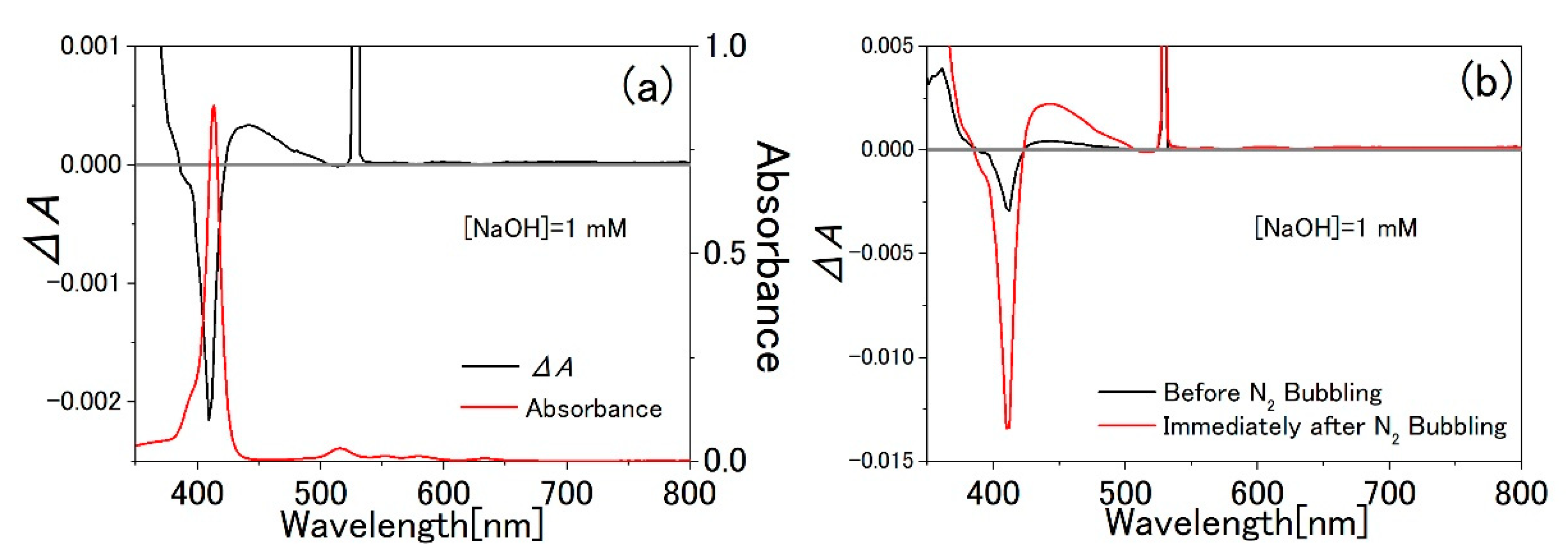
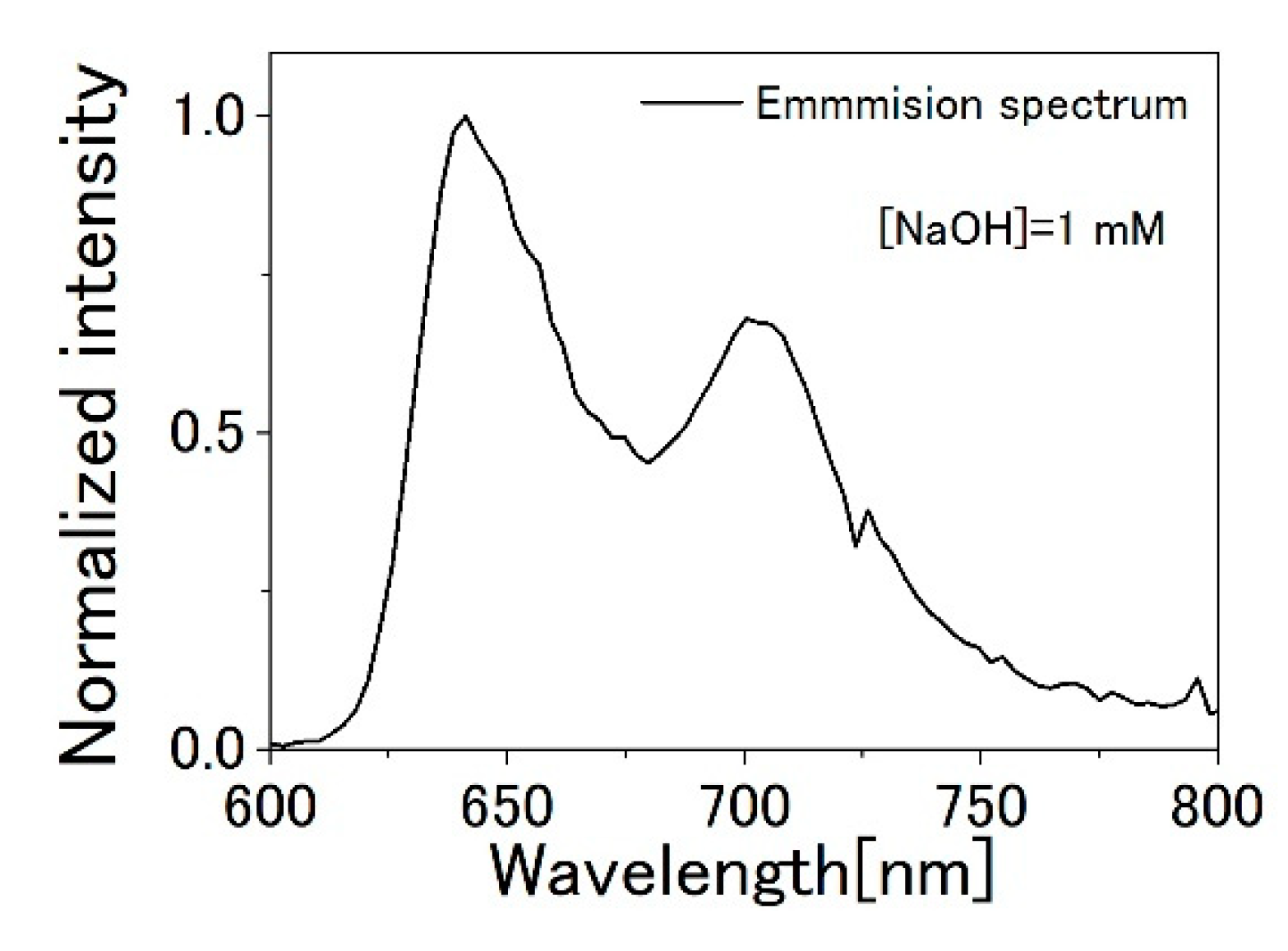
3.1. TPPS Aqueous Solution

Figure 2a shows the DAS of the F-monomer aqueous solution with [NaOH] = 1 mM induced by 532 nm laser irradiation. Δ A is the absorbance change of the sample. This result reproduces the previous one [1]. Figure 2b shows the DAS of the F-monomer aqueous solution before and immediately after N2 bubbling. Immediately after N2 bubbling for 40 min, the amount of the absorbance change | Δ A | at 412 nm increased by 4.6 times and decreased with time. This result suggests that the observed DAS was caused by the triplet state formation. The triplet excited-state absorption and SA effects were enhanced by the extension of the triplet state lifetime due to the decrease in dissolved oxygen. N2 bubbling did not significantly change the shapes of the absorption spectra and DAS of the sample. Δ A at 532 nm was not able to be observed because the probe light was transmitted through the notch filter (532 nm).
Figure 3a shows the DAS of the F-monomer aqueous solution with [NaOH] = 1 mM with the 808 nm laser irradiation. Figure 3b shows the DAS observed without the 808 nm laser irradiation. Because the DAS observed with laser irradiation was similar in the shape to that observed without the laser, it is presumed that DAS was not induced by the laser irradiation or below the detectable limit. This means that DAS cannot be observed without real excitation of the F-monomer.

3.2. PVA Sample

Figure 4 shows the presently (a) and previously (b) observed absorption spectra and DAS of the PVA sample with the predominant absorption of F-monomer. Unlike the previous result, the broad increase in absorption was clearly observed. This result indicates that the absorbance change was not caused by the TPPS aggregation induced by the light gradient force. This is discussed in more detail in the discussion section.

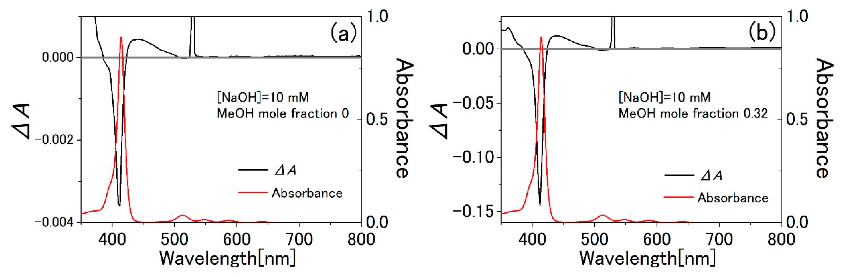
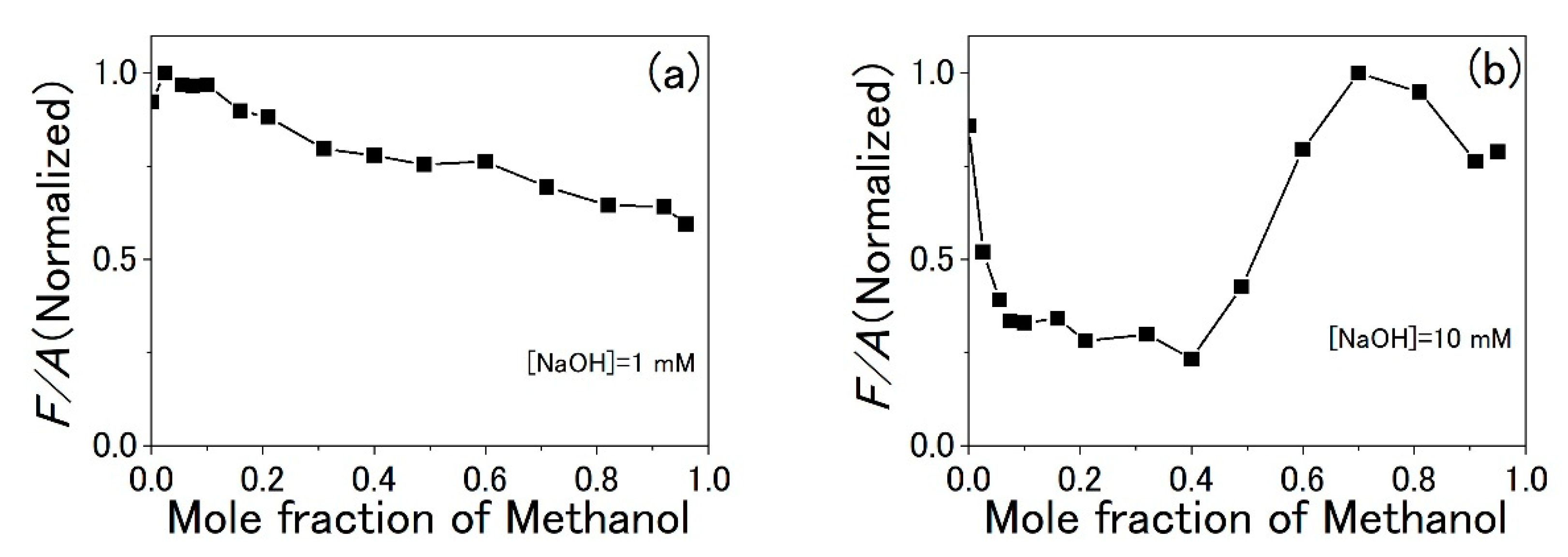
3.3. Water-Methanol Mixed Solution of TPPS

Figure 5a shows the dependence of the absorption change of the water-MeOH mixed solution of F-monomer at 412 nm on the mole fraction of MeOH when the NaOH concentration is 1 mM. The 532 nm laser was used as the pump light. Because | Δ A | at 412 nm is proportional to the absorbance A at the excitation wavelength of 532 nm, we do not compare | Δ A | but | Δ A | / A . The A tended to decrease with the mole fraction of MeOH. As the proportion of MeOH to water was increased, | Δ A | / A was decreased. | Δ A | / A is 0.17 times larger when the mole fraction of MeOH is 0.96 than when it is 0. The decrease in | Δ A | / A can be explained by the increase in the dissolved oxygen concentration by mixing methanol. As the dissolved oxygen concentration increases, the lifetime of the triplet state of the F-monomer becomes short, and the triplet excited-state absorption and SA effects decrease.
Figure 5b shows the dependence of | Δ A | / A of the water-MeOH mixed solution of F-monomer on the mole fraction of MeOH when the NaOH concentration is 10 mM. Because A tended to decrease with the mole fraction of MeOH, | Δ A | needs to be corrected. By mixing MeOH with the F-monomer aqueous solution, | Δ A | / A increased up to more than 50 times. Compared with when the mole fraction of MeOH is 0, | Δ A | / A is 57 and 0.27 times when it is 0.32 and 0.95, respectively. The dissolved oxygen concentration is considered to decrease by adding MeOH and NaOH. Figure 6 shows the DAS of the mixed solution of F-monomer when the mole fraction of MeOH is (a) 0 and (b) 0.32. Mixing MeOH did not significantly change the shapes of the absorption spectra and DAS. | Δ A | / A of the F-monomer aqueous solution with [NaOH] = 10 mM was approximately two times larger than that of the F-monomer aqueous solution with [NaOH] = 1 mM (Figure 2a and Figure 6a).
Even when the F-monomer solution was irradiated with the pump on and the probe off, a signal was observed around Q band (Figure 7). Because the shape of the signal is the same as the emission spectrum of F-monomer [5,39], the signal is due to the fluorescence of F-monomer. The well-known method [40] was used to compare the fluorescence quantum yields of each sample. The fluorescence quantum yields of two different samples, Φ 1 and Φ 2 , are related by the equation
Φ 1 Φ 2 = ( F 1 A 1 ) ( A 2 F 2 ) ( n 1 2 n 2 2 )
where F 1 and F 2 are the fluorescence intensity (area under the emission peak) of each sample, A 1 and A 2 are the absorbance at the excitation wavelength of each sample, and n 1 and n 2 are the refractive indices of the solvent of each sample. Because the refractive indices of water and methanol are almost the same, in the present experiment, it can be approximated as
Φ 1 Φ 2 = ( F 1 A 1 ) ( A 2 F 2 ) .
Therefore, it is simply expressed by
Φ F A .
Figure 8 shows the dependence of normalized F / A of the water-MeOH mixed solution of F-monomer with [NaOH] = 1 mM (a) and with [NaOH] = 10 mM (b) on the mole fraction of MeOH. For [NaOH] = 1 mM, F / A monotonically decreases with the mole fraction of MeOH, while that for [NaOH] = 10 mM decreases when | Δ A | / A at 412 nm is large (Figure 5b and Figure 8b).

4. Discussion

In the previous report [1], the broad increase in absorption over 425 nm to 505 nm was attributed to the J-aggregates formation. To explain the previous consideration, the theory of excitation energy of J-aggregates will be briefly explained. Excitation energy E k of the one-dimensional aggregates composed of the molecules with excitation energy E 0 and transition dipole moment M can be written as
E k = E 0 + 2 J cos ( k π N + 1 )
J = M 2 4 π ε r 3 ( 1 3 cos 2 θ 0 )
where ε is the permittivity of the medium surrounding J-aggregates, N is the number of molecules in a single aggregate, r is the distance between the molecules and θ 0 is the angle between the transition dipole moment of the component TPPS molecule and the axis connecting the centers of the transition dipoles [41]. Equation (4) shows the dependence E k on N . In the case of TPPS, the values of E k = 1 of dimer ( N = 2 ), trimer ( N = 3 ) and tetramer ( N = 4 ) were estimated in ref. [1,42] to be 2.70 eV (459 nm), 2.63 eV (471 nm) and 2.60 eV (477 nm), respectively. Therefore, assuming that J-aggregates with lower N ( N < 20 ) than those observed in thermal equilibrium ( N > 20 ) was formed by light gradient force, the broad increase in absorption and the decrease in absorption at 412 nm can be explained. Furthermore, it was also explained that the broad increase in absorption of the PVA sample was smaller than the aqueous solution in the previous experiment (Figure 4b). This is because F-monomers in PVA matrix are fixed and cannot form J-aggregates by light gradient force.
However, it becomes clear that the origin of the absorption change is not J-aggregates formation induced by light gradient force because the broad increase in the absorption of the PVA sample was observed in the present experiment (Figure 4a). According to the report of theoretical calculations of gradient forces applied to dye molecules, light intensities of 10 MW / cm 2 are required to capture a single molecule [43]. Because the laser light with an optical intensity of 1.3 kW / cm 2 at 808 nm or less at 532 nm was used in this experiment, the present result that the dye molecules did not form aggregates is consistent with this report. As an order-of-magnitude estimation, if the gradient force potential ( 1 2 α E 2 ) at I = ε 0 2 E 2 c = 1.3   kW / cm 2 ( ε 0 : electric constant, E : optical electric field, c : speed of light) with the estimated molecular polarizability α of TPPS from its polarizability volume V = 1.46   nm 3 [2] and Equation (5) of ref. [2] is compared with thermal energy k B T of the molecule at room temperature ( T = 300 K) with the Boltzmann constant k B , 1 2 α E 2 k B T is roughly evaluated to be 10 7 . It is therefore reasonable that the Δ A signal indicating molecular aggregation beyond Δ A 10 6 was not observed as seen in Figure 3. To observe the capture of dye molecules, it is necessary to use a laser focused by an objective lens, for example. In fact, studies using high intensity lasers have shown results suggesting the capture of dye molecules and aggregates [44,45,46].
Let us discuss a possible reason why the broad increase in the absorption of the PVA sample was not observed in the previous experiment. S/N of the previously observed DAS of the PVA sample is lower than that in the present paper (Figure 4). This may be because of non-optimized sensitivity and AC gain settings of the 128-channel lock-in detector in the previous experiment. For an accurate measurement of DAS, S/N should have been improved with the optimal settings.
From the present experiment, it is clarified that the absorption change of the F-monomer aqueous solution with [NaOH] = 1 mM was induced by the triplet excited-state absorption and SA effects due to the triplet state formation as explained below. Immediately after N2 bubbling, | Δ A | at 412 nm of the F-monomer aqueous solution becomes 4.6 times larger (Figure 2b). N2 bubbling decreased the dissolved oxygen concentration and increased the triplet excited-state absorption and SA effects as a result of the extension of the triplet state lifetime. This conclusion is consistent with the previous reports of the transient absorption change of F-monomer [3,4,5]. We tried to measure DAS by avoiding the real excitation using an 808 nm laser, but no DAS was observed (Figure 3). This result suggests that the DAS induced by 532 nm laser irradiation was caused only by the real excitation. To summarize, a phenomenon such as “highly efficient” light-induced molecular aggregation [1,2] in bulk solution does not exist. One of the causes of the misinterpretation is that the triplet-triplet absorption spectrum of the TPPS monomer nearly coincides accidentally with that of the red-shifted energy of TPPS J-aggregates as observed in [42].
From the present experiment, we confirmed the reproducibility of the previously reported results in ref. [2] that | Δ A | / A of the water-MeOH mixed solution of F-monomer with the high proportion of MeOH to water (approximately 0.80 mole fraction of MeOH) was approximately 0.1 times that of the F-monomer aqueous solution (Figure 5). This result is consistent with the fact that the oxygen solubility in MeOH is approximately 20 times larger than that in water [47]. It is pointed out in ref. [47], that the reason why the oxygen solubility in MeOH is higher than that in water is that non-polar oxygen molecules are easily dissolved in less polar solvents.
Let us discuss that the dependence of | Δ A | / A of the water-MeOH mixed solution of the F-monomer on the mole fraction of MeOH. When the NaOH concentration is 1 mM, the DAS is presumed to be caused by the triplet state formation because the shape of the DAS is similar to that induced by the triplet state formation. The reason why | Δ A | / A decreases with the mole fraction of MeOH is explained as follows. Since the oxygen solubility in MeOH is approximately 20 times larger than that of water ref. [47], it is expected that the dissolved oxygen concentration increases as the proportion of MeOH to water increases. This is also supported by the ref. [48], where the dissolved oxygen concentration increases with the ratio [ethanol]/[water] in a mixed solvent of the two.
In contrast, it is difficult to explain the dependence of | Δ A | / A of the mixed solution of F-monomer on the mole fraction of MeOH when the NaOH concentration is 10 mM. Here, we introduce the hypothesis that the increase in absorption change is caused by the decrease in dissolved oxygen concentration. Because the shape of the DAS is similar to that induced by the triplet state formation (Figure 6b), the origin of the absorption change is considered to be that. Because | Δ A | / A increased by mixing MeOH, the lifetime of the triplet state probably extends by mixing MeOH. The result of F / A decrease with | Δ A | / A increase (Figure 5b and Figure 8b) is consistent with the interpretation of the extension of the triplet state lifetime: Longer triplet state lifetime decreases the ratio of the singlet states to the triplet states during the pump light irradiation. However, the cause of the extension of the triplet state lifetime is unclear. Because the shape of the absorption spectra of the aqueous solution and the mixed solution of the F-monomer is similar (Figure 6), the form of F-monomer unlikely changed. A possible cause is a change in solvent conditions, especially a decrease in dissolved oxygen concentration. It is possible that the equilibrium of complex formation between porphyrin π -electron orbital (acceptor) and lone-pair electrons in oxygen (donor) was changed depending on the dissolved oxygen concentration.
When the NaOH concentration is 10 mM, the mechanism of decrease in the dissolved oxygen concentration by mixing MeOH with the F-monomer aqueous solution is not clear. We just point out a possibly relevant exothermic reaction by mixing water and MeOH: The dependence of excess enthalpy of a water-MeOH mixed solvent on the mole fraction of MeOH reported in ref. [49,50] is similar to that of | Δ A | / A of the mixed solution of the F-monomer with [NaOH] = 10 mM at 412 nm on the mole fraction of MeOH (Figure 5b). It is reported that the excess enthalpy is at its maximum when the mole fraction of MeOH is approximately 0.3. It was concluded in ref. [50] that the exothermic reaction by mixing water and MeOH was caused by an increase in the number of the hydrogen bonds between water and MeOH molecules forming clusters.
The oxygen concentration should not decrease, however, only by increase in the number of hydrogen bonds because | Δ A | / A of the F-monomer aqueous solution with [NaOH] = 1 mM did not increase by mixing MeOH (Figure 5a). Hence, we focus on the effect of NaOH on the hydrogen bonds. The effect of NaOH on the hydrogen bond strength in a mixed solvent of water and ethanol (EtOH) has been investigated by observing the nuclear magnetic resonance of the proton in the hydroxy group of water and EtOH molecules [51]. It was concluded that Na + did not affect the hydrogen bond, while OH strengthened it. It is plausible that the dissolved oxygen concentration decreases as the hydrogen bonds are strengthened. Therefore, it is presumed that the decrease in the dissolved oxygen concentration in the F-monomer aqueous solution by mixing MeOH was induced by the increase in the number of the hydrogen bonds and strengthening of the hydrogen bonds when the NaOH concentration was 10 mM.

5. Conclusions

It had been considered that the origin of absorption change of the F-monomer aqueous solution was J-aggregates formation induced by light gradient force of laser [1,2]. The two main pieces of evidence were as follows. First, the increase in the broad absorption spectral range of the F-monomer aqueous solution was able to be attributed to J-aggregates ( N < 20 ) formation. Second, the broad increase in absorption of the PVA sample was smaller than that of the F-monomer aqueous solution. This result was consistent with the fact that F-monomer molecules in PVA matrix cannot freely move to form J-aggregates.
It is clarified here that the absorption change of the F-monomer aqueous solution with [NaOH] = 1 mM observed in the present experiment was induced by the triplet excited-state absorption and SA effects due to the triplet state formation. There are three pieces of evidence for the statement. (1); The shape of the DAS in ref. [1,2] is similar to that of the DAS reported in ref. [3,4,5], where the absorption changes were assigned to be induced by the triplet excited-state absorption and SA effect due to the triplet state formation in TPPS monomers. (2); The broad increase in absorption of the PVA sample was clearly observed in the present experiment (Figure 4a). (3); | Δ A | of the F-monomer aqueous solution with [NaOH] = 1 mM at 412 nm increased by 4.6 times immediately after N2 bubbling (Figure 2b). The lifetime of the triplet state was extended by the decrease in the dissolved oxygen concentration, and the triplet excited-state absorption and SA effects increased.
| Δ A | / A in the water-MeOH mixed solution of TPPS monotonically decreased with MeOH concentration at 1 mM NaOH and increased by a factor of more than 50 in the highest case at 10 mM NaOH. The former result can be explained by an increase in dissolved oxygen concentration [47], but the latter result is not readily explained at present. We would like to point out the possibility of a decrease in the dissolved oxygen concentration due to the addition of a tiny amount of NaOH. Although a further detailed study is required, our result opens a unique possibility of optical nonlinearity enhancement and of a structural change of mixed solution by adjusting the concentrations of alcohol and base/acid.

Author Contributions

Conceptualization, E.T.; methodology, E.T., S.I., and T.K.; validation, S.I.; formal analysis, S.I.; investigation, S.I.; writing—original draft preparation, S.I.; writing—review and editing, E.T. and T.K.; supervision, E.T. All authors have read and agreed to the published version of the manuscript.

Funding

This research received no external funding.

Institutional Review Board Statement

Not applicable.

Informed Consent Statement

Not applicable.

Data Availability Statement

Data are contained within the article.

Acknowledgments

The authors acknowledge technical support and advice by Masayuki Shirakawa.

Conflicts of Interest

The authors declare no conflict of interest.

References

  1. Shirakawa, M.; Nakata, K.; Suzuki, M.; Kobayashi, T.; Tokunaga, E. Nonlinear absorption spectroscopy of porphyrin j-aggregates in aqueous solution: Evidence for control of degree of association by light-induced force. J. Phys. Soc. Jpn. 2017, 86, 044703. [Google Scholar] [CrossRef]
  2. Shirakawa, M.; Kobayashi, T.; Tokunaga, E. Solvent effects in highly efficient light-induced molecular aggregation. Appl. Sci. 2019, 9, 5381. [Google Scholar] [CrossRef]
  3. Tsukahara, S.; Watarai, H. Kinetics for acid-dissociation of tetraphenylporphinetetrasulfonate in the ground state measured by laser photolysis relaxation method. Phys. Chem. Chem. Phys. 2002, 4, 1592–1597. [Google Scholar] [CrossRef]
  4. Bonnet, R.; Ridge, R.J. Pulsed irradiation of water-soluble porphyrins. J. Chem. Soc. Faraday Trans. 1982, 78, 127–136. [Google Scholar] [CrossRef]
  5. Kalyanasundaram, K.; Neumann-Spallart, M. Photophysical and redox properties of water-soluble porphyrins in aqueous media. J. Chem. Phys. 1982, 86, 5163–5169. [Google Scholar] [CrossRef]
  6. Sreedhar, S.; Illyaskutty, N.; Sreedhanya, S.; Philip, R.; Muneera, C.I. An organic dye-polymer (phenol red-poly (vinyl alcohol)) composite architecture towards tunable-optical and -saturable absorption characteristics. J. Appl. Phys. 2016, 119, 193106. [Google Scholar] [CrossRef]
  7. Zhu, G.; Long, Y.; Ren, H.; Zhou, Y.; Zhang, L.; Shi, Z.; Shehzad, F.K.; Asif, H.M. Notable third-order optical nonlinearities realized in layer-by-layer assembled composite films by intercalation of porphyrin/polyoxometalate into layered double hydroxide. J. Phys. Chem. C 2016, 120, 22549–22557. [Google Scholar] [CrossRef]
  8. Gonçalves, P.J.; Sciuti, L.F.; Neto, N.M.B.; Silva, R.C.E.; Silveira-Alves, E., Jr.; Mendonça, C.R.; Zílio, S.C.; Borissevitch, I.E.; Boni, L.D. Effects of pH on the ultrafast transient absorption of iron (III) meso-tetrakis (4-N-methyl-pyridiniumyl) porphyrin (Fe3 + TMPyP) molecular complexes. J. Photochem. Photobiol. A 2021, 408, 113082. [Google Scholar] [CrossRef]
  9. Vijisha, M.V.; Ramesh, J.; Arunkumar, C.; Chandrasekharan, K. Impressive nonlinear optical responses of a cationic porphyrin derivative in a flexible all-polymer Bragg stack on optical Tamm mode coupling. J. Mater. Chem. C 2020, 8, 12689–12697. [Google Scholar] [CrossRef]
  10. Badran, H.A.; Abul-Hail, R.C.; Shaker, H.S.; Musa, A.I.; Hassan, Q.M.A. An all-optical switch and third-order optical nonlinearity of 3,4-pyridinediamine. Appl. Phys. B 2017, 123, 31. [Google Scholar] [CrossRef]
  11. Shi, Z.; Zhou, Y.; Zhang, L.; Mu, C.; Ren, H.; Hassan, D.; Yanga, D.; Asifa, H.M. New supramolecular compounds based on porphyrin and polyoxometalate: Synthesis, characterization and nonlinear optical and optical limiting properties. RSC Adv. 2014, 4, 50277–50284. [Google Scholar] [CrossRef]
  12. Sreekumar, G.; Frobel, P.G.L.; Muneera, C.I.; Sathiyamoorthy, K.; Vijayan, C.; Mukherjee, C. Saturable and reverse saturable absorption and nonlinear refraction in nanoclustered Amido Black dye–polymer films under low power continuous wave He–Ne laser light excitation. J. Opt. A Pure Appl. Opt. 2009, 11, 125204. [Google Scholar] [CrossRef]
  13. Srinivas, N.K.M.N.; Rao, S.V.; Rao, D.N. Saturable and reverse saturable absorption of Rhodamine B in methanol and water. J. Opt. Soc. Am. B 2003, 20, 2470. [Google Scholar] [CrossRef]
  14. Hirata, S.; Vacha, M. Large reverse saturable absorption at the sunlight power level using the ultralong lifetime of triplet excitons. J. Phys. Chem. Lett. 2017, 8, 3683–3689. [Google Scholar] [CrossRef]
  15. Wang, C.; Lystrom, L.; Yin, H.; Hetu, M.; Kilina, S.; McFarland, S.A.; Sun, W. Increasing the triplet lifetime and extending the ground-state absorption of biscyclometalated Ir(III) complexes for reverse saturable absorption and photodynamic therapy applications. Dalton Trans. 2016, 45, 16366–16378. [Google Scholar] [CrossRef] [PubMed]
  16. Blau, W.; Byrne, H.; Dennis, W.M. Reverse saturable absorption in tetraphenylporphyrins. Opt. Commun. 1985, 56, 25–29. [Google Scholar] [CrossRef]
  17. Wrthner, F.; Kaiser, T.E.; Saha-Mller, C.R. J-Aggregates: From serendipitous discovery to supramolecular engineering of functional dye materials. Angew. Chem. Int. Ed. 2011, 50, 3376–3410. [Google Scholar] [CrossRef] [PubMed]
  18. Kobayashi, T.; Misawa, K. Hierarchical structure of one-dimensional J-aggregates. J. Lumin. 1997, 72, 38–40. [Google Scholar] [CrossRef]
  19. Biroli, A.O.; Tessore, F.; Righetto, S.; Forni, A.; Macchioni, A.; Rocchigiani, L.; Pizzotti, M.; Carlo, G.D. Intriguing influence of −cooh-driven intermolecular aggregation and acid−base interactions with n,n-dimethylformamide on the second-order nonlinear-optical response of 5,15 push−pull diarylzinc(ii) porphyrinates. Inorg. Chem. 2017, 56, 6438–6450. [Google Scholar] [CrossRef] [PubMed] [Green Version]
  20. Ray, P.C.; Leszczynski, J. Nonlinear optical properties of highly conjugated push–pull porphyrin aggregates: Role of intermolecular interaction. Chem. Phys. Lett. 2006, 419, 578–583. [Google Scholar] [CrossRef]
  21. Bergendahl, L.T.; Paterson, M.J. Excited states of porphyrin and porphycene aggregates: Computation insights. Comput. Theor. Chem. 2014, 1040, 274–286. [Google Scholar] [CrossRef]
  22. Ogawa, T.; Tokunaga, E.; Kobayashi, T. Giant electrooptic response of excitons in porphyrin J-aggregates. Chem. Phys. Lett. 2005, 408, 186–191. [Google Scholar] [CrossRef]
  23. Lapes, M.; Petera, J.; Jirsa, M. Photodynamic therapy of cutaneous metastases of breast cancer after local application of meso-tetra-(para-sulphophenyl)-porphin (TPPS4). J. Photochem. Photobiol. B 1996, 36, 205–207. [Google Scholar] [CrossRef]
  24. Singh, S.; Aggarwal, A.; Bhupathiraju, N.V.S.; Dinesh, K.; Arianna, G.; Tiwari, K.; Drain, C.M. Glycosylated porphyrins, phthalocyanines, and other porphyrinoids for diagnostics and therapeutics. Chem. Rev. 2015, 115, 10261–10306. [Google Scholar] [CrossRef] [PubMed]
  25. Ethirajan, M.; Chen, Y.; Joshi, P.; Pandey, R.K. The role of porphyrin chemistry in tumor imaging and photodynamic therapy. Chem. Soc. Rev. 2011, 40, 340–362. [Google Scholar] [CrossRef] [PubMed]
  26. Nyman, E.S.; Hynninen, P.H. Research advances in the use of tetrapyrrolic photosensitizers for photodynamic therapy. J. Photochem. Photobiol. B 2004, 73, 1–28. [Google Scholar] [CrossRef] [PubMed]
  27. Bhaumik, J.; Mittal, A.K.; Banerjee, A.; Chisti, Y.; Banerjee, U.C. Applications of phototheranostic nanoagents in photodynamic therapy. Nano Res. 2015, 8, 1373–1394. [Google Scholar] [CrossRef]
  28. Hou, L.; Zhang, X.; Pijper, T.C.; Browne, W.R.; Feringa, B. Reversible photochemical control of singlet oxygen generation using diarylethene photochromic switches. J. Am. Chem. Soc. 2014, 136, 910–913. [Google Scholar] [CrossRef] [PubMed] [Green Version]
  29. Tian, J.; Ding, L.; Xu, H.; Shen, Z.; Ju, H.; Jia, L.; Bao, L.; Yu, J. Cell-specific and pH-activatable rubyrin-loaded nanoparticles for highly selective near-infrared photodynamic therapy against cancer. J. Am. Chem. Soc. 2013, 135, 18850–18858. [Google Scholar] [CrossRef]
  30. Ge, J.; Lan, M.; Zhou, B.; Liu, W.; Guo, L.; Wang, H.; Jia, Q.; Niu, G.; Huang, X.; Zhou, H.; et al. A graphene quantum dot photodynamic therapy agent with high singlet oxygen generation. Nat. Commun. 2014, 5, 4596. [Google Scholar] [CrossRef] [Green Version]
  31. Ochsner, M. Photophysical and photobiological processes in the photodynamic therapy of tumours. J. Photochem. Photobiol. B 1997, 39, 1–18. [Google Scholar] [CrossRef]
  32. Kalyanasundaram, K.; Gratzel, M. Light induced redox reactions of water soluble porphyrins, sensitization of hydrogen generation from water by zincporphyrin derivatives. Helv. Chim. Acta 1980, 63, 478–485. [Google Scholar] [CrossRef]
  33. McLendon, G.; Miller, D.S. Metalloporphyrins catalyse the photo-reduction of water to H2. J. Chem. Soc. Chem. Commun. 1980, 533–534. [Google Scholar] [CrossRef]
  34. Harriman, A.; Richoux, M. Photoproduction of hydrogen from reductive quenching of a water-soluble zinc porphyrin. J. Photochem. 1981, 15, 335–339. [Google Scholar] [CrossRef]
  35. Harriman, A.; Porter, G.; Richoux, M. Photosensitised reduction of water to hydrogen using water-soluble zinc porphyrins. J. Chem. Soc. Faraday Trans. 1981, 77, 833–844. [Google Scholar] [CrossRef]
  36. Okura, I.; Kim-Thuan, N. Hydrogen generation by visible light with tris-(2,2′-bipyridine) ruthenium dication. J. Mol. Catal. 1979, 5, 311–314. [Google Scholar] [CrossRef]
  37. Choi, M.Y.; Pollard, J.A.; Webb, M.A.; McHale, J.L. Counterion-dependent excitonic spectra of tetra(p-carboxyphenyl)porphyrin aggregates in acidic aqueous solution. J. Am. Chem. Soc. 2003, 125, 810–820. [Google Scholar] [CrossRef]
  38. Ishii, N.; Tokunaga, E.; Adachi, S.; Kimura, T.; Matsuda, H.; Kobayashi, T. Optical frequency- and vibrational time-resolved two-dimensional spectroscopy by real-time impulsive resonant coherent Raman scattering in polydiacetylene. Phys. Rev. A 2004, 70, 023811. [Google Scholar] [CrossRef] [Green Version]
  39. Akins, D.L.; Özçelik, S.; Zhu, H.; Guo, C. Fluorescence decay kinetics and structure of aggregated tetrakis(p-sulfonatophenyl)porphyrin. J. Phys. Chem. 1996, 100, 14390–14396. [Google Scholar] [CrossRef]
  40. Eaton, D.F. Reference materials for fluorescence measurement. Pure Appl. Chem. 1988, 60, 1107–1114. [Google Scholar] [CrossRef]
  41. Kobayashi, T. J-Aggregates, 1st ed.; World Scientific: Singapore, 1996; pp. 95–110. [Google Scholar]
  42. Nakata, K.; Kobayashi, T.; Tokunaga, E. Electric field-controlled dissociation and association of porphyrin J-aggregates in aqueous solution. Phys. Chem. Chem. Phys. 2011, 13, 17756–17767. [Google Scholar] [CrossRef] [PubMed]
  43. Kudo, T.; Ishihara, H. Proposed nonlinear resonance laser technique for manipulating nanoparticles. Phys. Rev. Lett. 2012, 109, 087402. [Google Scholar] [CrossRef]
  44. Tanaka, Y.; Yoshikawa, H.; Masuhara, H. Laser-induced self-assembly of pseudoisocyanine j-aggregates. J. Phys. Chem. C 2007, 111, 18457–18460. [Google Scholar] [CrossRef]
  45. Osborne, M.A.; Balasubramanian, S.; Furey, W.S.; Klenerman, D. Optically biased diffusion of single molecules studied by confocal fluorescence microscopy. J. Phys. Chem. B 1998, 102, 3160–3167. [Google Scholar] [CrossRef]
  46. Mototsuji, A.; Shoji, T.; Wakisaka, Y.; Murakoshi, K.; Yao, H.; Tsuboi, Y. Plasmonic optical trapping of nanometer-sized J-/H-dye aggregates as explored by fluorescence microspectroscopy. Opt. Express 2017, 25, 13617–13625. [Google Scholar] [CrossRef]
  47. Rollie, M.E.; Patonay, G.; Warner, I.M. Deoxygenation of solutions and its analytical applications. Ind. Eng. Chem. Res. 1987, 26, 1–6. [Google Scholar] [CrossRef]
  48. Bakalyar, S.R.; Bradley, M.P.T.; Honganen, R. The role of dissolved gases in high -performance liquid chromatography. J. Chromatogr. 1978, 158, 277–293. [Google Scholar] [CrossRef]
  49. Benjamin, L.; Benson, G.C. A deuterium isotope effect on the excess enthalpy of methanol-water solutions. J. Phys. Chem. 1963, 67, 858–861. [Google Scholar] [CrossRef]
  50. Wakisaka, A.; Abdoul-Carime, H.; Yamamoto, Y.; Kiyozumi, Y. Non-ideality of binary mixtures Water–methanol and water–acetonitrile from the viewpoint of clustering structure. J. Chem. Soc. Faraday Trans. 1998, 94, 369–374. [Google Scholar] [CrossRef]
  51. Nose, A.; Hojo, M.; Ueda, T. Effects of salts, acids, and phenols on the hydrogen-bonding structure of water−ethanol mixtures. J. Phys. Chem. B 2004, 108, 798–804. [Google Scholar] [CrossRef]
Figure 1. Experimental set-up for detecting change in transmitted probe light spectra induced by 532 nm laser irradiation (Ref.: reference signal, APDs: avalanche photodiodes).
Applsci 11 04892 g001
Figure 2. (a) Absorption spectrum (red line) and DAS (black line) of F-monomer aqueous solution with [NaOH] = 1 mM. (b) DAS of F-monomer aqueous solution before (black line) and immediately after (red line) N2 bubbling. A 532 nm laser was used as the pump.
Applsci 11 04892 g002
Figure 3. Absorption spectra (red line) and DAS (black line) of F-monomer aqueous solution with [NaOH] = 1 mM. DAS was observed with (a) and without (b) 808 nm laser. The small steps around 480 nm and 560 nm are artifacts of the 128-channel lock-in amplifier detector (due to the unbalanced base level at the unit boundary of the 128-channel lock-in, which consists of four 32-channel lock-in amplifier units).
Applsci 11 04892 g003
Figure 4. Absorption spectra (red line) and DAS (black line) of presently (a) and previously (b) prepared PVA sample. The 532 nm laser was used as the pump.
Applsci 11 04892 g004
Figure 5. Dependence of amount of | Δ A | / A of water-MeOH mixed solution of F-monomer with 1 mM NaOH (a) and 10 mM NaOH (b) on mole fraction of MeOH. The 532 nm laser was used as the pump. | Δ A | is amount of absorbance change at 412 nm. A is absorbance at excitation wavelength of 532 nm.
Applsci 11 04892 g005
Figure 6. Absorption spectra (red line) and DAS (black line) of water-MeOH mixed solution of F-monomer with 0 (a) and 0.32 (b) mole fraction of MeOH. NaOH concentration is 10 mM. A 532 nm laser was used as the pump.
Applsci 11 04892 g006
Figure 7. Emission spectrum (corrected for spectral sensitivity) of F-monomer aqueous solution with [NaOH] = 1 mM observed with 128-channel lock-in detector. The 532 nm laser was used as the pump.
Applsci 11 04892 g007
Figure 8. Dependence of F / A of water-MeOH mixed solution of F-monomer with [NaOH] = 1 mM (a) and 10 mM (b) on mole fraction of MeOH. The 532 nm laser was used as the pump.
Applsci 11 04892 g008
Table 1. Concentration of each component in TPPS aqueous solutions mixed with MeOH and NaOH.
[TPPS][NaOH]Mole Fraction of MeOH
20   µ M 1   mM 0.00 ~ 0.96
20   µ M 10   mM 0.00 ~ 0.95
Publisher’s Note: MDPI stays neutral with regard to jurisdictional claims in published maps and institutional affiliations.

