Article

Carbon Coatings Deposited on Prosthodontic Ni-Cr Alloy

1
Chair of Restorative Dentistry, Department of Dental Technology, Medical University of Lodz, 92-213 Lodz, Poland
2
Department of Material Science, Bauman Moscow State Technical University, 105005 Moscow, Russia
3
Institute of Materials Science and Engineering, Lodz University of Technology, 90-924 Lodz, Poland
*
Author to whom correspondence should be addressed.
Appl. Sci. 2021, 11(10), 4551; https://doi.org/10.3390/app11104551
Submission received: 8 March 2021 / Revised: 4 May 2021 / Accepted: 6 May 2021 / Published: 17 May 2021
(This article belongs to the Special Issue Advances of Diamond Carbon Coatings)

Abstract

:
The study discusses the results of investigations conducted on carbon coatings applied on a prosthodontic alloy Ni-Cr. Carbon coatings with the thickness of about 1000 nm were deposited by means of the RF PACVD method with a titanium interlayer applied by magnetron spray dispersion. The coatings underwent microscopic examinations, as well as structural tests with the use of Raman spectrometry, investigations of mechanical properties, adhesion and corrosion tests; also, the bacterial adhesion to the sample surface was determined. It can be inferred from the performed studies that the obtained carbon coatings exhibit mechanical properties which allow them to be used for prosthodontic elements. The coatings’ adhesion to the metallic substrate made of Ni-Cr alloy equaled about 150 mN. The examined coatings clearly improve the corrosion resistance and reduce the number of bacteria adhering to the sample surfaces. Taking all this into account, it can be stated that carbon coatings can be potentially applied to protect metal prosthetic restorations.

1. Introduction

Metals and their alloys constitute a significant percent of all the materials used in stomatology. They are applied both in prosthetics and orthodontics. Practically all elements of orthodontic apparati, such as fasteners and wires, are made of metals. This results from their properties. Metals are characterized by a proper rigidity and elasticity; they are also relatively easy to form by means of different methods. They are, however, most broadly used in prosthetics, for elements of permanent and mobile prostheses (splints, bridges, crowns, etc.). Such an extensive application of metals and their alloys results from the fact that, at present, there are no alternative materials which would exhibit comparable properties (especially the strength, elasticity and durability).
The oral cavity environment demonstrates corrosive properties in respect of metals. This is connected with the presence of a large amount of chloride ions, proteins and enzymes, as well as acidic metabolites of the dental plaque bacteria, and also bacteria-induced cathodic depolarization phenomena on the surface of the alloys. All this makes it impossible for any passivating basic metal alloy to resist biological corrosion. The side effects of the use of metals include body reactions such as allergic or cytotoxic reactions, or discomfort in the patient’s oral cavity [1,2,3,4,5,6]. Their source can be corrosion products, metal ions released in the corrosion process, which penetrate into surrounding tissue, as well as galvanic currents [7,8,9,10]. Another challenge posed by the oral cavity environment is the presence of bacteria and fungi. Prosthetic restorations often cover a significant part of the muco-bone basis, and so, the retention of the food remains, as well as the bacteria accumulation on the submucosal surface of the prosthesis and the formation of a biofilm on it, can cause not only biological corrosion of the metals but also inflammation of those areas of the mucous membrane. The microorganisms present in the oral cavity can cause corrosive destruction (MIC—microbially influenced/inducted corrosion) of metal prosthetic elements, implants, etc. [11,12]. This phenomenon refers even to titanium and its alloys, which are commonly treated as very well resistant to corrosion [13]. Therefore, an important issue is not only improvement of corrosion resistance but also minimization of the possibility of a biofilm formation.
Despite their many flaws, metals continue to be used, as, presently, there are no other materials which would possess comparable properties. The occurrence of an organism reaction very often refers to non-precious metal alloys (cobalt-chromium and nickel-chromium). These alloys do not demonstrate sufficient corrosion resistance in the oral cavity environment. However, due to economic reasons, they are commonly used in stomatology as a cheap alternative for the expensive precious metal alloys. A similar behavior is demonstrated by zinc, copper and silver as components of amalgams. Due to a significantly lower corrosion resistance of these metals, the latter cause a much stronger biological response of the body compared to the precious metal alloys. In order to improve their corrosion resistance, various types of surface modifications are applied, which make it possible to increase the biological tolerance of these alloys. The usual method involves coating with different layers, such as oxides, carbides or metal nitrides [14,15,16,17,18]. These coatings make it possible to improve the corrosion resistance and biocompatibility, and they reduce the bacterial adhesion [19,20,21]. They can significantly limit the release of metal ions into the surrounding environment [22]. Among the many available methods of coating synthesis, medical applications often involve covering the surfaces with layers deposited from the gaseous phase [15,23,24,25,26,27]. One of the most interesting techniques is the application of carbon layers [26,27]. Carbon is an element which characterizes in good corrosion resistance and biocompatibility. These properties largely depend on the form of the carbon—whether it has a diamond or graphite structure. Studies conducted by many authors show that such coatings have many advantageous properties, allowing their potential use in dental prosthetics [28,29]. Therefore, covering metal elements with layers containing this element should provide them with good properties in the oral cavity environment.
The aim of this study was to evaluate the quality of carbon layers deposited on prosthodontic Ni-Cr alloy in RF PACVD reactor equipped with a magnetron using titanium interlayer.

2. Materials and Methods

The possibilities of using protective coatings on metal surfaces are determined both by their biological and mechanical properties. On the one hand, protective coatings should reduce the metal alloy corrosion as well as the release of harmful corrosion products and metal ions from the alloys, being harmless themselves as well, and on the other hand, they should exhibit sufficiently good mechanical strength and adhesion, in order to remain on the surface of prosthetic restorations for at least a few years. That is why the present study mainly focuses on investigating those properties of carbon coatings.
Cylindrical samples made of Ni-Cr alloy, 8 mm in diameter and 7 mm in height, were prepared. The chemical composition of the alloy, determined using a fluorescent X-ray analysis method on a SIEMENS SRS 300 X-ray spectrometer, is shown in Table 1 surfaces of the specimens were subjected to grinding followed by mechanical polishing (Ra = 0.31 μm).
The samples were coated, by means of the magnetron sputtering method, with an interlayer of titanium, about 200 nm thick, and next, with the use of the RF PACVD method, with a carbon layer, about 1000 nm (1 μm) thick. The reaction chamber was pumped down to a pressure of 2 × 10⁻³ Pa. Deposition process provided 10 min etching in Ar plasma with self-bias—720 V. Process parameters are the following: power [W] for RF PACVD amounted to 50–150, power [W] for DC MS amounted to 2400–2700, methane flow [sccm] for RF PACVD and DC MS amounted to 0–30, argon flow [sccm] for RF PACVD amounted to 0–12, argon flow [sccm] for DC MS amounted to 9. The deposition time of Ti interlayer was 12 min, whereas the final DLC coating was synthesized for 30 min. More details regarding the deposition technology of gradient Ti-C coatings can be found elsewhere [30].
The samples prepared in this way were then subjected to the following tests:
  • Observation of the surface and the cross-section of the sample by means of an electron scanning microscope HITACHI S-3000N.
  • Determination of the chemical composition of the coating with surface element distribution by the EDS method, by means of the NORAN INSTRUMENTS system cooperating with an electron scanning microscope HITACHI S-3000N.
  • Chemical structure of the synthesized coatings was determined using inVia confocal micro-Raman spectrometer (Renishaw), working with 532 nm wavelength and power of 2.5 mW.
  • Mechanical properties. Hardness were determined using Berkovich indenter trihedral pyramid shape with an angle equaling 65.3° and the investigation was performed according to ISO Standards [31] on a MTS NANO INSTRUMENTS G-200 nanoindeter. The results were registered and analyzed using the TestWorksPro 4 Software. The measurement was performed with the CSM (continuous stiffness measurement) [32,33] method.
  • Adhesion to substrate. Examinations were carried out by the scratch test method using a sapphire cone-shaped penetrator [34]. Rounding radius of the tip was 1 μm. A 2 mm scratch was made by gradually increasing penetrator load of 0 to 200 mN.
  • Corrosion tests—conducted in a chemical environment of deoxidized artificial saliva in the form of a Fusayama–Meyer solution with the composition shown in Table 2 at 37 °C in the ST1 thermostatic chamber (POL-EKO), the measurements were carried out on the PGSTAT 30 galvanostat (Autolab) with GPES and FRA control software v. 4.9.
  • Corrosion tests in artificial saliva with microscopic examinations of the corrosion areas. The tests were carried out on five samples from each group.
  • Bacterial adhesion on the surface of the samples. To that end, the samples were sterilized in a steam autoclave at the temperature of 135 °C. The samples prepared in this way were then placed in a liquid culture of Escherichia coli bacteria (strain K12) and incubated at the temperature of 37 °C. Each sample was placed in a separate container with a solution, in which about 2 × 10³ bacteria were present. After 24 h of being kept in the incubator, they were removed from the bacteria cultures and subjected to preparation procedures aiming at strengthening the bacteria which had colonized the sample surfaces [35]. Next, the samples were sprayed with a thin layer of gold by means of a sputter JEE-4X Jeol. The number of bacteria was estimated under an electron scanning microscope HITACHI S-3000N on six samples covered with a DLC coating and six samples without a coating. Every sample underwent five measurements in randomly selected areas (150 × 100 μm), which gave a total of 30 measurements.

3. Results

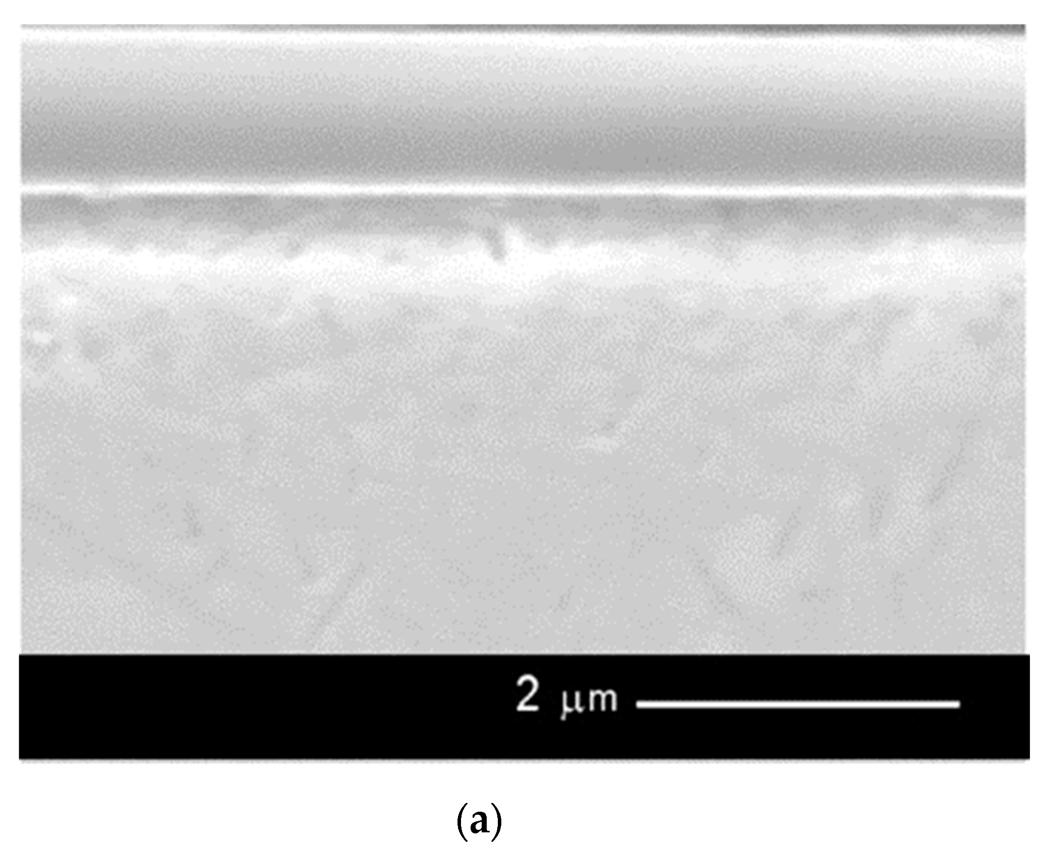
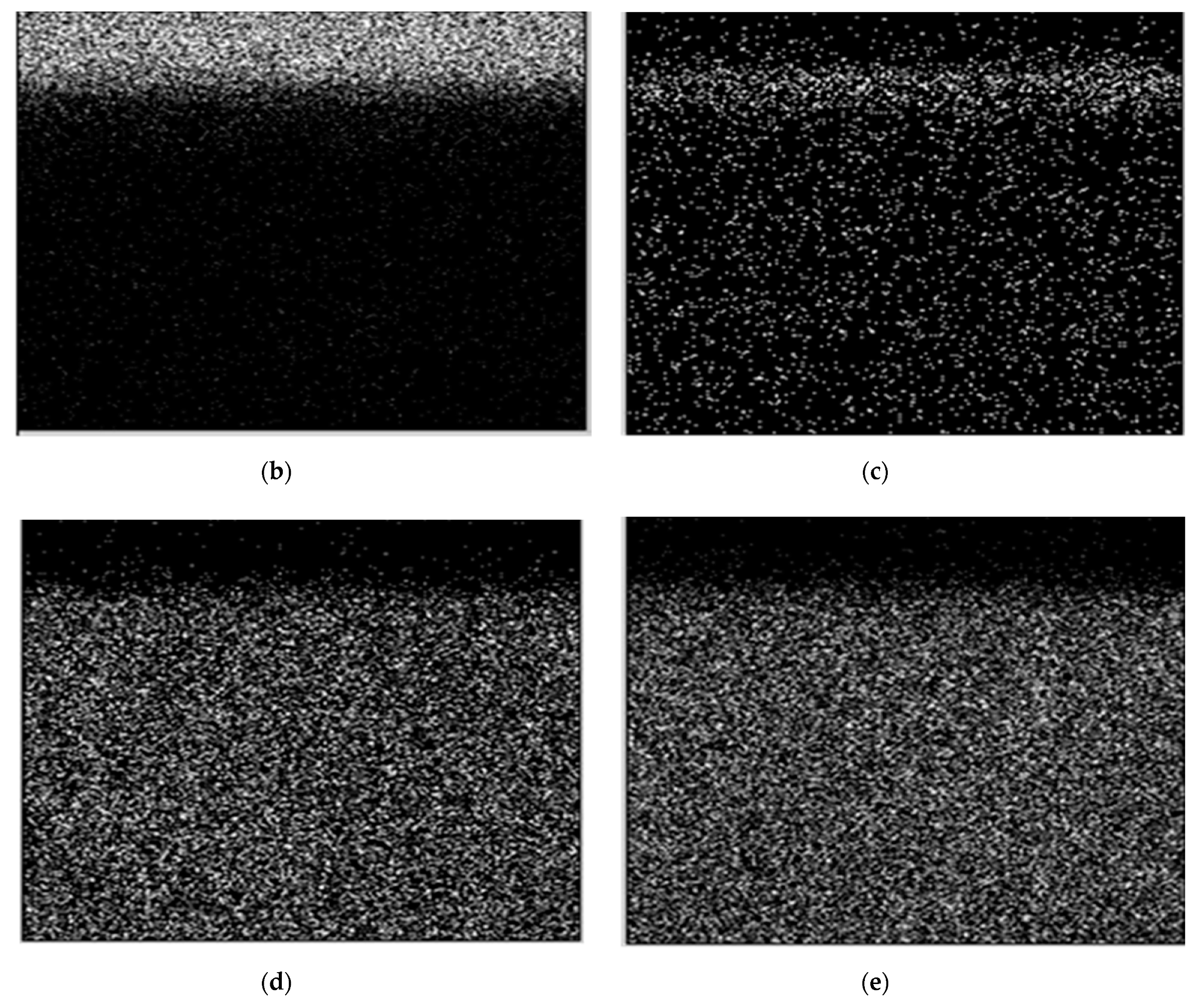
Figure 1 presents the surface of a sample with a carbon coating and an EDS spectrogram made on that surface, whereas Figure 2 shows a microscopic image of the cross-section of the sample together with the surface distributions of carbon, titanium, nickel and chromium.
Microscopic observations showed that carbon layers on prosthodontic Ni-Cr alloy are solid and tight, without surface defects. X-ray microanalysis showed that only carbon exists in the examined layers, there are no other impurities. Therefore, this layer may be considered as a pure carbon layer.
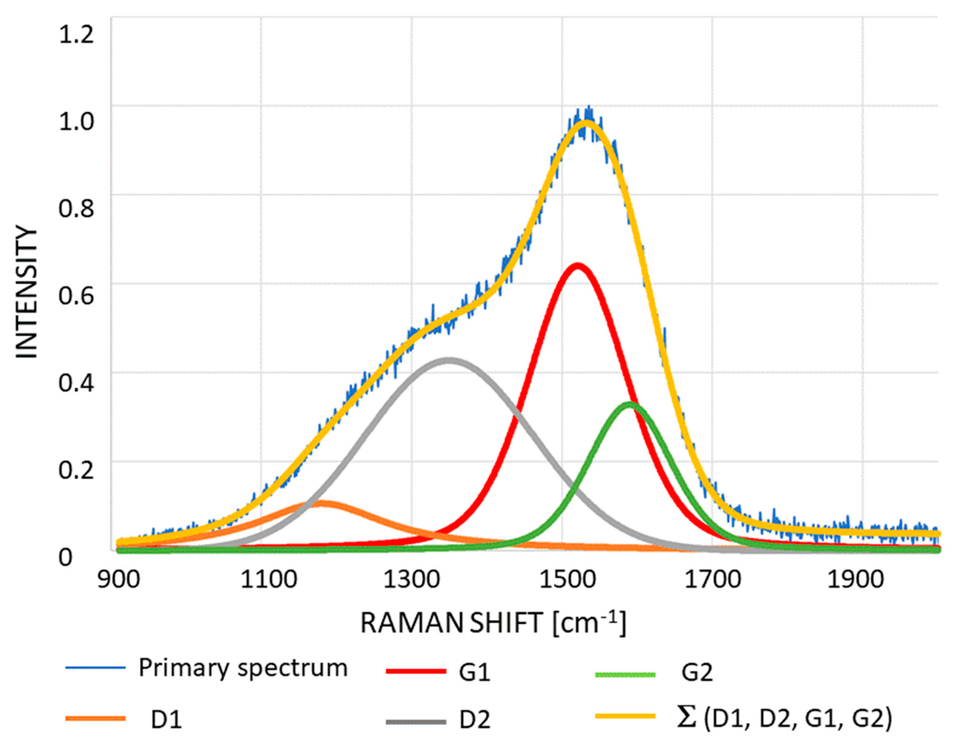
Figure 3 presents typical Raman spectra of manufactured carbon coatings with their deconvolution employing four-peak fitting method. Namely, D1, D2, G1 and G2 Gaussians, according to the literature [36]. The obtained de-convolution results resemble those, typical for diamond-like carbon coatings (DLC), widely presented in the literature [37].
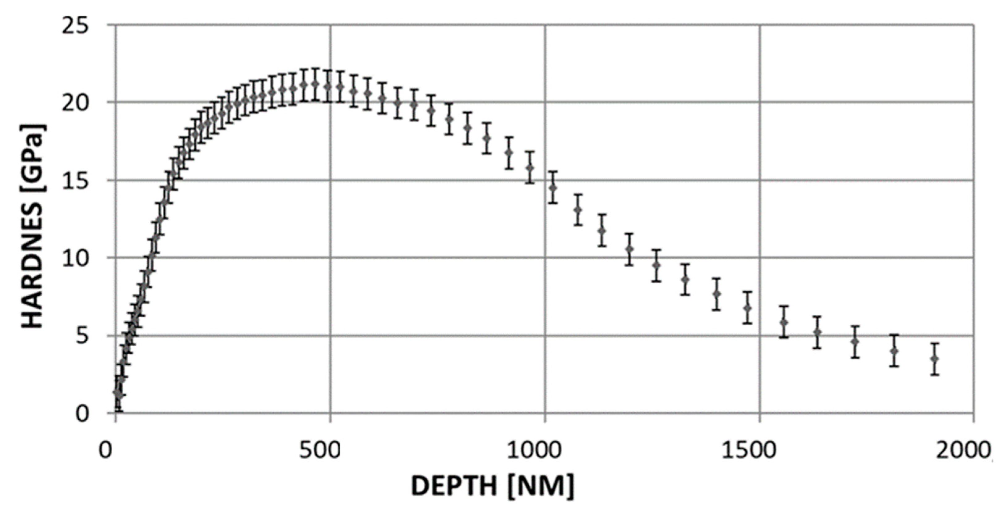
Figure 4 presents hardness the carbon coatings in the function of the depth of the forced-in penetrator. The hardness of layers amounted to 20–22 GPa. The hardness of the alloy amounted approximately 3 GPa.
Adhesion in the examined layers amounted to 150 mN. These values can be considered as entirely satisfactory. During exploitation of elements covered with carbon layers, layer should not be separated from a substrate.
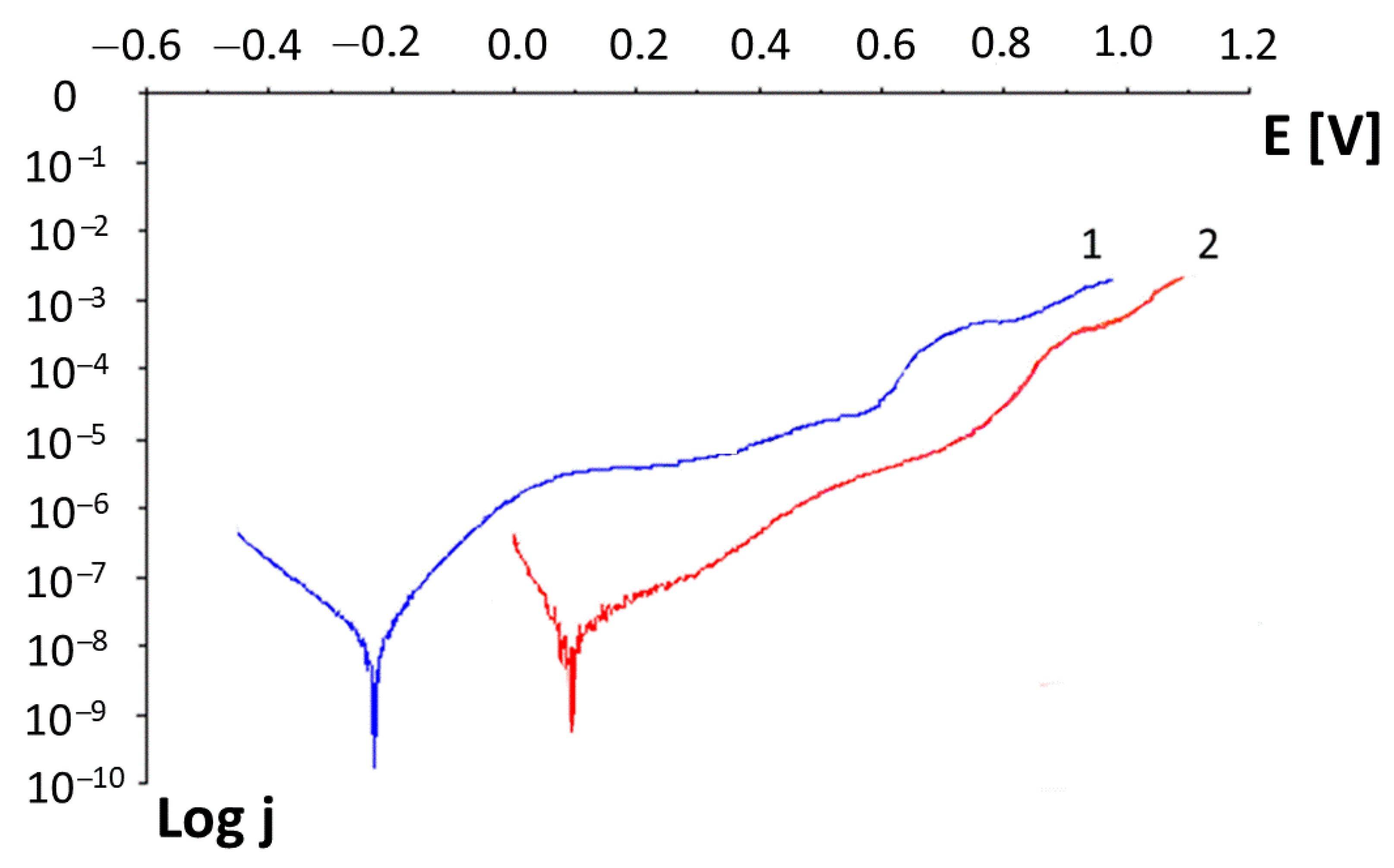
Figure 5 shows exemplary current-potential characteristics of samples with a carbon layer (1) and a sample without a coating (2). It is clear from the presented characteristics that the potential at which the process of intensive corrosive pulping of the passive layers begins is visibly shifted towards the anodic side for samples with carbon layers, compared with those without coatings. The linear density characteristics of the current intensity j from the polarization potential E were used to determine the breakthrough potentials of the passive layers Eb. The mean breakthrough potential values have been given in Table 3.
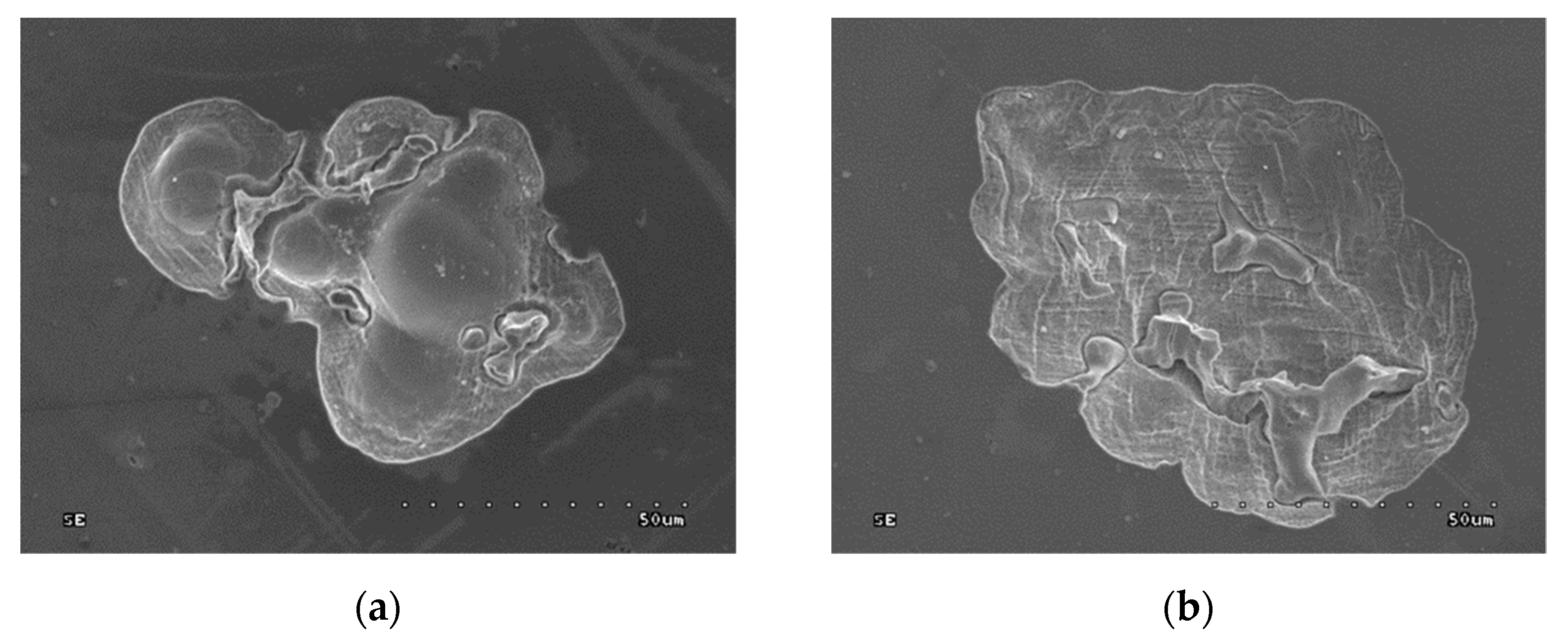
Figure 6 shows microscopic images of the corrosion areas after the tests.
After the corrosion examinations, corrosion pits appeared on the surface of the samples, 50–100 μm in size. An effect of the intensive corrosive processes is also removal of the carbon layer from the area near the formed pit, which is visible in Figure 6b.
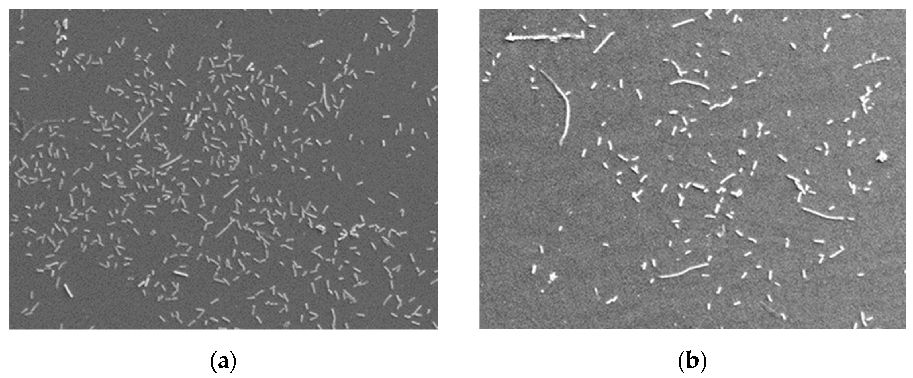
Figure 7 shows exemplary micro-images of the surface of the examined samples with adhering bacteria. Table 4 presents the number of bacteria adhering to the surfaces of the particular groups of samples.
The results were subjected to a statistical analysis, which used the one-way analysis of variance test (ANOVA) without repetitions (p < 0.05). For the evaluation of the normality of the distribution of the examined variables, the W Shapiro–Wilk test was applied, while, for the evaluation of the variance homogeneity—the Levene test. The performed statistical analysis demonstrated that the differences in the number of adhering bacteria on the surfaces of the samples with and without a carbon layer were statistically significant.

4. Discussion

Providing the coatings applied on prosthetic elements with the proper protective properties first of all requires the appropriate density (no cracks, scratches, pores, etc.), which will ensure anticorrosive properties. Fulfilling this condition will provide protection from the formation of corrosion products and their release into the oral cavity, as well as from ions transferred to it from the metallic substrate. The microscopic observations showed that the examined carbon coatings on prosthetic alloy Ni-Cr are monolithic and hermetic (Figure 1 and Figure 2), without surficial defects. Here, there are also no cracks, pores or delaminations. Therefore, it is possible to assume that they can provide the proper density, thus constituting a barrier separating the environment of the oral cavity of the patient from the alloy from which the prosthetic elements are made. The X-ray microanalysis demonstrated that, in the examined coatings, only carbon is present (Figure 1b), without any other additives. Therefore, we can state that this is a pure carbon coating. Unfortunately, based on the performed chemical composition tests, it is impossible to clearly establish the type of the coating. However, the Raman spectrometry made it possible to state that, in the examined coating, both nanocrystalline diamond and nanocrystalline graphite are present. On the cross-sections of the samples (Figure 1), one can clearly see a titanium interlayer located between the carbon layer and the substrate.
Protective coatings should also characterize in other properties so that they can fulfil their role. Such properties include the wear resistance. The latter mostly depends on the coatings’ hardness. The higher the hardness, the higher the wear resistance. The examined coatings characterize in hardness of about 20 GPa, with the hardness of the substrate alloy of about 3 GPa. The values obtained by other authors are similar [22,38]. The obtained hardness values of the test samples make it possible to provide significant wear resistance. While the wear resistance was not tested, it can be inferred from the reports of other authors that carbon coatings of this type characterize in high wear resistance, which is much higher than that of the substrate alloy. This is an additional advantage behind their use for prosthetic elements. There are not many literature reports referring to carbon coatings on prosthetic elements, yet tests performed on other alloys being applied in e.g., orthopedics exhibit elevated resistance of this type [22,38,39,40]. It should be noted that many alloys used in orthopedics have a composition which is very similar to that of the alloys applied for prosthetic and orthodontic elements used in the oral cavity. The operation conditions of e.g., hip joint endoprostheses are significantly harder than those of dental prosthetic restorations.
In the case of protective coatings, it is very, or even the most, important to provide them with the proper adhesion to the substrate, as only this will make it possible to fully take advantage of their properties. Even the best coatings which do not possess sufficient adhesion to the substrate are incapable of properly fulfilling their role. This is so, as each spalling of the layer causes exposition of the substrate material and loss of tightness, with all its consequences. In the examined layers, the adhesion equaled about 150 mN. These values can be treated as satisfactory, and they are comparable with those obtained by other authors on Co-Cr alloys and titanium alloys [38,39], while being higher than those obtained by Ji Li on titanium alloy Ti6Al4V [23]. During the operation of elements coated with carbon layers, the latter should not become separated from the substrate. It seems that the very good adhesion is a result of the use of a titanium interlayer. Batory et al. and Wei Yang et al. have also achieved improvement in the adhesion of carbon layers by applying a titanium interlayer [37,38].
The corrosion resistance tests carried out by means of the potentiodynamic method showed that carbon coatings shift the potential Ekor towards the anodic side by about 0.3 V. For the coated samples, the breakthrough potential of the passive layers Eb is about 0.2 V more anodic compared to the Eb for samples without a coating. This corrosion has a pitting character, which results from both the anodic potentiodynamic characteristics curves and the morphology of the sample surfaces examined with the use of a scanning electron microscope. No samples exhibited the occurrence of crevice corrosion. Based on the microscopic images, it can be assumed that the surface solubilization processes begin and the most intensively occur in the vicinity of the intermetallic phase inclusions present in the alloy. Summing up, it can be stated that carbon layers have a beneficial effect by increasing the corrosion resistance of the alloy. This is in agreement with the reports by other authors, in which different alloys coated with carbon layers are studied and which demonstrate an improvement of the corrosion resistance in a body fluid environment [25,37,38,39,40].
The studies of many authors have proved that there is a possibility to modify the surface of materials in order to limit the bacteria’s ability to colonize them [41]. The measurements of the number of bacteria adhering to the alloy surface showed that there were much fewer of them on surfaces covered with a carbon coating. These differences are statistically significant. It can be stated that the carbon layer greatly reduced the number of bacteria adhering to the surface of the examined alloy. The obtained results are in agreement with those obtained by other authors [42,43,44,45] who have also demonstrated that carbon coatings on different alloys reduce the number of adhering bacteria.
It seems that these results are very advantageous from the point of view of the prophylactics in inflammatory complications connected with the introduction of prosthetic restorations into the oral cavity. The colonization of the surface of prosthetic restorations also leads to disadvantageous changes in the oral cavity. The surface of prostheses, directly after their introduction into the oral cavity, is colonized by bacteria, which form a biofilm—dental plaque. Undoubtedly, one of the pathogenic factors leading to the development of mucous membrane inflammation of the oral cavity is the dental plaque and the bacteria which form it. The harmful operation of bacteria and the products of their metabolism can lead to the development of mucous membrane inflammation of the oral cavity being in contact with the elements of the prosthesis. For example, an inflammatory reaction of the gums, which are in contact with the edges of prosthetic crowns, are observed.
One of the important factors affecting the corrosion resistance and the number of adhering bacteria is surface roughness. In the present studies, smooth polished surfaces were used, which is in agreement with the dental practice, where the metal prosthesis surfaces undergo polishing, due to e.g., hygienic reasons.
Full characteristics of the obtained layers should also consider the release of the metal ions from the substrate into the body fluids. However, based on the corrosion tests, we can assume that an improvement in the corrosion resistance should limit the number of ions transported into the body fluid environment and thus the surrounding tissue. Such a supposition is confirmed by the studies of Ji Li et al., who have demonstrated that carbon layers on titanium alloys significantly reduce the release of harmful ions into the surrounding tissue [23]. In addition, J.S. Viswanathan, applying carbon layers in his research, has improved the corrosion resistance of alloy Ni-Ti and increased its biocompatibility [25]. Reducing the corrosion and thus also the number of released metal substrate ions can also, as in the case of e.g., Ti(CN) type coatings, contribute to the improvement in biocompatibility of coated metal elements [19,20,21,22].

5. Conclusions

It can be concluded from the performed studies that the mechanical properties of the obtained carbon coatings make them suitable for prosthetic elements. The applied method makes it possible to obtain continuous homogeneous coatings with no visible defects which could weaken their structure and worsen their properties. Both their hardness and wear resistance, confirmed in other studies, should ensure their proper functions. The coatings’ adhesion to the metal substrate made of alloy Ni-Cr is good enough to prevent their exposition during the operation in the oral cavity. Coatings significantly improve the corrosion resistance, which should reduce allergic and cytotoxic reactions of the body. It can be clearly seen that the applied carbon coatings reduce the adhesion of bacteria to the alloy surface, and thus also to the metal prosthetic elements. This property will improve the hygienic conditions in the oral cavity and can contribute to a significant limitation of the microbiological corrosion. Taking all this into account, we can state that carbon coatings can be potentially used for the protection of metal prosthetic restorations.

Author Contributions

Conceptualization, Z.K. and L.K.; methodology, Z.K. and L.K.; validation, Z.K., M.S. and L.K.; formal analysis, L.K.; investigation, Z.K. and L.K.; writing—original draft preparation, Z.K. and L.K.; writing—review and editing, M.S. and L.K.; visualization, Z.K. and L.K.; supervision, M.S.; project administration, L.K. All authors have read and agreed to the published version of the manuscript.

Funding

This research was funded by Medical University of Lodz and Lodz University of Technology.

Institutional Review Board Statement

Not applicable.

Informed Consent Statement

Not applicable.

Data Availability Statement

The data that support the findings of this study are available from the L.K. upon reasonable request.

Conflicts of Interest

The authors declare no conflict of interest.

References

  1. Lee, J.-J.; Song, K.-Y.; Ahn, S.-G.; Choi, J.-Y.; Seo, J.-M.; Park, J.-M. Evaluation of effect of galvanic corrosion between nickel-chromium metal and titanium on ion release and cell toxicit. J. Adv. Prosthodont. 2015, 7, 172–177. [Google Scholar] [CrossRef] [Green Version]
  2. Holm, C.; Morisbak, E.; Kalfoss, T.; Dahl, J.E. In vitro element release and biological aspects of base–metal alloys for metal-ceramic applications. Acta Biomater. Odontol. Scand. 2015, 1, 2–4. [Google Scholar] [CrossRef] [Green Version]
  3. Milheiro, A.; Nozaki, K.; Kleverlaan, C.J.; Muris, J.; Miura, H.; Feilzer, A.J. In vitro cytotoxicity of metallic ions released from dental alloys. Odontology 2016, 104, 136–142. [Google Scholar] [CrossRef] [PubMed]
  4. Chandar, S.; Kotian, R.; Madhyastha, P.; Kabekkodu, S.P.; Rao, P. In vitro evaluation of cytotoxicity and corrosion behavior of commercially pure titanium and Ti-6Al-4V alloy for dental implants. J. Indian Prosthodont. Soc. 2017, 17, 35–40. [Google Scholar]
  5. Mc Ginley, E.L.; Fleming, G.J.P.; Moran, G.P. Development of a discriminatory biocompatibility testing model for non-precious dental casting alloys. Dent. Mater. 2011, 27, 1295–1306. [Google Scholar] [CrossRef]
  6. Ramírez-Ledesma, A.L.; Roncagliolo, P.; Álvarez-Pérez, M.A.; Lopez, H.F.; Juárez-Islas, J.A. Corrosion Assessment of an Implantable Dental Co-Cr Alloy in Artificial Saliva and Biocompatibility Behavior. J. Mater. Eng. Perform. 2020, 29, 1657–1670. [Google Scholar] [CrossRef]
  7. Pochrząst, M.; Marciniak, J.; Wróbel, K.; Bączkowski, B. Electrochemical Properties of Ni-Cr and Co-Cr Alloys Used in Prosthodontics. Solid State Phenom. 2011, 183, 143–148. [Google Scholar] [CrossRef]
  8. Schmutz, P.; Quach-Vu, N.-C.; Gerber, I. Metallic Medical Implants: Electrochemical Characterization of Corrosion Processes. Electrochem. Soc. Interface 2020, 17, 35–40. [Google Scholar] [CrossRef]
  9. Nierlich, J.; Papageorgiou, S.N.; Bourauel, C.; Hültenschmidt, R.; Bayer, S.; Stark, H.; Keilig, L. Corrosion behavior of dental alloys used for retention elements in prosthodontics. Eur. J. Oral Sci. 2016, 124, 287–294. [Google Scholar] [CrossRef] [Green Version]
  10. Mercieca, S.; Conti, C.M.; Buhagiar, J.; Camilleri, J. Assessment of corrosion resistance of cast cobalt- and nickel-chromium dental alloys in acidic environments. J. Appl. Biomater. Funct. Mater. 2018, 16, 47–54. [Google Scholar] [CrossRef] [PubMed] [Green Version]
  11. Mystkowska, J.; Ferreira, J.A.; Leszczyńska, K.; Chmielewska, S.; Dąbrowski, J.R.; Wieciński, P.; Kurzydłowski, K. Biocorrosion of 316LV steel used in oral cavity due to Desulfotomaculum nigrificans bacteria. J. Biomed. Mater. Res. B Appl. Biomater. 2017, 105, 222–229. [Google Scholar] [CrossRef] [PubMed]
  12. Mystkowska, J.; Niemirowicz-Laskowska, K.; Łysik, D.; Tokajuk, G.; Dąbrowski, J.R.; Bucki, R. The Role of Oral Cavity Biofilm on Metallic Biomaterial Surface Destruction–Corrosion and Friction Aspects. Int. J. Mol. Sci. 2018, 19, 743. [Google Scholar] [CrossRef] [Green Version]
  13. Adya, N.; Alam, M.; Ravindranath, T.; Mubeen, A.; Saluja, B. Corrosion in titanium dental implants: Literature review. J. Indian Prosthodont. Soc. 2005, 53, 126–131. [Google Scholar] [CrossRef]
  14. Wu, F.; Chen, T.; Wang, H.; Liu, D. Effect of Mo on Microstructures and Wear Properties of In Situ Synthesized Ti(C, N)/Ni-Based Composite Coatings by Laser Cladding. Materials 2017, 10, 1047. [Google Scholar] [CrossRef] [Green Version]
  15. Nematia, A.; Saghafia, M.; Khamseh, S.; Alibakhshic, E.; Zarrintajd, P.; Saebe, M.R. Magnetron-sputtered TixNy thin films applied on titanium-based alloys for biomedical applications: Composition-microstructure-property relationships. Surf. Coat. Technol. 2018, 349, 251–259. [Google Scholar] [CrossRef]
  16. Asri, R.I.M.; Harun, W.S.W.; Samykano, M.; Lah, N.A.C.; Ghani, S.A.C.; Tarlochan, F.; Raza, M.R. Corrosion and surface modification on biocompatible metals: A review. Mater. Sci. Eng. 2017, 77, 1261–1274. [Google Scholar] [CrossRef] [Green Version]
  17. Jamesh, M.I.; Li, P.; Bilek, M.M.; Boxman, R.L.; McKenzie, D.R.; Chu, P.K. Evaluation of corrosion resistance and cytocompatibility of graded metal carbon film on Ti and NiTi prepared by hybrid cathodic arc/glow discharge plasma-assisted chemical vapor deposition. Corros. Sci. 2015, 97, 126–138. [Google Scholar] [CrossRef]
  18. Wang, G.; Zreiqat, H. Functional Coatings or Films for Hard-Tissue Applications. Materials 2010, 3, 3994–4050. [Google Scholar] [CrossRef] [Green Version]
  19. Banaszek, K.; Wiktorowska-Owczarek, A.; Kowalczyk, E.; Klimek, L. Possibilities of applying Ti (C, N) coatings on prosthetic elements—Research with the use of human endothelial cells. Acta Bioeng. Biomater. 2016, 18, 119–126. [Google Scholar]
  20. Banaszek, K.; Klimek, L.; Zgorzynska, E.; Swarzynska, A.; Walczewska, A. Cytotoxicity of titanium carbonitride coatings for prostodontic alloys with different amounts of carbon and nitro gen. Biomed. Mater. 2018, 13, 045003. [Google Scholar] [CrossRef]
  21. Calderon, S.; Alves, C.F.A.; Manninen, N.K.; Cavaleiro, A.; Carvalho, S. Electrochemical Corrosion of Nano-Structured Magnetron-Sputtered Coatings. Coatings 2019, 9, 682. [Google Scholar] [CrossRef] [Green Version]
  22. Banaszek, K.; Klimek, L. Ti(C, N) as Barrier Coatings. Coatings 2019, 9, 432. [Google Scholar] [CrossRef] [Green Version]
  23. Li, J.; Wang, K.; Li, Z.; Tu, J.P.; Jin, G.; Su, J.; Zhai, B. Mechanical tests, wear simulation and wear particle analysis of carbon-based nanomultilayer coatings on Ti6Al4V alloys as hip prostheses. RSC Adv. 2018, 8, 6849–6857. [Google Scholar] [CrossRef] [Green Version]
  24. Jachowicz, M.; Kaczmarek, Ł.; Rylski, A.; Przybylski, K.; Danielewski, M.; Wendler, B. New type AlMo-, AlTi- or Si-based magnetron sputtered protective coatings on metallic substrates. J. Mater. Process. Technol. 2006, 175, 427–432. [Google Scholar]
  25. Kaczmarek, Ł.; Wendler, B.; Siniarski, D.; Rylski, A.; Bieliński, D.; Dobrowolski, O.; Lipiński, P. Oxidation resistance of refractory γ-TiAlW coatings. Surf. Coat. Technol. 2007, 201, 167–6170. [Google Scholar] [CrossRef]
  26. Viswanathan, S.; Mohan, L.; Chakraborty, M.; Mandal, C.; Bera, P.; Aruna, S.T.; Anandan, C. Carbon plasma immersion ion implantation and DLC deposition on Ni−Ti alloy. Mater. Manuf. Process. 2018, 33, 1121–1127. [Google Scholar] [CrossRef]
  27. Rajib, P. Diamond-Like-Carbon Coatings for Advanced Biomedical Applications. Glob. J. Nanomed. 2017, 5, 1–5. [Google Scholar]
  28. Grabarczyk, J.; Batory, D.; Louda, P.; Couvrat, P.; Kotela, I.; Bąkowicz-Mitura, K. Carbon coatings for medical implants. J. Achiev. Mater. Manuf. Eng. 2007, 20, 107–110. [Google Scholar]
  29. Mitura, E.; Niedzielska, A.; Niedzielski, P.; Klimek, L.; Rylski, A.; Mitura, S.; Moll, J. The properities of carbon layers deposited onto titanium substrates. Diam. Relat. Mater. 1996, 5, 998–1001. [Google Scholar] [CrossRef]
  30. Jamesh, M.I.; Boxman, R.L.; Bilek, M.M.; Kocer, C.; Hu, T.; Zhang, X.; McKenzie, D.R.; Chu, P.K. Effects of pulse voltage and deposition time on the adhesion strength of graded metal/carbon films deposited on bendable stainless-steel foils by hybrid cathodic arc—Glow discharge plasma assisted chemical vapor deposition. Appl. Surf. Sci. 2016, 366, 535–544. [Google Scholar] [CrossRef]
  31. Batory, D.; Blaszczyk, T.; Clapa, M.; Mitura, S. Investigation of anti-corrosion properties of Ti:C gradient layers manufactured in hybrid deposition system. Mater. Sci. 2008, 43, 3385–3391. [Google Scholar] [CrossRef]
  32. PN-EN-ISO 14577-1, -2, -3. Metals—Apparatus Testing of Hardness and Material Properties of Indenter Indentation; International Organization for Standardization: Geneva, Switzerland, 2003. [Google Scholar]
  33. Li, X.; Bharat, B. A review of nanoindentation continuous stiffness measurement technique and its applications. Mat. Charact 2002, 48, 11–36. [Google Scholar]
  34. Oliver, W.C.; Pharr, G.M. An improved technique for determining hardness and elastic modulus using load and displacement sensing indentation experiments. J. Mater. Res. 1992, 7, 1564–1583. [Google Scholar] [CrossRef]
  35. Huang, L.; Lu, J.; Xu, K. Elasto-plastic deformation and fracture mechanism of a diamond-like carbon film deposited on a Ti–6Al–4V substrate in nano-scratch test. Thin Solid Film. 2004, 466, 175–182. [Google Scholar]
  36. Banaszek, K.; Szymanski, W.; Pietrzyk, B.; Klimek, L. Adhesion of E. coli Bacteria Cells to Prosthodontic Alloys Surfaces Modified by TiO2 Sol-Gel Coatings. Adv. Mater. Sci. Eng. 2013, 2013, 179241. [Google Scholar] [CrossRef] [Green Version]
  37. Tai, F.C.; Lee, S.C.; Chen, J.; Wei, C.; Chang, S.H. Multipeak fitting analysis of Raman spectra on DLCH film. J. Raman Spectrosc. 2009, 40, 1055–1059. [Google Scholar] [CrossRef]
  38. Kaczorowski, W.; Świątek, H.; Łuczak, K.; Głuszek, M.; Cłapa, M. Impact of Plasma Pre-Treatment on the Tribological Properties of DLC Coatings on PDMS Substrates. Materials 2021, 14, 433. [Google Scholar] [CrossRef] [PubMed]
  39. Batory, D.; Szymański, W.; Cłapa, M. Mechanical and tribological properties of gradient a-C:H/Ti coatings. Mater. Sci. Pol. 2013, 31, 415–423. [Google Scholar] [CrossRef]
  40. Yang, W.; Gao, Y.; Guo, P.; Xu, D.; Hu, L.; Wang, A. Adhesion, biological corrosion resistance and biotribological properties of carbon films deposited on MAO coated Ti substrates. J. Mech. Behav. Biomed. Mater. 2020, 101, 1–6. [Google Scholar] [CrossRef]
  41. Cano, D.; Lousa, A.; Esteve, J.; Ferrer-Anglada, N. Low Wear and Low Friction DLC Coating with Good Adhesion to CoCrMo Metal Substrates. Phys. Status Solidi 2018, 255, 1800255. [Google Scholar] [CrossRef]
  42. Jakubowski, W.; Ślósarczyk, A.; Paszkiewicz, Z.; Szymański, W.; Walkowiak, B. Bacterial colonisation of bioceramic surfaces. Adv. Appl. Ceram. 2008, 107, 217–221. [Google Scholar] [CrossRef]
  43. Liu, C.Q.; Zhao, Y.; Liu, S.; Wang, E.W. Abel: Reduction of bacterial adhesion on modified DLC coatings. Colloids Surf. B Biointerfaces 2018, 61, 182–187. [Google Scholar] [CrossRef]
  44. Robertson, S.N.; Gibson, D.; Mac Kay, W.G.; Reid, S.; Williams, C.; Birney, R. Investigation of the antimicrobial properties of modified multilayer diamond-like carbon coatings on 316 stainless steel. Surf. Coat. Technol. 2017, 314, 72–78. [Google Scholar] [CrossRef] [Green Version]
  45. Marciano, F.R.; Bonetti, L.F.; Mangolin, J.F.; Da-Silva, N.S.; Corata, E.J.; Trava-Airoldia, V.J. Investigation into the antibacterial property and bacterial adhesion of diamond-like carbon films. Vacuum 2011, 85, 662–666. [Google Scholar] [CrossRef]
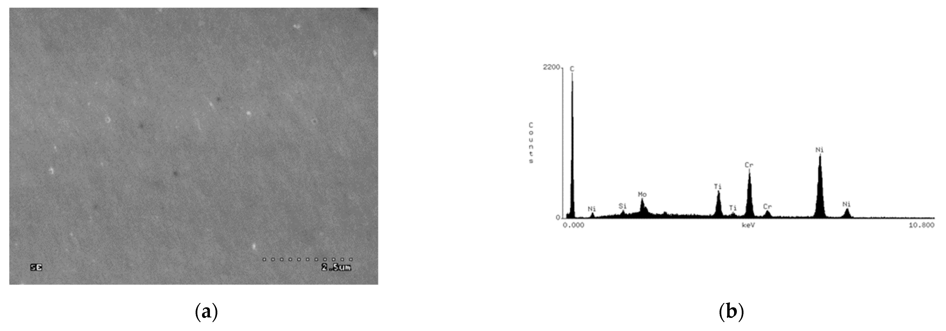
Figure 1. Surface of a sample with a carbon coating (a) and an EDS spectrogram from the sample surface (b).
Applsci 11 04551 g001
Figure 2. Microscopic image of a sample’s cross-section with the surface element distributions (a) microscopic image, (b) surface distribution of carbon, (c) surface distribution of titanium, (d) surface distribution of nickel, (e) surface distribution of chromium.
Applsci 11 04551 g002aApplsci 11 04551 g002b
Figure 3. Raman spectra of examined samples.
Applsci 11 04551 g003
Figure 4. Hardness in the function of the depth of the forced-in penetrator.
Applsci 11 04551 g004
Figure 5. Exemplary current-potential characteristics; 1–Ni-Cr alloy sample with a carbon layer, 2–Ni-Cr alloy sample without a coating.
Applsci 11 04551 g005
Figure 6. Image of the pits after corrosion tests, (a) sample without a coating, (b) sample with a coating.
Applsci 11 04551 g006
Figure 7. Sample surfaces with adhering bacteria (a) Ni-Cr alloy sample without a coating. (b) Ni-Cr alloy sample with a carbon layer.
Applsci 11 04551 g007
Table 1. Composition of Ni-Cr alloy.
Element Percentage by Weight (wt.%)
CrMoSiFeCoMnNi
24.798.891.571.330.170.13rest
Table 2. Chemical composition of artificial saliva solution.
KClNaClCaCl2·2H2ONaH2PO4·2H2ONa2S·9H2OUreaTriple Distilled Water
0.4 g0.4 g0.906 g0.69 g0.005 g1 g1 dm3
Table 3. Mean values of breakthrough potentials of the examined samples Eb.
SampleMean ValueStandard Deviation
Ni-Cr with a coating0.619 V0.010
Ni-Cr without a coating0.823 V0.003
Table 4. The number of bacteria adhering to the surfaces (150 × 100 μm) of the particular groups of samples.
SampleAverage ValueStandard Deviation
Ni-Cr with a coating8410
Ni-Cr without a coating17312
Publisher’s Note: MDPI stays neutral with regard to jurisdictional claims in published maps and institutional affiliations.