Case Report

Complications of Teeth Affected by Molar-Incisor Malformation and Pathogenesis According to Microbiome Analysis

1
Department of Pediatric Dentistry, School of Dentistry, Kyung Hee University, Seoul 02447, Korea
2
Department of Pediatric Dentistry, Kyung Hee University Dental Hospital, Seoul 02447, Korea
3
Department of Pediatric Dentistry, Kyung Hee University Dental Hospital at Gangdong, Seoul 05278, Korea
*
Author to whom correspondence should be addressed.
Appl. Sci. 2021, 11(1), 4; https://doi.org/10.3390/app11010004
Submission received: 13 October 2020 / Revised: 18 December 2020 / Accepted: 18 December 2020 / Published: 22 December 2020
(This article belongs to the Special Issue Oral Microbial Communities and Oral Health)

Abstract

:
A molar-incisor malformation (MIM) is a recently reported dental anomaly that causes premature loss of the first molar with severe dentoalveolar infection. However, there has been no research on the pathogenesis yet. The aim of this study was to report the clinical process of MIMs and investigate the pathogenesis by conducting a microbiome analysis. An eight-year-old girl was diagnosed with MIM and after two years, four permanent first molars were sequentially extracted due to severe dentoalveolar infection. We recorded the patient`s clinical progress and collected oral microbiome samples from the extracted teeth with MIM and sound teeth as controls. The sites of microbiome sampling were represented by five habitats in two groups. Group (1) was the perio group: ① supragingival plaque, ② subgingival plaque, and ③ a pical abscess; and group (2) was the endo group: ④ coronal pulp chamber and ⑤ root canal. The perio group was composed predominantly of genera Streptococcus, Veilonella, and Leptotrichia. Spirochetes appeared in one sample from a severe periodontal abscess. Aggregatibacter actinomyces were not identified. In the endo groups, pulp necrosis was observed in all MIM and the genera Peptostreptococcus and Parvimonas predominated. In conclusion, MIM teeth caused localized tooth-related periodontitis with pulp necrosis rather than localized juvenile periodontitis, resulting in a poor prognosis, and timely extraction is highly recommended.

1. Introduction

A molar-incisor malformation (MIM) is a recently reported dental anomaly of the permanent first molars, deciduous molars, and permanent maxillary central incisors [1]. MIM anomalies of the permanent first molars and deciduous molars may be characterized by normal crowns with a constricted cervical region and thin, narrow, and short roots, while the affected maxillary central incisors may exhibit a hypoplastic enamel notch near the cervical third of the clinical crown [2]. Although the etiology of MIMs remains uncertain, it is thought to be attributable to systemic disease associated with the neural system during infancy [3,4,5,6].
MIM was named by Lee et al. [1] in 2014. In addition, there have been papers published this dental anomaly as ‘Root malformation associated with a cervical mineralized diaphragm’ or ‘Molar root-incisor malformation’. According to Vargo et al. [3] in 2020, a total 87 cases have been published in articles so far, and all except one case have been affected by the permanent first molars. The average diagnostic age was nine years.
MIM has been mistaken for molar-incisor hypomineralization (MIH) until recently. MIM has similarities with and differences from MIH. The similarities include the involved teeth; both MIM and MIH appear most frequently in the maxillary central incisors and the permanent first molars [1,2]. There are differences, however, in the affected tissues. In MIH, the enamel is affected, whereas in MIM, dentin and cementum are affected. In incisors affected by MIM, the cervical enamel notch may appear. The exact cause of both is unknown, and treatment is difficult [3]. Since it has been the biggest problem in children’s oral health recently, it is important to identify the correct treatment.
MIM teeth are associated with complications such as dentoalveolar infections, early exfoliation, space loss, spontaneous pain, impaction, and poor incisor esthetics [3]. Dentoalveolar infection is the most common complication of MIM. In severe cases, inflammation surrounds the entire roots, resulting in an abscess, fistula, and vertical mobility. A MIM with complications is usually extracted due to the poor prognosis reported in previous case studies [1,7,8]. The early loss of the first permanent molar can negatively affect occlusal stability and craniofacial development [9,10].
Various factors can affect the development of a dentoalveolar infection, and the oral microbiome is one of them. The oral microbiome is described as a group of resident oral microorganisms in the host. Oral diseases, such as dental caries and periodontal disease are caused by dysbiosis of the host and oral microbiome [11]. Next-generation sequencing (NGS) is an effective method used to conduct research on the microbiome [12]. NGS is a molecular technique that reads more sequences faster and more economically and is widely used to identify microorganisms.
In this study, we analyzed the oral microbiomes around the MIM teeth to provide evidence for the pathogenesis of the complications because the oral microbiome is associated with oral health and oral disease. The aim of this study was to report the clinical process of MIM teeth and investigate the pathogenesis by microbiome analysis.

2. Materials and Methods

2.1. Ethics

The guidelines in the Helsinki Declaration were followed in this investigation. This study was approved by the Institutional Review Board of the School of Dentistry, Kyung Hee University (KHDIRB1606-6). The participants were recruited from patients in the Department of Pediatric Dentistry, School of Dentistry, Kyung Hee University. Written consent for participation in the study was obtained from the parent and child.

2.2. Clinical Progress

An eight-year-old girl visited our dental clinic for a dental eruption problem. Her medical history included a premature birth at nine months and she was taking medication for attention deficit hyperactivity disorder (ADHD). Radiographic and clinical examinations revealed root deformation of the first molars, primary molars, and cervical notch of the maxillary central incisors (Figure 1). The maxillary first molars were locked on the distal surface of the maxillary primary second molar and the mandibular first molars were mesially displaced due to the early exfoliation of the mandibular primary second molars. A lingual arch was set and periodic checks followed.
When the patient was 10, severe dentoalveolar infection and fistula occurred on the mandibular left first molar (Figure 2). Orthodontic analysis was conducted for treatment planning. She already had a space discrepancy and we decided to extract all permanent first molars affected by MIM and perform orthodontic treatment after the eruption of the secondary molars. Thereafter, the four MIM first molars were extracted sequentially and they showed twisted roots with granulation tissue (Figure 3). The crown and root of the maxillary left molar and mandibular right molar were fractured during the extraction. One year later, the second molars erupted and were relatively well-aligned.

2.3. Sampling

The samples were MIM teeth and the non-MIM teeth were the controls. The MIM samples included four permanent first molars and maxillary central incisors as one sample, and the non-MIM teeth including the maxillary left premolars and mandibular right premolars as one sample.
The oral microbiome was obtained from five habitats around the teeth in the two groups. The perio group included ① a supragingival plaque, ② subgingival plaque, and ③ an apical abscess, and the endo group included ④ a coronal pulp chamber and ⑤ a root canal (Figure 4). The perio samples were collected before the extractions, and the endo samples were collected after the extractions. All samples are listed in Table 1. The sample identification (ID) numbers refer to the two-digit FDI notations.
  • Perio group
    Supragingival plaque: The patient was requested not to brush her mouth 24 h prior to sample collection. Supragingival dental plaque samples were collected by rubbing a sterile cotton swab across the cervicobuccal area of the teeth twice or three times under pressure [13].
    Subgingival plaque: The subgingival plaque samples were collected by inserting sterile endodontic paper points (sized 30; two paper points per site) into the gingival sulci or periodontal pocket for 10 s right after isolation and supragingival plaque removal [14].
    Periapical abscess: The periapical abscess sample was obtained via aspiration. The swollen area was aspirated with a syringe fitted with a 16-gauge needle and expressed into a sterile vial [15].
  • Endo group
    Coronal pulp chamber and ⑤ root canal: The extracted tooth was cleaned with 30% hydrogen peroxide [16]. Aseptic techniques such as sterile burs were used to access the pulp space. Bacteriological samples of the pulp chamber were collected immediately after crown access. Pulp remnant and infected dentin were collected using a sterilized spoon excavator and sterilized paper points were inserted to absorb the remaining fluid containing microorganisms. In the root canal, two sequential new paper points were placed at the same level and utilized to soak up the fluid in the canal. Each paper point was held in position for 30 s. Sterilized endodontic files were then used to collect the infected root canal dentin severally. Only the tip area was collected into the tube by cutting.
The samples were placed in sterile 1.5 mL microcentrifuge tubes and frozen for storage at −80 °C. DNA was extracted from the clinical samples using the FastDNA SPIN Kit for Soil (MP Biomedicals, Santa Ana, CA, USA) according to the manufacturer’s instructions. The concentration and purity of the DNA samples were determined using an ND-1000 NanoDrop spectrophotometer (Thermo Scientific, Waltham, MA, USA) by measuring the absorbance at 260 and 280 nm.

2.4. PCR Amplification and Illumina Sequencing

PCR amplification of the extracted DNA was performed using primers targeting regions between V3 and V4 of the 16S rRNA gene. For bacterial amplification, 341F primers (5′TCGTCGGCAGCGTC-AGATGTGTATAAGAGACAG-CCTACGGGNGGCWGCAG-3′; the target region primer) and 805R (5′-GTCTCGTGGGCTCGGAGATGTGTATAAGAGACAG-GACTACHVGGGTATCTAATCC-3) were used. Amplifications were conducted under the following conditions: initial denaturation at 95 °C for 3 min, followed by 25 cycles of denaturation at 95 °C for 30 s, primer annealing at 55 °C for 30 sec, and extension at 72 °C for 30 s, with a final elongation at 72 °C for 5 min. Secondary amplification for attaching the Illumina Nextera barcode was then performed using the i5 forward primer (5′AATGATACGGCGACCACCGAGATCTACAC-XXXXXXXX-TCGTCGGCAGCGTC-3′; X indicates the barcode region) and i7 reverse primer (5′-CAAGCAGAAGACGGCATACGAGAT-XXXXXXXXAGTCTCGTGGGCTCGG-3). The conditions for the secondary amplification were similar to the earlier one except that the amplification cycles were set to 8.
The PCR products were confirmed using 2% agarose gel electrophoresis and visualized by the Gel Doc system (BioRad, Hercules, CA, USA). The amplified products were purified via the QIAquick PCR purification kit (Qiagen, Valencia, CA, USA). Equal concentrations of purified products were pooled together and short fragments (non-target products) were removed with the Ampure bead kit (Agencourt Bioscience, MA, USA). The quality and size of the products were assessed on a Bioanalyzer 2100 (Agilent, Palo Alto, CA, USA) using a DNA 7500 chip. Mixed amplicons were pooled, and sequencing was conducted at Chunlab, Inc. (Seoul, Korea) using the Illumina MiSeq Sequencing System (Illumina, San Diego, CA, USA) according to the manufacturer`s instructions.

2.5. MiSeq Pipeline Method

The processing of the raw reads started with a quality check and filtering of the low quality (<Q25) reads using Trimmomatic 0.32. After passing the QC check, the paired-end sequence data were merged using PandaSeq. The primers were then trimmed with ChunLab’s in-house program at a similarity cutoff of 0.8. The noise was removed from the sequences using Mothur’s pre-clustering program, which merges the sequences and extracts unique sequences allowing up to two differences between the sequences. The EzTaxon database was used for taxonomic assignments using BLAST 2.2.22 and pairwise alignment was used to calculate similarity. Uchime and the non-chimeric 16S rRNA database from EzTaxon were used to detect chimerism in the reads with best hit similarity rates below 97%. Sequence data were then clustered using CD-Hit and UCLUST, and alpha diversity analysis was conducted.

3. Results

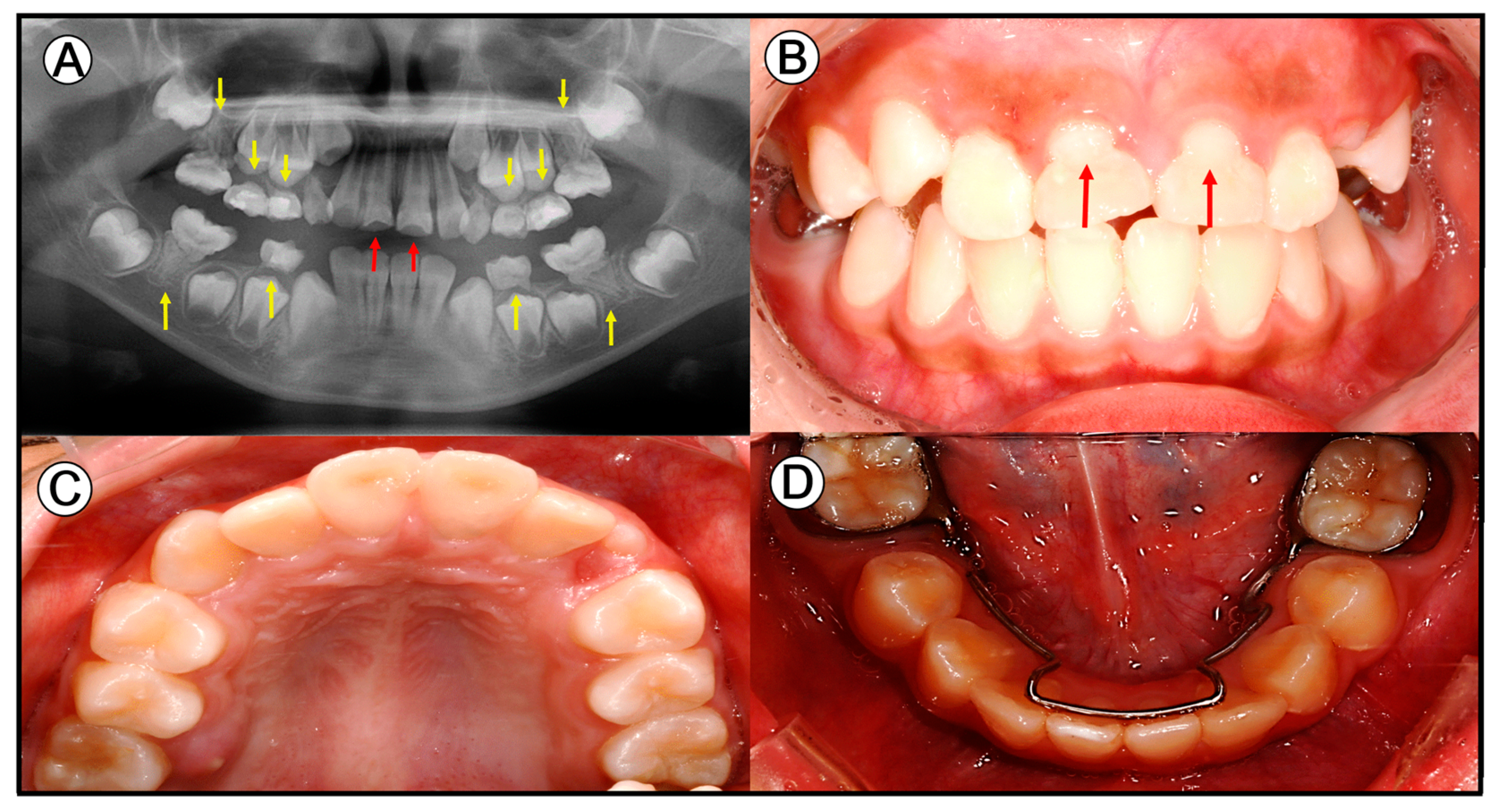
3.1. Clinical Information

The left MIM (ID 26 and 36) had a severe dentoalveolar abscess with a fistula and was extracted with abundant granulation tissue. The right MIM (ID 16 and 46) showed relatively less granulation tissue. The MIM had short, twisted, and fused abnormal roots (Figure 3). ID 26 and 46 were fractured during the extraction. The fractured surfaces were roughly calcified and there was a small hole connecting the pulp chamber and the root canal. The coronal pulp chamber and root canal were opened after extraction and necrotized, liquefied, gray, and slurried pulp and infected dentin were revealed. The incisors affected by the MIM (ID 11) and the non-MIM teeth (ID Nor) were clinically sound.

3.2. Taxonomic Identification and Operational Taxonomical Unit (OTU) Assessment of Diversity

An average of 18,621, 19,051, 19,952, 18,136, and 17,402 sequence reads was generated from the supragingival, subgingival, apical abscess, coronal pulp chamber, and root canal samples, respectively, after filtering (Table 2). Operational taxonomical units (OTUs), which is an operational definition of a species or group of species often used only when DNA sequence data are available, were utilized as a measure of diversity [17]. In the present study, OTUs were defined at the 3% divergence (97% similarity) threshold using the average neighbor clustering algorithm. The average numbers of OTUs were 165, 199, 185, 56, and 65, respectively. There were 3–4 times more OTUs in the perio group than in the endo group. Within the supragingival plaque and subgingival plaque, the MIM mandibular right first molar (ID 46-1 and 2) showed the lowest number of OTUs and the non-MIM teeth (ID Nor-1 and 2) showed higher than average OTUs.
The α-diversity in the samples was assessed by the Ace, Chao 1, and Shannon diversity indices at a 3% distance range (Table 2). Diversity is an indicator of richness (abundance) and evenness (composition) in the samples. The average Ace index was 185.62, 236.09, 215.64, 78.97, and 111.86, respectively. The average Chao 1 index was 181.92, 230.68, 224.18, 77.72, and 83.9, respectively. The average Shannon index was 3.47, 3.58, 3.24, 2, and 2.2, respectively. Diversity showed a similar pattern to the OTUs between and within the groups.

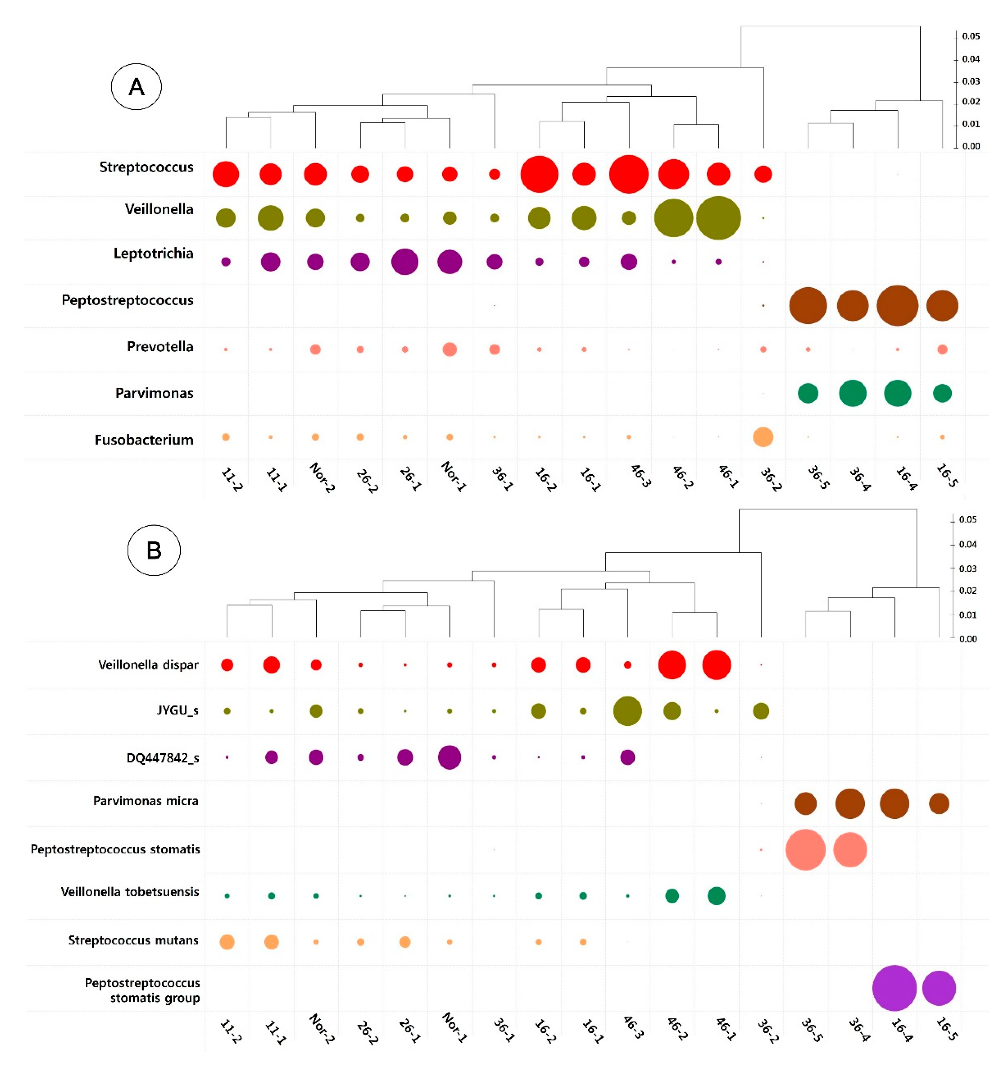
3.3. Taxonomic Identification of the Oral Microbiome

A total of seven major bacterial phyla (Spirochaetes, Firmicutes, Fusobacteria, Actinobacteria, Proteobacteria, Bacteriodetes, and Saccharibacteria_TM7) were identified at a higher than 1% frequency in all the plaque samples (Figure 5A). The perio group showed an average of 5.7 phyla. Only sample 36-2 showed seven phyla, including Spirochaetes. Seven samples showed six phyla and six samples showed five phyla. The endo group showed an average of 3.5 phyla and sample 36-4 showed two phyla.
Figure 5B shows that at the genus level, Veillonella, Streptococcus, and Leptotrichia were present at 5% frequency in almost all the perio group samples. Parvimonas, Peptostreptococcus, Atopobium, and Dialister were present in all endo group samples. Other genera were found in several samples in relation to the habitat or tooth. Aggregatibacter actinomyces were not identified.
Figure 5C shows the analysis of the endo group samples at a frequency of 1%. Bulleidia was found in all four samples. Fusobacterium, Mogibacterium, Prevotella, and Olsenella were present in three samples. HQ441590_g was found in two samples, while Shuttleworthia, Alloprevotella, Eubacterium_g10, Selenomonas, and Oribacterium were present in one sample.

3.4. Heatmap Analysis

In general, there was a distinct difference between the perio and endo groups. The perio group showed genera Streptococcus, Veillonella, and Leptotrichia (Figure 6A). Especially, Fusobacterium was found in a higher proportion in sample 36-2 compared to that in the other samples of the perio group. The endo group showed significant proportions of gram-positive anaerobic cocci species, such as Parvimonas micra and Peptostreptococcus stomatitis (group) (Figure 6B).

4. Discussion

The patient showed the stereotypic characteristics of a MIM patient. Based on our study in 2014 [1], the average age of 12 MIM patients at the initial visit was about 7.8 years (range 4–13 years) and it was the same with this patient. This seems to be due to the eruption of the MIM teeth including the permanent first molars. The patient in this study was female and the sex ratio was even in a previous study [1]. However, in the study by Kim et al. [8], there were 24 males out of 38, and in the study by Vargo et al. [3], there was a male predominance at 56.3%. The gender ratio is still controversial.
Interestingly, the patient had a medical history of systemic disease, preterm birth at nine months, and medication for ADHD. Reportedly, almost 95% of the MIM patients had diseases during infancy [8]. One of 12 in Lee’s study [1], one of one in McCreedy’s 2015 study [18], and four of 30 in Wright’s 2016 study [19] were born preterm. In addition, meningitis, spina bifida, cerebral cyst, cephalohematoma, seizures, hydrocephalus, and other diseases were present in MIM patients [3,8,19]. According to a recent review, “the exact aetiology of this condition is unknown. However, the fact that root malformations are limited to isolated teeth suggests that a non-genetic, environmental factor related to past medical history could be the cause” [3]. The authors suggested a possible epigenetic association, which needs further investigation.
In this patient, all the permanent first molars, the deciduous molars, and the maxillary central incisors were MIM teeth. This case seemed more serious compared to those in previous studies [3,8,19]. MIM teeth occurred in the permanent first molar, and often only in the mandible or in the maxilla. In many cases, the primary second molar was affected, but rarely the primary first molar or the permanent second molar. In the future, a classification for MIM is needed. The prevalence of incisors affected by MIM was about half [1] or one-third [3].
Sampling was the most crucial part for identifying the microbiome by collecting plaque and obtaining accurate data. If the sampling is incorrect, the disease-related bacteria may not be accurately identified. In this study, the sampling method applied was the one used by researchers in previous studies [13,14,15]. The NGS method MiSeq was developed in 2011 by Illumina. The characteristics of MiSeq consists of minimization while maintaining the chemistry of HiSeq (Illumina) and is suitable for reading V3 and V4 of 16S rRNA applied to microbial community analysis [20].
A total of 17 samples were categorized into the perio and endo groups. The perio group had higher average OTUs and alpha diversity compared to the endo group. At the genus level, Streptococcus, Veillonella, and Leptotrichia were present in most of the perio group samples, but not in the endo group. These genera were usually found in healthy Korean preschool children [21].
Particularly, sample 36-2 with a clinically severe dentoalveolar abscess showed only the presence of the Spirochetes phylum. Spirochetes were present in subgingival plaques and elevated in advanced periodontal disease and endodontic infection [22]. Treponema socranskii and Treponema medium appeared at 1% frequency in sample 36-2. Treponema genera belonging to oral Spirochetes were isolated from the periodontal inflammation site [23].
In the heatmap, the perio group was subtly divided into three clusters: (1) 11-2, 11-1, Nor-2, 26-2, 26-1, Nor-1, and 36-1; (2) 16-2, 16-1, 46-3, 46-2, and 46-1; and (3) 36-2. The first group included the MIM-incisor and non-MIM teeth, and the maxillary left MIM-molar. The incidence of Leptotrichia, Prevotella, and Fusobacterium was relatively high. The second group included the right MIM-molars with relatively high Streptococcus and Veillonella. Sample 36-2 showed a relatively large number of Fusobacterium. Fusobacterium is an absolute anaerobic gram-negative bacterium with a long filament, which is isolated from periodontal tissue and can aggregate with other oral bacteria [24]. It is one of the signs of acute dentoalveolar infection [15].
Among the endo group, samples 16-4, 16-5, and 36-5 showed four phyla and 36-4 showed two phyla. Saccharibacteria_TM7 and Proteobacteria was not present as compared to the Perio group. At the genus level, Parvimonas, Peptostreptococcus, Atopobium, and Dialister were present in all the endo group samples. The anaerobic gram-positive cocci Parvimonas and Peptostreptococcus have been identified from caries dentin, infected pulp, and complex infections such as dental root canals, progressive periodontitis, and dental abscesses [15,25]. Atopobium was originally identified as an anaerobic lactic acid bacillus in a periodontal pocket. Dialister is a gram-negative bacterium, which is an absolute anaerobic and found in root canal infections and periodontal infections [25]. In addition, Shuttleworthia, Bulleidia, Mogibacterium, and Olsenella, identified at 1% frequency, are present in the root canals in endodontic-periodontal lesion [25]. Regarding species, the endo group showed significant numbers of gram-positive anaerobic cocci species, such as Peptostreptococcus stomatitis and Parvimonas micra.
According to the new classification of periodontal disease in 2018, general periodontitis is different from MIM complications because they usually start after middle-age [26]. In addition, early-onset periodontitis (EOP), localized juvenile periodontitis (LJP), or aggressive periodontitis (AP), categorized as periodontitis in the 2018 classification, were also different [27]. Although the location in the molar and incisor, and the sudden periodontal abscess were similar to MIM complications, MIM complications develop before puberty while EOP, LJP, and AP develop during puberty. AP was associated with Aggregatibacter actinomyces, but it was not identified in the MIM-complicated molar.
In the 2018 classification, “Other conditions affecting the periodontium” included periodontal abscess, endodontic-periodontal lesions, and localized tooth-related factors [28]. In the localized tooth-related factors, tooth anatomy such as cervical enamel projections, enamel pearls, and developmental grooves could enhance plaque retention [29,30]. Due to the inability to keep these areas clean, it was always accompanied by a poor prognosis despite adequate treatment. According to previous studies, the root malformation of MIM resembled cervical enamel projections, enamel pearls, and developmental grooves and cementum deformation were also confirmed [2]. Biofilm was present in the cervical lower part of the crown in the study by Witt et al. [31]. Periodontitis in MIM seemed to begin as localized tooth-related periodontitis and on further progression, it was assumed to be a periodontal abscess with a severe anatomic alteration.
Pulp necrosis occurs in all teeth affected by MIM regardless of the severity of the periodontitis. Pulp necrosis of MIM teeth could spontaneously occur due to the lack of nutrients and oxygen supply [32]. The anatomical characteristics of MIM seem to cause pulp necrosis and localized tooth-related periodontitis independently, and when combined, an endo-perio lesion might form. If a purulent bacterial infection was added to this, a periodontal abscess could occur [32]. There are two case reports of successful endodontic treatment of MIM teeth. Yue and Kim [9] successfully performed endodontic treatment of a MIM mandibular left permanent first molar of a 13-year-old boy and Byun et al. [10] treated MIM-suspected maxillary central incisors endodontically in a 12-year-old boy. Even if pulp treatment was performed, the prognosis is poor because the periodontal disease is difficult to treat. The long-term course of these cases should be determined.
Early diagnosis, the timely extraction of a MIM, and orthodontic treatment might be considered the treatment of choice, as suggested in previous studies [7,8,33,34,35,36]. This was because the prognosis for anatomical periodontitis is poor. Although there were two successful cases of endodontic treatment in the previously published papers [9,10], as long as the biofilm causing periodontal disease persists, as in the study by Witt et al. [31], it will cause recurring periodontitis. Rather, extraction of the MIM-molar at the proper time when the secondary molar can move mesially and considering orthodontic treatment may prove to be of long-term benefit to the patient [37].
There were limitations to this research. First, clinical tests including vitality tests, periodontal examination, and a periapical radiographic examination were not done at every visit. Second, microbiological analyses should be done in more MIM patients or more non-MIM patients for comparison.

5. Conclusions

The results of this study confirmed that the dentoalveolar infection of a MIM was different from localized juvenile periodontitis. It seemed that pulp necrosis and localized tooth-related periodontitis occurred spontaneously over time after the eruption of the MIM and developed into severe endo-perio lesions or a periodontal abscess. We carefully suggest that the treatment of choice was the extraction of the MIM at the appropriate time.

Author Contributions

Conceptualization, H.-S.L. and S.-C.C.; Methodology, H.-S.L.; software, H.J.K.; validation, K.L.; formal analysis, O.H.N.; investigation, S.-C.C.; resources, M.-S.K.; data duration, H.-S.L.; writing—original draft preparation, H.-S.L. and S.-C.C.; writing—review and editing, H.-S.L. and S.-C.C.; Supervision, project administration, funding acquisition, H.-S.L. All authors have read and agreed to the published version of the manuscript.

Funding

This research was supported by the Basic Science Research Program of the National Research Foundation of Korea (NRF), funded by the Ministry of Education, Science, and Technology (NRF-2016R1C1B1015005 and 2020R1C1C1006937).

Acknowledgments

We thank to Je Seon Song and Soo-Hyun Kim for stating the MIM research together and Jung-Wook Kim for inspiring us.

Conflicts of Interest

The authors declare no conflict of interest.

References

  1. Lee, H.S.; Kim, S.H.; Kim, S.O.; Lee, J.H.; Choi, H.J.; Jung, H.S.; Song, J.S. A new type of dental anomaly: Molar-incisor malformation (MIM). Oral Surg. Oral Med. Oral Pathol. Oral Radiol. 2014, 118, 101–109. [Google Scholar] [CrossRef]
  2. Lee, H.S.; Kim, S.H.; Kim, S.O.; Choi, B.J.; Cho, S.W.; Park, W.; Song, J.S. Microscopic analysis of molar--incisor malformation. Oral Surg. Oral Med. Oral Pathol. Oral Radiol. 2015, 119, 544–552. [Google Scholar] [CrossRef]
  3. Vargo, R.J.; Reddy, R.; Da Costa, W.B.; Mugayar, L.R.F.; Islam, M.N.; Potluri, A. Molar-incisor malformation: Eight new cases and a review of the literature. Int. J. Paediatr. Dent. 2020, 30, 216–224. [Google Scholar] [CrossRef]
  4. Pavlic, A.; Vrecl, M.; Jan, J.; Bizjak, M.; Nemec, A. Case report of a molar-root incisor malformation in a patient with an autoimmune lymphoproliferative syndrome. BMC Oral Health 2019, 19, 49. [Google Scholar] [CrossRef]
  5. Zschocke, J.; Schossig, A.; Bosshardt, D.D.; Karall, D.; Glueckert, R.; Kapferer-Seebacher, I. Variable expressivity of TCTEX1D2 mutations and a possible pathogenic link of molar-incisor malformation to ciliary dysfunction. Arch. Oral Biol. 2017, 80, 222–228. [Google Scholar] [CrossRef]
  6. Qari, H.; Kessler, H.; Narayana, N.; Premaraj, S. Symmetric multiquadrant isolated dentin dysplasia (SMIDD), a unique presentation mimicking dentin dysplasia type 1b. Oral Surg. Oral Med. Oral Pathol. Oral Radiol. 2017, 123, e164–e169. [Google Scholar] [CrossRef]
  7. Kim, M.J.; Song, J.S.; Kim, Y.J.; Kim, J.W.; Jang, K.T.; Hyun, H.K. Clinical Considerations for Dental Management of Children with Molar-Root Incisor Malformations. J. Clin. Pediatr. Dent. 2020, 44, 55–59. [Google Scholar] [CrossRef]
  8. Kim, J.E.; Hong, J.K.; Yi, W.J.; Heo, M.S.; Lee, S.S.; Choi, S.C.; Huh, K.-H. Clinico-radiologic features of molar-incisor malformation in a case series of 38 patients: A retrospective observational study. Medicine 2019, 98, e17356. [Google Scholar] [CrossRef]
  9. Yue, W.; Kim, E. Nonsurgical Endodontic Management of a Molar-Incisor Malformation-affected Mandibular First Molar: A Case Report. J. Endod. 2016, 42, 664–668. [Google Scholar] [CrossRef]
  10. Byun, C.; Kim, C.; Cho, S.; Baek, S.H.; Kim, G.; Kim, S.G.; Kim, S.-Y. Endodontic Treatment of an Anomalous Anterior Tooth with the Aid of a 3-dimensional Printed Physical Tooth Model. J. Endod. 2015, 41, 961–965. [Google Scholar] [CrossRef]
  11. Marsh, P.D. In Sickness and in Health-What Does the Oral Microbiome Mean to Us? An Ecological Perspective. Adv. Dent. Res. 2018, 29, 60–65. [Google Scholar] [CrossRef]
  12. Belibasakis, G.N.; Bostanci, N.; Marsh, P.D.; Zaura, E. Applications of the oral microbiome in personalized dentistry. Arch. Oral Biol. 2019, 104, 7–12. [Google Scholar] [CrossRef]
  13. Lee, H.S.; Lee, J.H.; Kim, S.O.; Song, J.S.; Kim, B.I.; Kim, Y.J. Comparison of the oral microbiome of siblings using next-generation sequencing: A pilot study. Oral Dis. 2016, 22, 549–556. [Google Scholar] [CrossRef]
  14. Camelo-Castillo, A.J.; Mira, A.; Pico, A.; Nibali, L.; Henderson, B.; Donos, N.; Inmaculada, T. Subgingival microbiota in health compared to periodontitis and the influence of smoking. Front. Microbiol. 2015, 6, 119. [Google Scholar] [CrossRef] [Green Version]
  15. Santos, A.L.; Siqueira, J.F., Jr.; Rocas, I.N.; Jesus, E.C.; Rosado, A.S.; Tiedje, J.M. Comparing the bacterial diversity of acute and chronic dental root canal infections. PLoS ONE 2011, 6, e28088. [Google Scholar] [CrossRef] [Green Version]
  16. Hsiao, W.W.; Li, K.L.; Liu, Z.; Jones, C.; Fraser-Liggett, C.M.; Fouad, A.F. Microbial transformation from normal oral microbiota to acute endodontic infections. BMC Genom. 2012, 13, 345. [Google Scholar] [CrossRef] [Green Version]
  17. Blaxter, M.; Mann, J.; Chapman, T.; Thomas, F.; Whitton, C.; Floyd, R.; Abebe, E. Defining operational taxonomic units using DNA barcode data. Philos. Trans. R. Soc. Lond. B Biol. Sci. 2005, 360, 1935–1943. [Google Scholar] [CrossRef]
  18. McCreedy, C.; Robbins, H.; Newell, A.; Mallya, S.M. Molar-incisor Malformation: Two Cases of a Newly Described Dental Anomaly. J. Dent. Child. 2016, 83, 33–37. [Google Scholar]
  19. Wright, J.T.; Curran, A.; Kim, K.J.; Yang, Y.M.; Nam, S.H.; Shin, T.J.; Hyun, H.-K.; Kim, Y.-J.; Lee, S.-H.; Kim, J. Molar root-incisor malformation: Considerations of diverse developmental and etiologic factors. Oral Surg. Oral Med. Oral Pathol. Oral Radiol. 2016, 121, 164–172. [Google Scholar] [CrossRef]
  20. Qin, N.; Li, D.; Yang, R. Next-generation sequencing technologies and the application in microbiology--A review. Wei Sheng Wu Xue Bao 2011, 51, 445–457. [Google Scholar]
  21. Lee, S.E.; Nam, O.H.; Lee, H.S.; Choi, S.C. Diversity and homogeneity of oral microbiota in healthy Korean pre-school children using pyrosequencing. Acta Odontol. Scand. 2016, 74, 335–336. [Google Scholar] [CrossRef]
  22. Sakamoto, M.; Siqueira, J.F., Jr.; Rocas, I.N.; Benno, Y. Diversity of spirochetes in endodontic infections. J. Clin. Microbiol. 2009, 47, 1352–1357. [Google Scholar] [CrossRef] [Green Version]
  23. Baumgartner, J.C.; Khemaleelakul, S.U.; Xia, T. Identification of spirochetes (treponemes) in endodontic infections. J. Endod. 2003, 29, 794–797. [Google Scholar] [CrossRef]
  24. Han, Y.W. Fusobacterium nucleatum: A commensal-turned pathogen. Curr. Opin. Microbiol. 2015, 23, 141–147. [Google Scholar] [CrossRef] [Green Version]
  25. Gomes, B.P.; Berber, V.B.; Kokaras, A.S.; Chen, T.; Paster, B.J. Microbiomes of Endodontic-Periodontal Lesions before and after Chemomechanical Preparation. J. Endod. 2015, 41, 1975–1984. [Google Scholar] [CrossRef]
  26. Needleman, I.; Garcia, R.; Gkranias, N.; Kirkwood, K.L.; Kocher, T.; Iorio, A.D.; Moreno, F.; Petrie, A. Mean annual attachment, bone level, and tooth loss: A systematic review. J. Periodontol. 2018, 89 (Suppl. S1), S120–S139. [Google Scholar] [CrossRef] [PubMed]
  27. Fine, D.H.; Patil, A.G.; Loos, B.G. Classification and diagnosis of aggressive periodontitis. J. Periodontol. 2018, 89 (Suppl. S1), S103–S119. [Google Scholar] [CrossRef] [Green Version]
  28. Caton, J.G.; Armitage, G.; Berglundh, T.; Chapple, I.L.C.; Jepsen, S.; Kornman, K.S.; Mealey, B.L.; Papapanou, P.N.; Sanz, M.; Tonetti, M.S. A new classification scheme for periodontal and peri-implant diseases and conditions—Introduction and key changes from the 1999 classification. J. Periodontol. 2018, 89 (Suppl. S1), S1–S8. [Google Scholar] [CrossRef]
  29. Ercoli, C.; Caton, J.G. Dental prostheses and tooth-related factors. J. Periodontol. 2018, 89 (Suppl. S1), S223–S236. [Google Scholar] [CrossRef]
  30. Romeo, U.; Palaia, G.; Botti, R.; Nardi, A.; Del Vecchio, A.; Tenore, G.; Polimeni, A. Enamel pearls as a predisposing factor to localized periodontitis. Quintessence Int. 2011, 42, 69–71. [Google Scholar]
  31. Witt, C.V.; Hirt, T.; Rutz, G.; Luder, H.U. Root malformation associated with a cervical mineralized diaphragm--a distinct form of tooth abnormality? Oral Surg. Oral Med. Oral Pathol. Oral Radiol. 2014, 117, e311–e319. [Google Scholar] [CrossRef] [Green Version]
  32. Herrera, D.; Retamal-Valdes, B.; Alonso, B.; Feres, M. Acute periodontal lesions (periodontal abscesses and necrotizing periodontal diseases) and endo-periodontal lesions. J. Periodontol. 2018, 89 (Suppl. S1), S85–S102. [Google Scholar] [CrossRef] [Green Version]
  33. Neo, H.L.; Watt, E.N.; Acharya, P. Molar-incisor malformation: A case report and clinical considerations. J. Orthod. 2019, 46, 343–348. [Google Scholar] [CrossRef]
  34. Yoshiaki, N.; Erika, K.; Ayako, O.; Ryoko, O.; Mieko, S.; Yasuko, T.; Chieko, T.; Kazumune, A.; Hideki, D.; Tamotsu, S.; et al. Oral micobiome in Four Female Centenarians. Appl. Sci. 2020, 10, 5312. [Google Scholar]
  35. Yoshiaki, N.; Erika, K.; Noboru, K.; kaname, N.; Akihiro, Y.; Nobuhiro, H. The Oral Microbiome of Healthy Japanese People at the Age of 90. Appl. Sci. 2020, 10, 6450. [Google Scholar]
  36. Hwang, J.Y.; Lee, H.-S.; Choi, J.; Nam, O.H.; Kim, M.S.; Choi, S.C. The Oral Microbiome in Children with Black Stained Tooth. Appl. Sci. 2020, 10, 8054. [Google Scholar] [CrossRef]
  37. Cervino, G.; Cicciù, M.; Biondi, A.; Bocchieri, S.; Herford, A.S.; Laino, L.; Fiorillo, L. Antibiotic Prophylaxis on Third Molar Extraction: Systematic Review of Recent Data. Antibiotics 2019, 8, 53. [Google Scholar] [CrossRef] [Green Version]
Figure 1. Panoramic radiographic and clinical photos at the first visit. (A) In the 8-year-old girl, all the primary molars, first molars (yellow arrows), and anterior teeth (red arrows) were affected by MIM. There was a loss of space due to early exfoliation of the mandibular primary second molars. (BD) A mandibular lingual arch was set for space maintenance. Morphological abnormalities (red arrows) were observed in the maxillary central incisors.
Applsci 11 00004 g001
Figure 2. Clinical photos. (A) Two years later, a severe dentoalveolar abscess was observed on the mandibular left first molar. (B) The full depth of the distobuccal region was probed and a fistula was seen (yellow arrow).
Applsci 11 00004 g002
Figure 3. Extracted teeth affected with MIM. (A) The maxillary right first molar (ID 16) showed twisted and fused roots after cleaning. (B) The maxillary left first molar (ID 26) showed a fractured crown and root with granulation tissue and an abnormal calcified surface between the crown and root. (C) The mandibular right first molar (ID 46) showed a small canal (red arrow) on the fractured surface. (D) The mandibular left first molar (ID 36) showed abundant granulation tissue and pulp necrosis in the pulp chamber.
Applsci 11 00004 g003
Figure 4. Sampling of normal and MIM teeth. Perio group: ① supragingival plaque, ② subgingival plaque, and ③ apical abscess. Endo group: ④coronal pulp chamber and ⑤ root canal.
Applsci 11 00004 g004
Figure 5. Relative abundance of oral bacteria at the phylum and genus levels. (A) Relative abundance of major bacterial phyla with >1% frequency in all samples. (B) Relative abundance of major bacterial genera with >5% frequency in all samples. (C) Relative abundance of major bacterial genera with >1% frequency in the endodontic samples.
Applsci 11 00004 g005
Figure 6. Heatmap analysis at the genus (A) and species (B) level with >3% frequency. The perio group showed genera Streptococcus, Veilonella, Leptotrichia, Prevotella, and Fusobacterium. The endo group showed gram-positive anaerobic cocci species, such as Parvimonas micra and Peptostreptococcus stomatitis.
Applsci 11 00004 g006
Table 1. Sample identification (ID).
GroupLocationMaxillary Central IncisorsMaxillary Right First MolarMaxillary Left First MolarMandibular Left First MolarMandibular Right First MolarNon-MIM Teeth
1116263646Nor
Perio1Supragingival plaque11-116-126-136-146-1Nor-1
2Subgingival plaque11-216-226-236-246-2Nor-2
3Apical abscess 46-3
Endo4Coronal pulp chamber 16-4*36-4**
5Root canal 16-5*36-5**
* The crown and root were separated during the extraction; ** Samples were taken but DNA extraction failed due to DNA degradation or insufficient amount.
Table 2. Taxonomic identification and assessment of α-diversity.
GroupSample IDTaxonomic Identificationα-Diversity
Number of Final ReadsNumber of OTUs *AceChao 1Shannon
Nor-118092183201.84193.623.52
Perio11-119109155181.941843.32
16-118517161178.61171.123.71
26-118293196219.54206.503.67
36-118317193212.38206.043.88
46-119395104119.42130.252.74
Average18621165185.62181.923.47
Nor-219134206250.28243.273.70
11-218492262319.99304.783.84
16-219032185217.45216.953.54
26-218528201213.69207.564.01
36-219629206238.47248.503.58
46-219491133176.67163.002.78
Average19051199236.09230.683.58
46-319952185215.64224.183.24
Endo16-4170335267.6161.432.01
36-4192385990.32941.98
Average181365678.9777.722.00
16-5162336273.6869.802.23
36-51857068150.0398.002.17
Average1740265111.8683.92.2
* OTUs: operational taxonomical units.
Publisher’s Note: MDPI stays neutral with regard to jurisdictional claims in published maps and institutional affiliations.

