Article

The Effect of Insertion Angles and Depths of Dental Implant on the Initial Stability

1
School of Dentistry, College of Dentistry, China Medical University, Taichung 404, Taiwan
2
Department of Biomedical Engineering, Hungkuang University, Taichung 433, Taiwan
3
Department of Bioinformatics and Medical Engineering, Asia University, Taichung 413, Taiwan
*
Author to whom correspondence should be addressed.
Appl. Sci. 2020, 10(9), 3112; https://doi.org/10.3390/app10093112
Submission received: 23 March 2020 / Revised: 19 April 2020 / Accepted: 26 April 2020 / Published: 29 April 2020
(This article belongs to the Special Issue Implant Dentistry)

Abstract

:
The objective of this study was to evaluate the effect of the measurement directions of the Periotest value (PTV) and implant stability quotient (ISQ) of dental implant fixtures inserted at various angles and depths. Five groups were organized: vertically inserted implants in an evencrestal position; implants inserted at a 17° tilt in a distal subcrestal position; implants inserted at a 17° tilt in a mesial supracrestal position; implants inserted at a 30° tilt in a distal subcrestal position; and implants inserted at a 30° tilt in a mesial supracrestal position. The PTV and ISQ were measured along four directions (buccal, lingual, mesial, and distal directions) in all specimens. The PTV and ISQ exhibited a highly negative correlation when the fixture was vertically inserted. Compared to the implants inserted vertically and at the distal subcrestal position, the fixture with a greater tilt and inserted at the mesial supracrestal position had a lower stability. The PTV and ISQ of the tilted implants were affected by the measurement direction, inserted angulation and depth.

1. Introduction

Dental implant is one of the mainstream approaches for treating the problem of missing teeth. Unlike a conventional bridge, dental implants do not damage the adjacent teeth, are aesthetically pleasing, and provide sufficient strength [1,2,3]. When the implant root is first inserted into the alveolar bone, part of the bone tissue attaches to the dental implant fixture, and the connection between the bone and the fixture becomes denser after healing and undergoing osseointegration with the fixture surface. Typically, osseointegration is a slow process that requires 3–6 months, and the primary stability of the dental implant fixture after its insertion has been confirmed to be a crucial factor affecting osseointegration [4,5]. Several studies have noted that a high initial or primary stability of the dental implant fixture after insertion into the alveolar bone leads to superior osseointegration and a high postoperative implant survival rate [4,5,6]. Current clinical practice employs three measures of the primary stability of dental implant fixtures, namely the implant stability quotient (ISQ), the Periotest value (PTV), and the maximum insertion torque (ITV) of the inserted fixture [7,8]. Both ISQ and PTV are widely used because of their postoperative implant stability tracking capabilities.
At one time, dentists mostly used tissue-supported complete dentures for patients with edentulism; however, this approach caused residual ridge resorption, which reduced the retention of the tissue-supported complete denture and impaired long-term treatment [9,10,11]. The researchers stated that many treatment options are available for patients with edentulism [12], such as using shorter dental implant fixtures [13], performing sinus lift surgery on the maxilla to increase the height of the bone [14], or using zygomatic implants [15]. In addition, the tilted insertion of dental implant fixtures, or tilted implants, offer patients a more conservative option. In a study of patients with edentulism, Krekmanov et al. [16] evaluated the success rate of tilted and vertical insertion of the dental implant fixtures. They documented that implants could be tilted backward by 25°–35° and 30°–35°, respectively, while at the same time avoiding their insertion into the mental foramen and maxillary sinus. In a meta-analysis in 2014, Chrcanovic et al. [17] commented that the angulation of clinically tilted implants is typically between 11° and 45°. Malo et al. [18] proposed the technique of all-on-4, which offers an alternative treatment for patients with edentulism. The technique involves using four dental implant fixtures in the maxilla and mandible to support the prosthetic structure. Because of the anatomy of the jawbone, and to avoid inserting the fixture into the maxillary sinus, tilted implants must be used to acquire more implant anchorage. Regarding inserting fixtures in the mandible, care should be taken to avoid damaging the inferior alveolar nerve. In addition, tilted implants are also frequently used in clinical treatment with complete-arch prostheses to reduce the cantilever length.
Bone quality and quantity, anatomical limitations, and the patient’s insertion position are used for clinical references to determine the insertion depth of the dental implant fixture. Degidi et al. [19] contended that inserting implants in a subcrestal position can reduce the risk of future fixture exposure. Barros et al. [20] inserted eight subcrestal and equicrestal fixtures at various depths on both sides of the mandibles of nine dogs. Their experiment demonstrated that subcrestally inserting implants resulted in a more aesthetically pleasing emergence profile, as well as superior treatment in the aesthetic areas. However, Todescan et al. [21] noted that deep fixture insertion was more likely to cause crestal bone resorption over time, and Rojas-Vizcaya et al. [22] contended that inserting tilted implants at various depths affected the pocket depth and the degree of marginal bone loss.
Despite these investigations concerning the effects of insertion depth on the primary stability of dental implant fixtures [19,20,21,23], few studies have assessed the effects of various angulations of tilted implants. Therefore, this study examined the PTV and ISQ of the primary stability of dental implant fixtures with respect to (1) measurement direction (mesial, distal, buccal, and lingual directions), (2) insertion angulation (vertical, 0°, 17° and 30°), and (3) the insertion depth of the fixture (i.e., insertion to the evencrestal, distal subcrestal, and mesial supracrestal positions).

2. Materials and Methods

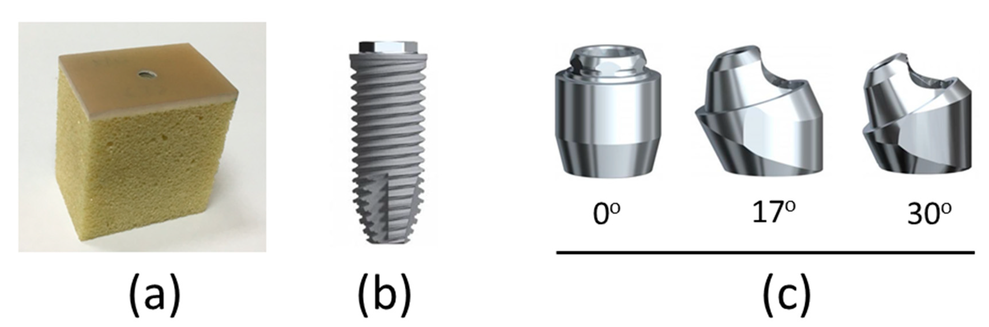
2.1. Preparation of the Artificial Foam Bone Specimen and Dental Implant Components

The artificial foam bone specimens, composed of rigid cellular polyurethane blocks (Sawbones, Vashon, WA, USA) and representing trabecular bone with an elastic modulus of 137 MPa (model 1522-12), were attached to 2-mm-thick synthetic cortical shells (model 3401-01), with an elastic modulus of 16.7 GPa (Figure 1a). Commercial dental implants (4-mm diameter, 11.5-mm length; Branemark Systems NobelSpeedy Groovy, Nobel Biocare AB, Gothenburg, Sweden) were used in this study (Figure 1b). Multiunit abutments (Nobel Biocare AB) were connected to the implants. Abutments with three different inclinations of 0°, 17° and 30° were used in this study (Figure 1c).

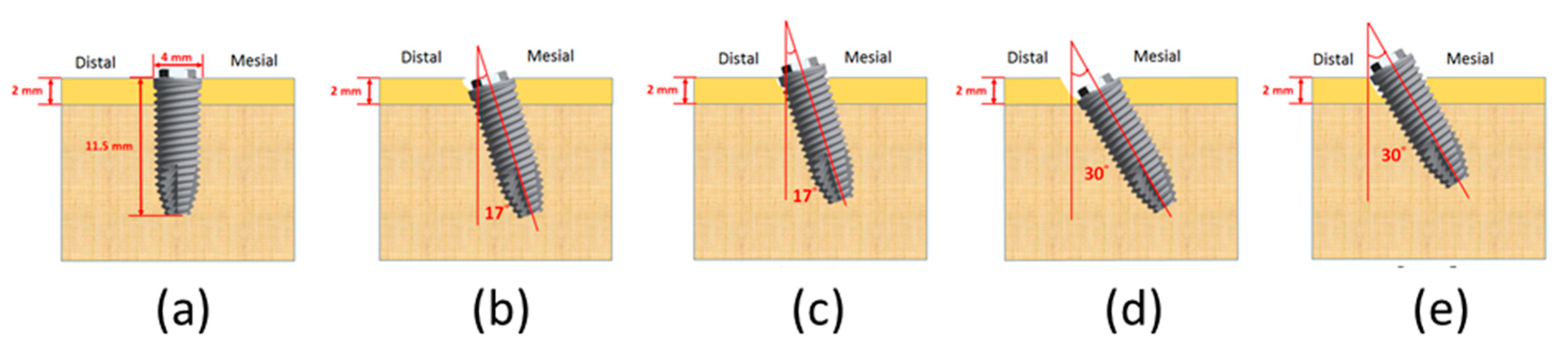
2.2. Insertion of the Dental Implant at Various Angulations and Depths
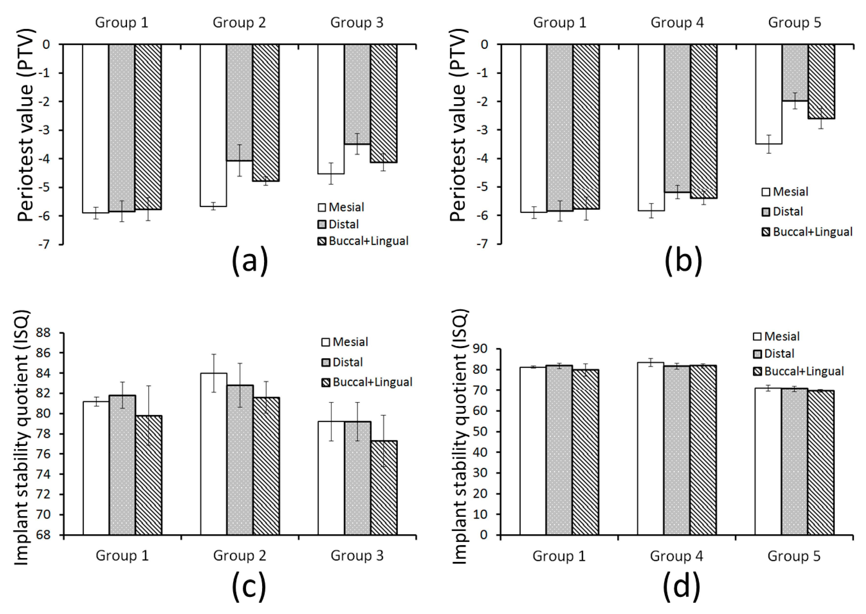
Before inserting the dental implant fixture, pilot holes were drilled into each artificial bone specimen, as per manufacturer instructions. A slope vise was used to precisely control the drilling angulation. Five groups were organized according to angulation and insertion depth (Figure 2), and each group included five specimens:
Group 1:
Vertical implant in an evencrestal position.
Group 2:
17° tilted implant in a distal subcrestal position.
Group 3:
17° tilted implant in a mesial supracrestal position.
Group 4:
30° tilted implant in a distal subcrestal position.
Group 5:
30° tilted implant in a mesial supracrestal position.

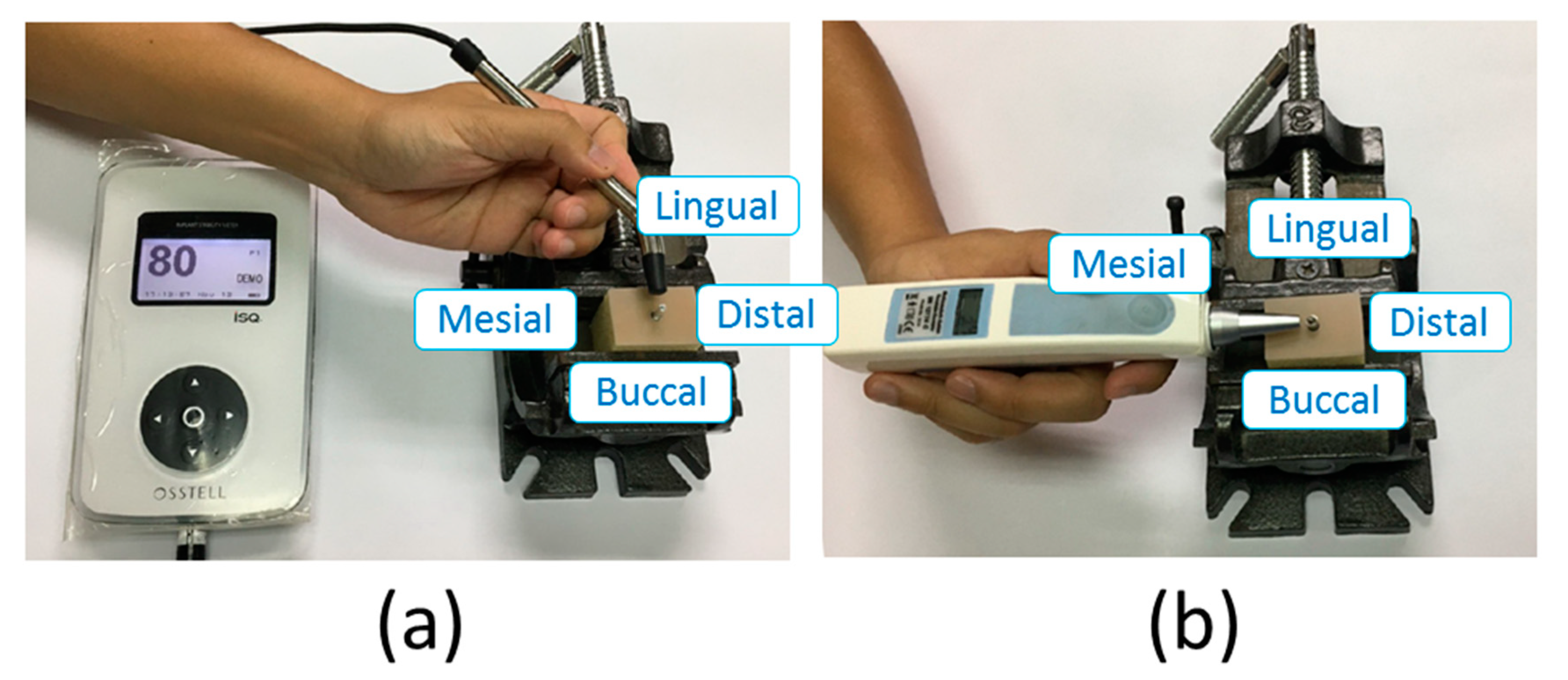
2.3. Measuring the PTV and ISQ

The PTV was determined by connecting multiunit abutments to implants and then measuring the mobility of the implants using the Periotest device (Medizintechnik Gulden, Bensheim, Germany). The abutment screw was inserted to 35 N/cm using a dental implant wrench. Measures along four directions (lingual, buccal, mesial, and distal direction) of the PTV were recorded (Figure 3). The tip of the Periotest device was placed perpendicular to the abutment, at a distance of 2 mm. Moreover, the Osstell ISQ™ (Osstell ISQ, Osstell AB, Gothenborg, Sweden) wireless resonance frequency analyzer was used to measure the ISQ value. The smart peg (Type 1, Osstell AB) was placed on top of the implants. The ISQs along four directions (lingual, buccal, mesial, and distal directions) were also measured.

2.4. Statistical Analysis

All statistical analyses were conducted using SPSS Version 19 (IBM Corporation, Armonk, NY, USA), and the significance level was set to p < 0.05. The following three statistical methods were used to assess the objectives investigated in this study.
(1) To assess the effects of various measurement directions (buccal, lingual, mesial, and distal directions) on the PTV and ISQ of the fixture, a one-way ANOVA was used to analyze whether the results of the ISQ and PTV measurements along the four directions were different, and the Tukey test was used for post hoc pairwise comparison when a difference was discerned. The correlation between ISQ and PTV was then presented using the Pearson correlation coefficient.
(2) To assess effects of various insertion angles (0°, 17° and 30°) on the PTV and ISQ of the fixture, a one-way ANOVA was conducted to determine whether a statistically significant difference existed between the PTVs and ISQs in groups 1, 2 and 4, and the Tukey Test was used for post hoc pairwise comparison when a difference was discovered. The same procedure was applied to compare the differences between groups 1, 3 and 5.
(3) To assess the effects of various insertion methods (evencrestal, subcrestal, and supracrestal positions) on the PTV and ISQ of the fixture, a one-way ANOVA was conducted to determine whether a statistically significant difference existed between the PTVs and ISQs in groups 1, 2 and 4, and the Tukey Test was subsequently used for post hoc pairwise comparison when a difference was discovered. The same procedure was applied to compare the differences between groups 1, 4 and 5.

3. Results

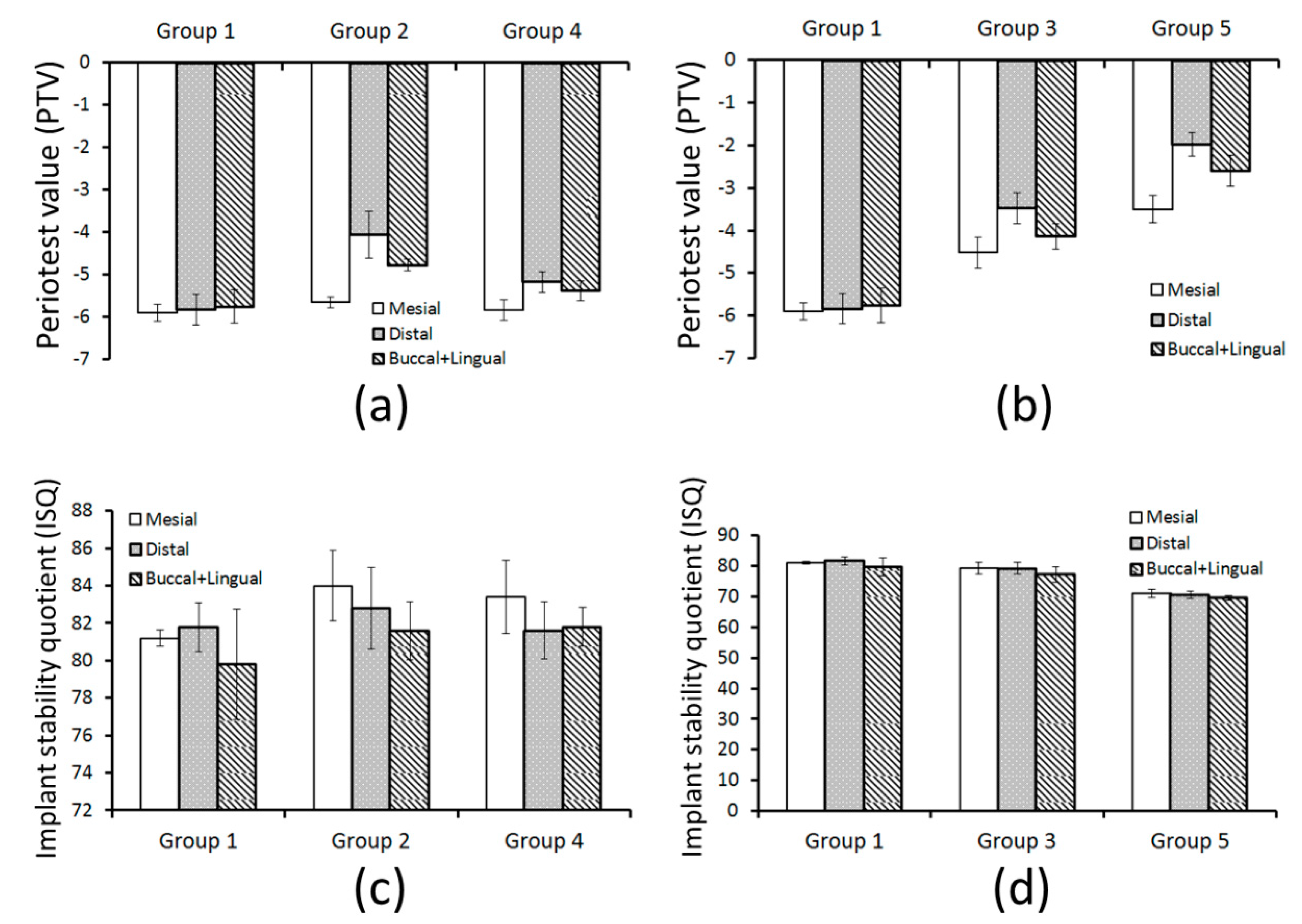
3.1. Differences and Correlations among the Four Measurement Directions of the PTV and ISQ

The average value measured along the four directions represents the ISQ of the dental implant fixture (Table 1), in accordance to the results of one-way ANOVA, revealing no statistically significant difference in the ISQs along the four directions.
PTVs measured from the buccal and lingual directions in each group were compared using t tests. The results indicated no significant difference between PTVs measured along the buccal and lingual directions in the five groups; thus, the mean of the buccal and lingual directions was used as the value. Next, one-way ANOVA was used to compare the PTVs measured along the three groups (Table 2).
PTV was not significantly different along any particular direction in Group 1; therefore, a correlation analysis was performed on the mean PTV and ISQs along the four directions. The results of the Pearson correlation coefficient test indicated a high negative correlation between ISQ and PTV (r = −0.85, p = 0.04; Table 3). In addition, no other obvious correlations among all other combinations in the other four groups were observed.

3.2. Differences of PTV and ISQ among the Three Inserted Angulation

Regarding the effect of the three angles (vertical, 17° and 30° insertion angles) of the implants on the PTV (Figure 4a,b), the groups that had the implants inserted into the distal subcrestal position had no significant difference compared with the PTV along the mesial direction, using the PTV of Group 1 as a reference. The measurement results of the PTV along the distal direction indicated that the 17° tilted implant had a higher PTV than vertical and 30° tilt implants. The PTV of the 30° tilted implant had no significant difference compared with that of the vertical implant. Regarding the mean PTV of the buccal and lingual group, the PTV of the 17° tilted implant was higher than that of the vertical and 30° tilted implants. The PTV of the 30° titled implant exhibited no significant difference compared with that of the vertical implant. In the groups that inserted the fixture into the mesial supracrestal position, the PTV of the 30° titled implants was significantly higher than all the directions of the 17° titled implants and vertical implants.
Regarding the effect of the tilt angles (vertical, 17° and 30°) on the ISQ of the fixture (Figure 4c,d), the mean along the four directions was used to indicate the effect of insertion angle on ISQ because the ISQ was not affected by measurement direction in all groups. Using the ISQ of Group 1 as the reference, for the groups that had the fixtures inserted into the distal subcrestal position, no significant difference was evident among three groups. For the groups that had the fixtures inserted into the mesial supracrestal position, vertical insertion and the 17° tilted angle did not exhibit any significant difference in their ISQs. However, the ISQs of the vertically inserted and 17° titled implants were significantly higher than the ISQ of the 30° titled implant.

3.3. Differences of PTV and ISQ among the Three Inserted Depth

Regarding the effect of insertion depth on the fixtures on the PTV (Figure 5a,b), the implant PTVs measured along the mesial direction for the groups with 17° tilted implants were examined using the PTV of Group 1 as a reference. The PTV of the vertical evencrestal position and distal subcrestal position did not differ significantly; however, the PTVs were both significantly lower than those at the mesial supracrestal position. For the measurement results of the PTV at the distal direction, the implant at the vertical evencrestal position exhibited a lower PTV compared with that at the distal subcrestal position and mesial supracrestal position. The implant PTV between the distal subcrestal position and mesial supracrestal position exhibited no significant difference. Regarding the mean of the buccal and lingual directions group, the implant PTV at the mesial supracrestal position was higher than that at the distal subcrestal position and vertical evencrestal position. The distal subcrestal position also yielded a higher implant PTV compared with the vertical evencrestal position. Similarly, the implant PTV of the groups with 30° tilted implants measured along the mesial direction were examined using the PTV of Group 1 as a reference. Group 1 and Group 4 did not exhibit any significant difference in their implant PTVs from all directions, whereas both Groups 1 and 4 yielded greater PTVs than Group 5 from all directions.
Regarding the effect of three insertion depths of the implant on ISQ (Figure 5c,d), because the ISQ was not affected by measurement direction, the mean of the four directions was used to represent the effect of insertion angle on ISQ. Using the ISQ in Group 1 as a reference, for the groups with 17° tilted implants, the implant ISQ at the distal subcrestal position was significantly higher than that at the mesial supracrestal position. The ISQ at the vertical evencrestal position displayed no significant difference with those in Groups 2 and 3. Similarly, using the ISQ in Group 1 as a reference, for the groups with 30° tilted implants, the distal subcrestal position and vertical evencrestal position displayed no significant difference in their implant ISQ. However, the implant ISQ at the distal subcrestal position and vertical evencrestal position was significantly higher than that at the mesial supracrestal position.

4. Discussion

Dental implant is one of the major treatments for patients with full or partial edentulism, and the degree of primary stability affects the survival rate of the dental implant. Numerous studies have measured the primary stability of vertical implants [24,25,26,27]; however, studies on the primary stability of tilted implants are rare. In fact, this study was the first to explore the effect of insertion angle and insertion depth of tilted implants, as well as the effect of measurement direction on the PTV and ISQ of implant primary stability, aiming to provide a reference for clinical dentists during implant operations. When the dental implant fixtures were inserted at various tilt angles, the PTV of the primary implant stability depended on the measurement direction. Therefore, the tapping direction for the PTV should be carefully selected during the clinical test.
In 1999, Mattsson et al. [28] used tilted implants to treat patients with severe atrophic edentulous maxillae, noting that tilting the implants at 30°–40° could prevent bone grafting. Rosén et al. [29] used 4–6 tilted implants to treat 33 patients with severe atrophic edentulous maxillae, and achieved a 97% success rate in long-term follow-up (average of 10 years). Pancko et al. [30] used tilted implants to treat patients with severe alveolar atrophy because of edentulism in the posterior mandible, achieving a 99% success rate. The use of all-on-4 for complete denture fabrication had become increasingly popular for edentulism treatment. This surgical technique was proposed by Malo et al. [18] and mainly involved inserting two dental implant fixtures axially in the anterior region and two tilted implant fixtures in the posterior region. Malo noted that all-on-4 had four advantages: avoidance of bone grafting, a high success rate, superior hygienic maintenance, and immediate loading. Malo et al. [31] evaluated the success rate of patients who underwent the all-on-4 treatment; 324 patients underwent mandibular reconstruction using 1296 fixtures, and the success rate of the fixtures 7 years after the surgery was 96.6%. Because of the high surgical success rate of all-on-4, an increasing number of dental implant fixtures had been inserted into the jawbone at titled angles.
In laboratories, acquiring fresh human jawbones with similar bone quality and quantity is extremely difficult [32,33]. Scholars had used fresh animal bones for biomechanical experiments with artificial implants [20,34,35]; however, the material properties of each bone specimen were different. Some scholars [26,32,36,37], as well as the ASTM regulations [38], contended that artificial foam bones were suitable materials for evaluating the biomechanical properties of artificial implants. Therefore, this study referred to experiments by scholars [26,36,37] and conducted experiments that used cellular polyurethane blocks. This study referred to the literature and selected the 137-MPa elastic modulus for artificial cancellous bone and artificial cortical bones with 2-mm thickness [36,37,39]. The temperature during inserting implant was not measured due to the artifial bone specimens used in this study. However, several researchers indicated that the high temperature during drilling or inserting implant may induce surrounding bone thermonecrosis [40,41]. The implant stability might be affected by the necrosis bone.
The three common methods for the clinical measurement of the primary stability of implants [8,36,42] are ISQ, PTV, and ITV. ITV can only be measured after inserting the dental implant fixture into the jawbone, whereas PTV and ISQ can be measured repeatedly; thus, PTV and ISQ are frequently used for the clinical assessment of changes in the fixture stability of patients during follow-up visits [43]. In addition, Han et al. [44] noted that the primary stability measured using ISQ and PTV should have more clinical implications compared to ITV and removal torque value. Aparicio et al. [45] contended that using only one approach to measure primary stability is not sufficient; therefore, this study used both ISQ and PTV to assess the primary stability of dental implants.
Many dental implant fixture manufacturers had launched tilted implant products [17]. Chrcanovic et al. analyzed numerous clinical studies on tilted implants before 2014 and discovered that the angles ranged from 11° to 45° [17]. In addition, scholars had noted in many clinical studies on maxilla and mandibles [18,23,31,46,47] that the angle of the tilted implants was less than 45°. Therefore, two angled abutments (17° and 30°) were chosen for the experiment in this study. The adopted insertion depth was based on the clinical study of Rojas-Vizcaya and Zadeh [22], which mentioned two levels of insertion depth for titled implants, namely the distal subcrestal position and the mesial supracrestal position. Inserting implants at the distal subcrestal position may lead to deep pockets, whereas inserting them at the mesial supracrestal position may cause mucosal recession and exposure of the textured portions of implants. Therefore, the evencrestal position, distal subcrestal position, and mesial supracrestal position were chosen as the insertion depths for the experiment in this study.
Investigation of the correlation between the PTV and ISQ of vertical inserted fixtures (Group 1) revealed that PTV and ISQ were highly correlated (r = −0.85, p = 0.04). This also confirmed the results of the experiments by Lachmann et al. [48] and Han et al. [44], noting that the PTV and ISQ of inserted fixtures displayed a highly negative correlation (r = −0.8–0.9 and r = −0.97, respectively).
The experimental results demonstrated that ISQ for both tilted implants (17° and 30°) and vertical implants measured in each test direction exhibited no significant difference (Table 1). This may be because the tilted implants have had different contact areas with the superior artificial cortical bone at different depths, but their contact areas with the inner artificial cancellous bones were almost the same. Some studies had noted that ISQ was less affected by the superior cortical bone and was more affected by the inner cancellous bone [36]. Therefore, ISQ exhibited no significant differences in each test direction, regardless of the degree of implant tilt. Additionally, in a clinical report from 2014, Elramady et al. [49] conducted primary stability tests on tilted implants in patients with edentulism using various directions, and their results also indicated no significant difference between the ISQ of the buccal–lingual and mesial–distal directions.
Regarding the PTV measurement results of tilted implants along all directions, the PTV measured from the distal side was significantly smaller than that measured from the mesial side, in both 17° and 30° tilted implants (Table 2, Figure 4 and Figure 5). The reason may be that more skin on the mesial side was removed following the dental implant manufacturer’s suggestion for pilot holes, resulting in a gap between the platform and the bone. When the dental implant fixture was tapped from the distal direction (from the distal side to the mesial side), it thus fell toward the mesial side. The gap at the mesial side caused a longer rebound time for the fixture, resulting in a greater PTV value, and thus, a less stable result, as indicated by the PTV of the primary stability.
Regarding the effect of tilted implants on the PTV, Aparicio et al. [45] performed 59 vertical implants and 41 fixture placements at 30° on patients with edentulism and compared the results that indicated that tilted implants yielded a greater PTV than vertical implants. In other words, the stability of tilted implants was inferior to that of vertical implants. However, some studies have noted that tilted implants do not affect stability. Wentaschek et al. [43] inserted 60 implants (40 vertical implants and 20 tilted implants) into 10 patients with partially edentulous mandibles, and concluded that no significant difference was evident between the ISQ and PTV of the tilted and axial implants. The main reason for this may be the inconsistent size of the dental implant fixtures.
Regarding the effects of insertion depth on the primary stability of dental implant fixtures, this study discovered that supracrestally placed implants exhibited lower stability than subcrestally placed implants, a result similar to those in other studies. Bergkvist et al. [50] and Cannizzaro et al. [51] also mentioned that inserting the implants at 1 mm–2 mm toward the subcrestal position yielded a more stable implant ISQ. However, some studies contended that the insertion depth of the implant does not affect primary stability. Romanos et al. [52] noted in a clinical study that the PTVs of crestally and subcrestally placed implants were −1.77 ± 3.26 and 1.77 ± 3.57, respectively, and neither exhibited any significant difference.
As for the effect of fixture-abutment connection type on the initial stability of the dental implant, da Costa Valente et al. [53] indicated that the ISQ did not reveal statistically significant differences between the dental implant with external and internal hexagon connection. In this study, the external hexagon implants were selected due to this type is the most common used for all-on-4 treatment. However, the effect of insertion angles and depths of the internal hexagon dental implants on the initial stability should be further investigated.
This study has several limitations. First, because fresh human jawbone with consistent bone quality is difficult to obtain, artificial bones were selected for the experiment in this study. Only one degree of artificial bone quality was simulated in this study, and the heterogeneous features of real human bones were thus not simulated. Second, the heating effect during drilling or inserting the implant was ignored in this study, due to the use of artificial bone in this study. Third, only one size of fixture was simulated in this study, and the experiment was performed using only three levels of insertion depth and angle degrees. Fourth, only two primary stability indicators of the fixture were measured in this study, and the stress and strain distribution on the marginal bone were not considered.

5. Conclusions

Porous artificial foam bones were used in this study to determine the effects of insertion angle and depth on the primary stability of dental implant fixtures. The findings of this study are as follows:
(1) Correlation between the PTV and ISQ of fixture: the two primary stability indicators displayed a highly negative correlation when the fixture was inserted vertically.
(2) Effect of insertion angle on the primary stability of fixtures: when the fixture was inserted at the mesial supracrestal position, the greater tilt angle resulted in less stability (a greater PTV or lower ISQ). When the fixture was inserted at the distal subcrestal position, its primary stability was less affected by the insertion angle.
(3) Effect of insertion depth on the primary stability of fixtures: inserting a vertical implant at the evencrestal position resulted in greater stability (lower PTV) than at the distal position and mesial supracrestal position. Inserting a 17°- or 30°-tilted implant at the distal subcrestal position yielded greater stability (a lower PTV and greater ISQ) than at the mesial supracrestal position.
(4) The ISQ of the dental implant fixtures was not affected by the measurement direction when a tilted implant was inserted; however, PTV was affected by the measurement direction.

Author Contributions

Concept/Design: H.-C.W. and J.-T.H. Data analysis: H.-C.W., M.-T.T., and J.-T.H. Drafting article: H.-C.W., M.-T.T., and J.-T.H. Funding secured by J.-T.H. Data collection: H.-C.W. and J.-T.H. All authors have read and agreed to the published version of the manuscript.

Funding

P This study was supported by the Ministry of Science and Technology, Taiwan (MOST 108-2813-C-039-015-E) and China Medical University, Taiwan (CMU 107-S-09).

Acknowledgments

Not applicable.

Conflicts of Interest

The authors declare no conflict of interest.

References

  1. Bouchard, P.; Renouard, F.; Bourgeois, D.; Fromentin, O.; Jeanneret, M.; Beresniak, A. Cost-effectiveness modeling of dental implant vs. bridge. Clin. Oral Implant. Res. 2009, 20, 583–587. [Google Scholar] [CrossRef]
  2. Branemark, P.-I. Osseointegrated implants in the treatment of the edentulous jaw. Experience from a 10-year period. Scand. J. Plast. Reconstr. Surg. Suppl. 1977, 2, 16. [Google Scholar]
  3. Hebel, K.; Gajjar, R.; Hofstede, T. Single-tooth replacement: Bridge vs. implant-supported restoration. J. Can. Dent. Assoc. 2000, 66, 435–438. [Google Scholar]
  4. Javed, F.; Ahmed, H.B.; Crespi, R.; Romanos, G.E. Role of primary stability for successful osseointegration of dental implants: Factors of influence and evaluation. Interv. Med. Appl. Sci. 2013, 5, 162–167. [Google Scholar] [CrossRef]
  5. Lioubavina-Hack, N.; Lang, N.P.; Karring, T. Significance of primary stability for osseointegration of dental implants. Clin. Oral Implant. Res. 2006, 17, 244–250. [Google Scholar] [CrossRef]
  6. Javed, F.; Romanos, G.E. The role of primary stability for successful immediate loading of dental implants. A literature review. J. Dent. 2010, 38, 612–620. [Google Scholar] [CrossRef]
  7. Atsumi, M.; Park, S.-H.; Wang, H.-L. Methods used to assess implant stability: Current status. Int. J. Oral Maxillofac. Implant. 2007, 22, 743–754. [Google Scholar]
  8. Mistry, G.; Shetty, O.; Shetty, S.; Singh, R.D. Measuring implant stability: A review of different methods. J. Dent. Implant. 2014, 4, 165. [Google Scholar] [CrossRef]
  9. Atwood, D.A. Reduction of residual ridges: A major oral disease entity. J. Prosthet. Dent. 1971, 26, 266–279. [Google Scholar] [CrossRef]
  10. Barão, V.; Delben, J.; Lima, J.; Cabral, T.; Assunção, W.G. Comparison of different designs of implant-retained overdentures and fixed full-arch implant-supported prosthesis on stress distribution in edentulous mandible–a computed tomography-based three-dimensional finite element analysis. J. Biomech. 2013, 46, 1312–1320. [Google Scholar] [CrossRef] [Green Version]
  11. Chen, J.; Ahmad, R.; Suenaga, H.; Li, W.; Swain, M.; Li, Q. A comparative study on complete and implant retained denture treatments–a biomechanics perspective. J. Biomech. 2015, 48, 512–519. [Google Scholar] [CrossRef]
  12. De Vico, G.; Bonino, M.; Spinelli, D.; Schiavetti, R.; Sannino, G.; Pozzi, A.; Ottria, L. Rationale for tilted implants: FEA considerations and clinical reports. ORAL Implantol. 2011, 4, 23. [Google Scholar]
  13. Sivolella, S.; Stellini, E.; Testori, T.; Di Fiore, A.; Berengo, M.; Lops, D. Splinted and unsplinted short implants in mandibles: A retrospective evaluation with 5 to 16 years of follow-up. J. Periodontol. 2013, 84, 502–512. [Google Scholar] [CrossRef]
  14. Silva, L.D.; De Lima, V.; Faverani, L.; De Mendonca, M.; Okamoto, R.; Pellizzer, E. Maxillary sinus lift surgery—With or without graft material? A systematic review. Int. J. Oral Maxillofac. Surg. 2016, 45, 1570–1576. [Google Scholar] [CrossRef] [Green Version]
  15. Aparicio, C.; Manresa, C.; Francisco, K.; Claros, P.; Alández, J.; González-Martín, O.; Albrektsson, T. Zygomatic implants: Indications, techniques and outcomes, and the zygomatic success code. Periodontol. 2000 2014, 66, 41–58. [Google Scholar] [CrossRef]
  16. Krekmanov, L.; Kahn, M.; Rangert, B.; Lindström, H. Tilting of posterior mandibular and maxillary implants for improved prosthesis support. Int. J. Oral Maxillofac. Implant. 2000, 15, 405–414. [Google Scholar]
  17. Chrcanovic, B.R.; Albrektsson, T.; Wennerberg, A. Tilted versus axially placed dental implants: A meta-analysis. J. Dent. 2015, 43, 149–170. [Google Scholar] [CrossRef]
  18. Maló, P.; Rangert, B.; Nobre, M. “All-on-Four” immediate-function concept with Brånemark System® implants for completely edentulous mandibles: A retrospective clinical study. Clin. Implant Dent. Relat. Res. 2003, 5, 2–9. [Google Scholar] [CrossRef]
  19. Degidi, M.; Perrotti, V.; Shibli, J.A.; Novaes, A.B.; Piattelli, A.; Iezzi, G. Equicrestal and subcrestal dental implants: A histologic and histomorphometric evaluation of nine retrieved human implants. J. Periodontol. 2011, 82, 708–715. [Google Scholar] [CrossRef]
  20. Barros, R.R.; Novaes, A.B., Jr.; Muglia, V.A.; Iezzi, G.; Piattelli, A. Influence of interimplant distances and placement depth on peri-implant bone remodeling of adjacent and immediately loaded Morse cone connection implants: A histomorphometric study in dogs. Clin. Oral Implant. Res. 2010, 21, 371–378. [Google Scholar] [CrossRef]
  21. Todescan, F.F.; Pustiglioni, F.E.; Imbronito, A.V.; Albrektsson, T.; Gioso, M. Influence of the microgap in the peri-implant hard and soft tissues: A histomorphometric study in dogs. Int. J. Oral Maxillofac. Implant. 2002, 17, 467–472. [Google Scholar]
  22. Rojas-Vizcaya, F.; Zadeh, H.H. Minimizing the discrepancy between implant platform and alveolar bone for tilted implants with a sloped implant platform: A clinical report. J. Prosthet. Dent. 2018, 119, 319–324. [Google Scholar] [CrossRef]
  23. Maló, P.; de Araújo Nobre, M.; Lopes, A.; Francischone, C.; Rigolizzo, M. “All-on-4” immediate-function concept for completely edentulous maxillae: A clinical report on the medium (3 years) and long-term (5 years) outcomes. Clin. Implant Dent. Relat. Res. 2012, 14, e139–e150. [Google Scholar] [CrossRef]
  24. Barikani, H.; Rashtak, S.; Akbari, S.; Badri, S.; Daneshparvar, N.; Rokn, A. The effect of implant length and diameter on the primary stability in different bone types. J. Dent. 2013, 10, 449. [Google Scholar]
  25. Hong, J.; Lim, Y.J.; Park, S.O. Quantitative biomechanical analysis of the influence of the cortical bone and implant length on primary stability. Clin. Oral Implant. Res. 2012, 23, 1193–1197. [Google Scholar] [CrossRef]
  26. Huang, H.L.; Chang, Y.Y.; Lin, D.J.; Li, Y.F.; Chen, K.T.; Hsu, J.T. Initial stability and bone strain evaluation of the immediately loaded dental implant: An in vitro model study. Clin. Oral Implant. Res. 2011, 22, 691–698. [Google Scholar] [CrossRef]
  27. Pommer, B.; Hof, M.; Fädler, A.; Gahleitner, A.; Watzek, G.; Watzak, G. Primary implant stability in the atrophic sinus floor of human cadaver maxillae: Impact of residual ridge height, bone density, and implant diameter. Clin. Oral Implant. Res. 2014, 25, e109–e113. [Google Scholar] [CrossRef]
  28. Mattsson, T.; Köndell, P.-Å.; Gynther, G.W.; Fredholm, U.; Bolin, A. Implant treatment without bone grafting in severely resorbed edentulous maxillae. J. Oral Maxillofac. Surg. 1999, 57, 281–287. [Google Scholar] [CrossRef]
  29. Rosén, A.; Gynther, G. Implant treatment without bone grafting in edentulous severely resorbed maxillas: A long-term follow-up study. J. Oral Maxillofac. Surg. 2007, 65, 1010–1016. [Google Scholar] [CrossRef]
  30. Pancko, F.; Dyer, J.; Weisglass, S.; Kraut, R.A. Use of tilted implants in treatment of the atrophic posterior mandible: A preliminary report of a novel approach. J. Oral Maxillofac. Surg. 2010, 68, 407–413. [Google Scholar] [CrossRef]
  31. Maló, P.; de Araújo Nobre, M.; Lopes, A.; Ferro, A.; Gravito, I. A ll-on-4® Treatment Concept for the Rehabilitation of the Completely Edentulous Mandible: A 7-Year Clinical and 5-Year Radiographic Retrospective Case Series with Risk Assessment for Implant Failure and Marginal Bone Level. Clin. Implant Dent. Relat. Res. 2015, 17, e531–e541. [Google Scholar]
  32. Elfar, J.; Stanbury, S.; Menorca, R.M.G.; Reed, J.D. Composite bone models in orthopaedic surgery research and education. J. Am. Acad. Orthop. Surg. 2014, 22, 111. [Google Scholar]
  33. Singh, A.; Sharma, R.; Sharma, R.; Musmade, D. Challenges in cadaver availability for learning and research in medical sciences. Int. J. Med. Clin. Res. 2011, 2, 67–71. [Google Scholar]
  34. Engelke, W.; Decco, O.A.; Cura, A.C.; Maldonado, I.; Crippa, F.G. Influence of implant diameter in the displacement of dental implants in trabecular bovine bone under a static lateral load: Experimental results and computational modeling. Implant Dent. 2014, 23, 635–640. [Google Scholar] [CrossRef]
  35. Manresa, C.; Bosch, M.; Echeverría, J.J. The comparison between implant stability quotient and bone-implant contact revisited: An experiment in B eagle dog. Clin. Oral Implant. Res. 2014, 25, 1213–1221. [Google Scholar] [CrossRef]
  36. Hsu, J.T.; Fuh, L.J.; Tu, M.G.; Li, Y.F.; Chen, K.T.; Huang, H.L. The effects of cortical bone thickness and trabecular bone strength on noninvasive measures of the implant primary stability using synthetic bone models. Clin. Implant Dent. Relat. Res. 2013, 15, 251–261. [Google Scholar] [CrossRef]
  37. Liu, C.; Tsai, M.-T.; Huang, H.-L.; Chen, M.Y.-C.; Hsu, J.-T.; Su, K.-C.; Chang, C.-H.; Wu, A.Y.-J. Relation between insertion torque and bone–implant contact percentage: An artificial bone study. Clin. Oral Investig. 2012, 16, 1679–1684. [Google Scholar] [CrossRef]
  38. American Society for Testing and Materials A. ASTM F1839-08 Standard Specification for Rigid Polyurethane Foam for Use as a Standard Material for Testing Orthopedic Devices and Instruments. Available online: https://www.techstreet.com/standards/astm-f1839-08-2016?product_id=1931970 (accessed on 29 March 2020).
  39. Miyamoto, I.; Tsuboi, Y.; Wada, E.; Suwa, H.; Iizuka, T. Influence of cortical bone thickness and implant length on implant stability at the time of surgery—Clinical, prospective, biomechanical, and imaging study. Bone 2005, 37, 776–780. [Google Scholar] [CrossRef]
  40. Di Fiore, A.; Sivolella, S.; Stocco, E.; Favero, V.; Stellini, E. Experimental Analysis of Temperature Differences During Implant Site Preparation: Continuous Drilling Technique Versus Intermittent Drilling Technique. J. Oral Implantol. 2018, 44, 46–50. [Google Scholar] [CrossRef]
  41. Lucchiari, N.; Frigo, A.C.; Stellini, E.; Coppe, M.; Berengo, M.; Bacci, C. In vitro assessment with the infrared thermometer of temperature differences generated during implant site preparation: The traditional technique versus the single-drill technique. Clin. Implant Dent. Relat. Res. 2016, 18, 182–191. [Google Scholar] [CrossRef]
  42. Swami, V.; Vijayaraghavan, V.; Swami, V. Current trends to measure implant stability. J. Indian Prosthodont. Soc. 2016, 16, 124. [Google Scholar] [CrossRef] [PubMed]
  43. Wentaschek, S.; Hartmann, S.; Walter, C.; Wagner, W. Six-implant-supported immediate fixed rehabilitation of atrophic edentulous maxillae with tilted distal implants. Int. J. Implant Dent. 2017, 3, 35. [Google Scholar] [CrossRef] [Green Version]
  44. Han, H.-C.; Lim, H.-C.; Hong, J.-Y.; Ahn, S.-J.; Han, J.-Y.; Shin, S.-I.; Chung, J.-H.; Herr, Y.; Shin, S.-Y. Primary implant stability in a bone model simulating clinical situations for the posterior maxilla: An in vitro study. J. Periodontal Implant Sci. 2016, 46, 254–265. [Google Scholar] [CrossRef] [PubMed] [Green Version]
  45. Aparicio, C.; Perales, P.; Rangert, B. Tilted implants as an alternative to maxillary sinus grafting: A clinical, radiologic, and periotest study. Clin. Implant Dent. Relat. Res. 2001, 3, 39–49. [Google Scholar] [CrossRef]
  46. Di, P.; Lin, Y.; Li, J.-H.; Luo, J.; Qiu, L.-X.; Chen, B.; Cui, H.-Y. The All-on-Four implant therapy protocol in the management of edentulous Chinese patients. Int. J. Prosthodont. 2013, 26, 509–516. [Google Scholar] [CrossRef] [Green Version]
  47. Lopes, A.; Maló, P.; de Araujo Nobre, M.; Sanchez-Fernández, E. The N obel G uide® A ll-on-4® Treatment Concept for Rehabilitation of Edentulous Jaws: A Prospective Report on Medium-and Long-Term Outcomes. Clin. Implant Dent. Relat. Res. 2015, 17, e406–e416. [Google Scholar] [CrossRef]
  48. Lachmann, S.; Jäger, B.; Axmann, D.; Gomez-Roman, G.; Groten, M.; Weber, H. Resonance frequency analysis and damping capacity assessment: Part 1: An in vitro study on measurement reliability and a method of comparison in the determination of primary dental implant stability. Clin. Oral Implant. Res. 2006, 17, 75–79. [Google Scholar] [CrossRef]
  49. Elramady, M.M.; Sayed, A.M.; Awadalla, M.A.; Mohamed, F.S.; Nassef, T.M. Measuring primary stability for the inclined implants retaining mandibular overdenture using resonance frequency. In Proceedings of the 2014 Cairo International Biomedical Engineering Conference (CIBEC), Giza, Egypt, 11–13 December 2014; pp. 83–86. [Google Scholar]
  50. Bergkvist, G.; Nilner, K.; Sahlholm, S.; Karlsson, U.; Lindh, C. Immediate loading of implants in the edentulous maxilla: Use of an interim fixed prosthesis followed by a permanent fixed prosthesis: A 32-month prospective radiological and clinical study. Clin. Implant Dent. Relat. Res. 2009, 11, 1–10. [Google Scholar] [CrossRef]
  51. Cannizzaro, G.; Leone, M.; Consolo, U.; Ferri, V.; Esposito, M. Immediate functional loading of implants placed with flapless surgery versus conventional implants in partially edentulous patients: A 3-year randomized controlled clinical trial. Int. J. Oral Maxillofac. Implant. 2008, 23, 867–875. [Google Scholar]
  52. Romanos, G.E.; Aydin, E.; Gaertner, K.; Nentwig, G.H. Long-term results after subcrestal or crestal placement of delayed loaded implants. Clin. Implant Dent. Relat. Res. 2015, 17, 133–141. [Google Scholar] [CrossRef]
  53. Da Costa Valente, M.L.; de Castro, D.T.; Shimano, A.C.; Lepri, C.P.; dos Reis, A.C. Analysis of the influence of implant shape on primary stability using the correlation of multiple methods. Clin. Oral Investig. 2015, 19, 1861–1866. [Google Scholar] [CrossRef] [PubMed]
Figure 1. (a) Artificial foam bone specimen; (b) NobelSpeedy Groovy; (c) Multiunit abutments.
Applsci 10 03112 g001
Figure 2. (a) Group 1: vertical implant in an evencrestal position; (b) Group 2: 17° tilted implant in a distal subcrestal position; (c) Group 3: 17° tilted implant in a mesial supracrestal position; (d) Group 4: 30° tilted implant in a distal subcrestal position; (e) Group 5: 30° tilted implant in a mesial supracrestal position.
Applsci 10 03112 g002
Figure 3. (a) Measuring the implant stability quotient (ISQ) by Osstell ISQ device; (b) measurement of the Periotest value (PTV) by Periotest device.
Applsci 10 03112 g003
Figure 4. PTV (a,b) and ISQ (c,d) measurements of Groups 1, 2 and 4 and Groups 1, 3 and 5.
Applsci 10 03112 g004
Figure 5. PTV (a,b) and ISQ (c,d) measurements of Groups 1, 2 and 3 and Groups 1, 4 and 5.
Applsci 10 03112 g005
Table 1. ISQ in the five groups.
GroupImplant Angulations and Inserted TypeMeasured Direction (Mean ± SD)p Value *Mean of the Four Direction
MesialDistalBuccalLingual
1vertical implant with evencrestal position81.2 ± 0.44781.8 ± 1.30979.8 ± 2.9579.8 ± 2.950.31480.7 ± 1.91
217° tilted implant with distal subcrestal position84 ± 1.8782.8 ± 2.16881.4 ± 1.34181.8 ± 1.7890.14982.5 ± 1.79
317° tilted implant with mesial supracrestal position79.2 ± 1.92479.2 ± 1.92477.4 ± 2.06877.2 ± 2.490.34678.3 ± 2.24
430° tilted implant with distal subcrestal position83.4 ± 1.94981.6 ± 1.51782.2 ± 1.30382.6 ± 1.3410.46882.5 ± 1.6
530° tilted implant with mesial supracrestal position71 ± 1.41470.6 ± 1.29170.0 ± 0.070.2 ± 0.4470.20770.6 ± 0.86
* One-way ANOVA was used on values measured along four directions (mesial, distal, buccal, and lingual directions).
Table 2. PTV in the five groups.
GroupImplant Angulations and Inserted TypeMeasured Direction (Mean ± SD)p Value *
Mesial (Mean ± SD) Distal (Mean ± SD)Mean (B&L) (Mean ± SD)
1vertical implant with evencrestal position−5.9 ± 0.2 a−5.84 ± 0.358 a−5.76 ± 0.397 a0.922
217° tilted implant with distal subcrestal position−5.66 ± 0.134 a−4.06 ± 0.55 b−4.78 ± 0.144 c<0.001
317° tilted implant with mesial supracrestal position−4.52 ± 0.37 a−3.48 ± 0.35 b−4.13 ± 0.297 a0.001
430° tilted implant with distal subcrestal position−5.86 ± 0.251 a−5.18 ± 0.239 b−5.39 ± 0.238 b0.004
530° tilted implant with mesial supracrestal position−3.5 ± 0.316 a−1.98 ± 0.277 b−2.6 ± 0.354 c<0.001
* One-way ANOVA was used on values measured along three directions (mesial, distal, and the mean of buccal and lingual directions); Post hoc pairwise comparisons were conducted using Tukey tests; means with the same letter (a or b or c) were not significantly different at the 0.05 level.
Table 3. Correlations between the ISQ and the PTV along various measurement directions in the five groups.
GroupImplant Angulations and Inserted TypeCorrelation (r and p) between the ISQ and Different Measurement Direction of PTV
Mean of Four DirectionMesial DirectionDistal DirectionMean of Buccal and Lingual Directions
1vertical implant with evencrestal positionr = −0.85
(p = 0.04)
217° tilted implant with distal subcrestal position r = 0.384
(p = 0.523)
r = 0.178
(p = 0.775)
r = −0.949
(p = 0.014)
317° tilted implant with mesial supracrestal position r = −0.673
(p = 0.213)
r = −0.142
(p = 0.82)
r = −0.345
(p = 0.57)
430° tilted implant with distal subcrestal position r = −0.968
(p = 0.007)
r = −0.711
(p = 0.178)
r = −0.636
(p = 0.249)
530° tilted implant with mesial supracrestal position r = −0.200
(p = 0.747)
r = 0.285
(p = 0.642)
r = −0.292
(p = 0.634)

