1. Introduction
Stand-up paddle boarding (SUP) is a Hawaiian paddle sport derived from surfing. Unlike traditional surfing, during which participants sit on a board until a wave arrives, SUP requires that boarders stand on a board and use a paddle to push themselves on water. The variations of SUP include flat-water paddling for outdoor recreation, fitness, or sightseeing, paddle-board racing, and paddle-board yoga. By 2003, the Encyclopedia of Surfing had never reported on SUP [
1]; however, a few years later, according to the SUP industry statistics, the sales revenue of stand-up paddle boards exceeded US
$720 million [
2]. SUP has become the world’s fastest growing water leisure activity [
3,
4]. This water activity can be performed in different environments, such as calm lakes, rivers, or open oceans, and people typically participate in SUP for one or more hours. Participants must learn how to use the power of various body parts to paddle in different environments [
5] and maintain dynamic stability at all times. To maintain ideal paddle posture, muscles in trunks, upper limbs, and buttocks and knee and ankle joints are activated and coordinated throughout movement and constantly stimulate balance perceptual receptors to maintain stable paddling when standing on an unstable board. Continually and correctly performing a correct paddle action when balancing the interference from the board buoyancy and the blade resistance is challenging. Contracting core muscles is necessary to establish a stable basis for paddle movements that require constant muscle activation and sufficient strength to maintain long-term activation. The goal of SUP is using a kinetic train to transfer the core strength to upper limbs to enhance propulsion power.
Many websites and reports claim that long-term participation in SUP can improve muscle strength, fitness, core muscle stability, and balance as well as reducing lower back pain. Although SUP is also generally regarded as an effective full-body workout, few studies are in progress, and little scientific evidence can confirm these benefits. Studies related to SUP have included only research on topics such as sports injury epidemiology [
6,
7,
8], body composition analysis, and respiratory metabolism [
9,
10,
11,
12]. Other studies have analyzed the effectiveness of exercise training intervention [
13,
14]. One biomechanics study [
15] used surface electromyography (EMG) to observe muscle activity during SUP on a training machine and at sea, demonstrating that muscle activation during the water-based test started sooner and was maintained longer than that during the dynamometer test. They concluded that the training effect on water was more beneficial than that on the training machine because of longer myoelectric signal activation [
15]. These studies have mostly focused on professional paddle boarders and SUP competitions rather than recreational paddle boarders or beginners.
During performing long-distance leisure paddling, paddlers must often alternate between kneeling and standing positions in accordance with personal physical conditions. Until now, the biomechanical understanding of paddling in these two postures remains limited. Regardless of whether SUP is considered from the perspective of exercise training or exercise fatigue, understanding the difference between the two postures is necessary. Therefore, this study examined the muscle group activation in various parts of the body by using EMG signals during SUP in kneeling and standing positions.
2. Materials and Methods
2.1. Participants
The study enrolled 16 college students, who received 8 weeks of basic SUP training. Their average age was 23.1 ± 1.8 years. Their average height and weight were 175.22 ± 5.30 cm and 70.83 ± 10.58 kg, respectively. Each participant was informed regarding the testing procedure before a formal test to ensure that they clearly understood the content and purpose of the research before testing. The study was conducted in accordance with the Declaration of Helsinki, and the protocol was approved by the Ethics Committee of Kaohsiung Medical University Chung-Ho Memorial Hospital (KMUHIRB-E(I)-20170042).
2.2. Equipment
2.2.1. SUP Board and Paddle
This study used an inflatable SUP board that was 294.6 cm long, 81.3 cm wide, and 11.5 cm thick and had a weight of 9.4 kg and a maximum load capacity of 95 kg (WILDAIR 11’6", SAFE SUP, Italy). A two-section carbon fiber paddle was used with an adjustable length from 165 to 220 cm, a weight of 610 g, and a paddle area of 580 cm2. For the standing position, the paddle length was adjusted to be 3–4 cm taller than that of the participant in accordance with the recommendation of Laird Hamilton (the pioneer of SUP). For the kneeling position, the paddle was adjusted to the shortest length.
2.2.2. Wireless EMG System (Delsys Trigno)
In this study, the Trigno Wireless Electromyography System (Delsys Inc., Natick, Massachusetts, USA) was used to collect data with a sampling frequency of 2000 Hz. In addition, a Trigno wireless sensor’s integrated three-axis accelerometer was used to segment the paddling stages. An EMG system was used on the following muscles on the dominant side of the body: the upper-body muscle group included the pectoralis major, latissimus dorsi, lower trapezius, biceps brachii, triceps brachii, and wrist flexor and extensor; the trunk and lower body muscles included the rectus abdominis, external oblique muscle, erector spinae, multifidus, gluteus maximus, rectus femoris, biceps femoris, tibialis anterior, and gastrocnemius. The EMG sensors were placed along the longitudinal midline of the target muscle in accordance with the recommendations of the Surface Electromyography for the Noninvasive Assessment of Muscles project (SENIAM) [
16].
2.3. Procedure
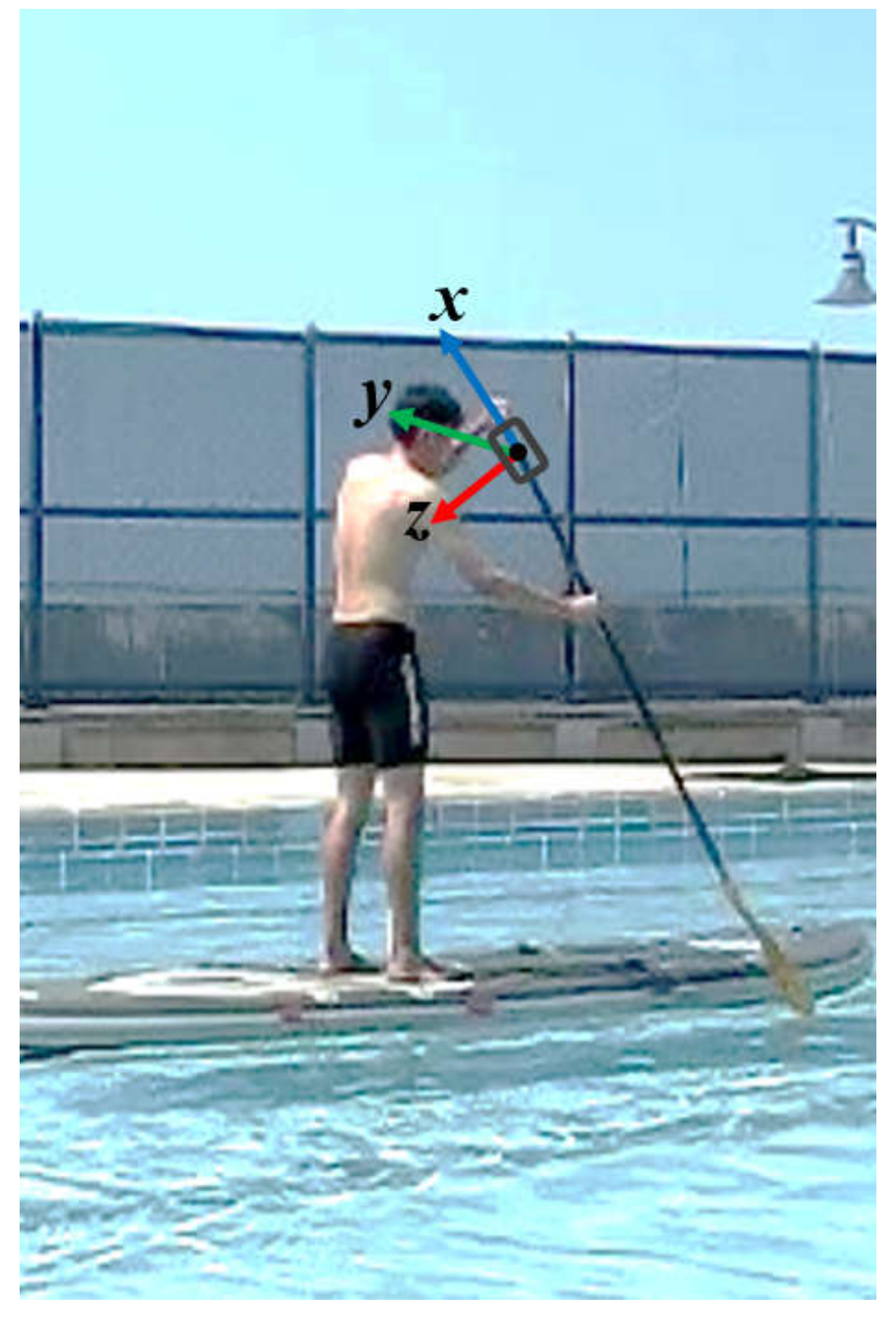
Before starting the experiment, participants shaved body hair if necessary and lightly abraded their skin to provide low noise levels for EMG recordings. Wireless EMG sensors were then fixed longitudinally over the muscle bellies and wrapped with a 3M Tagaderm film (3M Deutschland GmbH) for waterproofing. In addition, the accelerometer was placed 5 cm below the handle of the paddle. The positive direction of the acceleration x-axis was upward, the y-axis was toward the inside of the paddle, and the z-axis was toward the rear (see
Figure 1). This setup was used to detect four stroke phases for signal segmentation.
The testing site was an outdoor swimming pool that was 25 m long. To maintain the forward movement when paddling, participants alternated sides after three strokes. Each participant paddled back and forth twice in a high kneeling position and a standing position in a random order. Participants were instructed to paddle at a comfortable speed to simulate a real paddling situation. Generally, data for 5–6 strokes could be recorded in a 25 m swimming pool. Data in the acceleration and deceleration periods were discarded. Finally, only one data set captured, when the participants moved just halfway across the pool, was adopted for analysis. A total of four trips contained four records.
2.4. Data Analysis
First, one SUP stroke movement was separated into four phases: catch, pull, exit, and recovery. “Catch” refers to the process of placing the paddle blade in water. The “pull” phase is the process, in which the blade is completely immersed in water and is swung backward to generate the forward power [
17]. In the “exit” and “recovery” phases, the blade is pulled out of water and returned to the starting position before starting the next catch phase. In this study, accelerometer data were used to segment the four phases (exit, recovery, catch, and pull) during one stroke. For example, we analyzed stroke data for right-handed participants when they paddled on the right side. At this moment, they placed their left hands (grip hands) on top of the paddle and held a shaft with their right hand to provide propulsion force. Data were treated as a cycle from the beginning of the exit period to the end of the next pull period. The time for one paddle cycle was normalized to 0%–100%. The EMG sensors received raw EMG data and first removed frequencies less than 3 Hz and greater than 1000 Hz. After signal rectification, a 20–450 Hz bandpass filter and a 60 Hz notch filter were used. Finally, a root mean square (RMS) envelope with a window of 0.125 seconds was used. The average and maximum EMG activation values (μV) for each muscle were then calculated. The time series analysis of each muscle activation was based on 50% of the peak activation value. Here, the same peak value was used for kneeling and standing. The period greater than 50% was defined as the main activation period, and that less than 50% was the nonactivation period. The starting and termination of activation for each muscle group were defined using the starting and ending times of the main activation period, respectively, and all muscle activation and termination times were presented as a percentage of the paddle cycle (% paddle cycle).
2.5. Statistical Analyses
The SPSS statistical software version 20.0 (IBM Corp., Armonk, NY, USA) was used for statistical analyses. A paired t test was used to determine differences in the average and maximum EMG activation values of each muscle group as well as the starting and termination activation times between the standing and kneeling positions. The statistical significance was set to p < 0.05.
3. Results
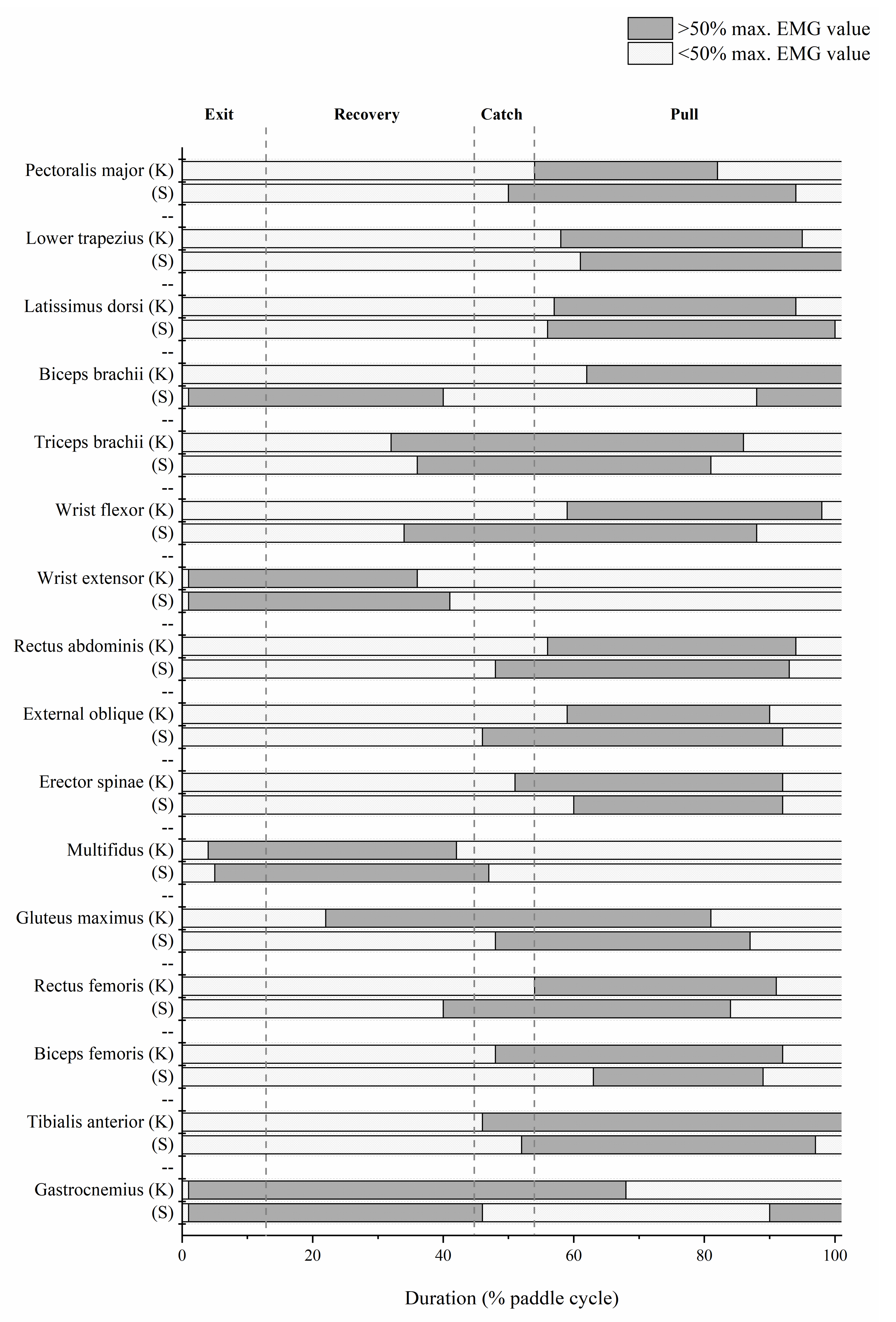
Figure 2 illustrates the on–off muscle timing in the two paddling positions, and
Table 1 analyzes the time difference in the activation sequence. A significant EMG difference was observed between the positions in starting activation times in the biceps, wrist flexor, external oblique abdominis, erector spinae, gluteus maximus, and rectus femoris and those in the termination times in the biceps, wrist flexor, rectus femoris, and gastrocnemius.
Table 2 and
Table 3 present the average and maximum EMG activation values of each muscle group in the two positions, respectively. The difference in average EMG activation value for each muscle group was nonsignificant in different postures. However, the biceps tended to have a larger maximum EMG activation value in the kneeling position than in the standing position. The maximum EMG activation values for the triceps, external oblique abdominis, and gastrocnemius were larger in the standing position than in the kneeling position.
4. Discussion
Although SUP is a recreational activity, the training and fatigue effects on each muscle group caused by sustained paddling on an unstable water surface should not be underestimated. Although this study could not directly demonstrate these effects (including training or fatigue effects), the changes after long-term muscle use were evident. To provide relevant information to practitioners, the possible effects of different postures on each muscle group are described as follows.
4.1. Trunk Muscle Group
Regarding the activation timing (
Figure 2), both the rectus abdominis and external oblique abdominis muscles were activated in advance in the catch stage when standing.
Table 1 indicates that the rectus abdominis was first activated at 56.91% and the external oblique abdominis initiated muscle firing at 54.92% of the cycle time. By contrast, in the kneeling position, the two muscles started to be activated only during the pull period (60.73% in the rectus abdominis and 62.25% in the external oblique muscle). We inferred that, in a relatively unstable stance, the core must be stabilized in advance to facilitate the limb strength transmission. According to the study conducted by Ruess et al. (2013), when a paddling action was performed on an unstable water surface, core muscles were first activated early in the pull phase to provide posture stability and standing balance, and then thigh and shoulder muscles continued to be activated late in the pull phase to provide the driving force for propulsion [
15]. The core muscle group was located at the body’s center of gravity (COG), affected the functional efficiency of the kinetic chain and enhanced the transmission and control of the limb strength [
18]. By contrast, erector spine muscles were activated earlier in the kneeling position than in the standing position (53.42% vs. 58.42%,
p < 0.05) and were more likely to be activated in the catch stage (
Figure 2). We inferred that the catch action can be performed using more torso rotation in the standing position whereas in the kneeling position the paddle can only be inserted into the water by arching the back and extending the arm, necessitating the recruitment of the erector spinae during the catch phase to facilitate this action.
As shown in
Table 3, the difference in the maximal activation values for the external oblique abdominis muscle in the kneeling and standing positions was significant (2.53 vs. 5.04 μV,
p < 0.05), and a larger external oblique abdominis muscle firing was observed in the standing position. This echoes the finding that the catch action can be accomplished using more torso rotation in the standing position. As a result, during the pull period, torso rotation provided by the external oblique abdominis muscle can be fully used to enhance the paddling force.
4.2. Shoulder and Upper Limb Muscles
The maximum activation value of the biceps muscle in the kneeling position was significantly higher than that in the standing position (59.05 vs. 28.44 μV;
Table 3), and the activation time series were also different between the positions. As illustrated in
Figure 2, the biceps muscles mainly acted during the recovery phase in the standing position and during the pull phase in the kneeling position. Because the degree of torso rotation was small in the kneeling position, we inferred that the pulling force mainly originated from the arm. The biceps was activated to help pull the arm from the front to the ventral side of the body to perform the paddle stroke. By contrast, in the standing position, the biceps muscle was mainly activated from the late pull phase to the recovery phase. We presumed that it played a role in shoulder flexion by assisting the opposite-side upper hand to lift the paddle. The triceps muscle mainly acted from the later stage of the recovery phase to the middle stage of the pull phase in both positions. A higher maximum activation value was observed in the standing position than in the kneeling position (25.70 vs. 10.61 μV;
Table 3). This is unsurprising, because SUP players are often trained to straighten their arms and twist their torsos while paddling. This inevitably increases the activation of the triceps, but this action is difficult to perform while kneeling, thereby causing this difference. In addition, the difference between the kneeling and standing postures in movement patterns from the exit to recovery phases may be caused by the length of the paddle. A longer paddle is harder to control than a shorter paddle, and this influences the wrist flexor muscle when standing. As shown in
Table 1, the starting time of the wrist flexor muscle in the standing position was 39.17%, whereas that in the kneeling position was 58.33%, indicating that the paddling in the standing posture required an earlier and more precise wrist movement to operate the paddle in the later stage of recovery.
4.3. Lower Limb Muscles
As depicted in
Table 3, the gastrocnemius muscle activation was higher in the standing position and must start in the pull phase (
Figure 2). We speculated that this is due to the higher COG accompanied by larger swaying motions [
19] in the standing position, which increased the need of maintaining balance. The gluteus maximus muscle of the single joint muscle group began to be recruited during the recovery period when kneeling (
Figure 2). The causes may be similar to those of the erector spinae. In this case, the hip joint hyperextended to assist the straight back in extending the paddle as far as possible in front of the body to facilitate the next step of inserting the paddle into the water. In the pull phase, in response to the instability of the SUP board, most of the lower limb muscles, including the gluteus maximus, rectus femoris, biceps femoris, and tibialis anterior muscle, were activated simultaneously.
4.4. Application in SUP Course Teaching
Although the difference was nonsignificant, the abdominal muscles had higher maximum activation values, when the participants were in the standing position, and a significant difference was observed in the external oblique abdominis. Behm et al. (2005) proposed that exercise that effectively enhances core muscles should be performed on an unstable surface [
20]. In this study, the stance instability substantially increased in the standing position. To maintain stability and balance in an unstable state, the human body exhibited a higher degree of muscle activation than it did in the steady state. In other words, the combination of an unstable plane and a muscle strength training can increase the degree of muscle activation and improve the training effectiveness. The research has confirmed that six weeks of SUP simulator training significantly improved core muscle group endurance [
14] and that the core muscle group strength significantly increased static and dynamic balance ability [
21]. Ruess et al. (2013) demonstrated that dynamic balance ability can be significantly improved after 30 minutes of SUP training on water [
10]. Furness et al. [
7] also demonstrated that elite stand-up paddlers had more favorable static balance than recreational paddlers did, indicating that an SUP workout can achieve core strengthening after long- or short-term training to improve balance ability. In the kneeling position, because trunk and lower limb movements are limited, the main source of paddling power is the upper arm force. Thus, more upper arm muscle group activation is required in the kneeling position. These results demonstrated that muscle use is adjusted in different postures. SUP participants can achieve full-body or particular training through this sport. For SUP instructors, muscle activation requirements in different postures can be used as evaluation indicators of training effectiveness and references for training program design. The evidence can also be used to develop a common approach to teaching or coaching standards in paddle sports. For example, for SUP practitioners, who often have pain or injuries due to incorrect movement in the shoulder and elbow joints when paddling, gaining an understanding of muscle groups activated in each phase can help instructors adjust excessive arm use in trainees and recommend the use of the whole body to minimize injury risk during SUP. Moreover, the evidence in this study suggested that switching SUP postures when fatigued can help paddlers complete challenges requiring longer distances.
5. Conclusions
The SUP in the kneeling position increased the demand for the biceps, whereas the standing position increased the demand for the core and lower limb muscles. In various outdoor water environments, paddlers can switch between postures at any time for amusement. The knowledge that SUP posture changes can activate different muscle groups can enhance training efficiency and provide reference for designing individualized training programs.
6. Patents
This section is not mandatory but may be added, if there are patents resulting from the work reported in this manuscript.
Author Contributions
Conceptualization of the study, F.-H.T., W.-L.W., and Y.-Y.H.; methodology, software, and validation, F.-H.T., W.-L.W., and Y.-Y.H.; formal analysis, Y.-J.C. and J.-M.L.; investigation and experimental work, Y.J.C. and J.-M.L.; resources, F.-H.T. and W.-L.W.; data curation and statistics, Y.-J.C. and J.-M.L.; writing of the original draft preparation, F.-H.T., Y.-J.C., and J.-M.L.; writing of review and editing, all authors; visualization, Y.J.C. and J.-M.L.; supervision, F.-H.T., W.-L.W., and Y.-Y.H.; project administration, Y.-Y.H. All authors have read and agreed to the published version of the manuscript.
Funding
The study was supported by grants from the NSYSU-KMU Joint Research Project (#NSYSUKMU106-P025) and the Ministry of Science and Technology (MOST) (#MOST 108-2221-E-992-094-)
Conflicts of Interest
The authors declare no conflicts of interest.
References
- Warshaw, M. The Encyclopedia of Surfing, 1st ed.; Harcourt Inc.: Orlando, FA, USA, 2003. [Google Scholar]
- Helliker, K. Surf’s Up: The Rise of Stand-up Paddle Boards. Available online: https://www.wsj.com/articles/SB10001424052748703720504575377023651849234 (accessed on 20 August 2017).
- Hammer, S. Catch the wave of stand-up paddling. Provid. J. 2011, 5, 3. [Google Scholar]
- The Outdoor Foundation. 2015 Special Report on Paddlesports; The Coleman Company: Washington, WA, USA, 2015. [Google Scholar]
- Schram, B. Stand up paddle boarding: An analysis of a new sport and recreational activity. PhD Thesis, Bond University, Gold Coast, Australia, 2015. [Google Scholar]
- Walker, C.; Nichols, A.; Forman, T. A survey of injuries and medical conditions affecting stand-Up paddle surfboarding participants. Clin. J. Sports Med. 2010, 20, 144–145. [Google Scholar]
- Furness, J.; Olorunnife, O.; Schram, B.; Climstein, M.; Hing, W. Epidemiology of Injuries in Stand-Up Paddle Boarding. Orthop. J. Sports Med. 2017, 5, 2325967117710759. [Google Scholar] [CrossRef] [PubMed]
- Klontzas, M.E.; Hatzidakis, A.; Karantanas, A.H. Imaging findings in a case of stand up paddle surfer’s myelopathy. BJR Case Rep. 2015, 1, 20150004. [Google Scholar] [CrossRef] [PubMed]
- Schram, B.; Hing, W.; Climstein, M. Profiling the sport of stand-up paddle boarding. J. Sports Sci. 2015, 34, 937–944. [Google Scholar] [CrossRef] [PubMed]
- Ruess, C.; Kristen, K.H.; Eckelt, M.; Mally, F.; Litzenberger, S.; Sabo, A. Stand up Paddle Surfing-An Aerobic Workout and Balance Training. Procedia Eng. 2013, 60, 62–66. [Google Scholar] [CrossRef]
- Schram, B.; Hing, W.; Climstein, M. Laboratory-and Field-Based Assessment of Maximal Aerobic Power of Elite Stand-Up Paddle-Board Athletes. Int. J. Sports Physiol. Perform 2016, 11, 28–32. [Google Scholar] [CrossRef] [PubMed]
- Suari, Y.; Schram, B.; Ashkenazi, A.; Gann-Perkal, H.; Berger, L.; Reznikov, M.; Shomrat, S.; Kodesh, E. The Effect of Environmental Conditions on the Physiological Response during a Stand-Up Paddle Surfing Session. Sports 2018, 6, 25. [Google Scholar] [CrossRef] [PubMed]
- Osti, F.R.; de Souza, C.R.; Teixeira, L.A. Improvement of Balance Stability in Older Individuals by On-Water Training. J. Aging Phys. Act. 2018, 26, 222–226. [Google Scholar] [CrossRef] [PubMed]
- Schram, B.; Hing, W.; Climstein, M. The physiological, musculoskeletal and psychological effects of stand up paddle boarding. BMC Sports Sci. Med. Rehabil 2016, 8, 32. [Google Scholar] [CrossRef] [PubMed]
- Ruess, C.; Kristen, K.H.; Eckelt, M.; Mally, F.; Litzenberger, S.; Sabo, A. Activity of Trunk and Leg Muscles during Stand up Paddle Surfing. Procedia Eng. 2013, 60, 57–61. [Google Scholar] [CrossRef]
- SENIAM Recommendations for sensor locations on individual muscles. Available online: http://seniam.org/sensorlocation.htm (accessed on 12 January 2018).
- Michael, J.S.; Smith, R.; Rooney, K.B. Determinants of kayak paddling performance. Sports Biomech. 2009, 8, 167–179. [Google Scholar] [CrossRef] [PubMed]
- Shinkle, J.; Nesser, T.W.; Demchak, T.J.; McMannus, D.M. Effect of core strength on the measure of power in the extremities. J. Strength Cond Res. 2012, 26, 373–380. [Google Scholar] [CrossRef] [PubMed]
- Kaufman, C.; Hibbert, J.E. Shoulder, Hip, and Trunk Kinematics Vary with Posture during Stand Up Paddle Boarding; Southern California Conferences for Undergraduate Research: San Marcos, California, USA, 2019. [Google Scholar]
- Behm, D.G.; Leonard, A.M.; Young, W.B.; Bonsey, W.A.C.; MacKionon, S.N. Trunk muscle electromyographic activity with unstable and unilateral exercises. J. Strength Cond. Res. 2005, 19, 193–201. [Google Scholar] [PubMed]
- Norwood, J.T.; Anderson, G.S.; Gaetz, M.B.; Twist, P.W. Electromyographic activity of the trunk stabilizers during stable and unstable bench press. J. Strength Cond Res. 2007, 21, 343–347. [Google Scholar] [PubMed]
© 2020 by the authors. Licensee MDPI, Basel, Switzerland. This article is an open access article distributed under the terms and conditions of the Creative Commons Attribution (CC BY) license (http://creativecommons.org/licenses/by/4.0/).


