Review

Developments in Antibiotic-Eluting Scaffolds for the Treatment of Osteomyelitis

1
School of Clinical Medicine, University of Cambridge, Cambridge CB2 0QQ, UK
2
Department of Trauma and Orthopaedic Surgery, Addenbrooke’s Hospital, University of Cambridge, Cambridge CB2 0QQ, UK
*
Author to whom correspondence should be addressed.
Appl. Sci. 2020, 10(7), 2244; https://doi.org/10.3390/app10072244
Submission received: 2 March 2020 / Revised: 19 March 2020 / Accepted: 24 March 2020 / Published: 26 March 2020
(This article belongs to the Special Issue Implantable Medical Device)

Abstract

:
Osteomyelitis is a devastating disease caused by the infection of bone tissue and is associated with significant morbidity and mortality. It is treated with antibiotic therapy and surgical debridement. A high dose of systemic antibiotics is often required due to poor bone penetration and this is often associated with unacceptable side-effects. To overcome this, local, implantable antibiotic carriers such as polymethyl methacrylate have been developed. However, this is a non-biodegradable material that requires a second surgery to be removed. Attention has therefore shifted to new antibiotic-eluting scaffolds which can be created with a range of unique properties. The purpose of this review is to assess the level of evidence that exists for these novel local treatments. Although this field is still developing, these strategies seem promising and provide hope for the future treatment of chronic osteomyelitis.

Graphical Abstract

1. Osteomyelitis

Osteomyelitis is an inflammatory bone disease caused by infection. It is most commonly associated with skin commensals such as the bacterium Staphylococcus aureus [1]. These organisms can be introduced by a variety of routes. The most common of these is trauma, with infection rates as high as 16% being reported in open long-bone fractures [2,3]. Other causes include joint arthroplasty and diabetic foot disease, both of which are becoming increasingly more prevalent as populations age [4,5,6]. Osteomyelitis can also result from the haematogenous spread of bacteria and this is especially important to consider in children. Fortunately, the incidence of bloodborne osteomyelitis has dramatically reduced following the introduction of systemic antibiotics [7].
Patients with osteomyelitis present with a variety of symptoms. Some of these are relatively non-specific, such as malaise, fatigue, chills, delayed healing and pyrexia [1]. Others tend to be more localised: pain at the site of infection, swelling and erythema [1]. Recognition of these features should prompt clinicians to obtain blood cultures, a full set of blood tests, inflammatory markers, plain x-ray films and a magnetic resonance imaging (MRI) scan [1,8]. Notably, the symptoms of osteomyelitis can either present themselves acutely, or chronically. The distinction between these subsets is pivotal to treatment.
In acute osteomyelitis, patients are treated empirically with high-dose intravenous antibiotics [1]. Typically, a broad-spectrum agent is initiated and then refined by culture results [1]. The standard course of treatment lasts four to six weeks [1,9]. However, once there is dead bone, bone abscesses or biofilm formation, complete surgical debridement is also required [10]. This is usually the case in chronic osteomyelitis [10]. Details of the debridement surgery depend on the Cierny–Mader classification and clinical context (Figure 1) [11]. This is a staging system for osteomyelitis based on the anatomical boundaries of infection and the physiological status of the host. Intra-operative samples should always be taken for microbiology and histology assessment [10].

2. Local Treatments

Because bone is an inflexible tissue, following bone loss such as in trauma and debridement surgery, any bony defect will remain and fill with haematoma [12]. This provides an ideal environment for bacteria to multiply and establish chronic infection and/or a biofilm. These bacteria go on to release osteolytic cytokines and osteonecrosis factors which evoke a powerful inflammatory response [13]. Over time, this can further damage the underlying bone and its blood supply. The result is a highly resilient biofilm in an area of increasingly poor antibiotic access. It has been reported that to treat these cases, a 10 to 100 times increase in antibiotic concentrations is required [14]. The use of high-dose systemic antibiotic treatment is expensive and associated with an increased risk of side effects [14,15]. It is therefore important to prevent chronic infection occurring. One way in which this can be done is by filling the bony defect(s) with a bone graft, material such as polymethyl methacrylate or a scaffold to prevent haematoma formation. In the United States (US) alone, the annual cost of treating bone defects has been estimated to be $5 billion [16].

2.1. Polymethyl Methacrylate

The most popular local treatment is an antibiotic-eluting material called polymethyl methacrylate (PMMA) or ‘bone cement’ [17]. This has been referred to as the gold-standard of chronic osteomyelitis treatment [17,18]. Experimental studies have shown that PMMA can deliver in the order of 200 times the amount that systemic antibiotics are able to [19]. In animals, Evans and Nelson (1993) found that PMMA beads had a 100% success rate at preventing the recurrence of osteomyelitis [20]. However, randomised controlled trials in humans have failed to identify a difference in outcome between PMMA and systemic antibiotics [21,22,23]. In addition, the high temperatures generated during the preparation of PMMA can degrade its antibiotic content and induce thermal necrosis. This means certain antibiotics such as tetracyclines cannot be used [18]. Others have described a rare ‘bone cement implantation syndrome’ with PMMA use [24]. This is thought to be due to a leachable MMA monomer that causes local tissue toxicity and systemic effects such as hypoxia and confusion [24]. Perhaps the most concerning disadvantages of PMMA, is that the majority of the antibiotic loaded is not released and every patient requires a second operation for it to be removed [17]. This increases morbidity, hospital costs, recovery time and the risk of perioperative complications such as infection. Attention has therefore shifted to alternative local treatments such as resorbable antibiotic scaffolds.

2.2. Scaffolds

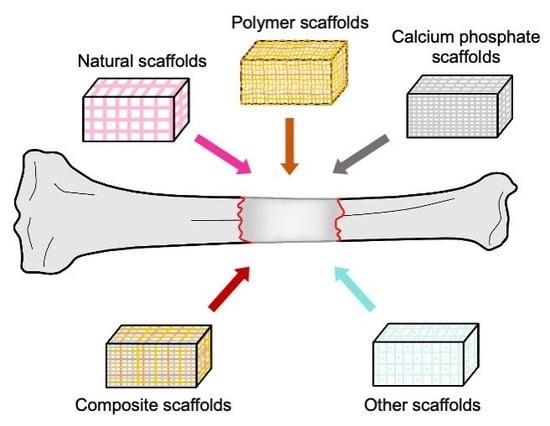
A bone scaffold is a three-dimensional (3D) matrix that fills defective bone and facilitates repair. In the context of tissue engineering, many of these scaffolds are biodegradable and capable of osteoconduction, osteoinduction and osteogenesis [25]. These are defined as the abilities to guide reparative bone growth, encourage osteoblastic differentiation and contribute living bone cells respectively. In addition, they can deliver anti-inflammatory drugs and antibiotics [26]. This enables high concentrations to be delivered directly to an affected site without systemic side effects. It also overcomes any issues with drug bioavailability, compliance and allows for a sustained release pattern [26]. Antibiotic-eluting scaffolds have been classified as natural, synthetic or composite for the purpose of this article.

3. Properties of an Antibiotic-Eluting Scaffold

3.1. Biocompatability

One of the most important properties of any bone graft is biocompatibility [27]. This helps to prevent a severe inflammatory response that may impair healing or cause rejection [27]. However, human leukocyte antigen (HLA) matching is infrequently performed for bone allografts [28,29]. This is in spite of growing evidence to suggest that allosensitisation occurs. Currently, the long-term effect of HLA sensitisation on bone graft survival is unknown. In comparison, autografts derived from the patient are inherently biocompatible but require a harvest surgery to be obtained. Synthetic and composite scaffolds represent an alternative therapy. These scaffolds can be designed and produced ex vivo, to mimic native tissue and have as little immunogenicity as possible [30].

3.2. Biodegradability

The long-term presence of non-biodegradable scaffolds has been associated with impaired bone formation, difficult radiological assessment of bone healing, inflammation and prolonged drug release [25]. For example, the non-biodegradable material PMMA has been shown to elute low levels of antibiotic up to five years post-implantation [31]. Extended exposure to low-dose, sub-therapeutic levels of antibiotic is a major risk factor for bacterial resistance. In the aforementioned case, gentamicin-resistant staphylococci were recovered from the surface of the PMMA beads [30]. In another study that used gentamicin-eluting PMMA, 90% of the bacteria isolated were found to be resistant compared to 16% for plain cement [32]. It has also been suggested that chronic, unnecessary exposure to drugs such as gentamicin may cause nephrotoxicity, even at a low dose [33]. Those with renal impairment are especially at risk [33]. Moreover, when complete elution from a non-biodegradable scaffold is eventually achieved, the material acts as a foreign body and can become colonised, leading to recurrent infections [34]. In these cases, a second surgery is needed to remove the material. On the other hand, biodegradable scaffolds completely unload their antibiotic content over a defined time period and do not linger in the body. This avoids many of the disadvantages mentioned above and eliminates the need for retrieval surgery. Biodegradability is therefore a key property of a successful bone scaffold [26]. It is important that the rate of degradation is balanced with the rate of bone formation.

3.3. Mechanical and Structural Properties

Given that bone scaffolds are implanted in areas of dead space, they should have mechanical properties consistent with that anatomical site to prevent the risk of fracture [27]. However, scaffolds must also exhibit sufficient porosity to allow cellular penetration, angiogenesis and the transport of oxygen, nutrients and waste products [35,36]. In addition, a porous spatial arrangement enables drug release from deep inside the material. It has been shown that in vivo, that macroporous antibiotic-loaded calcium phosphates out-perform microporous variants due to a higher antibiotic release rate (up to 13x more) [37]. This is not the case with PMMA which exhibits surface-level diffusion [38]. There is therefore a trade-off between mechanical strength and porosity.

3.4. Bone Growth

Other desirable properties of scaffolds include osteoconduction, osteoinduction and osteogenesis [25,39,40]. Osteoconduction is the ability to guide reparative bone growth. This is generally an intrinsic property and may be achieved by using a porous structure [41]. Osteoinduction on the other hand is the ability to encourage osteoblastic differentiation. This is an extrinsic process driven by the release of growth factors such as bone morphogenic proteins (BMPs) [41]. Osteogenesis, or the ability to contribute living cells, may be achieved by impregnating scaffolds with a variety of cell types [42].

3.5. Manufacturing Properties

The ideal bone scaffold must also be cost-effective, easy-to-use and scalable. One way the production of scaffolds can be maximised, is through the use of three-dimensional printing. Three-dimensional printers are thought to decrease long-term purchase costs once the high set up and maintenance expenses have been recompensed. Antibiotic-eluting scaffolds can be produced using this method if a thermostable drug such as tobramycin is used [43]. Many of these materials have been shown to be efficacious in both in vitro and in vivo models of osteomyelitis [43,44]. In the latter study, 3D-printed resorbable calcium phosphate scaffolds containing sitafloxacin and rifampin outperformed gentamicin-laden PMMA when bacterial colonization outcomes and bone growth were assessed [44]. Another benefit of 3D-printing is that it allows for the fabrication of patient-specific designs which completely fill the bone void. An alternative approach to achieve this is to manufacture injectable scaffolds that harden in vivo [45].

4. The Choice of Antibiotic

A successful local antibiotic must have good tissue penetration and predictable pharmacokinetics [46]. The choice of drug is also guided by the clinical context. For example, in cases of osteomyelitis that are culture positive for methicillin-resistant Staphylococcus aureus (MRSA), a vancomycin-eluting scaffold should be used [6,47]. If the organism is unknown or the treatment is prophylactic, one may instead opt for a broader-spectrum drug such as tobramycin which is effective against both gram-positive and gram-negative bacteria [48]. One must also consider patient-specific factors when choosing an antibiotic. These include allergy, co-morbidities, susceptibility to side-effects and the dose required. In some reports, authors have instead used a combination-based approach. This involves impregnating a single scaffold with a mixture of synergistic antibiotics, such as gentamicin, vancomycin and clindamycin [45,49]. The use of combination therapy is thought to increase the likelihood of eradication and minimise the risk of antibiotic resistance [50]. It is therefore likely to be used more frequently as this field develops.

5. Natural Scaffolds

Given the importance of biocompatibility and biodegradability, natural scaffolds such as collagen, hyaluronic acid, cellulose and chitosan may seem like sensible biomaterials to use in osteomyelitis [26]. Natural polymers are biologically active and typically promote excellent cell adhesion and growth [27]. However, they are also immunologically active and carry the risk of rejection [26]. In addition, they generally have poor mechanical properties which limit their use in load-bearing orthopaedic applications [26,27].

5.1. Collagen

Collagen is the most abundant protein in mammalian tissue and forms a key component of the extracellular matrix [51]. It is also the most widely explored natural biomaterial [34]. A recent systematic review of 413 patients treated with antibiotic-loaded collagen sponges (fleeces) reported an overall success rate of 91% [52]. However, the authors also identified a moderate to high risk of bias in these studies [52]. Moreover, the pharmacokinetic profiles of these materials showed an average local antibiotic concentration that was above the bacterial minimum inhibitory concentration (MIC), for only five days [52]. This rapid antibiotic release rate of collagens has long been documented. In a study by Sorensen et al. (1990) for example, the authors found that their collagen sponge eluted 95% of its gentamicin in the first day in vitro [53]. It is also important to note that like other natural scaffolds, collagen is a weak biomaterial. To improve its mechanical strength collagen can be crosslinked, or more effectively, it can be combined with synthetic materials to form composite scaffolds [54,55].

5.2. Chitosan

Polysaccharides are a further group of natural biomaterials and include chitosan, a derivative of chitin which is found in arthropod skeletons. Chitosan shares many of the advantages and disadvantages as collagen-based scaffolds. For example, it is also mechanically weak and has a compressive strength one to two orders lower than cancellous bone [56]. Chitosan has hydrophilic elements and a positive charge which enables it to interact with negatively charged polymers, macromolecules and certain polyanions [26]. This means that it lacks structural stability in aqueous environments—a critical requirement of any bone scaffold [26,56]. On the other hand, these properties enable chitosan to form a ‘hydrogel’ that can be exploited for drug-delivery and nanoencapsulation purposes [26,56]. Some authors even attribute the cationic nature of chitosan with an intrinsic antimicrobial ability [57]. Chitosan is therefore an unsuitable bone substitute when used alone, but can be very useful when blended with other materials to improve its mechanical and structural properties when hydrated. For this reason, chitosan-based composites are being increasingly used in bone tissue engineering [58].

6. Synthetic Scaffolds

Synthetic scaffolds are a large, rapidly expanding category of biomaterials. These includes calcium phosphates and synthetic polymers.

6.1. Polymers

The most commonly used synthetic polymers are polyurethane (PUR) and saturated aliphatic polyesters such as poly(lactic acid) (PLA), poly(glycolic acid) (PGA), poly(lactic-co-glycolic acid) (PLGA) and poly(carprolactones) (PCL) [17,26,59]. Like natural scaffolds, these products are generally biocompatible and biodegradable [59]. However, synthetic polymers have longer release rates and can be produced under more controlled manufacturing methods [35]. This means that they have predictable physicomechanical properties and can be engineered, for example, by adjusting the molecular weight, to yield specific desired characteristics [35]. The versality of these scaffolds is their largest advantage [26]. One disadvantage though, is that compared to native bone, synthetic polymers have relatively poor mechanical properties [59]. This makes them unsuitable for use in high load-bearing areas [26]. Moreover, they degrade by hydrolysis which produces carbon dioxide. This has been reported to lower the surrounding pH, resulting in tissue necrosis [60,61]. In scaffolds that rapidly dissolve, this change in pH may even be large enough to reduce the functional efficacy of the local antibiotic [17]. For these reasons, antibiotic-eluting synthetic polymers remain the focus of in vitro and in vivo experiments [45,61,62].
An example of the utility of these materials is provided by Li et al. (2010) [62]. This group used biodegradable PUR scaffolds impregnated with free-base vancomycin, to treat 40 rats with post-traumatic osteomyelitis [62]. Using this method, the authors found the bacterial load at four weeks (measured in colony forming units) was significantly lower than that of negative controls [62]. The effectiveness of this resorbable scaffold was comparable to PMMA which is the current standard of care [62]. In another study by McLaren et al. (2014), a biodegradable PLGA scaffold was used to deliver gentamicin and clindamycin to 30 sheep with osteomyelitis [45]. These animals were sacrificed at two- and six-weeks post-implantation to look for evidence of infection. No bacteria were isolated from animals treated with antibiotic-eluting scaffolds, but Staphylococcus aureus was successfully isolated from control groups [45]. The authors also showed that at 13 weeks the scaffold material had fully degraded [45]. Any area of the defect that was not filled with new bone contained cartilaginous tissue that would be expected to eventually turn into mineralised bone [45]. Synthetic polymers are therefore promising biomaterials. They are contained in many recent composites that have been used in clinical trials as discussed below.

6.2. Calcium Phosphates

Calcium-based bone substitutes are a particularly large group of materials used in bone tissue engineering [35]. These include calcium sulfates, tricalcium phosphate (TCP), hydroxyapatite (HA) and biphasic calcium phosphates (TCP + HA). Unlike natural scaffolds and synthetic polymers, the chemical and operation properties of calcium products are similar to the mineral phase of bone [63]. The high mechanical stiffness (Young’s modulus), hardness, brittleness and low elasticity make calcium substitutes appropriate for bone regeneration [63]. Compared with human bone, the compressive strength of calcium phosphates is generally much higher, though they have a lower tensile strength, fracture toughness and increased fragility [63,64]. Calcium-based scaffolds are also biocompatible, osteoconductive and bioactive, meaning that they are capable of forming a biological interface with host tissue [65]. This can help to prevent implant dislocation. However, it has been postulated that the dissolution of calcium sulfate, like other synthetics, leads to an acidic microenvironment which may cause inflammation [66]. There is also evidence to suggest that some products which are marketed as being resorbable, may still persist over several years in some patients [67]. This is not ideal as the defect should be replaced by new, stronger bone in all cases.
The potential of antibiotic-eluting, biodegradable, calcium-based scaffolds in chronic osteomyelitis has been long recognised [68]. As a result, there is a wealth of literature in this area. Importantly, this has led to numerous clinical trials with positive results (Table 1). It has also inspired companies to mass-produce calcium-based products. Many of these, such as OSTEOSET®-T (resorbable alpha hemihydrate calcium sulfate + tobramycin) have received approval by the Food and Drug Administration (FDA) in the US. However, a recent systematic review of 484 patients has highlighted that many of these papers have a significant risk of bias [69]. The authors therefore warn that while these results may seem promising, they are currently inconclusive [69]. Of those listed in Table 1, only McKee et al. (2010) conducted a prospective randomised trial and even then, no blinding was put in place [70]. There is therefore a need for more recent, larger-scale randomised-control trials in this area [69]. Care should also be taken when extrapolating the results of studies based on osteomyelitis, to other contexts. It has recently been shown that antibiotic-impregnated calcium sulfate beads do not improve outcomes in periprosthetic joint infections, for example [71].

7. Composite Scaffolds

Composite scaffolds are the current focus of bone tissue engineering [35]. They combine different materials with unique properties in an attempt to overcome the deficiencies of the individual constituents. For example, bio-ceramic composites are made of calcium-based scaffolds and synthetic polymers. This allows the composite to have the typical mechanical strength, bioactivity and osteoconductive properties associated with calcium substitutes, but also overcome their fragility issues [26]. Other common preparations include calcium-based or synthetic polymer scaffolds, with natural polymers such as collagen or chitosan. Recently, it has been shown in vitro that beta-TCP-collagen composites have a significantly higher loading capacity and a steadier release rate of gentamicin and vancomycin compared to TCP and HA granules [75]. Theoretically, composites can incorporate any number of the above materials. They have even been made with other antimicrobials such as bioactive glass and various metal ions [76,77]. The relative proportions of each constituent material can drastically change features such as the drug release profile, bacterial inhibition zone and ultimately, the outcome in vivo [77,78]. Considering the above, there is therefore a near-infinite number of possible composites. This is therefore a very promising field of bone tissue engineering. However, as a relatively new advancement, there are no clinical studies on antibiotic-eluting composites for osteomyelitis. Recent experimental studies are highlighted in Table 2. It must be noted that the transfer of in vitro results to in vivo studies is difficult, even in animals.

8. Other Antimicrobial Materials

In addition to antibiotic-loaded bone scaffolds, there are also other antimicrobial biomaterials including bioactive glass [82]. This is a biodegradable, osteoconductive, osteoinductive and osteogenic material that kills bacteria by leaching ionic dissolution products from its surface [83,84]. This changes the local osmotic pressure and pH such that it is hostile to microbial growth [84]. It has also been shown to be angiogenic in vitro [85]. However, disadvantages of bioactive glass include brittleness and a low fracture toughness [86]. Bioactive glass S53P4 has been the focus of a large multinational study of 116 patients across six countries with chronic osteomyelitis [87]. This research reported a cure rate of 90% at a median follow-up length of 31 months [87]. There is some evidence that the antibacterial effect of bioactive glass can be further improved by doping them with metal ions such as zinc, strontium or silver [87]. These materials in themselves have antimicrobial properties [88]. In a recent study be Mestres et al. (2010), for example, magnesium-containing cements were shown to have intrinsic antimicrobial activity and were sufficient alone, to significantly improve the health state of animals [37]. Other antimicrobials include enzyme-loaded scaffolds and polymeric nanoparticles [89,90]. The latter is especially notable, as no bacterial resistance was observed towards these materials in vitro after 20 serial passages (1300 bacterial generations) [90]. On the other hand, resistance occurred after only a few passages for clinically relevant antibiotics [90]. The usefulness of nanoparticles in bone tissue engineering has been summarised by several authors [91,92].

9. Conclusions

Antibiotic-eluting scaffolds present many advantages over other local methods for the treatment of osteomyelitis. This is a dynamic field that has seen many exciting advances. Natural, synthetic and composite bone substitutes have all been developed, some of which have moved into clinical trials. Perhaps the most promising of these are the biodegradable, antibiotic-eluting composite scaffolds. These can be designed to incorporate the best features of all available biomaterials and can be 3D-printed, therefore allowing scalability and/or patient-specificity [93]. Composites have come at a time when the field of bone tissue engineering is moving away from the concept of an ‘ideal’ bone substitute, towards applications that depend on the clinical context [94,95,96]. There is still a need for large-scale randomised control trials in this area before definitive conclusions can be reached. It also remains to be fully explored how antibiotic-eluting composite scaffolds can be made to interact with other pathways involved in bone repair, namely growth factor signaling and cell-based therapies.

Author Contributions

Conceptualization, H.K. and W.S.K.; Methodology, H.K. and A.K.; Formal Analysis, H.K.; Investigation, H.K. and A.K.; Writing-Original Draft Preparation, H.K.; Writing-Review & Editing, H.K., A.K., and W.S.K.; Visualization, H.K.; Supervision, W.S.K.; Project Administration, W.S.K. All authors have read and agreed to the published version of the manuscript.

Funding

This research received no external funding.

Conflicts of Interest

The authors declare no conflict of interest.

References

  1. Lazzarini, L.; Mader, J.T.; Calhoun, J.H. Osteomyelitis in long bones. JBJS 2004, 86, 2305–2318. [Google Scholar] [CrossRef] [PubMed]
  2. Jiang, N.; Ma, Y.F.; Jiang, Y.; Zhao, X.Q.; Xie, G.P.; Hu, Y.J.; Qin, C.H.; Yu, B. Clinical characteristics and treatment of extremity chronic Osteomyelitis in Southern China. Medicine 2015, 94, e1874. [Google Scholar] [CrossRef] [PubMed]
  3. Leonidou, A.; Kiraly, Z.; Gality, H.; Apperley, S.; Vanstone, S.; Woods, D.A. The effect of the timing of antibiotics and surgical treatment on infection rates in open long-bone fractures: A 6-year prospective study after a change in policy. Strateg. Trauma Limb Reconstr. 2014, 9, 167–171. [Google Scholar] [CrossRef] [Green Version]
  4. Springer, B.D.; Cahue, S.; Etkin, C.D.; Lewallen, D.G.; McGrory, B.J. Infection burden in total hip and knee arthroplasties: An international registry-based perspective. Arthroplast. Today 2017, 3, 137–140. [Google Scholar] [CrossRef] [PubMed] [Green Version]
  5. Hatzenbuehler, J.; Pulling, T.J. Diagnosis and management of osteomyelitis. Am. Fam. Physician 2011, 84, 1027–1033. [Google Scholar] [PubMed]
  6. Panteli, M.; Giannoudis, P.V. Chronic osteomyelitis: What the surgeon needs to know. EFORT Open Rev. 2016, 1, 128–135. [Google Scholar] [CrossRef]
  7. Klenerman, L. A history of osteomyelitis from the Journal of Bone and Joint Surgery: 1948 to 2006. JBJS 2007, 89, 667–670. [Google Scholar] [CrossRef] [Green Version]
  8. Beaman, F.D.; Von Herrmann, P.F.; Kransdorf, M.J.; Adler, R.S.; Amini, B.; Appel, M.; Arnold, E.; Bernard, S.A.; Greenspan, B.S.; Lee, K.S.; et al. Appropriateness Criteria ® Suspected Osteomyelitis, Septic Arthritis, or Soft Tissue Infection (Excluding Spine and Diabetic Foot) Expert Panel on Musculoskeletal Imaging. J. Am. Coll. Radiol. 2017, 14, 326–337. [Google Scholar] [CrossRef] [Green Version]
  9. NHS Osteomyelitis. Available online: https://www.nhs.uk/conditions/osteomyelitis/ (accessed on 25 February 2020).
  10. Ferguson, J.; Wong, T.H.N.; Atkins, L.B.; McNally, M. Osteomyelitis. BMJ Best Pract. 2018, 1–44. [Google Scholar]
  11. Cierny, G.; Mader, J.T.; Penninck, J.J. A clinical staging system for adult osteomyelitis. Clin. Orthop. Relat. Res. 2003, 414, 7–24. [Google Scholar] [CrossRef] [Green Version]
  12. McNally, M.; Nagarajah, K. Osteomyelitis. Orthop. Trauma 2010, 24, 416–429. [Google Scholar] [CrossRef]
  13. Fritz, J.M.; McDonald, J.R. Osteomyelitis: Approach to diagnosis and treatment. Phys. Sportsmed. 2008, 36, 50–54. [Google Scholar] [CrossRef] [PubMed] [Green Version]
  14. Nelson, C.L. The Current Status of Material Used for Depot Delivery of Drugs. Clin. Orthop. Relat. Res. 2004, 427, 72–78. [Google Scholar] [CrossRef]
  15. Gogia, J.; Meehan, J.; Di Cesare, P.; Jamali, A. Local Antibiotic Therapy in Osteomyelitis. Semin. Plast. Surg. 2009, 23, 100–107. [Google Scholar] [CrossRef] [PubMed] [Green Version]
  16. Perez, J.R.; Kouroupis, D.; Li, D.J.; Best, T.M.; Kaplan, L.; Correa, D. Tissue Engineering and Cell-based Therapies for Fractures and Bone defects. Front. Bioeng. Biotechnol. 2018, 31, 105. [Google Scholar] [CrossRef] [PubMed] [Green Version]
  17. Inzana, J.A.; Schwarz, E.M.; Kates, S.L.; Awad, H.A. Biomaterials approaches to treating implant-associated osteomyelitis. Biomaterials 2016, 81, 58–71. [Google Scholar] [CrossRef] [Green Version]
  18. Van Vugt, T.A.G.; Arts, J.J.; Geurts, J.A.P. Antibiotic-Loaded Polymethylmethacrylate Beads and Spacers in Treatment of Orthopedic Infections and the Role of Biofilm Formation. Front. Microbiol. 2019, 10, 1626. [Google Scholar] [CrossRef]
  19. Wahlig, H.; Dingeldein, E.; Bergmann, R.; Reuss, K. The release of gentamicin from polymethylmethacrylate beads. An experimental and pharmacokinetic study. JBJS 1978, 60B, 270–275. [Google Scholar] [CrossRef] [Green Version]
  20. Evans, R.P.; Nelson, C.L. Gentamicin-impregnated polymethylmethacrylate beads compared with systemic antibiotic therapy in the treatment of chronic osteomyelitis. Clin. Orthop. Relat. Res. 1993, 295, 37–42. [Google Scholar] [CrossRef]
  21. Barth, R.E.; Vogely, H.C.; Hoepelman, A.I.M.; Peters, E.J.G. “To bead or not to bead?” Treatment of osteomyelitis and prosthetic joint-associated infections with gentamicin bead chains. Int. J. Antimicrob. Agents 2011, 38, 371–375. [Google Scholar] [CrossRef] [PubMed]
  22. Nelson, C.L.; Evans, R.P.; Blaha, J.D.; Calhoun, J.; Henry, S.L.; Patzakis, M.J. A comparison of gentamicin-impregnated polymethylmethacrylate bead implantation to conventional parenteral antibiotic therapy in infected total hip and knee arthroplasty. Clin. Orthop. Relat. Res. 1993, 295, 96–101. [Google Scholar] [CrossRef]
  23. Shih, H.-N.; Shih, L.-Y.; Wong, Y.-C. Diagnosis and Treatment of Subacute Osteomyelitis. J. Trauma Infect. Crit. Care 2005, 58, 83–87. [Google Scholar] [CrossRef] [PubMed]
  24. Donaldson, A.J.; Thomson, H.E.; Harper, N.J.; Kenny, N.W. Bone cement implantation syndrome. Br. J. Anaesth. 2009, 102, 12–22. [Google Scholar] [CrossRef] [PubMed] [Green Version]
  25. Yuan, N.; Rezzadeh, K.S.; Lee, J.C. Biomimetic Scaffolds for Osteogenesis. Recept. Clin. Investig. 2015, 2, 898. [Google Scholar]
  26. Dorati, R.; DeTrizio, A.; Modena, T.; Conti, B.; Benazzo, F.; Gastaldi, G.; Genta, I. Biodegradable scaffolds for bone regeneration combined with drug-delivery systems in osteomyelitis therapy. Pharmaceuticals 2017, 10, 96. [Google Scholar] [CrossRef] [Green Version]
  27. O’Brien, F.J. Biomaterials & scaffolds for tissue engineering. Mater. Today 2011, 14, 88–95. [Google Scholar]
  28. Ward, W.G.; Gautreaux, M.D.; Lippert, D.C.; Boles, C. HLA sensitization and allograft bone graft incorporation. Clin. Orthop. Relat. Res. 2008, 466, 1837–1848. [Google Scholar] [CrossRef] [Green Version]
  29. O’Sullivan, E.D.; Battle, R.K.; Zahra, S.; Keating, J.F.; Marson, L.P.; Turner, D.M. Allosensitization Following Bone Graft. Am. J. Transplant. 2017, 17, 2207–2211. [Google Scholar] [CrossRef] [Green Version]
  30. Janoušková, O. Synthetic Polymer Scaffolds for Soft Tissue Engineering. Physiol. Res. 2018, 67, 335–348. [Google Scholar] [CrossRef]
  31. Neut, D.; van de Belt, H.; van Horn, J.R.; van der Mei, H.C.; Busscher, H.J. Residual gentamicin-release from antibiotic-loaded polymethylmethacrylate beads after 5 years of implantation. Biomaterials 2003, 24, 1829–1831. [Google Scholar] [CrossRef]
  32. Hope, P.G.; Kristinsson, K.G.; Norman, P.; Elson, R.A. Deep infection of cemented total hip arthroplasties caused by coagulase-negative staphylococci. JBJS 1989, 71, 851–855. [Google Scholar] [CrossRef] [Green Version]
  33. Stravinskas, M.; Horstmann, P.; Ferguson, J.; Hettwer, W.; Nilsson, M.; Tarasevicius, S.; Petersen, M.M.; McNally, M.A.; Lidgren, L. Pharmacokinetics of gentamicin eluted from a regenerating bone graft substitute in vitro and clinical release studies. JBJS 2016, 5, 427–435. [Google Scholar]
  34. Kendall, R.W.; Duncan, C.P.; Smith, J.A.; Ngui-Yen, J.H. Persistence of bacteria on antibiotic loaded acrylic depots: A reason for caution. Clin. Orthop. Relat. Res. 1996, 329, 273–280. [Google Scholar] [CrossRef]
  35. Wheelton, A.; Mace, J.; Khan, W.S.; Anand, S. Biomaterials and Fabrication to Optimise Scaffold Properties for Musculoskeletal Tissue Engineering. Curr. Stem Cell Res. Ther. 2016, 11, 578–584. [Google Scholar] [CrossRef] [PubMed] [Green Version]
  36. Mehdizadeh, H.; Sumo, S.; Bayrak, E.S.; Brey, E.M.; Cinar, A. Three-dimensional modeling of angiogenesis in porous biomaterial scaffolds. Biomaterials 2013, 34, 2875–2887. [Google Scholar] [CrossRef] [PubMed]
  37. Mestres, G.; Fernandez-Yague, M.A.; Pastorino, D.; Montufar, E.B.; Canal, C.; Manzanares-Céspedes, M.C.; Ginebra, M.P. In vivo efficiency of antimicrobial inorganic bone grafts in osteomyelitis treatments. Mater. Sci. Eng. C Mater. Biol. Appl. 2019, 97, 84–95. [Google Scholar] [CrossRef]
  38. Rasyid, H.N.; Soegijoko, S. Influence of soluble fillers in improving porosity of handmade antibiotic-impregnated polymethyl methacrylate (PMMA) beads: An in-vitro study. Malays. Orthop. J. 2016, 10, 6–10. [Google Scholar]
  39. Schlickewei, C.W.; Yarar, S.; Rueger, J.M. Eluting antibiotic bone graft substitutes for the treatment of osteomyelitis in long bones. A review: Evidence for their use? Orthop. Res. Rev. 2014, 6, 71. [Google Scholar] [CrossRef] [Green Version]
  40. Van Der Stok, J.; Van Lieshout, E.M.M.; El-Massoudi, Y.; Van Kralingen, G.H.; Patka, P. Bone substitutes in the Netherlands—A systematic literature review. Acta Biomater. 2011, 7, 739–750. [Google Scholar] [CrossRef] [Green Version]
  41. Ghassemi, T.; Shahroodi, A.; Ebrahimzadeh, M.H.; Mousavian, A.; Movaffagh, J.; Moradi, A. Current concepts in scaffolding for bone tissue engineering. Arch. Bone Jt. Surg. 2018, 6, 90–99. [Google Scholar]
  42. Hosseini, F.S.; Soleimanifar, F.; Ardeshirylajimi, A.; Vakilian, S.; Mossahebi-Mohammadi, M.; Enderami, S.E.; Khojasteh, A.; Zare Karizi, S. In vitro osteogenic differentiation of stem cells with different sources on composite scaffold containing natural bioceramic and polycaprolactone. Artif. Cells Nanomed. Biotechnol. 2019, 47, 300–307. [Google Scholar] [CrossRef] [PubMed] [Green Version]
  43. Shim, J.H.; Kim, M.J.; Park, J.Y.; Pati, R.G.; Yun, Y.P.; Kim, S.E.; Song, H.R.; Cho, D.W. Three-dimensional printing of antibiotics-loaded poly-ε-caprolactone/poly(lactic-co-glycolic acid) scaffolds for treatment of chronic osteomyelitis. Tissue Eng. Regen. Med. 2015, 12, 283–293. [Google Scholar] [CrossRef]
  44. Trombetta, R.P.; Ninomiya, M.J.; El-Atawneh, I.M.; Knapp, E.K.; Bentley, K.L.M.; Dunman, P.M.; Schwarz, E.M.; Kates, S.L.; Awad, H.A. Calcium phosphate spacers for the local delivery of sitafloxacin and rifampin to treat orthopedic infections: Efficacy and proof of concept in a mouse model of single-stage revision of device-associated osteomyelitis. Pharmaceutics 2019, 11, 94. [Google Scholar] [CrossRef] [PubMed] [Green Version]
  45. McLaren, J.S.; White, L.J.; Cox, H.C.; Ashraf, W.; Rahman, C.V.; Blunn, G.W.; Goodship, A.E.; Quirk, R.A.; Shakesheff, K.M.; Bayston, R.; et al. A biodegradable antibiotic-impregnated scaffold to prevent osteomyelitis in a contaminated in vivo bone defect model. Eur. Cell. Mater. 2014, 27, 332–349. [Google Scholar] [CrossRef] [PubMed]
  46. Ford, C.A.; Cassat, J.E. Advances in the local and targeted delivery of anti-infective agents for management of osteomyelitis. Expert Rev. Anti Infect. Ther. 2017, 15, 851–860. [Google Scholar] [CrossRef]
  47. Cao, Z.; Jiang, D.; Yan, L.; Wu, J. In vitro and in vivo drug release and antibacterial properties of the novel vancomycin-loaded bone-like hydroxyapatite/poly amino acid scaffold. Int. J. Nanomed. 2017, 12, 1841–1851. [Google Scholar] [CrossRef] [Green Version]
  48. Scott, C.P.; Higham, P.A.; Dumbleton, J.H. Effectiveness of bone cement containing tobramycin. An in vitro susceptibility study of 99 organisms found in infected joint arthroplasty. J. Bone Jt. Surg. Br. 1999, 81, 440–443. [Google Scholar] [CrossRef]
  49. Seidenstuecker, M.; Mrestani, Y.; Neubert, R.H.H.; Bernstein, A.; Mayr, H.O. Release Kinetics and Antibacterial Efficacy of Microporous β-TCP Coatings. J. Nanomater. 2013, 2013, 1–8. [Google Scholar] [CrossRef]
  50. Zhao, X.; Drlica, K. Restricting the Selection of Antibiotic-Resistant Mutants: A General Strategy Derived from Fluoroquinolone Studies. Clin. Infect. Dis. 2001, 33, S147–S156. [Google Scholar] [CrossRef] [Green Version]
  51. Shoulders, M.D.; Raines, R.T. Collagen Structure and Stability. Annu. Rev. Biochem. 2009, 78, 929–958. [Google Scholar] [CrossRef] [Green Version]
  52. Van Vugt, T.A.G.; Walraven, J.M.B.; Geurts, J.A.P.; Arts, J.J.C. Antibiotic-Loaded Collagen Sponges in Clinical Treatment of Chronic Osteomyelitis: A Systematic Review. J. Bone Jt. Surg. Am. 2018, 100, 2153–2161. [Google Scholar] [CrossRef] [PubMed] [Green Version]
  53. Sørensen, T.S.; Sørensen, L.I.; Merser, S. Rapid release of gentamicin from collagen sponge: In vitro comparison with plastic beads. Acta Orthop. 1990, 61, 353–356. [Google Scholar] [CrossRef]
  54. Hapach, L.A.; VanderBurgh, J.A.; Miller, J.P.; Reinhart-King, C.A. Manipulation of in vitro collagen matrix architecture for scaffolds of improved physiological relevance. Phys. Biol. 2015, 12, 061002. [Google Scholar] [CrossRef] [PubMed]
  55. Zhang, D.; Wu, X.; Chen, J.; Lin, K. The development of collagen based composite scaffolds for bone regeneration. Bioact. Mater. 2018, 3, 129–138. [Google Scholar] [CrossRef] [PubMed]
  56. Levengood, S.K.L.; Zhang, M. Chitosan-based scaffolds for bone tissue engineering. J. Mater. Chem. B 2014, 2, 3161–3184. [Google Scholar] [CrossRef] [PubMed]
  57. Goy, R.C.; De Britto, D.; Assis, O.B.G. A review of the antimicrobial activity of chitosan. Polimeros 2009, 19, 241–247. [Google Scholar] [CrossRef]
  58. Islam, M.M.; Shahruzzaman, M.; Biswas, S.; Nurus Sakib, M.; Rashid, T.U. Chitosan based bioactive materials in tissue engineering applications—A review. Bioact. Mater. 2020, 5, 164–183. [Google Scholar] [CrossRef]
  59. Rezwan, K.; Chen, Q.Z.; Blaker, J.J.; Boccaccini, A.R. Biodegradable and bioactive porous polymer/inorganic composite scaffolds for bone tissue engineering. Biomaterials 2006, 27, 3413–3431. [Google Scholar] [CrossRef]
  60. Liu, H.; Slamovich, E.B.; Webster, T.J. Less harmful acidic degradation of poly(lactic-co-glycolic acid) bone tissue engineering scaffolds through titania nanoparticle addition. Int. J. Nanomed. 2006, 1, 541–545. [Google Scholar] [CrossRef] [Green Version]
  61. Hafeman, A.E.; Zienkiewicz, K.J.; Carney, E.; Litzner, B.; Stratton, C.; Wenke, J.C.; Guelcher, S.A. Local delivery of tobramycin from injectable biodegradable polyurethane scaffolds. J. Biomater. Sci. Polym. Ed. 2010, 21, 95–112. [Google Scholar] [CrossRef] [Green Version]
  62. Li, B.; Brown, K.V.; Wenke, J.C.; Guelcher, S.A. Sustained release of vancomycin from polyurethane scaffolds inhibits infection of bone wounds in a rat femoral segmental defect model. J. Control. Release 2010, 145, 221–230. [Google Scholar] [CrossRef] [PubMed]
  63. Bagde, A.D.; Kuthe, A.M.; Quazi, S.; Gupta, V.; Jaiswal, S.; Jyothilal, S.; Lande, N.; Nagdeve, S. State of the Art Technology for Bone Tissue Engineering and Drug Delivery. IRBM 2019, 40, 133–144. [Google Scholar] [CrossRef]
  64. Thavornyutikarn, B.; Chantarapanich, N.; Sitthiseripratip, K.; Thouas, G.A.; Chen, Q. Bone tissue engineering scaffolding: Computer-aided scaffolding techniques. Prog. Biomater. 2014, 3, 61–102. [Google Scholar] [CrossRef] [PubMed] [Green Version]
  65. Dorozhkin, S.V. Calcium orthophosphate-based bioceramics. Materials 2013, 6, 3840–3942. [Google Scholar] [CrossRef] [PubMed] [Green Version]
  66. Chen, W.L.; Chen, C.K.; Lee, J.W.; Lee, Y.L.; Ju, C.P.; Lin, J.H.C. Structure, properties and animal study of a calcium phosphate/calcium sulfate composite cement. Mater. Sci. Eng. C 2014, 37, 60–67. [Google Scholar] [CrossRef] [PubMed]
  67. Ferguson, J.Y.; Dudareva, M.; Riley, N.D.; Stubbs, D.; Atkins, B.L.; McNally, M.A. The use of a biodegradable antibiotic-loaded calcium sulphate carrier containing tobramycin for the treatment of chronic osteomyelitis: A series of 195 cases. Bone Jt. J. 2014, 96B, 829–836. [Google Scholar] [CrossRef] [PubMed]
  68. McKee, M.D.; Wild, L.M.; Schemitsch, E.H.; Waddell, J.P. The use of an antibiotic-impregnated, osteoconductive, bioabsorbable bone substitute in the treatment of infected long bone defects: Early results of a prospective trial. J. Orthop. Trauma 2002, 16, 622–627. [Google Scholar] [CrossRef]
  69. Vugt, T.A.G.; Geurts, J.; Arts, J.J. Clinical Application of Antimicrobial Bone Graft Substitute in Osteomyelitis Treatment: A Systematic Review of Different Bone Graft Substitutes Available in Clinical Treatment of Osteomyelitis. BioMed Res. Int. 2016, 2016, 6984656. [Google Scholar] [CrossRef] [Green Version]
  70. McKee, M.D.; Li-Bland, E.A.; Wild, L.M.; Schemitsch, E.H. A Prospective, Randomized Clinical Trial Comparing an Antibiotic-Impregnated Bioabsorbable Bone Substitute With Standard Antibiotic-Impregnated Cement Beads in the Treatment of Chronic Osteomyelitis and Infected Nonunion. J. Orthop. Trauma 2010, 24, 483–490. [Google Scholar] [CrossRef]
  71. Flierl, M.A.; Culp, B.M.; Okroj, K.T.; Springer, B.D.; Levine, B.R.; Della Valle, C.J. Poor Outcomes of Irrigation and Debridement in Acute Periprosthetic Joint Infection With Antibiotic-Impregnated Calcium Sulfate Beads. J. Arthroplast. 2017, 32, 2505–2507. [Google Scholar] [CrossRef]
  72. Fleiter, N.; Walter, G.; Bösebeck, H.; Vogt, S.; Büchner, H.; Hirschberger, W.; Hoffmann, R. Clinical use and safety of a novel gentamicin-releasing resorbable bone graft substitute in the treatment of osteomyelitis/osteitis. Bone Jt. Res. 2014, 3, 223–229. [Google Scholar] [CrossRef] [PubMed]
  73. Humm, G.; Noor, S.; Bridgeman, P.; David, M.; Bose, D. Adjuvant treatment of chronic osteomyelitis of the tibia following exogenous trauma using OSTEOSET®-T: A review of 21 patients in a regional trauma centre. Strateg. Trauma Limb Reconstr. 2014, 9, 157–161. [Google Scholar] [CrossRef] [PubMed] [Green Version]
  74. McNally, M.A.; Ferguson, J.Y.; Lau, A.C.K.; Diefenbeck, M.; Scarborough, M.; Ramsden, A.J.; Atkins, B.L. Single-stage treatment of chronic osteomyelitis with a new absorbable, gentamicin-loaded, calcium sulphate/hydroxyapatite biocomposite: A prospective series of 100 cases. Bone Jt. J. 2016, 98B, 1289–1296. [Google Scholar] [CrossRef] [PubMed] [Green Version]
  75. Roth, K.E.; Maier, G.S.; Schmidtmann, I.; Eigner, U.; Hübner, W.D.; Peters, F.; Drees, P.; Maus, U. Release of Antibiotics Out of a Moldable Collagen-β-Tricalciumphosphate-Composite Compared to Two Calcium Phosphate Granules. Materials 2019, 12, 4056. [Google Scholar] [CrossRef] [PubMed] [Green Version]
  76. Cheng, T.; Qu, H.; Zhang, G.; Zhang, X. Osteogenic and antibacterial properties of vancomycin-laden mesoporous bioglass/PLGA composite scaffolds for bone regeneration in infected bone defects. Artif. Cells Nanomed. Biotechnol. 2017, 46, 1–13. [Google Scholar] [CrossRef] [PubMed] [Green Version]
  77. Zhang, P.; Qin, J.; Zhang, B.; Zheng, Y.; Yang, L.; Shen, Y.; Zuo, B.; Zhang, F. Gentamicin-loaded silk/ nanosilver composite scaffolds for MRSA-induced chronic osteomyelitis. R. Soc. Open Sci. 2019, 6, 2. [Google Scholar] [CrossRef] [Green Version]
  78. Zhou, J.; Zhou, X.G.; Wang, J.W.; Zhou, H.; Dong, J. Treatment of osteomyelitis defects by a vancomycin-loaded gelatin/β-tricalcium phosphate composite scaffold. Bone Jt. Res. 2018, 7, 46–57. [Google Scholar] [CrossRef]
  79. Wang, Q.; Chen, C.; Liu, W.; He, X.; Zhou, N.; Zhang, D.; Gu, H.; Li, J.; Jiang, J.; Huang, W. Levofloxacin loaded mesoporous silica microspheres/nanohydroxyapatite/ polyurethane composite scaffold for the treatment of chronic osteomyelitis with bone defects. Sci. Rep. 2017, 7, 1–13. [Google Scholar]
  80. Kamboj, N.; Rodríguez, M.A.; Rahmani, R.; Gokuldoss Prashanth, K.; Hussainova, I. Bioceramic scaffolds by additive manufacturing for controlled delivery of the antibiotic vancomycin. Proc. Est. Acad. Sci. 2019, 68, 185–190. [Google Scholar] [CrossRef]
  81. Kuang, Z.; Dai, G.; Wan, R.; Zhang, D.; Zhao, C.; Chen, C.; Li, J.; Gu, H.; Huang, W. Osteogenic and antibacterial dual functions of a novel levofloxacin loaded mesoporous silica microspheres/nano-hydroxyapatite/polyurethane composite scaffold. Genes Dis. 2019. [Google Scholar] [CrossRef]
  82. Coraça-Huber, D.C.; Fille, M.; Hausdorfer, J.; Putzer, D.; Nogler, M. Efficacy of antibacterial bioactive glass S53P4 against S. aureus biofilms grown on titanium discs in vitro. J. Orthop. Res. 2014, 32, 175–177. [Google Scholar] [CrossRef] [PubMed]
  83. Rahaman, M.N.; Day, D.E.; Sonny Bal, B.; Fu, Q.; Jung, S.B.; Bonewald, L.F.; Tomsia, A.P. Bioactive glass in tissue engineering. Acta Biomater. 2011, 7, 2355–2373. [Google Scholar] [CrossRef] [PubMed] [Green Version]
  84. Drago, L.; Toscano, M.; Bottagisio, M. Recent evidence on bioactive glass antimicrobial and antibiofilm activity: A mini-review. Materials 2018, 11, 326. [Google Scholar] [CrossRef] [PubMed] [Green Version]
  85. Day, R.M. Bioactive glass stimulates the secretion of angiogenic growth factors and angiogenesis in vitro. Tissue Eng. 2005, 11, 768–777. [Google Scholar] [CrossRef]
  86. Fu, Q.; Saiz, E.; Rahaman, M.N.; Tomsia, A.P. Toward Strong and Tough Glass and Ceramic Scaffolds for Bone Repair. Adv. Funct. Mater. 2013, 23, 5461–5476. [Google Scholar] [CrossRef]
  87. Lindfors, N.; Geurts, J.; Drago, L.; Arts, J.J.; Juutilainen, V.; Hyvönen, P.; Suda, A.J.; Domenico, A.; Artiaco, S.; Alizadeh, C.; et al. Antibacterial bioactive glass S53P4 for chronic bone infections—A multinational study. Adv. Exp. Med. Biol. 2017, 971, 81–92. [Google Scholar]
  88. Nandi, S.K.; Mahato, A.; Kundu, B.; Mukherjee, P. Doped Bioactive Glass Materials in Bone Regeneration. Adv. Tech. Bone Regen. 2016, 13, 276–327. [Google Scholar]
  89. Mueller, B.; Treccani, L.; Rezwan, K. Antibacterial active open-porous hydroxyapatite/lysozyme scaffolds suitable as bone graft and depot for localized drug delivery. J. Biomat. Appl. 2017, 31, 1123–1134. [Google Scholar] [CrossRef]
  90. Gupta, A.; Landis, R.F.; Li, C.H.; Schnurr, M.; Das, R.; Lee, Y.W.; Yazdani, M.; Liu, Y.; Kozlova, A.; Rotello, V.M. Engineered Polymer Nanoparticles with Unprecedented Antimicrobial Efficacy and Therapeutic Indices against Multidrug-Resistant Bacteria and Biofilms. J. Am. Chem. Soc. 2018, 140, 12137–12143. [Google Scholar] [CrossRef]
  91. Vieira, S.; Vial, S.; Reis, R.L.; Oliveira, J.M. Nanoparticles for bone tissue engineering. Biotechnol. Prog. 2017, 33, 590–611. [Google Scholar] [CrossRef] [Green Version]
  92. Levingstone, T.J.; Herbaj, S.; Dunne, N.J. Calcium Phosphate Nanoparticles for Therapeutic Applications in Bone Regeneration. Nanomaterials 2019, 9, 1570. [Google Scholar] [CrossRef] [PubMed] [Green Version]
  93. Zhou, Z.; Yao, Q.; Li, L.; Zhang, X.; Wei, B.; Yuan, L.; Wang, L. Antimicrobial Activity of 3D-Printed Poly(ε-Caprolactone) (PCL) Composite Scaffolds Presenting Vancomycin-Loaded Polylactic Acid-Glycolic Acid (PLGA) Microspheres. Med. Sci. Monit. 2018, 24, 6934–6945. [Google Scholar] [CrossRef] [PubMed]
  94. Haugen, H.J.; Lyngstadaas, S.P.; Rossi, F.; Perale, G. Bone grafts: Which is the ideal biomaterial? J. Clin. Periodontol. 2019, 46, 92–102. [Google Scholar] [CrossRef] [PubMed]
  95. Janicki, P.; Schmidmaier, G. What should be the characteristics of the ideal bone graft substitute? Combining scaffolds with growth factors and/or stem cells. Injury 2011, 42, S77–S81. [Google Scholar] [CrossRef]
  96. Sohn, H.S.; Oh, J.K. Review of bone graft and bone substitutes with an emphasis on fracture surgeries. Biomater. Res. 2019, 23, 2. [Google Scholar] [CrossRef] [Green Version]
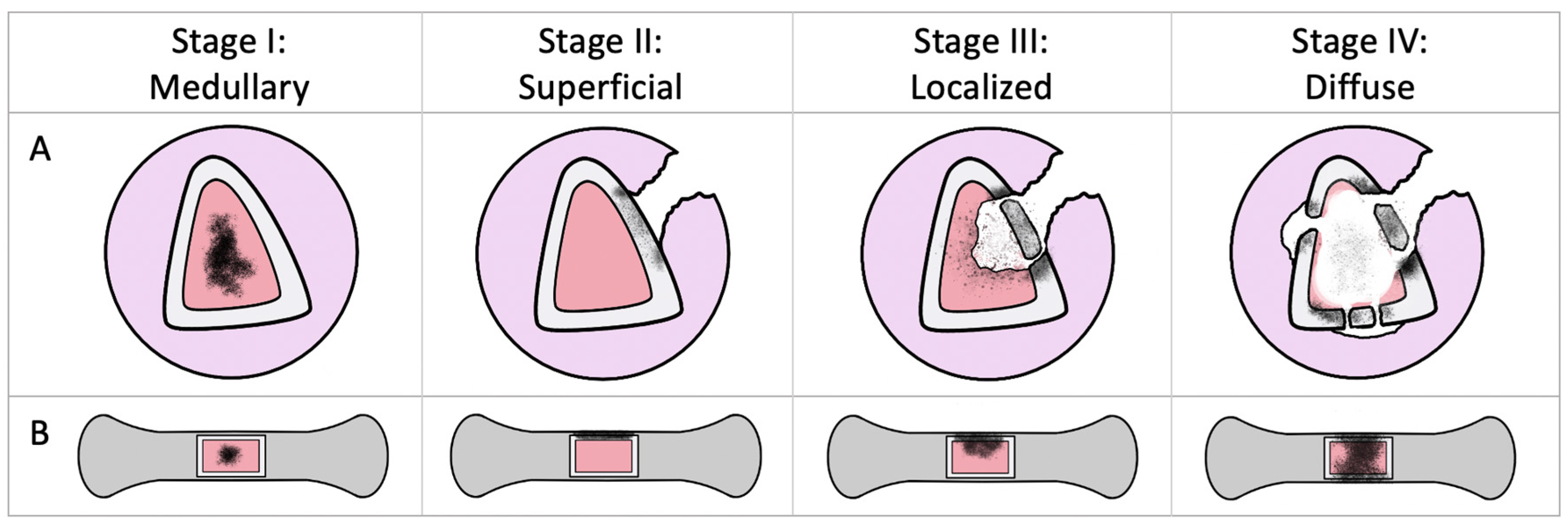
Figure 1. Simplified Cierny–Mader classification. Panel A: cross-sectional view of osteomyelitis in a long bone. Panel B: a longitudinal view with a window of bone removed. Stage 1 (medullary) osteomyelitis is confined to the medullary cavity. Stage 2 (superficial) involves the cortical bone and most often originates from direct inoculation or contiguous infection. Stage 3 (localised) involves both cortical and medullary bone. Stage 4 (diffuse) involves the entire thickness of the bone with a loss of stability. This figure was based on the original Cierny–Mader classification [11].
Applsci 10 02244 g001
Table 1. Clinical studies focusing on antibiotic-eluting, resorbable, calcium-based scaffolds for chronic osteomyelitis.
AuthorsNumber of PatientsMaterialSystemic Antibiotics UsedMean Follow-up (yrs)Eradication RateOther Outcomes
McKee et al., 2002 [68]25OSTEOSET®-T (calcium sulfate + tobramycin)Yes2.392%12% fracture, 32% wound leak, 36% had autologous bone grafting.
McKee et al., 2010 [70]15OSTEOSET®-T (calcium sulfate + tobramycin)Yes3.286% (same result as PMMA)14% fracture, 21% wound leak, 33% underwent further surgical procedures.
Fleiter et al., 2014 [72]20HERAFILL® G (calcium sulfate + calcium carbonate + gentamicin)No0.580%No adverse outcomes reported, sufficient gentamicin elution rates measured.
Humm et al., 2014 [73]21OSTEOSET®-T (calcium sulfate + tobramycin)Yes1.395%33.3% wound discharge, 100% union rate, 24% delayed wound-healing or pin-site infections.
Ferguson et al., 2014 [67]195OSTEOSET ®-T (calcium sulfate + tobramycin)Yes3.791%4.7% fracture (at a mean of 1.9 years), 15.4% wound leak, radiographic bone filling absent in 36.6%, partial in 59% and complete in 8%.
McNally et al., 2016 [74]100CERAMENT® G (calcium sulfate + hydroxyapatite + gentamicin)Yes1.696%3% fracture, 6% wound leak.
Table 2. Experimental studies focusing on antibiotic-eluting, composite scaffolds for chronic osteomyelitis.
AuthorsStudy TypeMaterialsMain Finding [s]
Cheng et al., 2017 [76]In vitroBioglass + PLGA + vancomycinSupported the fewest viable bacteria compared to controls after 24 h of S. aureus culture. Effect was maintained even after 6 cycles of exposure.
Wang et al., 2017 [79] In vivo (rabbits)Silica microspheres + nano-HA + polyurethane + levofloxacin (lev)Increased bone formation compared to controls and lev-PMMA at 6- and 12-weeks. After this time, the scaffold began to degrade.
Zhou et al., 2018 [78]In vivo (rabbits)Gelatin + β-TCP + vancomycinAt 8 weeks, the radiological and histopathological severities were significantly better than controls (7.3× and 3.66× respectively).
Kamboj et al., 2019 [80]In vitroSilicon–calcium silicate + polycaprolactone + vancomycin (3D-printed)Observed a two-step, controlled antibiotic release profile: ~50% during the first 40 h, then sustained release of 20% over the next 6 days.
Kuang et al., 2019 [81]In vitroSilica microspheres + nano-HA + polyurethane + levofloxacinObserved increased osteogenic differentiation of bone marrow stem cells at 14 days, a lower number of bacterial colony units at 12 days, decreased apoptosis of osteoblast precursors and decreased microbial adhesion compared to controls.
Zhang et al., 2019 [77]In vivo (rats)Silk + nanosilver + gentamicinLower colony count at 3 weeks compared to controls. Four of the six cases in this group inhibited bacterial growth completely.