Article

4-Hexylresorcinol Exhibits Different Characteristics to Estrogen

1
Department of Oral and Maxillofacial Surgery, College of Dentistry, Gangneung-Wonju National University, Gangneung 28644, Korea
2
Department of Oral and Maxillofacial Surgery, School of Dentistry, Jeonbuk National University, Jeonju 54896, Korea
3
Sericultural and Apicultural Division, National Institute of Agricultural Science, Rural Development Administration, Wanju 55365, Korea
4
Department of Oral Biochemistry, College of Dentistry, Gangneung-Wonju National University, Gangneung 28644, Korea
5
Biomedical, Surgical and Oral Sciences Department, Maxillofacial and Dental Unit, School of Dentistry, University of Milan, 20122 Milan, Italy
6
School of Biochemistry and Cell Biology, BK21 Plus KNU Biomedical Convergence Program, Skeletal Diseases Analysis Center, Korea Mouse Phenotyping Center (KMPC), Kyungpook National University, Daegu 41944, Korea
*
Author to whom correspondence should be addressed.
Appl. Sci. 2020, 10(5), 1737; https://doi.org/10.3390/app10051737
Submission received: 28 January 2020 / Revised: 19 February 2020 / Accepted: 27 February 2020 / Published: 3 March 2020

Abstract

:
4-Hexylresorcinol (4HR) has been used as a food additive and antiseptic. The aim of this study was to evaluate whether the application of 4HR in breast cancer cells and ovariectomized rats showed estrogen-like effects. MCF-7 and SK-BR-3 cells were treated by solvent, 1–100 µM bisphenol-A (BPA), or 1–100 µM 4HR, respectively. 3-(4, 5-Dimethylthiazole-2-yl)- 2,5-diphenyltetrazolium bromide (MTT) assay and Western blot for extracellular signal-regulated kinase-1/-2 (ERK1/2), phosphorylated ERK1/2 (p-ERK1/2), estrogen receptor-α (ERα), and ERβ were done. As an in vivo study, ovariectomized rats (n = 15) received solvent, 125 mg/kg of 4HR, or 10 µg of 17-β estradiol via daily subcutaneous injection for 7 days. Blood samples were obtained for evaluation of prolactin levels. Pituitary glands and uteruses were biopsied for histological evaluation and Western blot analysis. Compared with the control group, the application of 4HR decreased the proliferation of MCF-7 and SK-BR-3 cells, while the application of BPA increased (p < 0.05). The application of BPA increased the expression of ERα, ERβ, and p-ERK1/2, but 4HR did not change the expression of ERα, ERβ, or p-ERK1/2 in MCF-7 cells. In an animal model, the 4HR group showed similar levels of ERα, ERβ, and prolactin expression in the pituitary gland compared to the solvent only group, while the estradiol group showed higher levels. Serum prolactin levels were similar between the 4HR and solvent only groups. Taken together, 1–100 µM 4HR did not show BPA-like behavior in MCF-7 cells, and 125 mg/kg of 4HR daily subcutaneous injection for 7 days did not demonstrate estradiol-like effects in ovariectomized rats. Collectively, 4HR has no estrogen-like effects on both ERα-positive cells and estrogen-deficient rat models.

1. Introduction

4-Hexylresorcinol (4HR) has been widely used as an antiseptic [1] and food additive [2]. In the case of antiseptics, usage of 4HR has been decreased because of substitute development. However, 4HR is still used as an antibrowning agent because it is a strong inhibitor of tyrosinase [3]. Recently, other effects of 4HR have come into the limelight. 4HR has anticancer properties via inducing cancer cell apoptosis [4,5] and has been studied as an additive for graft materials because of its inhibitory effect on the nuclear factor kappa-light-chain-enhancer of activated B cells (NF-κB) pathway [6] and foreign body giant cell formation [7]. When 4HR is added to bone graft material, mineralization is increased [6,7]. 4HR helps macrophages to transform M2-type and angiogenesis [8]. Accordingly, 4HR incorporation is a new strategy for immunomodulation. However, increased bone mass and angiogenesis by 4HR may be similar to the actions of estrogen. Estrogen-like materials have been studied as endocrine disruptor and breast cancer inducer [9,10]. Differentiation of 4HR from other xenoestrogens is important for usage of 4HR as immunomodulation agent.
Xenoestrogen is the chemical that mimics the action of estrogen and results in endocrine system disruption [9,10]. Xenoestrogen may pollute and destroy the natural endocrine system. Many types of mutation may be associated with abuse of, or contamination with, xenoestrogen [10]. Xenoestrogen is a member of a group of organic phenolic compounds with a benzene ring [11]. Bisphenol A (BPA) is a representative chemical [9,10]. Global fear for xenoestrogen has sharply increased recently [12]. Even resveratrol in wine, which is a kind of phenolic compound, is also suspected to be a xenoestrogen [13]. However, there has been no clear evidence between wine drinking and endocrine system destruction.
4HR is also a type of phenolic compound. The binding affinity of 4HR to estrogen receptors (ER) has been reported as weak, but the application of 1–10 µM of 4HR showed increased activity of estrogen responsive promoters in MCF-7 cells [14]. There has been no report of endocrine system disruption by 4HR in human trials until now, despite its wide usage as a food additive. However, 1000 mg/kg body weight/day of 4HR oral administration induced all rats’ death during the first week of the administration, and the autopsy demonstrated a decreased spermatogenesis in 4 out of 10 animals [15]. The influence of the male genital system is a feature of xenoestrogen [16]. Thus, 4HR cannot be excluded from the list of potential xenoestrogens. According to National Health Institute (NIH), United States of America database [15], there has been no available information about the half-life and serum concentration of 4HR in humans because 4HR has been approved in its primary indication as topical agent and its systemic exposure is rare due to poor absorption. Oral gargling is supposed to be used daily, and chronic exposure to 4HR may have unknown risks. Accordingly, 4HR has been listed as a banned component in oral gargling since 2014 by the US Food and Drug Administration (FDA) [17].
However, there has been no convincing evidence for 4HR as a xenoestrogen. 4HR can induce cellular apoptosis in a dose-dependent and cellular type-dependent manner [4,18]. Thus, the inhibition of the spermatogenesis may be a complication of lethal dosage. Many types of anticancer drugs may influence rapidly regenerated normal cells, including sperm [19]. 4HR also has anticancer effects [4,5]. The increased expression of estrogen-responsive promoter in MCF-7 cells may be a nonspecific response of 4HR or cross-activity mediated by an alternative signal pathway, excluding ER. Actually, there was no check of ERα protein level or downstream signal changes in the previous report [14].
In this study, the effect of 4HR on breast cancer cells and ovariectomized animals was evaluated with regard to its potential estrogen-like effects. First, the effect of 4HR application on ER-positive and ER-negative breast cancer cells was evaluated and compared to other xenoestrogens, such as BPA and estrogen. Second, the expression level of ER was evaluated at the protein level. Third, the activation of the extracellular signal-regulated kinase (ERK) pathway by 4HR application was checked and compared to other xenoestrogens and estrogen. Finally, 4HR was administered to the ovariectomized rats. Blood prolactin levels and prolactin expression levels in the pituitary gland were measured and compared to estradiol application.

2. Materials and Methods

2.1. Cell Cultures and 4HR and Xenoestrogen Treatment

SK-BR-3 (Korean Cell Line Bank No. 30030) and MCF-7 (Korean Cell Line Bank No. 30022) were suspended in RPMI 1640 supplemented with 10% fetal bovine serum (FBS) (Euroclone, United Kingdom), 50 U/mL of penicillin G, 50 μg/mL of streptomycin sulfate, 2 g/L sodium carbonate, and 0.11 g/L sodium pyruvate. 4HR and BPA were purchased from Sigma-Aldrich (St. Louis, MO, USA). 17-β Estradiol was purchased from Santa Cruz Biotechnology (Santa Cruz, CA, USA). MCF-7 cells were ER-positive breast cancer cells, and SK-BR-3 cells were ER-negative. 17-β Estradiol or BPA were used as the positive control. Reagent was solubilized by dimethyl sulfoxide (DMSO) and diluted by culture medium.

2.2. MTT Assay

The concentration of cells per well was approximately 8000 in 6-well multi-plates, which were allowed to attach for 24 h. To analyze cellular growth by administration of 4HR or BPA, we treated the cells with 1, 10, and 100 μM of 4HR or BPA. The control was cells treated by solvent only. SK-BR-3 cells or MCF-7 cells were grown with 4-HR or BPA for 48 and 72 h. After incubation, the medium was removed. Then, yellow tetrazolium salt and 3-(4, 5-dimethylthiazole-2-yl)- 2,5-diphenyltetrazolium bromide (MTT) solution (Cell proliferation kit I; Roche Molecular Biochemicals, Pleasanton, CA, USA) were applied for 4 h. Formazan crystals were solubilized overnight, and the product was quantified by measuring the absorbance at 590 nm using a Victor multi-label counter (Perkin-Elmer-Wallac, Freiburg, Germany). The untreated control was set as 1, and relative activity of each group was calculated.

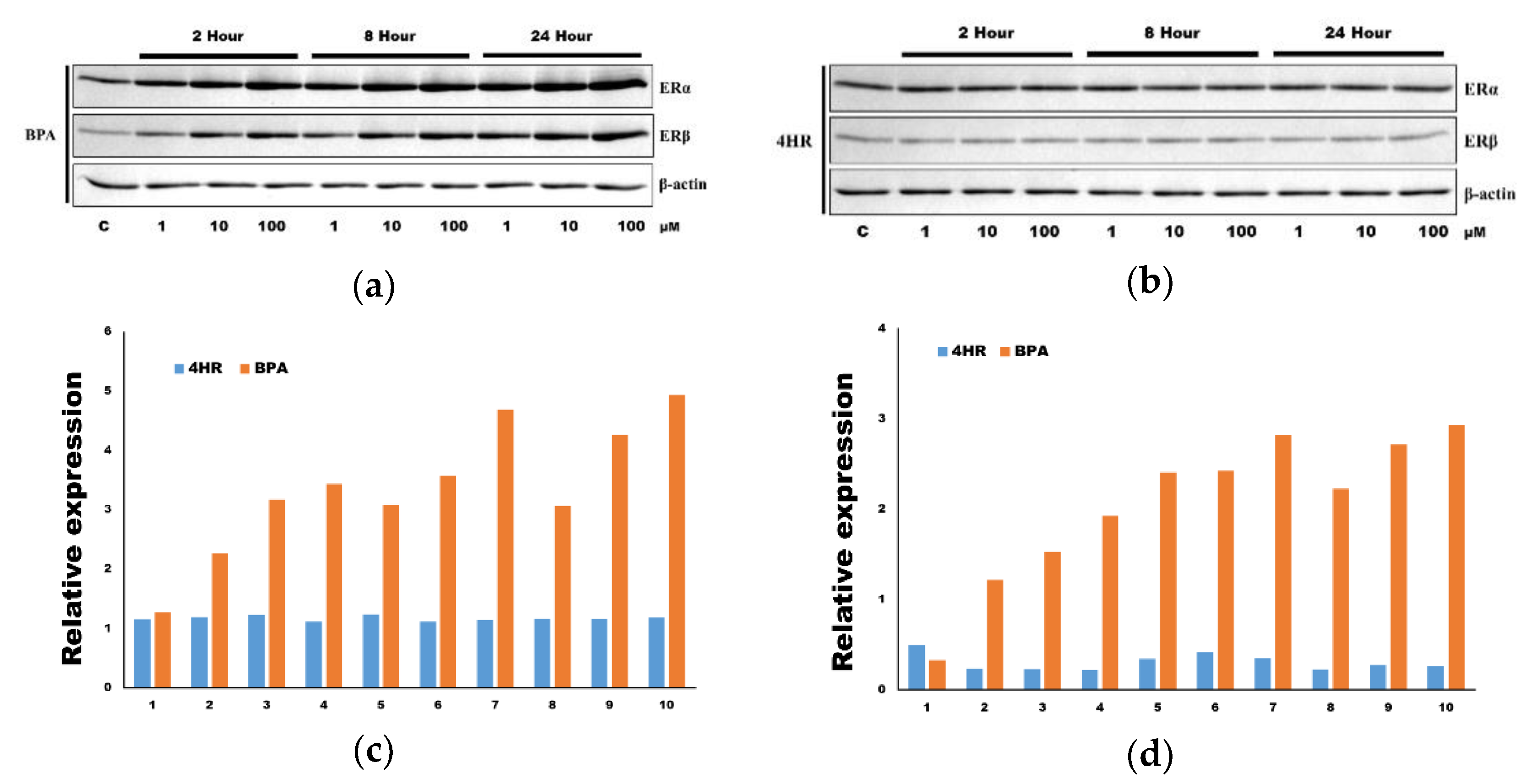
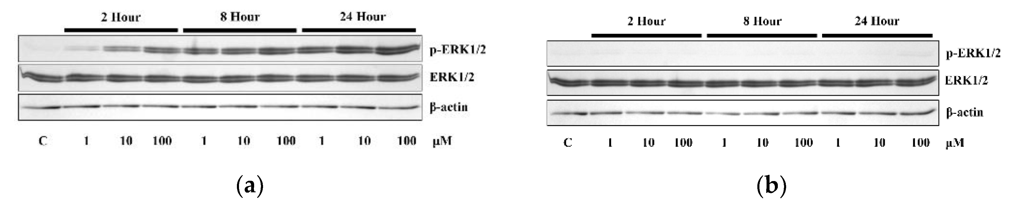
2.3. Western Blotting for ERK1/2, p-ERK1/2, ERα, and ERβ

To analyze the protein expression level of ERK1/2 (Abcam, Cambridge, United Kingdom), p-ERK1/2 (phosphorylated at T202 for ERK1 and phosphorylated at T185 for ERK2, Abcam), ERα (Santa Cruz Biotech), and ERβ (Santa Cruz Biotech), MCF-7 cells were treated with 1, 10, and 100 μM of 4HR or BPA. The control was cells treated by solvent only. Cells were treated with each reagent at various concentrations or for different time periods. The cells were washed with phosphate-buffered saline (PBS), then treated with trypsin-ethylenediaminetetraacetic acid (trypsin-EDTA) for 1 min and washed with PBS. Cells were lysed with ice-cold RIPA buffer, sonicated for 10 sec, and samples should have been cooled on ice between bursts. The lysates were centrifuged at 12,000× g for 10 min at 4 °C. The protein concentration of the supernatant was quantified using Bradford assay. Equal amounts of sample proteins were separated by 15% sodium dodecyl sulfate-polyacrylamide gel electrophoresis (SDS-PAGE) to analyze the protein with protein marker. After the proteins were transferred from the gel to a nitrocellulose membrane, the membranes were blocked with 5% nonfat dry milk in TBS-T buffer (25 mM Tris-HCl, 150 mM NaCl, 0.1% Tween 20, pH 7.5) for 1 h. After being washed with TBS-T buffer three times, the membrane was incubated with the primary antibodies (dilution ratio = 1:500) and horseradish peroxidase-conjugated secondary antibodies for 1 h. Then, the protein bands were detected using an ECL kit according to the manufacturer’s instructions. Blots were imaged and quantified using a ChemiDoc XRS system (Bio-Rad Laboratories, Hercules, CA, USA).

2.4. Animals and Experimental Design

Eight-week-old Crl:CD (Sprague–Dawley) specific pathogen-free (SPF)/viral antibody free (VAF) outbred female rats (Orientbio Inc., Sungnam, Korea) were used in this study. All procedures were performed in accordance with guidelines for laboratory animal care and were approved by the Gangneung-Wonju National University for animal research (GWNU-2019-15). Fifteen rats (2–3 rats per cage) were housed and fed as previously described [8]. All rats stayed for 1 week for acclimation prior to experimentation. Each rat was ovariectomized for both sides of the ovary and rested for 2 weeks after operation. The decreased level of blood estrogen was confirmed by examining the blood samples.
The rats were divided into 3 groups, each with 5 rats. As a negative control, solvent only was used. As a positive control, 17-β estradiol was used. As an experimental group, rats were treated with 4HR. The dosage per day was 1 μg/kg for 17-β estradiol and 128 mg/kg for 4HR. Every agent was prepared just before injection. Each drug was injected subcutaneously for 7 days. One day after final injection, blood was sampled, and then all rats were sacrificed. Blood prolactin levels were measured in whole blood using the RayBio® rat prolactin enzyme-linked immunosorbent assay (ELISA) kit (RayBiotech, Norcross, GA, USA). The subsequent procedure was in accordance with the manufacturer’s protocol. Biopsy of the pituitary gland was also done. Histological and molecular biology analyses were performed.

2.5. Immunohistochemical Determination and Western Blot Analysis in Pituitary Gland Samples

To assess the expression of prolactin, ERα, and ERβ in the pituitary gland samples, we performed immunohistochemical staining using anti-prolactin (Abcam), anti-ERα (Santa Cruz Biotech), and anti-ERβ antibodies (Santa Cruz Biotech). Detailed procedure was in accord with our previous publications [8]. Briefly, the paraffin sections were prepared on silane-coated slides and antigen retrieval was done with proteolytic enzyme (1 mg porcine trypsin, Sigma-Aldrich). Then, the endogenous peroxidase activity was suppressed by H2O2 treatment. The slide was washed with PBS 3 times. After blocking procedure, the sections were treated with primary antibodies (dilution ratio 1:50). After washing process, a universal secondary antibody (Dako REAL™ EnVision™/HRP, Rabbit/Mouse; Dako North America Inc., Carpinteria, CA, USA) was conjugated under a humid chamber. After removing the unreacted secondary antibody, the slides were stained with a chromogen (Dako REAL™ DAB+ Chromogen and Dako REAL™ Substrate Buffer; Dako North America Inc.).
The tissues were placed into micro test tubes and stored at −70 °C overnight (n = 5 for each group). The tissues were vigorously homogenized in a tissue protein extraction reagent buffer with a protease inhibitor cocktail, and Western blot analysis was performed as previously reported [8].

2.6. Statistical Analysis

The comparison among groups was done with the analysis of variance. The Bonferroni method was used as a post hoc test. The level of significance was set as <0.05.

3. Results

3.1. The Application of 4HR Suppressed the Proliferation of Both MCF-7 and SK-BR-3 Cells

The application of BPA (1 to 100 μM) to MCF-7 cells showed slightly increased cellular proliferation compared to the solvent only group (Figure 1a). However, the application of 4HR with the same range of concentration showed significantly decreased cellular proliferation compared to the solvent only group. The same trends were observed in SK-BR-3 cells (Figure 1b). Taken together, BPA showed xenoestrogen effects on both cells as expected. However, 4HR had no proliferative effects on both ERα-positive and -negative cells.

3.2. The Application of 4HR Did Not Increase ERα Expression and p-ERK

The application of BPA (1 to 100 μM) to MCF-7 cells increased the expression of ERα and ERβ (Figure 2a). However, the application of 4HR (1 to 100 μM) to MCF-7 cells neither increased nor decreased the expression of both ERα and ERβ (Figure 2b). The expression of ERβ was much more increased compared to that of ERα in the application of BPA. The application of BPA increased relative expression level of ERα compared to that of 4HR (Figure 2c). The application of BPA increased relative expression level of ERα compared to that of 4HR (Figure 2d).
It has been known that xenoestrogen activates the ERK pathway in the presence of ERα [10,20]. The application of BPA (1 to 100 μM) to MCF-7 cells increased p-ERK1/2 (Figure 3a). However, the application of 4HR (1 to 100 μM) to MCF-7 cells showed no change of p-ERK1/2 level, indicating no activation of the ERK pathway (Figure 3b). The application of BPA slightly increased relative expression level of ERK1/2 compared to that of 4HR (Figure 3c). However, the difference between groups seemed to be not significant. The application of BPA increased relative expression level of p-ERK1/2 compared to that of 4HR (Figure 3d).

3.3. 4HR Did Not Increase Prolactin and ERα Expression in the Pituitary Gland

To further explore the xenoestrogen effect of 4HR, we tested 4HR’s effect in an estrogen-deficient ovariectomized rat model. Daily application of 1 μg/kg of 17-β-estradiol in the ovariectomized rats increased prolactin, ERα, and ERβ expression levels in the pituitary gland as expected (Figure 4a). Blood prolactin levels were also significantly increased compared to the solvent only group (p < 0.05, Figure 4b). However, daily application of 128 mg/kg for 4HR in the ovariectomized rats showed low levels of prolactin, ERα, and ERβ expression in the pituitary gland (Figure 4c). Accordingly, blood prolactin levels were similar to those of the solvent only group and significantly lower than the 17-β-estradiol group (p < 0.05, Figure 4b). These results further confirmed that 4HR had no xenoestrogen effects in an ovariectomized rat model.

4. Discussion

In this study, 4HR was tested as a potential xenoestrogen. However, 4HR showed different behavior to breast cancer cells or ovariectomized animals compared to estradiol or BPA. The application of BPA increased cellular proliferation in both MCF-7 cells and SK-BR-3 cells, which are ER-positive and -negative breast cancer cells, respectively (Figure 1a). By contrast, the application of 4HR showed significantly decreased proliferation in both types of cells (p < 0.05, Figure 1b). The application of BPA increased ERα, ERβ, and p-ERK1/2 expressions, but the application of 4HR did not significantly change those expressions (Figure 2 and Figure 3). In the animal model, the application of estradiol increased the expression of ERα, ERβ, and prolactin in the pituitary gland (Figure 4). However, the application of 4HR showed a similar pattern to the solvent only group (Figure 4). Based on these results, 4HR did not behave like estradiol or BPA in the range of tested concentrations.
The warning for endocrine system disruptors has been increased annually [9,10]. Many types of endocrine disruptors have been introduced and tested for their potential biohazards [14]. These agents include pesticides, such as dichlorodiphenyltrichloroethane (DDT), its metabolites, and many common industrial chemicals, such as polychlorinated biphenyls and BPA [21,22]. Human health effects from exposure to xenoestrogen include: precocious puberty, increased incidence of breast cancer, testicular cancer, prostate cancer, and reduced sperm count [9,14,21]. Long-term uptake of low-dosage xenoestrogen is deposited in adipocytes and may be released in fasting conditions and show estrogen effects [21]. Therefore, health professionals warn against xenoestrogen for both short-term high dosage uptake and long-term low dosage uptake [9,10]. However, the effect of long-term low dosage uptake is difficult to simulate in experimental conditions.
BPA is a well-known xenoestrogen [10]. BPA increases the proliferation of breast cancer cells [23], and our results also showed the same effect on MCF-7 cells (Figure 1a). However, 4HR was cytotoxic to breast cancer cells regardless of ER expression (Figure 1). 4HR is also cytotoxic to primary cultured oral melanoma cells [5] and SCC-9 cells [4] in the similar range of concentration to this study. However, its inhibitory effect is not significant in the normal human dermal fibroblasts [4]. According to NIH toxicology report, 4HR has shown anticancer effects in various types of cancers [15]. As 4HR increases the apoptotic stress mediated by mitochondrial proteins [18], cancer cells seem to be more influenced by 4HR administration. Based on these findings, there has been no evidence that 4HR might increase the incidence of breast cancer.
Several xenoestrogens increase the activity of ER in the absence of estradiol [24]. In this study, BPA increased ERα and ERβ expression in MCF-7 cells (Figure 2a). However, 4HR did not change ERα and ERβ expression (Figure 2b). A previous study demonstrated that 4HR increased luciferase activity, which was localized in the estrogen-responsive promoter region [14]. As the function of estrogen is broad, many genes are regulated by estrogen [25]. Some genes influenced by estrogen may also be influenced by other hormones, cytokines, or chemicals. Activation of ERα results in the increased phosphorylation of ERK [20,26]. In this study, BPA application increased p-ERK1/2 expression, but 4HR did not (Figure 3).
When animals receive an ovariectomy, blood estrogen levels decrease [27]. Accordingly, the sensitivity of parenteral administration of pseudoestrogen is increased [22,28]. Many types of estrogen-like chemicals increase the expression of ERα and the production of prolactin in the anterior pituitary gland [29,30]. In this study, the administration of estradiol showed significantly higher levels of blood prolactin compared to the 4HR and solvent only groups (p < 0.05, Figure 4b). The expression level of ERα, ERβ, and prolactin in the pituitary glands were higher in the estradiol group than those in the 4HR and solvent only groups (Figure 4c). The BPA-injected group (128 mg/kg) also showed similar findings to those of estradiol group (data not shown). The uterine epithelium was thicker in the estradiol groups than those of the 4HR and solvent only groups (Figure 4a). The thickness of uterine epithelium increased in the ovariectomized rats after the administration of BPA [29].
There have been several reports on the estrogen-like behavior of 4HR [14,15]. Lethal dosage of 4HR shows the inhibition of spermatogenesis in some animals [15]. In addition, 4HR showed spermicidal activity in in vitro tests [15]. 4HR has anticancer effects via inducing cellular apoptosis [4,5]. As a complication of anticancer drugs, the inhibition of spermatogenesis is frequent [19]. Thus, the inhibition of spermatogenesis by lethal dosage of 4HR might be due to its apoptosis-inducing effect, not due to endocrine system disruption. 4HR increases the estrogen-responsive promoter region [14]. However, too many genes are included in the estrogen-responsive promoter region [25]. For example, the expression of vascular endothelial growth factor (VEGF) is modulated by xenoestrogens through the ER-dependent pathway [31]. 4HR also increases VEGF expression, but through the hypoxia-inducible factor-independent pathway [8]. ER blocker could not change VEGF expression induced by 4HR (data not shown). In the same report, 4HR showed low affinity to ER [14].
In this study, the application of BPA increased ERα, ERβ, and p-ERK1/2 expressions, but the application of 4HR did not significantly change those expressions (Figure 2 and Figure 3). However, the level of their expression was lower in the 4HR group than those in the BPA group (Figure 2 and Figure 3). The anticancer effect of 4HR has been demonstrated in different types of oral cancer cells [4,5] and in the animal models [4]. Several mechanisms have been clarified. 4HR suppresses calcium oscillation in oral melanoma cells [4]. The suppression of calcium oscillation is important for mitochondrial function [32]. Accordingly, 4HR might suppress the mitochondrial function. Mitochondria are main source of ATP production and cancer cells have higher energy demands. Therefore, 4HR application might have a higher impact on cellular proliferation in the cancer cells rather than in the normal human dermal fibroblast (see supplementary data). The signaling pathways of ERα, ERβ, and ERK1/2 are associated with the carcinogenesis of breast cancer [33]. As the application of 4HR might not be associated with carcinogenesis of breast cancer, its application could not change the expression level of these proteins.
The limitation of this study was the design of the animal study. According to a previous report [34], the species of experimental animal might influence the experimental results. In addition, the dosage and the administration route are important in the experimental design. In this study, a single dosage (128 mg/kg) was used for 4HR experiment. This dosage was referenced from a previous publication on BPA [29]. The median lethal dose (LD50) for rats is reported as 550 mg/kg in the case of oral administration [15]. In future studies, wider variations of dosage in the different species may be required. In addition, the duration of administration was 7 days and relatively short. If 4HR is taken as a food additive, the animal study should be designed as a low-dosage and long-term exposure model. When reviewing a previous study, 63 or 125 mg/kg body weight/day of 4HR oral administration given to B6C3F1 mice for 102 weeks showed no evidence of carcinogenic activity in female mice but equivocal evidence in male mice [35]. The main concern on xenoestrogen is its potential carcinogenic effect on the breast [10,11,12]. In this study, any clue of carcinogenic effect by 4HR on breast cancer cells was not found. In addition, we recently clarified the effect of angiogenesis and osteogenesis by 4HR is mediated not by estrogen, but by transforming growth factor-β1 [18,36].

5. Conclusions

In this study, 4HR decreased proliferation of MCF-7 and SK-BR-3 cells. 4HR did not change ERα expression and did not activate the ERK pathway in MCF-7 cells. 4HR also did not increase blood prolactin levels and ERα and prolactin expression in the pituitary gland. Collectively, there was no evidence of xenoestrogen-like behavior for 4HR in this study.

Supplementary Materials

The following are available online at https://www.mdpi.com/2076-3417/10/5/1737/s1.

Author Contributions

Conceptualization, S.-G.K. and Y.-J.K.; methodology, H.S., Y.-J.K. and J.-H.O.; validation, D.-W.K.; formal analysis, Y.-Y.J.; investigation, S.-G.K.; data curation, J.-H.O.; writing—original draft preparation, S.-G.K.; writing—review and editing, U.G.; supervision, J.-Y.C.; funding acquisition, S.-G.K. All authors have read and agreed to the published version of the manuscript.

Funding

This study was carried out with the support of ‘‘Cooperative Research Program for Agriculture Science and Technology Development (Project no. PJ01313902)’’ Rural Development Administration, Republic of Korea.

Acknowledgments

Dah-Hyun Koo and Yi-sol Jeong are dental school students and helped with preliminary research.

Conflicts of Interest

The authors declare no conflict of interest.

References

  1. Evans, R.T.; Baker, P.J.; Coburn, R.A.; Fischman, S.L.; Genco, R.J. In vitro antiplaque effects of antiseptic phenols. J. Periodontol. 1977, 48, 156–162. [Google Scholar] [CrossRef]
  2. Kim, Y.H.; Kim, J.M.; Lee, J.S.; Gang, S.R.; Lim, H.S.; Kim, M.; Lee, O.H. Development and validation of an analytical method for the determination of 4-hexylresorcinol in food. Food Chem. 2016, 190, 1086–1092. [Google Scholar] [CrossRef]
  3. Ortiz-Ruiz, C.V.; Berna, J.; Rodriguez-Lopez, J.N.; Tomas, V.; Garcia-Canovas, F. Tyrosinase-catalyzed hydroxylation of 4-hexylresorcinol, an antibrowning and depigmenting agent: A kinetic study. J. Agric. Food Chem. 2015, 63, 7032–7040. [Google Scholar] [CrossRef]
  4. Kim, S.G.; Choi, J.Y. 4-hexylresorcinol exerts antitumor effects via suppression of calcium oscillation and its antitumor effects are inhibited by calcium channel blockers. Oncol. Rep. 2013, 29, 1835–1840. [Google Scholar] [CrossRef] [Green Version]
  5. Lee, S.W.; Kim, S.G.; Park, Y.W.; Kweon, H.; Kim, J.Y.; Rotaru, H. Cisplatin and 4-hexylresorcinol synergise to decrease metastasis and increase survival rate in an oral mucosal melanoma xenograft model: A preliminary study. Tumour Biol. 2013, 34, 1595–1603. [Google Scholar] [CrossRef]
  6. Song, J.Y.; Kim, S.G.; Park, N.R.; Choi, J.Y. Porcine bone incorporated with 4-hexylresorcinol increases new bone formation by suppression of the nuclear factor kappa B signaling pathway. J. Craniofac. Surg. 2018, 29, 1983–1990. [Google Scholar] [CrossRef]
  7. Kweon, H.; Kim, S.G.; Choi, J.Y. Inhibition of foreign body giant cell formation by 4-hexylresorcinol through suppression of diacylglycerol kinase delta gene expression. Biomaterials 2014, 35, 8576–8584. [Google Scholar] [CrossRef] [PubMed]
  8. Jo, Y.Y.; Kim, D.W.; Choi, J.Y.; Kim, S.G. 4-Hexylresorcinol and silk sericin increase the expression of vascular endothelial growth factor via different pathways. Sci. Rep. 2019, 9, 3448. [Google Scholar] [CrossRef] [PubMed] [Green Version]
  9. Chin, K.Y.; Pang, K.L.; Mark-Lee, W.F. A review on the effects of bisphenol A and its derivatives on skeletal health. Int. J. Med. Sci. 2018, 15, 1043–1050. [Google Scholar] [CrossRef] [PubMed] [Green Version]
  10. Shafei, A.; Ramzy, M.M.; Hegazy, A.I.; Husseny, A.K.; El-Hadary, U.G.; Taha, M.M.; Mosa, A.A. The molecular mechanisms of action of the endocrine disrupting chemical bisphenol A in the development of cancer. Gene 2018, 647, 235–243. [Google Scholar] [CrossRef] [PubMed]
  11. Mosconi, G.; Carnevali, O.; Franzoni, M.F.; Cottone, E.; Lutz, I.; Kloas, W.; Yamamoto, K.; Kikuyama, S.; Polzonetti-Magni, A.M. Environmental estrogens and reproductive biology in amphibians. Gen. Comp. Endocrinol. 2002, 126, 125–129. [Google Scholar] [CrossRef] [PubMed]
  12. Hutchinson, T.H.; Ankley, G.T.; Segner, H.; Tyler, C.R. Screening and testing for endocrine disruption in fish-biomarkers as “signposts,” not “traffic lights,” in risk assessment. Environ. Health Perspect. 2006, 114 (Suppl. 1), 106–114. [Google Scholar] [CrossRef] [PubMed] [Green Version]
  13. Jing, Y.; Hu, T.; Lin, C.; Xiong, Q.; Liu, F.; Yuan, J.; Zhao, X.; Wang, R. Resveratrol downregulates PCSK9 expression and attenuates steatosis through estrogen receptor α-mediated pathway in L02cells. Eur. J. Pharmacol. 2019, 855, 216–226. [Google Scholar] [CrossRef] [PubMed]
  14. Amadasi, A.; Mozzarelli, A.; Meda, C.; Maggi, A.; Cozzini, P. Identification of xenoestrogens in food additives by an integrated in silico and in vitro approach. Chem. Res. Toxicol. 2009, 22, 52–63. [Google Scholar] [CrossRef] [PubMed] [Green Version]
  15. Hexylresorcinol, Compound Summary, U.S. National Library of Medicine, NIH. Available online: https://pubchem.ncbi.nlm.nih.gov/compound/3610#section=Metabolism-Metabolites (accessed on 17 February 2020).
  16. Duan, P.; Hu, C.; Quan, C.; Yu, T.; Zhou, W.; Yuan, M.; Shi, Y.; Yang, K. 4-Nonylphenol induces apoptosis, autophagy and necrosis in Sertoli cells: Involvement of ROS-mediated AMPK/AKT-mTOR and JNK pathways. Toxicology 2016, 341–343, 28–40. [Google Scholar] [CrossRef] [PubMed]
  17. Food and Drug Administration. Consumer Antiseptic Wash Final Rule. Questions and Answers. 2017. Available online: https://www.fda.gov/Drugs/GuidanceComplianceRegulatoryInformation/Guidances/default.htm (accessed on 28 January 2020).
  18. Kim, M.K.; Yoon, C.S.; Kim, S.G.; Park, Y.W.; Lee, S.S.; Lee, S.K. Effects of 4-hexylresorcinol on protein expressions in RAW264.7 cells as determined by immunoprecipitation high performance liquid chromatography. Sci. Rep. 2019, 9, 3379. [Google Scholar] [CrossRef]
  19. Okada, K.; Fujisawa, M. Recovery of spermatogenesis following cancer treatment with cytotoxic chemotherapy and radiotherapy. World J. Mens Health 2019, 37, 166–174. [Google Scholar] [CrossRef]
  20. Lei, B.; Sun, S.; Zhang, X.; Feng, C.; Xu, J.; Wen, Y.; Huang, Y.; Wu, M.; Yu, Y. Bisphenol AF exerts estrogenic activity in MCF-7cells through activation of Erk and PI3K/Akt signals via GPER signaling pathway. Chemosphere 2019, 220, 362–370. [Google Scholar] [CrossRef]
  21. Bengtson, D.A.; Henshel, D.S. Environmental Toxicology and Risk Assessment: Biomarkers and Risk Assessment. 5th Volume; American Society for Testing and Materials: West Conshohocken, PA, USA, 1996. [Google Scholar]
  22. Bigsby, R.M.; Caperell-Grant, A.; Madhukar, B.V. Xenobiotics released from fat during fasting produce estrogenic effects in ovariectomized mice. Cancer Res. 1997, 57, 865–869. [Google Scholar]
  23. Zhao, Q.; Howard, E.W.; Parris, A.B.; Ma, Z.; Xing, Y.; Yang, X. Bisphenol AF promotes estrogen receptor-positive breast cancer cell proliferation through amphiregulin-mediated crosstalk with receptor tyrosine kinase signaling. PLoS ONE 2019, 14, e0216469. [Google Scholar] [CrossRef] [Green Version]
  24. Hess-Wilson, J.K.; Boldison, J.; Weaver, K.E.; Knudsen, K.E. Xenoestrogen action in breast cancer: Impact on ER-dependent transcription and mitogenesis. Breast Cancer Res. Treat. 2006, 96, 279–292. [Google Scholar] [CrossRef] [PubMed]
  25. Handel, A.E. Bioinformatics analysis of estrogen-responsive genes. Methods Mol. Biol. 2016, 1366, 29–39. [Google Scholar] [PubMed] [Green Version]
  26. Wang, T.; Liu, B.; Guan, Y.; Gong, M.; Zhang, W.; Pan, J.; Liu, Y.; Liang, R.; Yuan, Y.; Ye, L. Melatonin inhibits the proliferation of breast cancer cells induced by bisphenol A via targeting estrogen receptor-related pathways. Thorac. Cancer 2018, 9, 368–375. [Google Scholar] [CrossRef] [PubMed] [Green Version]
  27. Collins, F.L.; Rios-Arce, N.D.; Atkinson, S.; Bierhalter, H.; Schoenherr, D.; Bazil, J.N.; McCabe, L.R.; Parameswaran, N. Temporal and regional intestinal changes in permeability, tight junction, and cytokine gene expression following ovariectomy-induced estrogen deficiency. Physiol. Rep. 2017, 5, e13263. [Google Scholar] [CrossRef] [PubMed]
  28. Newbold, R.R.; Jefferson, W.N.; Padilla-Banks, E.; Haseman, J. Developmental exposure to diethylstilbestrol (DES) alters uterine response to estrogens in prepubescent mice: Low versus high dose effects. Reprod. Toxicol. 2004, 18, 399–406. [Google Scholar] [CrossRef] [PubMed]
  29. Goloubkova, T.; Ribeiro, M.F.; Rodrigues, L.P.; Cecconello, A.L.; Spritzer, P.M. Effects of xenoestrogen bisphenol A on uterine and pituitary weight, serum prolactin levels and immunoreactive prolactin cells in ovariectomized Wistar rats. Arch. Toxicol. 2000, 74, 92–98. [Google Scholar] [CrossRef]
  30. Ronchetti, S.A.; Miler, E.A.; Duvilanski, B.H.; Cabilla, J.P. Cadmium mimics estrogen-driven cell proliferation and prolactin secretion from anterior pituitary cells. PLoS ONE 2013, 8, e81101. [Google Scholar] [CrossRef] [Green Version]
  31. Buteau-Lozano, H.; Velasco, G.; Cristofari, M.; Balaguer, P.; Perrot-Applanat, M. Xenoestrogens modulate vascular endothelial growth factor secretion in breast cancer cells through an estrogen receptor-dependent mechanism. J. Endocrinol. 2008, 196, 399–412. [Google Scholar] [CrossRef] [Green Version]
  32. Li, X.; Zhang, S.; Liu, X.; Wang, X.; Zhou, A.; Liu, P. Dynamic analysis on the calcium oscillation model considering the influences of mitochondria. Biosystems 2018, 163, 36–46. [Google Scholar] [CrossRef]
  33. Whyte, J.; Bergin, O.; Bianchi, A.; McNally, S.; Martin, F. Key signalling nodes in mammary gland development and cancer. Mitogen-activated protein kinase signaling in experimental models of breast cancer progression and in mammary gland development. Breast Cancer Res. 2009, 11, 209. [Google Scholar] [CrossRef]
  34. Nagao, T.; Saito, Y.; Usumi, K.; Yoshimura, S.; Ono, H. Low-dose bisphenol A does not affect reproductive organs in estrogen-sensitive C57BL/6N mice exposed at the sexually mature, juvenile, or embryonic stage. Reprod. Toxicol. 2002, 16, 123–130. [Google Scholar] [CrossRef]
  35. Chhabra, R.S.; Huff, J.E.; Haseman, J.; Hall, A.; Baskin, G.; Cowan, M. Inhibition of some spontaneous tumors by 4-hexylresorcinol in F344/N rats and B6C3F1 mice. Fundam. Appl. Toxicol. 1988, 11, 685–690. [Google Scholar] [CrossRef]
  36. Choi, K.H.; Kim, D.W.; Lee, S.K.; Kim, S.G.; Kim, T.W. The administration of 4-hexylresorcinol accelerates orthodontic tooth movement and increases the expression level of bone turnover markers in ovariectomized rats. Int. J. Mol. Sci. 2020, 21, 1526. [Google Scholar] [CrossRef] [PubMed] [Green Version]
Figure 1. The results of MTT assay. (a) The application of BPA (1 to 100 μM) to MCF-7 cells showed increased value compared to solvent only group at 48 and 72 h after administration. However, the application of 4HR with the same range of concentration showed decreased value compared to the solvent only group at both 48 and 72 h after administration. (b) The same trends were observed in SK-BR-3 cells (* p < 0.05).
Applsci 10 01737 g001
Figure 2. The expression of estrogen receptors after the administration of BPA or 4HR. (a) The application of BPA (1 to 100 μM) to MCF-7 cells increased the expression of ERα and ERβ. (b) The application of 4HR (1 to 100 μM) to MCF-7 cells did neither increase nor decrease the expression of both ERα and ERβ. (c) Relative protein expression of ERα to β-actin. The application of BPA increased relative expression level of ERα compared to that of 4HR. (d) Relative protein expression of ERβ to β-actin. The application of BPA increased relative expression level of ERα compared to that of 4HR. Full length blots were available in Supplementary Figures S1 and S2.
Applsci 10 01737 g002
Figure 3. The expression of extracellular signal-regulated kinase-1/-2 (ERK1/2) and phosphorylated p-ERK1/2 (p-ERK1/2). (a) The application of BPA (1 to 100 μM) to MCF-7 cells increased the expression of p-ERK1/2. (b) The application of 4HR (1 to 100 μM) to MCF-7 cells showed no change of p-ERK1/2 expression. (c) Relative protein expression of ERK1/2 to β-actin. The application of BPA slightly increased relative expression level of ERK1/2 compared to that of 4HR. However, the difference between groups seemed to be not significant. (d) Relative protein expression of p-ERK1/2 to β-actin. The application of BPA increased relative expression level of p-ERK1/2 compared to that of 4HR. In the case of 4HR, p-ERK1/2 was barely detected. Full length blots were available in Supplementary Figures S3 and S4.
Applsci 10 01737 g003aApplsci 10 01737 g003b
Figure 4. The results of in vivo experiment. (a) Daily application of 1 μg/kg for 17-β-estradiol in the ovariectomized rats resulted in increased prolactin, ERα, and ERβ expression level in the pituitary gland. However, daily application of 128 mg/kg for 4HR and solvent only groups showed low levels of prolactin, ERα, and ERβ expression in the pituitary gland. Interestingly, both 17-β-estradiol group and 4HR group showed high levels of vascular endothelial growth factor (VEGF)-A expression in the pituitary gland. In addition, epithelial hyperplasia was only observed in the estradiol group (arrow) (bar = 20 μm). (b) Blood prolactin level. Blood prolactin levels were increased in the estradiol group compared to the 4HR and solvent only groups (* p < 0.05). (c) Western blot for pituitary gland tissue samples. Prolactin, ERα, and ERβ expression were increased in the estradiol group (Sol: solvent only group; Est: estradiol group). Full length blots were available in Supplementary Figure S5.
Applsci 10 01737 g004

