Review

Application of Organic-Inorganic Hybrids in Chemical Analysis, Bio- and Environmental Monitoring

1
Institute of Biochemistry, Saarland University, 66123 Saarbrücken, Germany
2
Process and Systems Engineering Center (PROSYS), Department of Chemical and Biochemical Engineering, Technical University of Denmark, 2800 Kgs. Lyngby, Denmark
3
NanoBioMedical Centre, Adam Mickiewicz University in Poznan, 61-614 Poznan, Poland
*
Authors to whom correspondence should be addressed.
Appl. Sci. 2020, 10(4), 1458; https://doi.org/10.3390/app10041458
Submission received: 29 January 2020 / Revised: 12 February 2020 / Accepted: 14 February 2020 / Published: 21 February 2020

Abstract

:
Organic-inorganic hybrids (OIH) are considered to be a powerful platform for applications in many research and industrial fields. This review highlights the application of OIH for chemical analysis, biosensors, and environmental monitoring. A methodology toward metrological traceability measurement and standardization of OIH and demonstration of the role of mathematical modeling in biosensor design are also presented. The importance of the development of novel types of OIH for biosensing applications is highlighted. Finally, current trends in nanometrology and nanobiosensors are presented.

1. Introduction

Development of novel materials for chemical analysis and biosensor application is one of the most significant challenges in today’s science and technology. Novel developed platforms applied for biosensors and chemical analyses are mostly produced from inorganic materials [1,2,3]. However, in some cases, where it is significantly important to use specific mechanical (e.g., flexibility) or optical (e.g., transmittance) properties, these developed materials should also include organic components, and as a consequence, specific polymeric and/or (bio)receptors are used. The possibility of combining properties of inorganic (e.g., high charge-carrier mobility, mechanical and thermal stability) and organic (e.g., low cost, flexibility, facile tuning of optical and electrical properties) components in a novel composite material with unique features has been a tremendous challenge for a long period.
Organic-inorganic hybrids (OIH) consist of two or more different components, typically inorganic material (metal/semiconductor/dielectric particle or bulk component) and organic components which could be organic functional groups or molecules, biomolecules, polymers, etc., [4,5,6,7,8]. These components are attached together by specific interactions that result in the synergistic enhancement of their specific properties. OIH have attracted a lot of attention in the fields of ceramics [9], polymer chemistry [10], organic and inorganic chemistry [11], physics and material engineering [12,13].
Interestingly, paint pigments, i.e., Prussian Blue (iron(III) hexacyanoferrate) dispersed in organic solvents can be readily considered to be the earliest representatives of organic-inorganic hybrids [14]. However, the “organic-inorganic hybrid” concept appeared only in the 1980s with the development of soft inorganic chemistry [14]. Since that time, the preparation, characterization, and applications of organic-inorganic hybrids have become a fast expanding area of research in materials and life science, nanotechnology, bio- and environmental monitoring. For instance, the above-mentioned Prussian Blue has been successfully employed in biosensor development because of its selective ability toward hydrogen peroxide (H2O2) reduction in the presence of O2 as a product of enzymatic activity [15].
In the past years OIH were developed on a large scale with a lot of applications in analytical chemistry, and in bio- and environmental monitoring [16,17,18]. One may note the tremendous increase in the number of publications and patents relating to the development of novel OIH. Such composites may be casted into thin films [19,20], nanofibers [21], nanoparticles [22], porous materials [23], and hierarchical nanostructures [24], which is extremely important, for instance, to design effective biosensing platforms with quick read-out capabilities for biomedicine and environmental applications. Recently, considerable efforts have been made toward the development of electrochemical transducers for glucose detection which finds applications for diagnostic purposes, viz., point of care or in the food and beverage industry. The possibility to use OIH based on gold (Au)-chitosan nanoparticles (NPs) [25], and nanofilms [26] was demonstrated for efficient detection of glucose at different concentration range. In these OIH, chitosan provides a biocompatible environment and temperature-related harsh physiological conditions. On the other hand, Au-NPs enhance the overall system stability, catalytic activity, and facilitate the electron transfer for hydrogen peroxide removal. The same effect was shown for another OIH based on polyaniline-Au-NPs [27]. Recently, Pt-porphyrin-encapsulated polylactic acid (PLA) NPs were synthesized and glucose biosensors with an improved detection range were developed [28]. Apart from glucose biosensors, OIH materials are widely used for the detection of cancer biomarkers [29], toxins [30], and ions of heavy metals [31]. Taking into account the importance of the detection of small molecular weight biomolecules that have a physiological meaning in complex real samples, the efficiency of OIH-based biosensors for lactate [32], glutamate [33], and dopamine analysis was highlighted [34]. Multifarious electrode materials, bioreceptors, organic supplements and immobilization techniques were employed for the detection of the mentioned bioanalytes in different media with various rates of success [35,36,37].
Remarkably, the quality of the novel OIH in general, and biosensors as a case study, must be determined by appropriate control measures. However, the general guidelines toward standardization and validation of OIH are currently under development. Furthermore, the use of mathematical approaches as a rule of thumb for an accurate and rapid process and/or design analysis and improvement is still lacking when developing novel materials and biosensors. Nonetheless, in the last decades various data analysis, process control, and optimization tools using computational intelligence have proven their efficiency and were successfully integrated in different fields of biotechnology. Increased use of modelling techniques, especially with first principles based models, for novel OIH and biosensors already at the development stage would significantly advance the understanding of the underlying mechanisms and the observed response. As a consequence, mathematical modelling could provide the necessary guidance toward efficient and rapid design and performance improvement, which could minimize the experimental efforts, reagent usage, and waste generation. Another aspect to be considered is the creation of a web-based database tool containing continuously updated experimental information on the novel OIH and biosensors designs, materials, reactions, physical and/or chemical properties etc. Such online data-based platforms would not only provide an easy access to the current state-of-the art but also facilitate the general progress in the biosensor and OIH research.
In addition to creation of databases for documentation of OIH and biosensors, systematic classification of available knowledge on such systems in mathematical models is also an important tool that can be highly beneficial for speeding up the future OIH and biosensor development. In essence, a mathematical model can represent complex relationships between different system variables that are of relevance, and can be used to simulate or predict the behavior of a system, depending on for example specific changes in system operating conditions. Combined with a suitable optimization algorithm, such a mathematical model can, for example, be used for in silico prediction of the best set of operating or design conditions for a system, which for a biosensor could translate in prediction of the biosensor architecture that has the highest reliability or stability.
This review describes OIH materials development and their application for chemical analysis, bio- and environmental monitoring. Mostly, we focus on OIH materials that found application in biosensors and nanobiosensors. More importantly, herein, we propose a methodology toward metrological traceability measurement and standardization of nano-based OIH (i) and demonstrate the usefulness of the application of mathematical tools and modeling techniques in biosensors/nanobiosensors development (ii).

2. Classification and Synthesis of Organic-Inorganic Hybrids (OIH)
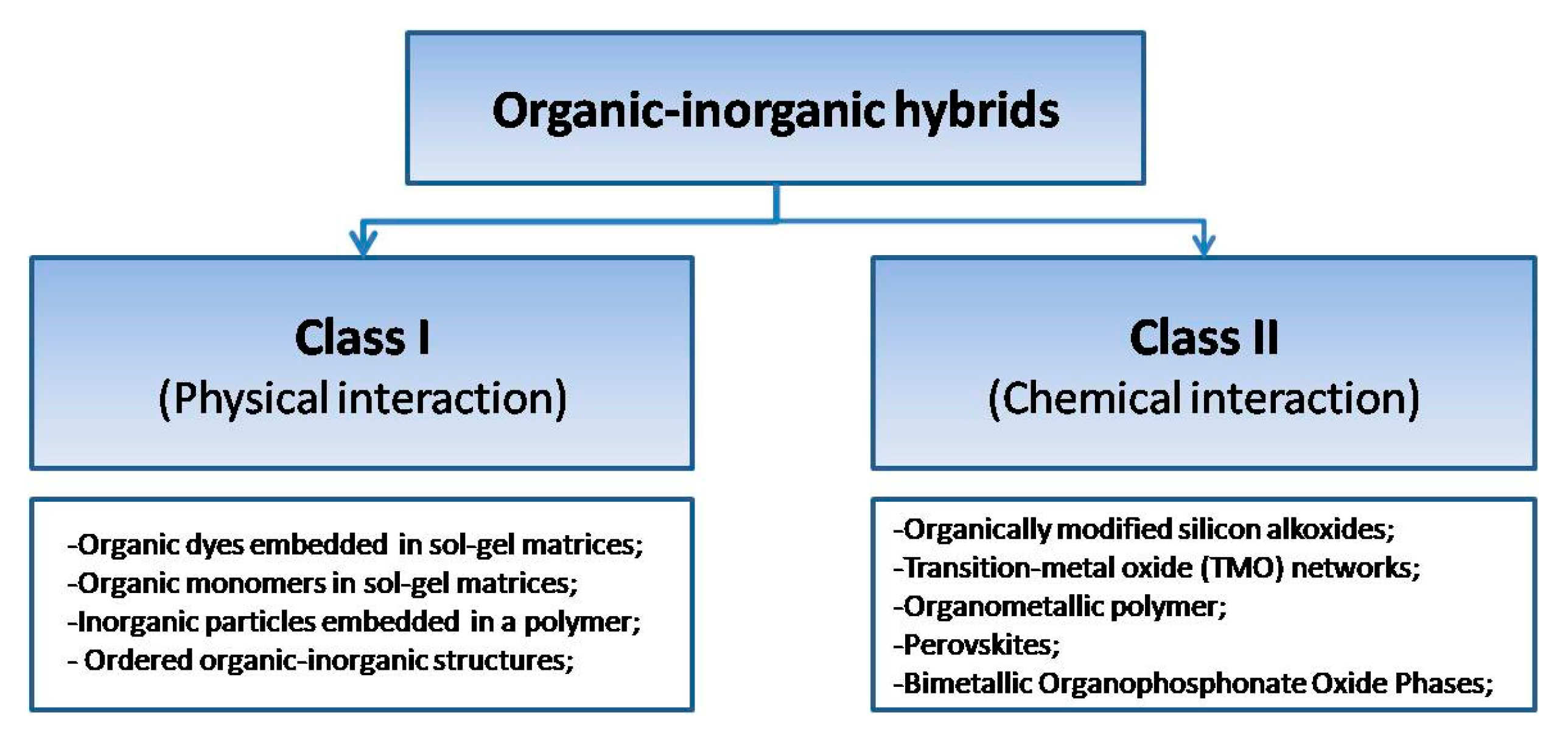
2.1. Classification

Depending on the nature of the organic-inorganic interface, hybrid materials can be classified into two main group: (i) hybrid systems where components have weak interaction (Van der Waals, electrostatic or hydrogen bonds); and (ii) organic-inorganic components that are linked by covalent or ionic-covalent chemical bonds [17].
The first type or class of OIH (Type/Class I) is characterized by the physical interaction (π–π interaction, hydrogen bonds formation etc.,) between organic and inorganic components. Notably, OIH materials of Class I occur only when organic and inorganic components both have some specific functional groups on their surface. Otherwise, OIH materials are in an inhomogeneous phase that results in phase separation between organic and inorganic components [38]. This class is mostly presented by various types of core-shell structures (organic-inorganic or vice versa), inorganic particles, or other nanostructures embedded into an organic (e.g., polymer) matrix which found application in biosensors [39], photocatalysis [40], and optoelectronics [41].
Type/Class II. In this type of hybrid materials, chemical bonding occurs between two different phases. The building blocks for class II OIH possess at least two distinct functional groups: metal-to-carbon links which are stable in the hydrolysis reactions and alkoxy groups (R-OM bonds) which may experience hydrolysis-condensation reactions in the presence of water [7]. This class of OIH has found a wide application in batteries [42], filtration [43], hydrophilic/hydrophobic materials [7]. For example, class II can be prepared by alkoxysilane (-Si(OR)3)-functionalized organic compound and telechelic organic component with functional moieties by co-condensation reaction. Other examples of OIH of class II are perovskites, metal-organic frameworks (MOF), and bimetallic organophosphate oxides.
Remarkably, both types/classes of OIH are intensively used for the development of biosensing tools (Figure 1). For biosensing, the design of OIH is determined by the requirements of the analytical task and operational modes of the final device.

2.2. Synthesis

2.2.1. Sol-Gel and Solvothermal Methods

There are numerous methods and approaches of hybrid materials synthesis. The first chemical method to allow the mixing of inorganic and organic components at the nanometric scale was the sol-gel process [44]. Sol-gel processes are a method of forming dispersed inorganic component in organic solvents, through the growth of metal-oxo polymer. The sol-gel approach is a simple and low-temperature method to produce OIH which allows to control the chemical composition of products. Compounds produced by the sol-gel process have many applications in superhydrophobic surfaces [45], electrolytes [46], biosensors [47], corrosion protection [48], etc.
Another method for preparing OIH, similar to the sol-gel process, is solvothermal synthesis [49]. The technique involves separation of organic and metal reagents using immiscible solvents in high temperatures, which can exceed the boiling point of the solvents.

2.2.2. (Self) Assembly Method

Unfortunately, most of OIH prepared via the sol-gel processes are not materials controlled on a meso-, or nano-scopic scale. Therefore, other synthesis strategies should be employed to control hybrid materials on the nanoscale.
The second strategy of hybrid materials synthesis is based on the (self)assembly of monodispersed nanoscale objects: nanoparticles (metal and semiconductor), core/shell nanocomposites, 1D and 2D nanomaterials [50,51]. These nanostructures can be modified by different type of organic molecules, functional dendrimers, biomolecules, polymers [2,52]. This synthesis strategy is often called “legochemistry,” because it allows one to build the hybrid assemblies by simply combining organic/inorganic materials. This method provides a number of advantages, such as better control of its structure on the nanoscale, and as a consequence it facilitates the tailoring of physical properties of the final material.

2.2.3. Supramolecular Template Method

Third methodology is based on the self-assembly properties of polymers and amphiphilic molecules which can create supramolecular OIH with tailored structure and morphology. This method enables the synthesis of OIH nanocomposites from inorganic nanoscale components in an organic matrix (polymer matrix). Combination of this method with the use of hexafunctional organosilanes, has yielded a new type of hybrid mesoporous solids. It was shown that this hybrids in the presence of surfactants poses high degree of porosity which allows to perform a secondary functionalization via post grafting [53].

2.2.4. Combined Method

The last approach is an integrative approach that combines all the above-mentioned methods. Among these methods, which can generate hybrid materials with hierarchical or other types of structures in a single stage, one may include nano-molding, reactive extrusion, electrospinning [54]. Recently, hybrid synthesis approach based on the combination of atomic layer deposition (ALD) and electrospinning (Figure 2) has been reported [55,56,57].
Electrospinning is a low-cost powerful technique that can deposit 1D organic nanofibers, such as collagen, polylactic acid (PLA), gelatine, and hyaluronic acid (HA) [54]. These materials are widely used in tissue engineering and medicine as 3D scaffolds for cell growth [56]. At the same time, these nanofibers can be a template for optical biosensor via ALD coating with photonic material [57,58]. ALD is a method that enables to deposit a broad class of materials with controlled properties. ALD provides the capability of introducing self-limited chemical reactions which lead to the formation of an ultrathin film even for complex morphologies, such as porous structures, nanoparticles, nanotubes etc., [40].
As recently reported, 1D electrospun polyacrylonitrile (PAN) nanofibers coated with a 30 nm ZnO ALD layer showed 1000-fold higher signal response versus planar ZnO nanolayers. The developed 1D ZnO hybrid nanostructures have been applied for optical gas sensing [57]. The obtained structures demonstrated high photoluminescence and great sensitivity to low ethanol concentration. ALD deposited ZnO/Al2O3 nanolaminates onto the organic polymer nanofibers are a new class of photonic materials with advanced structural and optical properties [57]. ALD technology has been improved to deposit ZnO/Al2O3 nanolaminate with a total thickness in the range of 20–40 nm by varying the single layer thickness from 0.5 nm to 10 nm. This methodology allowed to fabricate robust composites with high surface area and possess enhanced optical properties, which was applied for the development of biosensing photoluminescence transducers [50,57,58].
The same strategy can be applied for other materials to develop OIH for the bone tissue engineering (Figure 3). However, instead of using metal oxides, as an inorganic component, and PAN, as an organic one, we have used ALD boron nitrides (BN) and a biodegradable biopolymer, i.e., gelatin [56]. It has been demonstrated that BN effectively reinforces the gelatin matrix and increases the Young’s modulus. It has been also shown that the obtained hybrid is a promising nontoxic, biocompatible material for orthopedic applications [56].
Regardless of the synthesis method, wide spectra of OIH have found a great application in biosensors development with different rate of success. In this regard, it is worth to provide an overview and to classify OIH for biosensing application.

3. Biosensors as a Targeted Class of OIH

Biosensors are portable devices, able to selectively detect target biomolecules in low concentration range in the presence of a complex matrix (serum, blood, saliva, food samples, fermentation media, etc.,). Biosensors enable the conversion of biological functions (e.g., antigen-antibody interactions) into signal read-outs (e.g., change in optical, or electrical properties). This biosensor’s feature enables a wide range of practical applications from environmental monitoring to nanomedicine. The development and tailoring of new materials are important issues to develop novel biosensors with advanced properties. Large surface area, porosity, topography, and enhanced surface functionality can significantly improve the sensing properties of the hybrid material and strengthen the interaction between surface and adsorbate (bioanalyte) [59,60,61]. The use of nanostructured hybrid materials and/or nanocomposites for the development of biosensors, in this case nanobiosensors, gives the possibility to achieve higher analyte sensitivity and device miniaturization.
In recent years, researchers have investigated nano-based OIH (OInH) because of their new properties and applications they demonstrated. Generally speaking, all biosensors are a class of OIH because of the organic or bio-recognition elements (aptamers, antibodies, DNA/RNA etc.,) presented on the biosensor surface.
Biosensors can be prepared by means of different methods, i.e., drop casting and covalent immobilization [62,63,64,65]. Also, a conventional layer-by-layer (LbL) deposition methodology is widely utilized. The LbL fabrication approach offers a superior versatility toward production of the OIH assemblies, including polymer, colloids, and biomolecule coatings [66]. However, in some cases the manufacturing of LbL-enzymatic biosensors involves a number of complex, resource and time-consuming preparation steps. In addition, LbL assays are often suffering from mechanical instability that leads to insufficient signal reproducibility and irreversible changes in sensor architecture.
In order to solve the problems associated with the LbL preparation method, recently a novel one-step technique was proposed for the production of tailored enzymatic nanobiosensors that are instrumentally controlled and allow reproducible, spatial, and temporal resolution, simultaneous multi-analyte detection with high specificity [67]. However, it should be noted that regardless of the fabrication method, the general guidelines towards validation of the OIH in general, as well as towards standardization of biosensors and nanobiosensors as a case study are still under development (see section below titled “The role of nanoanalytics and nanometrology in OIH development”).
As previously defined, a biosensor is an analytical device that includes a combination of biological detecting elements and a transducer. Depending on the transducer’s signal (electrical, optical, mechanical etc.,) and biological recognition element (DNA, enzyme, antibody, molecular imprinted polymer etc.,), one may classify biosensors into different types. For instance, an electrochemical immunosensor means that this biosensor is based on antibody as a recognition element, and in this device linear (I-V), cyclic voltammetry (CV) signal, or electron impedance spectroscopy (EIS) are used.
OIH biosensors based on surface enhanced Raman spectroscopy (SERS) are broadly used in biomedicine applications. SERS is a very sensitive method in which monochromatic laser light excites vibrational modes in adsorbed molecules on a plasmon surface [59]. For instance, a poly(3,4-ethylenedioxythiophene) (PEDOT)-based 3D-OECT device decorated with gold nanoparticles (Au-NPs), as a SERS substrate, has been developed for the detection of dopamine (DA) [68]. This developed OECT/SERS biosensor featured a highly conductive PEDOT: PSS film, which enhanced the sensitivity for the electrochemical sensing of DA in the presence of multiple interferers, such as ascorbic (AA) and uric acids (UA). This biosensor provided an amperometric response to DA with a detection limit of 37 nM in the linear range from 50 nM to 100 μM.
Mahadeva et al. have developed the tin oxide (SnO2)-cellulose hybrids by adopting liquid phase deposition technique and applied it for the detection of urea at low concentration level. The growth of SnO2 nanocoating on the polymer films was revealed [69]. Furthermore, the enzyme (urease) was immobilized into this nanocomposite by physical absorption. The limit of detection for proposed urea biosensors was below a 50 mM concentration.
An electrochemical sensor based on self-doped polyaniline (SPAN) modified metal-organic framework (SPAN@UIO-66-NH2) with high electron mobility was developed. Electrochemical impedance spectroscopy (EIS) and cyclic voltammetry (CV) indicated that doping of SPAN enhanced the electron transfer rates of UIO-66-NH2 in SPAN@UIO-66-NH2. The optimized design of a Cd2+ sensor exhibited good linear response from 0.5 μg L−1 to 100 μg L−1, with a low detection limit of 0.17 μg L−1 (based on S/N = 3), excellent stability, and fast response. It showed decent reliability for detection of trace Cd2+ concentrations in natural lake and urine samples [70].
Among different types of biosensors, optical biosensors become more attractive for end-users because of their small dimensions, low weight, and portability. Furthermore focusing on optical biosensors, these devices do not require electric contacts and demonstrate high precision in measurements [71].
Among a number of different inorganic materials, ZnO is well-known and interesting because of its structure (different architectures i.e., nanoparticles, nanowires, etc.,), electrochemical (high isoelectric point (IEP) (pH~9.1)) and optical properties (high exciton binding energy and high PL at room temperature) [71,72]. Nanoscale ZnO materials offer many advantages for the development of novel biosensing platforms. 1D ZnO nanomaterials (ZnO nanorods and nanowires) have been intensively employed for in vitro biomedical detection (Figure 4) [2,72]. The unique optical properties of 1D ZnO nanostructures have led to extensive development and their integration with optoelectronics and nanophotonics. However, the full potential of 1D ZnO nanomaterials in biosensing have not been realized up to now because of some difficulties in linking ZnO with surface biofunctionalization. Polymer surface functionalization is considered to be the most promising to solve this drawback. Below, one may find several examples to illustrate the state-of-the-art based on polymer surface functionalization.
Thus, polydopamine (PDA) is a mussel-inspired polymer which was initially introduced by Lee et al. in 2007 [73]. Since that time, it has found a lot of applications in material chemistry, nanoscience, and bioengineering [74]. Because of the presence of several reactive groups the PDA structure enables further biomolecule immobilization for biosensors construction, as well as for the development of different biosensing interfaces, viz., glucose oxidase, sulfate-reducing bacteria orinterleukin-6 and human immunoglobulins introduced into/onto the polymer matrix through cross linking [74,75].
Interestingly, besides the intact PDA, ZnO-modified PDA nanostructures (ZnO/PDA) were used in biosensors [76,77]. Briefly, ZnO nanorods, deposited on conductive glass, were coated with PDA. Furthermore, a targeted protein (rabbit IgG) was immobilized on the surface of ZnO to form bioselective layer. Photoelectrochemical tests of the ZnO/PDA toward anti-rabbit IgG showed good sensitivity in the range 100 pg/mL to 500 ng/mL. In summary, combination of ZnO with PDA layers helped to improve optical, electronic, and sensitivity properties of ZnO/PDA toward targeted molecules. Besides, the PDA coating would prevent the photocorrosion of ZnO during photoelectrochemical procedures [77].
Because of the recent progress in nanoengineering, we may see that current research efforts pay more attention to nano-based hybrids. Using nano-based OIH in biosensor development enables to increase the biosensor stability and sensitivity, to enlarge the operating range by extension of lower and upper detection limit, to improve their shelf and storage time. Besides, the development of nanobiosensors provides an opportunity for the fabrication of small non-invasive sensing platforms whose production was considered to be impossible with older technology. On the one side, there is no doubt that nano-based OIH possess much better analytical microsystem characteristics compared to the measurement systems based on pure bulk materials. On the other side, it requires the use of novel methods of validation and “testing” of produced nanoanalytical measurement systems. Therefore, special approaches or methodologies of nanoanalysis and nanometrology for nano-based OIH and nanobiosensors should be systematized or developed.

4. The Role of Nanoanalytics and Nanometrology in Biosensors/Nanobiosensors Development

The unique properties of OIH make this class of materials popular in various application fields, including chemical analysis, SERS, biosensors, nanobiosensors, electrochemical sensors, spectroscopic sensor development, ELISA, immunoassays, molecular imprinted polymers (MIPs), etc. Meanwhile, there is still a great gap between nanohybrids development and their applications in the laboratory and industry. The reason for this situation is the absence of standardization procedures, poor validation, and metrology of the nano-based hybrid materials utilized for chemical analysis and biosensors development.
“I often say that when you can measure what you are speaking about, and express it in numbers, you know something about it; but when you cannot express it in numbers, your knowledge is of a meagre and unsatisfactory kind; it may be the beginning of knowledge, but you have scarcely, in your thoughts, advanced to the stage of science, whatever the matter may be” (Lord Kelvin, 1883) [78]. Today this concept can be summarized as follows: “To measure is equal to know.” In this regard, metrology as a global science of measurements and nanometrology as a part of metrology related to measurements at the nanoscale can directly affect our understanding of novel analytical systems.
Unfortunately, the analysis of the literature data for the last two decades indicates a clear lack of metrology at the nano-scale as a scientific standardization approach, see Figure 5A,B. However, the literature search related to the simple term “nanohybrids” revealed more than 16,600 scientific papers (Figure 5C) published during the same time evidencing the importance and significant impact of this research field in modern science.
Remarkably, the term nanometrology was already introduced in 2007 but the general guidelines toward nanomaterials validation and standardization in general and organic-inorganic nanohybrids (OInH) as a case study still do not exist. Moreover, the current challenges in nanometrology can be summarized as follows: (i) absence of new measurement platforms, test analytical methods, and methodologies working at the nanoscale; (ii) lack of analytical reference materials and documentation standards; (iii) lack of metrological traceability of the measurement that reduces confidence in the accuracy of our analyses and predictions; (iv) measurements in complex environments, i.e., biological matrices, presence of buffers, detergents, or other compounds interfering with a measured signal species (in case of biosensors-electrochemical species).
Obviously, the standardization of nanohybrids is necessary not only to improve our scientific understanding of the underlying fundamental processes responsible for the generated analytical signals, but is also closely linked to the quality control concepts of the systems to promote their industrial applications. Thus, the general role of nanometrology can be summarized by the following formula [79,80,81,82,83,84,85]:
NM = SU + QC + Ap,
where NM—nanometrology; SU—improved scientific understanding on the object; QC—quality control; Ap—application.
Nanometrology plays a crucial role in the production process of nanomaterials and devices with a high degree of accuracy and reliability for different applications. Not accepting this fact can lead to serious mistakes in evaluating results. A current challenge in nanometrology is to establish the criteria for reliability and accuracy of nanomaterials in general and OInH as a case study followed by protocols and approaches to measure them.
Unfortunately, nanometrology today is exclusively based on measurement of geometrical features, viz., size, shape, and roughness of an inorganic component at the nanoscale [86,87,88,89,90,91,92,93], Figure 6. However, to control the roughness (the disorder in the layers) or the shape at nano-scale only makes little sense as compared to the significance of these parameters at the macro- or micro-scales.
It should be noted that measuring at the nanoscale is a technological and scientific challenge that makes metrology at the nanoscale special. Thus, there is a great difference in metrological requirements toward macro-, micro-, and nano-scaled objects. Therefore, the measurement techniques and metrological strategies developed for bulk materials cannot be simply applied for standardization of nano-based structures. Instead, a strong adaption or optimization of the conventional methods and development of the novel methodological approaches is highly desirable. Nanoanalytics, as a global scientific approach, play a great role in this process [96,97].
The term nanoanalytics was initially introduced by Prof. Zolotov [98,99]. Later on, the main concepts, elements, and peculiarities of the nanoanalytics were re-reviewed and summarized by Prof. Shtykov et al. [100]. Recently, the role of nanoanalytics and nanoanalysis as a tool in the development of the modern OInH was highlighted [97]. It is important to stress that two different conceptual strategies are distinguished in nanoanalytics; the first one refers to the methods applied for studying of elemental composition of nano-based hybrids. Another one and more seldomly used strategy in nanoanalytics is to combine several functions in one methodology [98,99]. Thus, the tandem of several techniques and appropriate optimized methodologies, i.e., scanning electron microscopy (SEM) coupled with energy dispersive X-ray microanalysis (EDX), transmission electron microscopy (TEM), atomic force microscopy (AFM), etc., can be readily applied at once for analysis of OInH to get closer insights on the nature of the redistricted analytical signals from the targeted nano-objects [97].
Remarkably, the great challenge in nanoanalytics belongs to the nanoanalysis of the OInH with polymeric or biological component. Thus, the techniques with nanoscale lateral resolution, such as standard SEM or TEM techniques usually provide no chemical information [99]. At the same time, there is no single analytical method or methodology which is able to do this. Therefore, the analyst working with OIH/OInH has to use and optimize a set/tandem of several techniques of molecular analysis and then summarize their analytical merit. Thus, typically the tandem between Fourier transform infrared spectroscopy (FTIR), RAMAN spectroscopy, surface plasmon resonance (SPR), and mass spectrometry (MS)-based techniques can be applied [97]. However, these methods are unable to visualize/image single biomolecules or a targeted polymer with appropriate accuracy at nano-scale and do not allow a local activation or manipulation of such molecules directly in buffer solutions or in real matrices.
In some cases, the above-mentioned problem can be party solved by means of an AFM-based methodology (i.e., local activation or manipulation of biomolecules). This method can be seriously considered as a crucial nanoanalytical tool for characterization of OInH in terms of surface homogeneity (Figure 7) and three-dimensional visualization of polymers or biomolecules (Figure 8).
However, in some cases AFM does not allow to distinguish surface homogeneity between the probe and the artifacts. Also, with regards to OInH analysis, AFM provides analytical information on the organic- or bio- component intermixed with the inorganic component over the template surfaces. For example, in biosensors fabrication it was proven that the original electrode exhibits numerous cavities (see Figure 8) which can be readily transferred to the active end-layer of the final nanohybrid.
Another limitation of the AFM technique is that it is impossible to visualize the changes in surface chemistry of the nano-object. Thus, the heterogeneous blend of polystyrene and low-density polyethylene was characterized exclusively in terms of changes in their height profile, Figure 9. Instead, the changes in chemical profile, the monitoring of the degree of surface impurities, the quantification of the deposited rates, and the verification of possible degradation or re-arrangement of polymer over the surface of the inorganic template would be a desirable alternative.
Notably, quantification of deposition rates for a thin layer of modifier (polymer or biomolecule) regardless of fabrication approaches is usually achieved by means of AT- and BT-cuts quartz crystal microbalances (QCMs), Figure 10 [104,105]. Resonance frequency shifts represent the weight of deposited nano-dimensional OIH structures with nanogram precision. The mass of the deposited nano-based OIH can be estimated using the Sauerbrey equation [106]. Remarkably, the rate of deposition is a very important criterion toward the control of the structure of the thin hybrids, their properties, and synthesis reproducibility [105]. In addition, during monitoring of the deposition process, the true values of the thin film thickness can be readily obtained. Moreover, QCM-based measurement systems are well suitable for the construction of biosensors recognizing affinity interactions [107,108].
Although QCMs were considered as very precise measurement nanoanalytical systems, they greatly suffer from non-linearity and hysteresis. In addition, the dependence of the resonance frequency on temperature makes QCM a technique that is strongly affected by irreproducible environmental factors.
Summarizing, metrology at the nanoscale is addressed for (i) dimensional analysis of thin films, and nanostructured objects, i.e., nanoparticulate surfaces [111,112] and (ii) characterization of the homogeneity/inhomogeneity profile of bio-, organic, and inorganic components over the surface of OIH. However, the standardization protocols and analytical strategies toward comprehensive molecular analysis of the biological and organic component of nanohybrids are almost completely missing. In this regards, special guidelines and evaluation standards for complete (i.e., organic/biomolecule and inorganic component) standardization of OIH/OInH must be developed and introduced in the near future.
To address this issue, recently, a novel nanoanalytical platform for characterization and validation of OInH, including nanobiosensors based on the laser-desorption ionization mass spectrometry (LDI-MS) method was proposed [97]. The workflow of the approach is summarized in Figure 11.
It should be noted, that industrial nanometrology requires both qualitative and quantitative measurements with a minimum number of controlled parameters. Therefore, we do believe that the LDI-MS platform can become a crucial nanoanalytical tool in the nanometrology field. This platform is expected to address several issues, i.e., chemical profiling, metrological traceability for chemical composition between individual layers of the hybrid system, signal to noise ratio (S/N), chemical inhomogeneity, and degree of surface impurities.
However, whereas the great potential of the LDI-MS platform applied for characterization and standardization of the chemical profiles of OIH/OInH has been highlighted already, the general guidelines associated with this approach toward biosensors/nanobiosensors validation are still under development. Thus, the fabrication of the certified references and standards of OIH/OInH and biosensors will be requested in the next step. Moreover, standard approaches for characterization of chemical roughness, degree of chemical homogeneity of the OIH/OInH, as well as definition of the screened position and calibration criteria must be addressed appropriately. Furthermore, it will be important to address the following issues: measurement trueness, precision, accuracy, measurement error, measurement uncertainty, traceability and find criteria for acceptable thresholds, and imaging [112,113]. The general methodology for a metrological traceability measurement protocol is summarized in Figure 12.
Summarizing, it is absolutely clear that there is no single and unique analytical assay that can be applied for complete standardization of the fabricated OIH/OInH in general and for biosensors/nanobiosensors as a case study. Instead, the tandem of several characterization methodologies, innovative analytical approaches, and well-established guidelines should assist the OIH/OInH and biosensors development. Remarkably, a correctly implemented tandem of analytical assays can be beneficial to improve our understanding on the observed physical effects, improve synthesis reproducibility of OIH/OInH from batch-to-batch, intra- and inter-day behavior, intra- and inter-day precision and repeatability of the obtained analytical response.

5. Using Computational Intelligence and Mathematical Simulation and Modeling in OIH

Driven by the desire of creating a robust and systematic approach and/or platform toward control and optimization of operating conditions and chemical processes themselves, mathematical models were successfully integrated into the chemical industry and became a crucial component at the process development stage. The main purpose of using such models is by means of built-in numerical relationships between system input and output parameters to provide the required and relevant information on the dynamic evolution of the variables of interest as a function of time and space. For bio-based processes such system state variables include the main components of biocatalytic or fermentation reactions (e.g., analyte concentration, biomass growth, etc.) combined with specific bioreactor performance metrics that require the introduction of physical (e.g., feed rate, temperature), chemical (e.g., pH, dissolved oxygen), biochemical (e.g., cells composition), and micro-biological (e.g., contamination) parameters [114]. It is also important to mention that the current research objective is based on promoting efficient measurement, monitoring, modeling, and control (M3C) strategies that were set by the European Federation of Biotechnology (EFB) and the European Society of Biochemical Engineering Science (ESBES). The progress and principles of automated measurement, monitoring, control, and optimization, as well as modeling and quality by design (QbD) techniques, currently applied in biotechnology were carefully reviewed in [114].
Development of models of bio-based systems or processes includes data-driven (black-box or empirical models), mechanistic (white-box or first-principles models), and a combination (grey-box models) of both approaches. Thus, when building up the correlations between system parameters (input-output relations) with minimized process knowledge, empirical models can be applied and be rather helpful for example for screening the behavior of OIH-based biosensors with different analytes. However, such models lack the versatility and possibility of achieving a fundamental understanding that is hidden behind the parameter correlations because of the dependency of empirical models on the specific dataset used at the model development stage. On the contrary, mechanistic models are based on recreating (partly) the fundamental principles behind the behavior of the system, and can be (re)structured according to the final purpose (e.g., process control, experimental data analysis, etc.). Since enzymes are frequently immobilized in biosensor applications, mathematical modelling of biocatalytic processes deserves most attention here. Therefore, depending on the aim and complexity of the enzymatic biosensor model it can be defined as catalyst, reaction, reactor, and/or process model. The catalyst model is a complex mathematical formulation (at molecular level) of the enzyme–substrate interactions, as well as the enzyme sensitivity, stability, and overall performance variation at different experimental conditions.
On the other hand, the complete system behavior including kinetics, heat, and mass transfer, physical (i.e., enzyme adsorption) and chemical reaction mechanisms can be reproduced and analyzed by means of the reaction, reactor, and process models [115]. Depending on the task and final purpose of the model, the user can either decide on creating an in-house built model with the possibility to adjust model complexity or can select models incorporated in commercially available software (e.g., DigiSim). As a consequence, in the past years various types of mathematical approaches and models were proposed, developed, and applied to study or reproduce the behavior of individual biosensor designs [116], OIH material interfaces [117], biocatalytic [118,119], and electrochemical processes [120]. Although, the in-house built models are more flexible toward introduction of various system parameters, states, and conditions [121], as well as in terms of application scope, the main drawback is that their development often requires the work of a multi-skilled team of experts which is a rather resource- and time-consuming process when considering to use such models on a daily basis [122,123]. To facilitate the set up and integration of modelling approaches and tools into OIH and biosensors research, the creation of an online platform that contains sufficient amount of relevant experimental data obtained for different OIH materials, biosensors designs, measurement techniques, etc., is strongly required. Therefore, in order to guarantee that mathematical models could be robust and accurate, standardization of novel OIH/biosensors fabrication, response characterization and screening methods is crucial, see Figure 13. Apart from standardization of data, another element that can significantly contribute to the more wide-spread use of mathematical models in this field is to make mathematical models publicly available, such that knowledge incorporated in such a model can be reused, either in an R&D or educational context.

6. Conclusions, Outlook, and Perspectives

In this review, we have discussed the significance of OIH materials and nano-based hybrids within the diverse fields of applications. To support the further developments in nanoscience in general and OIH/OInH as a case study, it is highly necessary to seriously concern nanometrology issue as the next step. Obviously, the progress in this field will push the industrial application of nano-based hybrid materials and biosensors. Mathematical modelling can support design and development of OIH/OInH, by offering the capability to predict and optimize the performance of a biosensor, followed by experimental validation. In this way, scarce experimental resources can be used more efficiently by targeting experimental work toward designs that were selected by the mathematical model as being of particular interest.
Therefore, driven by an increasing demand in easy-to-fabricate, accurate, and rapid-to-respond OIH-based materials and tools such as nanobiosensors, the use of mathematical modelling techniques and tools must become a part of a basic routine from the development stage till their application in various fields of biotechnology. Creating standardized workflows toward OIH characterization together with relevant experimental data collection and storage in one universal platform, opens up the possibilities toward cost- and time-effective design and system response screening. Therefore, the application of computational intelligence would only improve and facilitate the research and industrial integration of novel OIH materials.

Author Contributions

Y.E.S. has designed the general concept of this review, has coordinated the process and also was responsible for the nanometrology and biosensors validation part. D.S. and K.V.G. were responsible for the modeling part in O-I hybrid materials and biosensors as a case study. I.I. has written parts 2 and 3 and has coordinated the process together with Y.E.S. All authors have read and agreed to the published version of the manuscript.

Funding

This work of Y.E.S is part of the research program founded by the Deutsche Forschungsgemeinschaft (DFG, German Research Foundation, project 427949628). I.I. acknowledges the financial support from the NCN of Poland by the SONATA 11 project UMO-2016/21/D/ST3/00962 and by CanBioSe project H2020-MSCA-RISE-2017 (Project number: 778157).

Acknowledgments

The authors would like to thank Viktoriia Krasiukova (Ottawa, Canada) for proof-reading the paper.

Conflicts of Interest

The authors declare no conflict of interest.

Abbreviations

AAascorbic acid
AFMatomic force microscopy
ALDatomic layer deposition
ApApplication
BNboron nitrides
CVcyclic voltammetry
DAdopamine
EDXenergy dispersive X-ray microanalysis
EISelectron impedance spectroscopy
ELISAenzyme-linked immunosorbent assay
EFBEuropean Federation of Biotechnology
ESBESEuropean Society of Biochemical Engineering Science
FTIRFourier transform infrared spectroscopy
GOgraphene oxide
IEPisoelectric point
LbLlayer-by-layer
LDPElow-density polyethylene
LDI-MSlaser-desorption ionization mass spectrometry
MIPsmolecular imprinted polymers
MSmass spectrometry
M3Cmeasurement, monitoring, modeling and control
NRsnanorods
NPsnanoparticles
NMnanometrology
OIHorganic-inorganic hybrids
OInHorganic-inorganic nano-based hybrids
PLApolylactic acid
PANpolyacrylonitrile
PEDOTpoly(3,4-ethylenedioxythiophene)
PANpolyacrylonitrile
PDApolydopamine
QCMsquartz crystal microbalances
QbDquality by design
QCquality control
RAMANRAMAN spectroscopy
SPANself-doped polyaniline
SPAN@UIO-66-NH2metal-organic framework (SPAN@UIO-66-NH2)
SERSsurface enhanced Raman spectroscopy
S/Nsignal to noise
SUimproved scientific understanding on the object
SEMscanning electron microscopy3D-OECT—3d organic electrochemical transistor

References

  1. Pumera, M. Graphene in biosensing. Mater. Today 2011, 14, 308–315. [Google Scholar] [CrossRef]
  2. Viter, R.; Savchuk, M.; Starodub, N.; Balevicius, Z.; Tumenas, S.; Ramanaviciene, A.; Jevdokimovs, D.; Erts, D.; Iatsunskyi, I.; Ramanavicius, A. Photoluminescence immunosensor based on bovine leukemia virus proteins immobilized on the ZnO nanorods. Sens. Actuators B Chem. 2019, 285, 601–606. [Google Scholar] [CrossRef] [Green Version]
  3. Myndrul, V.; Viter, R.; Savchuk, M.; Koval, M.; Starodub, N.; Silamiķelis, V.; Smyntyna, V.; Ramanavicius, A.; Iatsunskyi, I. Gold coated porous silicon nanocomposite as a substrate for photoluminescence-based immunosensor suitable for the determination of Aflatoxin B1. Talanta 2017, 175, 297–304. [Google Scholar] [CrossRef] [PubMed]
  4. Huang, H.-H.; Orler, B.; Wilkes, G. Ceramers: Hybrid materials incorporating polymeric/oligomeric species with inorganic glasses by a sol-gel process. Polym. Bull. 1985, 14, 557–564. [Google Scholar] [CrossRef]
  5. Tamaki, R.; Chujo, Y. Synthesis of poly(vinyl alcohol)/silica gel polymer hybrids by in-situ hydrolysis method. Appl. Organomet. Chem. 1998, 12, 755–762. [Google Scholar] [CrossRef] [Green Version]
  6. Iyoku, Y.; Kakimoto, M.; Imai, Y. The preparation of poly(methylsilsesquioxane) network-polyimide hybrid materials by the sol-gel process and their properties. High Perform. Polym. 1994, 6, 43–52. [Google Scholar] [CrossRef]
  7. Judeinstein, P.; Sanchez, C. Hybrid organic–inorganic materials: a land of multidisciplinarity. J. Mater. Chem. 1996, 6, 511–525. [Google Scholar] [CrossRef]
  8. Shea, K.J.; Loy, D.A.; Small, J.H. Condensed aryl-bridged siliconates. New ladder and network ionomers. Chem. Mater. 1992, 4, 255–258. [Google Scholar] [CrossRef]
  9. Munch, E.; Launey, M.E.; Alsem, D.H.; Saiz, E.; Tomsia, A.P.; Ritchie, R.O. Tough, Bio-Inspired Hybrid Materials. Science 2008, 322, 1516–1520. [Google Scholar] [CrossRef] [Green Version]
  10. Kango, S.; Kalia, S.; Celli, A.; Njuguna, J.; Habibi, Y.; Kumar, R. Surface modification of inorganic nanoparticles for development of organic–inorganic nanocomposites—A review. Prog. Polym. Sci. 2013, 38, 1232–1261. [Google Scholar] [CrossRef]
  11. Furukawa, H.; Cordova, K.E.; O’Keeffe, M.; Yaghi, O.M. The Chemistry and Applications of Metal-Organic Frameworks. Science 2013, 341, 1230444. [Google Scholar] [CrossRef] [PubMed] [Green Version]
  12. Kagan, C.R. Organic-Inorganic Hybrid Materials as Semiconducting Channels in Thin-Film Field-Effect Transistors. Science 1999, 286, 945–947. [Google Scholar] [CrossRef] [PubMed]
  13. Wang, M.-S.; Xu, G.; Zhang, Z.-J.; Guo, G.-C. Inorganic–organic hybrid photochromic materials. Chem. Commun. 2010, 46, 361–376. [Google Scholar] [CrossRef]
  14. Faustini, M.; Nicole, L.; Ruiz-Hitzky, E.; Sanchez, C. History of Organic-Inorganic Hybrid Materials: Prehistory, Art, Science, and Advanced Applications. Adv. Funct. Mater. 2018, 28, 1704158. [Google Scholar] [CrossRef]
  15. Karyakin, A.A.; Karyakina, E.E.; Gorton, L. On the mechanism of H2O2 reduction at Prussian Blue modified electrodes. Electrochem. Commun. 1999, 1, 78–82. [Google Scholar] [CrossRef]
  16. Batool, R.; Rhouati, A.; Nawaz, M.H.; Hayat, A.; Marty, J.L. A Review of the Construction of Nano-Hybrids for Electrochemical Biosensing of Glucose. Biosensors 2019, 9, 46. [Google Scholar] [CrossRef] [PubMed] [Green Version]
  17. Follmann, H.D.M.; Naves, A.F.; Araujo, R.A.; Dubovoy, V.; Huang, X.; Asefa, T.; Silva, R.; Oliveira, O.N. Hybrid Materials and Nanocomposites as Multifunctional Biomaterials. Curr. Pharm. Des. 2017, 23. [Google Scholar] [CrossRef] [PubMed]
  18. Hosu, O.; Florea, A.; Cristea, C.; Sandulescu, R. Functionalized Advanced Hybrid Materials for Biosensing Applications. In Advanced Biosensors for Health Care Applications; Elsevier: Amsterdam, The Netherlands, 2019; pp. 171–207. [Google Scholar]
  19. Wang, L.; Zhang, Z.; Geng, L.; Yuan, T.; Liu, Y.; Guo, J.; Fang, L.; Qiu, J.; Wang, S. Solution-printable fullerene/TiS 2 organic/inorganic hybrids for high-performance flexible n-type thermoelectrics. Energy Environ. Sci. 2018, 11, 1307–1317. [Google Scholar] [CrossRef]
  20. Jin, X.; Zhang, X.; Fang, H.; Deng, W.; Xu, X.; Jie, J.; Zhang, X. Facile Assembly of High-Quality Organic-Inorganic Hybrid Perovskite Quantum Dot Thin Films for Bright Light-Emitting Diodes. Adv. Funct. Mater. 2018, 28, 1705189. [Google Scholar] [CrossRef]
  21. Silva, J.M.; Maturi, F.E.; Barud, H.S.; Constantino, V.R.L.; Ribeiro, S.J.L. New organic-inorganic hybrid composites based on cellulose nanofibers and modified Laponite. Adv. Opt. Technol. 2018, 7, 327–334. [Google Scholar] [CrossRef]
  22. Wang, L.; Zhang, Z.; Liu, Y.; Wang, B.; Fang, L.; Qiu, J.; Zhang, K.; Wang, S. Exceptional thermoelectric properties of flexible organic−inorganic hybrids with monodispersed and periodic nanophase. Nat. Commun. 2018, 9, 3817. [Google Scholar] [CrossRef] [PubMed]
  23. Kou, D.; Zhang, S.; Lutkenhaus, J.L.; Wang, L.; Tang, B.; Ma, W. Porous organic/inorganic hybrid one-dimensional photonic crystals for rapid visual detection of organic solvents. J. Mater. Chem. C 2018, 6, 2704–2711. [Google Scholar] [CrossRef]
  24. Zhan, Y.; Zhao, S.; Wan, X.; He, S. Hierarchical Fe3O4 -derived organic/inorganic hybrids constructed by modified bio-inspired functionalization: efficient adsorbents for water-soluble methylene blue and mechanism. J. Chem. Technol. Biotechnol. 2019, 94, 1638–1650. [Google Scholar] [CrossRef]
  25. Xue, M.-H.; Xu, Q.; Zhou, M.; Zhu, J.-J. In situ immobilization of glucose oxidase in chitosan–gold nanoparticle hybrid film on Prussian Blue modified electrode for high-sensitivity glucose detection. Electrochem. Commun. 2006, 8, 1468–1474. [Google Scholar] [CrossRef]
  26. Du, Y.; Luo, X.-L.; Xu, J.-J.; Chen, H.-Y. A simple method to fabricate a chitosan-gold nanoparticles film and its application in glucose biosensor. Bioelectrochemistry 2007, 70, 342–347. [Google Scholar] [CrossRef]
  27. Xian, Y.; Hu, Y.; Liu, F.; Xian, Y.; Wang, H.; Jin, L. Glucose biosensor based on Au nanoparticles–conductive polyaniline nanocomposite. Biosens. Bioelectron. 2006, 21, 1996–2000. [Google Scholar] [CrossRef]
  28. Pandey, G.; Chaudhari, R.; Joshi, B.; Choudhary, S.; Kaur, J.; Joshi, A. Fluorescent Biocompatible Platinum-Porphyrin–Doped Polymeric Hybrid Particles for Oxygen and Glucose Biosensing. Sci. Rep. 2019, 9, 5029. [Google Scholar] [CrossRef]
  29. Sinibaldi, A.; Danz, N.; Munzert, P.; Michelotti, F. Hybrid inorganic/organic photonic crystal biochips for cancer biomarkers detection. Opt. Laser Technol. 2018, 102, 227–232. [Google Scholar] [CrossRef]
  30. Myndrul, V.; Viter, R.; Savchuk, M.; Shpyrka, N.; Erts, D.; Jevdokimovs, D.; Silamiķelis, V.; Smyntyna, V.; Ramanavicius, A.; Iatsunskyi, I. Porous silicon based photoluminescence immunosensor for rapid and highly-sensitive detection of Ochratoxin A. Biosens. Bioelectron. 2018, 102, 661–667. [Google Scholar] [CrossRef]
  31. De Benedetto, G.; Di Masi, S.; Pennetta, A.; Malitesta, C. Response Surface Methodology for the Optimisation of Electrochemical Biosensors for Heavy Metals Detection. Biosensors 2019, 9, 26. [Google Scholar] [CrossRef] [Green Version]
  32. Çakıroğlu, B.; Demirci, Y.C.; Gökgöz, E.; Özacar, M. A photoelectrochemical glucose and lactose biosensor consisting of gold nanoparticles, MnO2 and g-C3N4 decorated TiO2. Sens. Actuators B Chem. 2019, 282, 282–289. [Google Scholar]
  33. Lu, Y.; Chen, Y.; Wang, Q.; Hao, X.; Liu, P.; Chu, X. Organic–Inorganic Hybrid Nanocomposites: A Novel Way to Immobilize l-Glutamate Oxidase with Manganese Phosphate. J. Inorg. Organomet. Polym. Mater. 2019. [Google Scholar] [CrossRef]
  34. Qiu, R.; Xu, Q.; Jiang, H.; Wang, X. A Novel Enzyme-Free Biosensor Based on Porous Core–Shell Metal Organic Frame Nanocomposites Modified Electrode for Highly Sensitive Detection of Uric Acid and Dopamine. J. Biomed. Nanotechnol. 2019, 15, 1443–1453. [Google Scholar] [CrossRef]
  35. Rathee, K.; Dhull, V.; Dhull, R.; Singh, S. Biosensors based on electrochemical lactate detection: A comprehensive review. Biochem. Biophys. Reports 2016, 5, 35–54. [Google Scholar] [CrossRef] [Green Version]
  36. Florescu, M.; David, M. Tyrosinase-Based Biosensors for Selective Dopamine Detection. Sensors 2017, 17, 1314. [Google Scholar] [CrossRef] [PubMed] [Green Version]
  37. Tavakolian-Ardakani, Z.; Hosu, O.; Cristea, C.; Mazloum-Ardakani, M.; Marrazza, G. Latest Trends in Electrochemical Sensors for Neurotransmitters: A Review. Sensors 2019, 19, 2037. [Google Scholar] [CrossRef] [Green Version]
  38. Sanchez, C.; Soler-Illia, G.J.; Ribot, F.; Lalot, T.; Mayer, C.R.; Cabuil, V. Designed Hybrid Organic−Inorganic Nanocomposites from Functional Nanobuilding Blocks. Chem. Mater. 2001, 13, 3061–3083. [Google Scholar] [CrossRef]
  39. Viter, R.; Savchuk, M.; Iatsunskyi, I.; Pietralik, Z.; Starodub, N.; Shpyrka, N.; Ramanaviciene, A.; Ramanavicius, A. Analytical, thermodynamical and kinetic characteristics of photoluminescence immunosensor for the determination of Ochratoxin A. Biosens. Bioelectron. 2018, 99, 237–243. [Google Scholar] [CrossRef]
  40. Pavlenko, M.; Coy, E.L.; Jancelewicz, M.; Załęski, K.; Smyntyna, V.; Jurga, S.; Iatsunskyi, I. Enhancement of optical and mechanical properties of Si nanopillars by ALD TiO 2 coating. RSC Adv. 2016, 6, 97070–97076. [Google Scholar] [CrossRef]
  41. Sessolo, M.; Bolink, H.J. Hybrid Organic-Inorganic Light-Emitting Diodes. Adv. Mater. 2011, 23, 1829–1845. [Google Scholar] [CrossRef]
  42. Sun, C.; Liu, J.; Gong, Y.; Wilkinson, D.P.; Zhang, J. Recent advances in all-solid-state rechargeable lithium batteries. Nano Energy 2017, 33, 363–386. [Google Scholar] [CrossRef] [Green Version]
  43. Arkas, M.; Allabashi, R.; Tsiourvas, D.; Mattausch, E.-M.; Perfler, R. Organic/Inorganic Hybrid Filters Based on Dendritic and Cyclodextrin “Nanosponges” for the Removal of Organic Pollutants from Water. Environ. Sci. Technol. 2006, 40, 2771–2777. [Google Scholar] [CrossRef]
  44. Manocha, L.M.; Yasuda, E.; Tanabe, Y.; Manocha, S.; Vashistha, D. Sol-gel processing of carbidic glasses. Bull. Mater. Sci. 2000, 23, 1–4. [Google Scholar] [CrossRef]
  45. Han, J.T.; Zheng, Y.; Cho, J.H.; Xu, X.; Cho, K. Stable Superhydrophobic Organic−Inorganic Hybrid Films by Electrostatic Self-Assembly. J. Phys. Chem. B 2005, 109, 20773–20778. [Google Scholar] [CrossRef] [PubMed]
  46. Yang, G.; Chanthad, C.; Oh, H.; Ayhan, I.A.; Wang, Q. Organic–inorganic hybrid electrolytes from ionic liquid-functionalized octasilsesquioxane for lithium metal batteries. J. Mater. Chem. A 2017, 5, 18012–18019. [Google Scholar] [CrossRef]
  47. Jeronimo, P.; Araujo, A.; Conceicaobsmmontenegro, M. Optical sensors and biosensors based on sol–gel films. Talanta 2007, 72, 13–27. [Google Scholar] [CrossRef]
  48. Wang, D.; Bierwagen, G.P. Sol–gel coatings on metals for corrosion protection. Prog. Org. Coat. 2009, 64, 327–338. [Google Scholar] [CrossRef]
  49. Forster, P.M.; Thomas, P.M.; Cheetham, A.K. Biphasic Solvothermal Synthesis: A New Approach for Hybrid Inorganic−Organic Materials. Chem. Mater. 2002, 14, 17–20. [Google Scholar] [CrossRef]
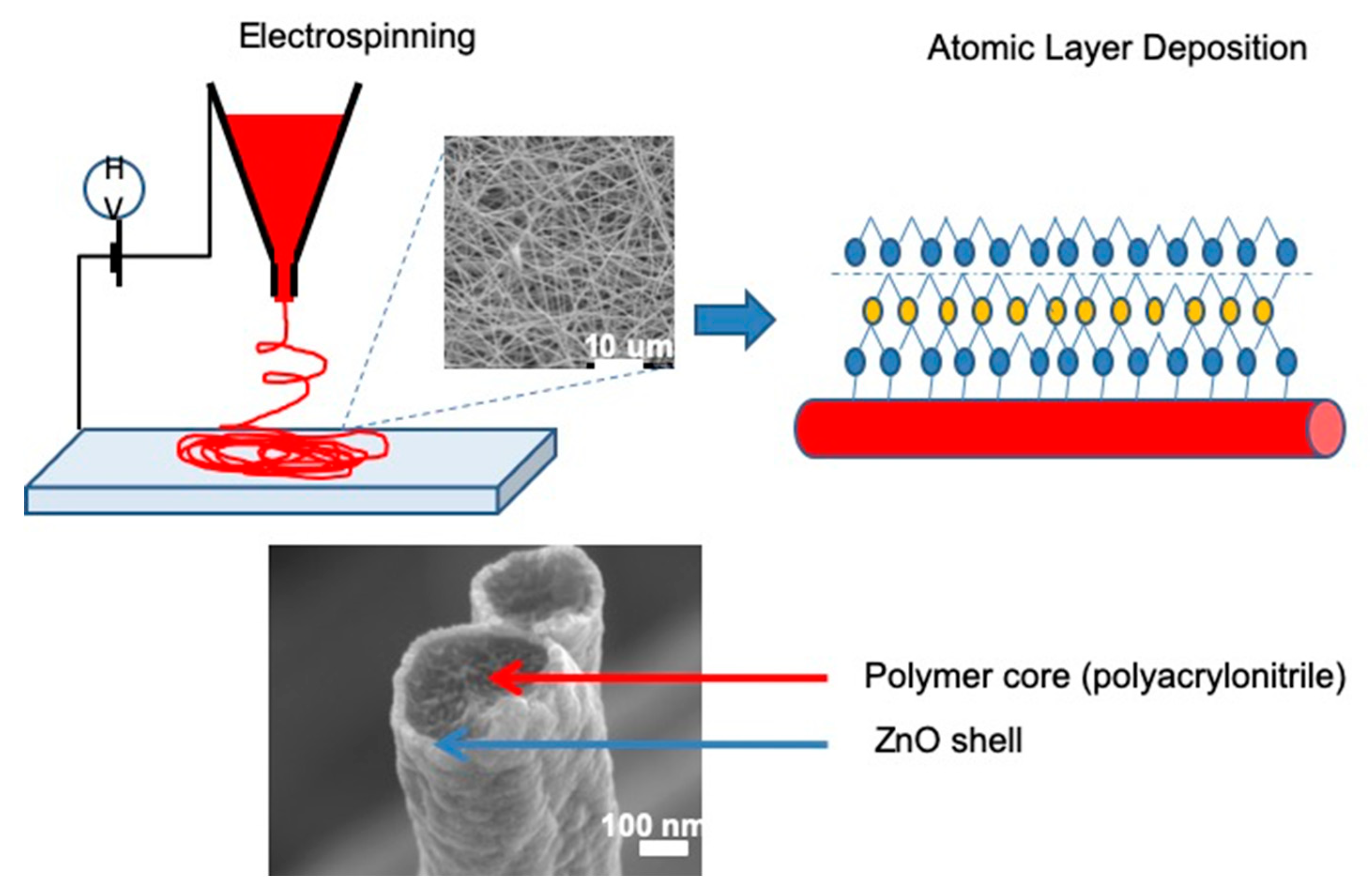
  50. Iatsunskyi, I.; Vasylenko, A.; Viter, R.; Kempiński, M.; Nowaczyk, G.; Jurga, S.; Bechelany, M. Tailoring of the electronic properties of ZnO-polyacrylonitrile nanofibers: Experiment and theory. Appl. Surf. Sci. 2017, 411, 494–501. [Google Scholar] [CrossRef]
  51. Dolbecq, A.; Mialane, P.; Lisnard, L.; Marrot, J.; Sécheresse, F. Hybrid Organic–Inorganic 1D and 2D Frameworks with ε-Keggin Polyoxomolybdates as Building Blocks. Chem. A Eur. J. 2003, 9, 2914–2920. [Google Scholar] [CrossRef]
  52. Shen, M.; Shi, X. Dendrimer-based organic/inorganic hybrid nanoparticles in biomedical applications. Nanoscale 2010, 2, 1596–1610. [Google Scholar] [CrossRef] [PubMed]
  53. Fattahi, P.; Yang, G.; Kim, G.; Abidian, M.R. A Review of Organic and Inorganic Biomaterials for Neural Interfaces. Adv. Mater. 2014, 26, 1846–1885. [Google Scholar] [CrossRef] [PubMed]
  54. Xue, J.; Wu, T.; Dai, Y.; Xia, Y. Electrospinning and Electrospun Nanofibers: Methods, Materials, and Applications. Chem. Rev. 2019, 119, 5298–5415. [Google Scholar] [CrossRef]
  55. Weber, M.; Tuleushova, N.; Zgheib, J.; Lamboux, C.; Iatsunskyi, I.; Coy, E.; Flaud, V.; Tingry, S.; Cornu, D.; Miele, P.; et al. Enhanced electrocatalytic performance triggered by atomically bridged boron nitride between palladium nanoparticles and carbon fibers in gas-diffusion electrodes. Appl. Catal. B Environ. 2019, 257, 117917. [Google Scholar] [CrossRef]
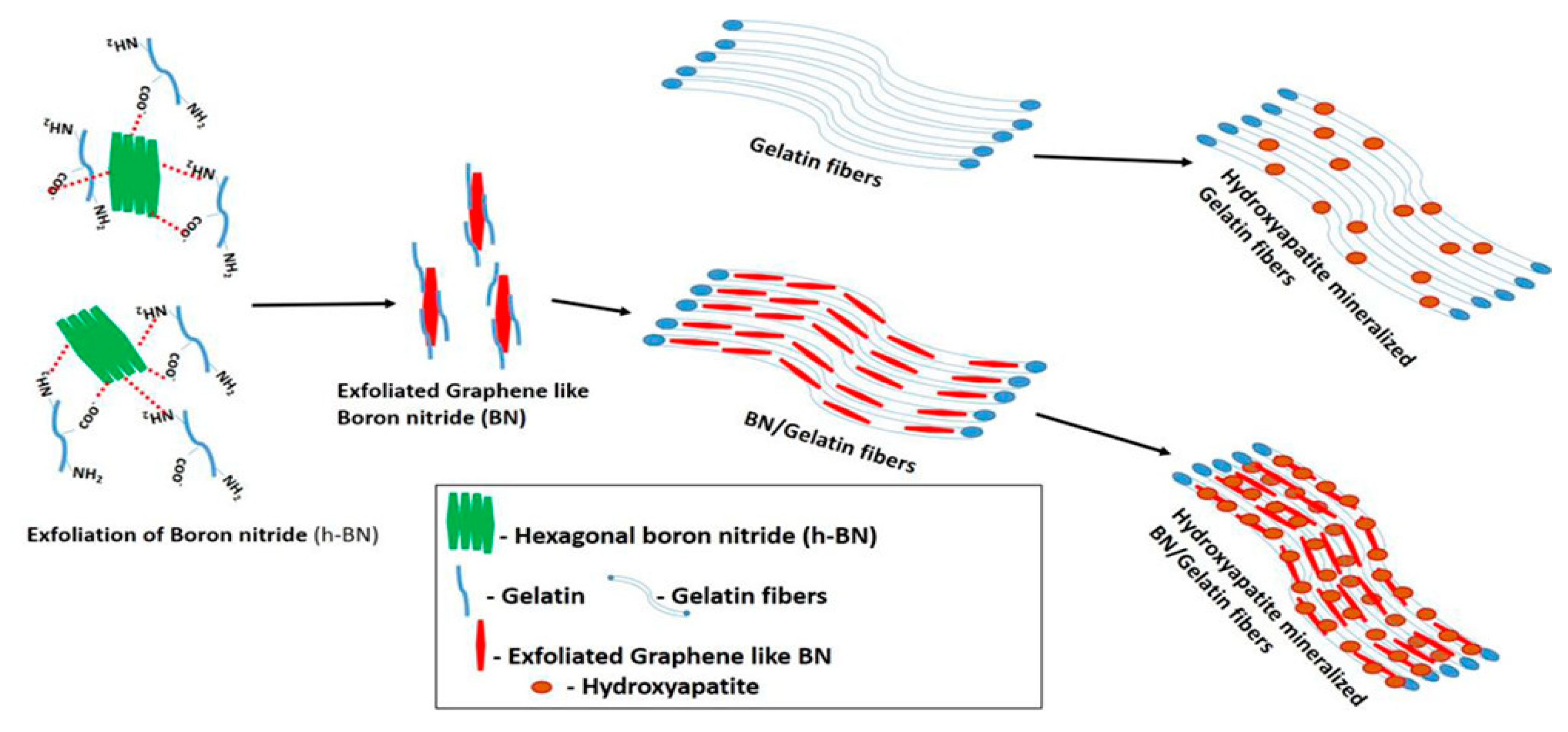
  56. Nagarajan, S.; Belaid, H.; Pochat-Bohatier, C.; Teyssier, C.; Iatsunskyi, I.; Coy, E.; Balme, S.; Cornu, D.; Miele, P.; Kalkura, N.S.; et al. Design of Boron Nitride/Gelatin Electrospun Nanofibers for Bone Tissue Engineering. ACS Appl. Mater. Interfaces 2017, 9, 33695–33706. [Google Scholar] [CrossRef]
  57. Viter, R.; Iatsunskyi, I.; Fedorenko, V.; Tumenas, S.; Balevicius, Z.; Ramanavicius, A.; Balme, S.; Kempiński, M.; Nowaczyk, G.; Jurga, S.; et al. Enhancement of Electronic and Optical Properties of ZnO/Al 2 O 3 Nanolaminate Coated Electrospun Nanofibers. J. Phys. Chem. C 2016, 120, 5124–5132. [Google Scholar] [CrossRef]
  58. Viter, R.; Abou Chaaya, A.; Iatsunskyi, I.; Nowaczyk, G.; Kovalevskis, K.; Erts, D.; Miele, P.; Smyntyna, V.; Bechelany, M. Tuning of ZnO 1D nanostructures by atomic layer deposition and electrospinning for optical gas sensor applications. Nanotechnology 2015, 26, 105501. [Google Scholar] [CrossRef]
  59. Graniel, O.; Iatsunskyi, I.; Coy, E.; Humbert, C.; Barbillon, G.; Michel, T.; Maurin, D.; Balme, S.; Miele, P.; Bechelany, M. Au-covered hollow urchin-like ZnO nanostructures for surface-enhanced Raman scattering sensing. J. Mater. Chem. C 2019, 7, 15066–15073. [Google Scholar] [CrossRef]
  60. Myndrul, V.; Iatsunskyi, I. Nanosilicon-Based Composites for (Bio)sensing Applications: Current Status, Advantages, and Perspectives. Materials (Basel) 2019, 12, 2880. [Google Scholar] [CrossRef] [Green Version]
  61. Pu, K. Biosensors and Bioimaging. ChemBioChem 2019, 20, 420–421. [Google Scholar] [CrossRef] [Green Version]
  62. Nguyen, H.H.; Lee, S.H.; Lee, U.J.; Fermin, C.D.; Kim, M. Immobilized enzymes in biosensor applications. Materials (Basel) 2019, 12, 121. [Google Scholar] [CrossRef] [Green Version]
  63. Sassolas, A.; Blum, L.J.; Leca-Bouvier, B.D. Immobilization strategies to develop enzymatic biosensors. Biotechnol. Adv. 2012, 30, 489–511. [Google Scholar] [CrossRef] [PubMed]
  64. Unnikrishnan, B.; Palanisamy, S.; Chen, S.-M. A simple electrochemical approach to fabricate a glucose biosensor based on graphene–glucose oxidase biocomposite. Biosens. Bioelectron. 2013, 39, 70–75. [Google Scholar] [CrossRef]
  65. Vasylieva, N.; Barnych, B.; Meiller, A.; Maucler, C.; Pollegioni, L.; Lin, J.-S.; Barbier, D.; Marinesco, S. Covalent enzyme immobilization by poly(ethylene glycol) diglycidyl ether (PEGDE) for microelectrode biosensor preparation. Biosens. Bioelectron. 2011, 26, 3993–4000. [Google Scholar] [CrossRef] [PubMed]
  66. Richardson, J.J.; Cui, J.; Björnmalm, M.; Braunger, J.A.; Ejima, H.; Caruso, F. Innovation in Layer-by-Layer Assembly. Chem. Rev. 2016, 116, 14828–14867. [Google Scholar] [CrossRef] [PubMed] [Green Version]
  67. Semenova, D.; Gernaey, K.V.; Morgan, B.; Silina, Y.E. Towards one-step design of tailored enzymatic nanobiosensors. Analyst 2020, 145, 1014–1024. [Google Scholar] [CrossRef] [PubMed]
  68. Chou, J.-A.; Chung, C.-L.; Ho, P.-C.; Luo, C.-H.; Tsai, Y.-H.; Wu, C.-K.; Kuo, C.-W.; Hsiao, Y.-S.; Yu, H.; Chen, P. Organic Electrochemical Transistors/SERS-Active Hybrid Biosensors Featuring Gold Nanoparticles Immobilized on Thiol-Functionalized PEDOT Films. Front. Chem. 2019, 7, 281. [Google Scholar] [CrossRef]
  69. Mahadeva, S.K.; Nayak, J.; Kim, J. Biosensor made with organic-inorganic hybrid composite: Cellulose-tin oxide. In Nanosensors, Biosensors, and Info-Tech Sensors and Systems 2011; Varadan, V.K., Ed.; SPIE: Bellingham, WA, USA, 2011; Volume 7980, pp. 114–118. [Google Scholar]
  70. Zhao, X.; Bai, W.; Yan, Y.; Wang, Y.; Zhang, J. Core-Shell Self-Doped Polyaniline Coated Metal-Organic-Framework (SPAN@UIO-66-NH 2) Screen Printed Electrochemical Sensor for Cd 2+ Ions. J. Electrochem. Soc. 2019, 166, B873–B880. [Google Scholar] [CrossRef]
  71. Tereshchenko, A.; Bechelany, M.; Viter, R.; Khranovskyy, V.; Smyntyna, V.; Starodub, N.; Yakimova, R. Optical biosensors based on ZnO nanostructures: advantages and perspectives. A review. Sens. Actuators B Chem. 2016, 229, 664–677. [Google Scholar] [CrossRef]
  72. Tamashevski, A.; Harmaza, Y.; Viter, R.; Jevdokimovs, D.; Poplausks, R.; Slobozhanina, E.; Mikoliunaite, L.; Erts, D.; Ramanaviciene, A.; Ramanavicius, A. Zinc oxide nanorod based immunosensing platform for the determination of human leukemic cells. Talanta 2019, 200, 378–386. [Google Scholar] [CrossRef] [Green Version]
  73. Lee, H.; Dellatore, S.M.; Miller, W.M.; Messersmith, P.B. Mussel-Inspired Surface Chemistry for Multifunctional Coatings. Science 2007, 318, 426–430. [Google Scholar] [CrossRef] [PubMed] [Green Version]
  74. Liebscher, J.; Mrówczyński, R.; Scheidt, H.A.; Filip, C.; Hădade, N.D.; Turcu, R.; Bende, A.; Beck, S. Structure of Polydopamine: A Never-Ending Story? Langmuir 2013, 29, 10539–10548. [Google Scholar] [CrossRef] [PubMed]
  75. Mrówczyński, R. Polydopamine-Based Multifunctional (Nano)materials for Cancer Therapy. ACS Appl. Mater. Interfaces 2018, 10, 7541–7561. [Google Scholar] [CrossRef] [PubMed]
  76. Stavra, E.; Petrou, P.S.; Koukouvinos, G.; Kiritsis, C.; Pirmettis, I.; Papadopoulos, M.; Goustouridis, D.; Economou, A.; Misiakos, K.; Raptis, I.; et al. Simultaneous determination of paraquat and atrazine in water samples with a white light reflectance spectroscopy biosensor. J. Hazard. Mater. 2018, 359, 67–75. [Google Scholar] [CrossRef]
  77. He, D.; He, X.; Yang, X.; Li, H.-W. A smart ZnO@polydopamine-nucleic acid nanosystem for ultrasensitive live cell mRNA imaging by the target-triggered intracellular self-assembly of active DNAzyme nanostructures. Chem. Sci. 2017, 8, 2832–2840. [Google Scholar] [CrossRef] [Green Version]
  78. Yang, Y.; Hu, W. Bifunctional polydopamine thin film coated zinc oxide nanorods for label-free photoelectrochemical immunoassay. Talanta 2017, 166, 141–147. [Google Scholar] [CrossRef]
  79. Zapato, L. Lord Kelvin Quotations. Kelvin Libr. 2008. [Google Scholar]
  80. Fissell, W.H. What Is Nanotechnology? Adv. Chronic Kidney Dis. 2013, 20, 452–453. [Google Scholar] [CrossRef]
  81. BSI Nanotechnologies—Terminology and Definitions for Nano-Objects Nanofibre and Nanoplate (ISO/TS 27687:2008); NSAI: Dublin, Ireland.
  82. Dean, S.; Mansoori, G.; Fauzi Soelaiman, T. Nanotechnology—An Introduction for the Standards Community. J. ASTM Int. 2005, 2, 13110. [Google Scholar] [CrossRef]
  83. Joint Committee for Guides in Metrology International vocabulary of basic and general terms in metrology (VIM). Int. Organ. 2004.
  84. Kim, H.-A.; Seo, J.-K.; Kim, T.; Lee, B.-T. Nanometrology and its perspectives in environmental research. Environ. Health Toxicol. 2014, 29, e2014016. [Google Scholar] [CrossRef] [PubMed] [Green Version]
  85. Bich, W.; Cox, M.G.; Dybkaer, R.; Elster, C.; Estler, W.T.; Hibbert, B.; Imai, H.; Kool, W.; Michotte, C.; Nielsen, L.; et al. Revision of the ‘Guide to the Expression of Uncertainty in Measurement’. Metrologia 2012, 49, 702–705. [Google Scholar] [CrossRef]
  86. Roebben, G.; Emons, H.; Reiners, G.; Decker, J.E.; Steele, A.G. Nanotechnology Standards; Murashov, V., Howard, J., Eds.; Nanostructure Science and Technology; Springer: New York, NY, USA, 2011; ISBN 978-1-4419-7852-3. [Google Scholar]
  87. Simonet, B.M.; Valcárcel, M. Monitoring nanoparticles in the environment. Anal. Bioanal. Chem. 2009, 393, 17–21. [Google Scholar] [CrossRef] [PubMed]
  88. Yacoot, A.; Koenders, L. Recent developments in dimensional nanometrology using AFMs. Meas. Sci. Technol. 2011, 22, 122001. [Google Scholar] [CrossRef]
  89. Postek, M.T.; Vladár, A.; Archie, C.; Ming, B. Review of current progress in nanometrology with the helium ion microscope. Meas. Sci. Technol. 2011, 22, 024004. [Google Scholar] [CrossRef] [Green Version]
  90. Borovikov, V.; Zangwill, A. Step-edge instability during epitaxial growth of graphene from SiC(0001). Phys. Rev. B 2009, 80, 121406. [Google Scholar] [CrossRef] [Green Version]
  91. Zangwill, A.; Vvedensky, D.D. Novel Growth Mechanism of Epitaxial Graphene on Metals. Nano Lett. 2011, 11, 2092–2095. [Google Scholar] [CrossRef] [Green Version]
  92. Birch, D.J.S.; Yip, P. Nanometrology. In Methods in Molecular Biology; Springer: Heidelberg, Germany, 2014; pp. 279–302. ISBN 9781627036481. [Google Scholar]
  93. Linsinger, T.P.J.; Roebben, G.; Solans, C.; Ramsch, R. Reference materials for measuring the size of nanoparticles. TrAC Trends Anal. Chem. 2011, 30, 18–27. [Google Scholar] [CrossRef]
  94. Zangwill, A. Modern Electrodynamics; Cambridge University Press: Cambridge, UK, 2012; ISBN 9781139034777. [Google Scholar]
  95. Whitehouse, D.J. Handbook of Surface and Nanometrology. IOP Publishing Ltd., CRC press: Boca Raton, FL, USA, 2003; ISBN 0750305835. [Google Scholar]
  96. Whitehouse, D.J. Handbook of Surface and Nanometrology; CRC Press: Boca Raton, FL, USA, 2010; ISBN 9780429140693. [Google Scholar]
  97. Semenova, D.; Silina, Y.E. The Role of Nanoanalytics in the Development of Organic-Inorganic Nanohybrids—Seeing Nanomaterials as They Are. Nanomaterials 2019, 9, 1673. [Google Scholar] [CrossRef] [Green Version]
  98. Zolotov, Y.A. Analytical chemistry: The day today. J. Anal. Chem. 2007, 62, 912–917. [Google Scholar] [CrossRef]
  99. Zolotov, Y.A. Nanoanalytics. J. Anal. Chem. 2010, 65, 1207–1208. [Google Scholar] [CrossRef] [Green Version]
  100. Shtykov, S. (Ed.) Nanoanalytics; Walter de Gruyter: Berlin, Germany, 2018; ISBN 9783110547207. [Google Scholar]
  101. Hansen, P.-E.; Roebben, G.; Babick, F.; Braun, A.; Dirscherl, K.; Hansen, P.-E.; Jennett, N.M. Introductory Guide to Nanometrology: Metrology for Dimensional Analysis of Thin Films; European Commission, Joint Research Centre: Belgium, Brussels, 2010; 64p, ISBN 978-0-9566809-1-4. [Google Scholar]
  102. Pilas, J.; Selmer, T.; Keusgen, M.; Schöning, M.J. Screen-Printed Carbon Electrodes Modified with Graphene Oxide for the Design of a Reagent-Free NAD + -Dependent Biosensor Array. Anal. Chem. 2019, 91, 15293–15299. [Google Scholar] [CrossRef] [PubMed]
  103. Ding, R.-F.; Yang, C.-W.; Huang, K.-Y.; Hwang, I.-S. High-sensitivity imaging with lateral resonance mode atomic force microscopy. Nanoscale 2016, 8, 18421–18427. [Google Scholar] [CrossRef]
  104. Okahata, Y.; Enna, G.; Ebato, H. Synthetic chemoreceptive membranes. Sensing bitter or odorous substances on a synthetic lipid multibilayer film by using quartz-crystal microbalances and electric responses. Anal. Chem. 1990, 62, 1431–1438. [Google Scholar] [CrossRef]
  105. Wu, T.; Zhang, H. Piezoelektrizität in zweidimensionalen Materialien. Angew. Chemie 2015, 127, 4508–4510. [Google Scholar] [CrossRef]
  106. Sauerbrey, G. Use of quartz vibration for weighing thin films on a microbalance. Z. Fur. Phys. 1959, 155, 206–222. [Google Scholar] [CrossRef]
  107. Kurosawa, S.; Hirokawa, T.; Kashima, K.; Aizawa, H.; Han, D.-S.; Yoshimi, Y.; Okada, Y.; Yase, K.; Miyake, J.; Yoshimoto, M.; et al. Detection of deposition rate of plasma-polymerized films by quartz crystal microbalance. Thin Solid Films 2000, 374, 262–267. [Google Scholar] [CrossRef]
  108. Pohanka, M. Overview of Piezoelectric Biosensors, Immunosensors and DNA Sensors and Their Applications. Materials (Basel) 2018, 11, 448. [Google Scholar] [CrossRef] [Green Version]
  109. Skládal, P. Piezoelectric quartz crystal sensors applied for bioanalytical assays and characterization of affinity interactions. J. Braz. Chem. Soc. 2003, 14, 491–502. [Google Scholar] [CrossRef] [Green Version]
  110. Lu, C.; Czanderna, A.W.; Townshend, A. Applications of piezoelectric quartz crystal microbalances. Anal. Chim. Acta 1987, 199, 279. [Google Scholar] [CrossRef]
  111. Logothetidis, S. Nanostructured Materials and Their Applications; Logothetidis, S., Ed.; NanoScience and Technology; Springer: Berlin/Heidelberg, Germany, 2012; ISBN 978-3-642-22226-9. [Google Scholar]
  112. Leach, R. Front-matter. In Fundamental Principles of Engineering Nanometrology; Elsevier: Amsterdam, The Netherlands, 2014; ISBN 9781455777532. [Google Scholar]
  113. Bell, S. Good Practice Guide No. 11—Introductory Guide to Uncertainty of Measurement; NPL: Teddington, UK, 2001; ISBN 1368-6550. [Google Scholar]
  114. Allen, D. Good Practice in Physical Interventions. A Guide for Staff and Managers; BILD Code of Practice for the Use of Physical Interventions. A Guide for Trainers and Commissioners of Training. J. Appl. Res. Intellect. Disabil. 2007, 20, 586–587. [Google Scholar] [CrossRef]
  115. Nicoletti, M.C.; Jain, L.C.; Giordano, R.C. Computational Intelligence Techniques as Tools for Bioprocess Modelling, Optimization, Supervision and Control. In Studies in Computational Intelligence; Springer: Berlin, Germany, 2009; pp. 1–23. ISBN 9783642018879. [Google Scholar]
  116. Sin, G.; Woodley, J.M.; Gernaey, K. V Application of modeling and simulation tools for the evaluation of biocatalytic processes: A future perspective. Biotechnol. Prog. 2009, 25, 1529–1538. [Google Scholar] [CrossRef] [PubMed]
  117. Baronas, R.; Ivanauskas, F.; Kulys, J. Mathematical Modeling of Biosensors; Springer: Heidelberg, Germany, 2010; ISBN 978-90-481-3243-0. [Google Scholar]
  118. HOFMANN, O.T.; ZOJER, E. Modelling Organic-Inorganic Hybrid Interfaces. In World Scientific Reference of Hybrid Materials; World Scientific Series in Nanoscience and Nanotechnology; World Scientific Publishing Company: Singapore, 2019; pp. 3–40. ISBN 978-981-327-053-4. [Google Scholar]
  119. Van Daele, T.; Gernaey, K.V.; Ringborg, R.H.; Börner, T.; Heintz, S.; Van Hauwermeiren, D.; Grey, C.; Krühne, U.; Adlercreutz, P.; Nopens, I. Application of iterative robust model-based optimal experimental design for the calibration of biocatalytic models. Biotechnol. Prog. 2017, 33, 1278–1293. [Google Scholar] [CrossRef] [PubMed]
  120. Ringborg, R.H.; Woodley, J.M. The application of reaction engineering to biocatalysis. React. Chem. Eng. 2016, 1, 10–22. [Google Scholar] [CrossRef] [Green Version]
  121. Roldan, A. Frontiers in first principles modelling of electrochemical simulations. Curr. Opin. Electrochem. 2018, 10, 1–6. [Google Scholar] [CrossRef]
  122. Semenova, D.; Zubov, A.; Silina, Y.E.; Micheli, L.; Koch, M.; Fernandes, A.C.; Gernaey, K.V. Mechanistic modeling of cyclic voltammetry: A helpful tool for understanding biosensor principles and supporting design optimization. Sens. Actuators B Chem. 2018, 259, 945–955. [Google Scholar] [CrossRef] [Green Version]
  123. Nabaei, V.; Chandrawati, R.; Heidari, H. Magnetic biosensors: Modelling and simulation. Biosens. Bioelectron. 2018, 103, 69–86. [Google Scholar] [CrossRef] [Green Version]
Figure 1. Classification of organic-inorganic hybrids (OIH) on the basis of the interaction between organic and inorganic components (original image).
Applsci 10 01458 g001
Figure 2. Schematic representation of core-shell (PAN-ZnO) fibers synthesis and results of scanning electron microscopy (SEM) analysis (original image). Note: the SEM images were obtained in the laboratory of the presented authors (SEM JEOL JSM—7001F, operating voltage—15 kV).
Applsci 10 01458 g002
Figure 3. The workflow of boron nitrides (BN)-gelatin nanofiber synthesis. Adapted and reproduced with permission from. Reproduced with permission from [56], Copyright American Chemical Society, 2017.
Applsci 10 01458 g003
Figure 4. (a) General ZnO-based optical biosensor scheme. Reproduced with permission from [71]. Copyright Elsevier, 2016. (b) Photoluminescence. Reproduced with permission from [72]. Copyright Elsevier, 2019. (c) Reflectance. Reproduced with permission from [76]. Copyright Elsevier, 2018, for detection of leukemic cells detection and paraquat/atrazine, respectively.
Applsci 10 01458 g004
Figure 5. The number of cited papers related to the terms highlighted above per year: ((A) Organic-inorganic hybrids metrology; (B) Nanometrology in hybrid materials; (C) Nanohybrids). Preliminary figures for the present decades indicate the relevance of the research field. (Compiled from the Web of Knowledge database search (www.webofknowledge.com), Nov. 2019).
Applsci 10 01458 g005
Figure 6. The relative relationship between different geometric components in conventional (A), micromechanics (B), and nanoengineering (C). Adapted with permission from ref. [94,95].
Applsci 10 01458 g006
Figure 7. Case study of geometry and pitch measurement of periodic structures obtained by atomic force microscopy (AFM): left—Illustration of light and AFM interaction with a periodic surface structure; right—typical profile images of line scans obtained with AFM. Reproduced and adapted with the permission from ref. [101].
Applsci 10 01458 g007
Figure 8. AFM three-dimensional profiles of the blank screen-printed carbon electrode (SPCE) (a), modified with graphene oxide (GO) and the cofactors (NAD+) (b), an additional layer of the enzymes L-lactate dehydrogenase (L-LDH, enzyme) and diaphorase (DIA) (c) and a membrane of cellulose acetate (CA) (d). The scanned area of the AFM images was 2 × 2 μm2. Reproduced with permission from [102], Copyright American Chemical Society, 2019.
Applsci 10 01458 g008
Figure 9. AFM images of a heterogeneous blend of polystyrene and low-density polyethylene (LDPE) in air using lateral resonance (LR) modes. Height images acquired with the LR mode (A); corresponding phase images (B); corresponding vertical deflection (quasi-static) images (C). Height profiles along the scan lines in image A (D). Reprinted and adapted with the permission from [103]. Copyright RSC Pub, 2009.
Applsci 10 01458 g009
Figure 10. The scheme of the quartz crystal microbalances (QCM) sensor (top) and resonator model (bottom). NOTE: xq—is the quartz wafer thickness; xf—is the film thickness; pqvq and pfvf are the acoustic impedance for the unit cross section area of the crystal and of the film, respectively. Adapted with permission of ref. [109,110] Copyright Elsevier, 1987.
Applsci 10 01458 g010
Figure 11. Schematic workflow for laser-desorption ionization mass spectrometry (LDI-MS) applied for characterization of the hybrid nanobiosensors towards design optimization. Reprinted with a permission from [97].
Applsci 10 01458 g011
Figure 12. The workflow of the methodology for validation of OIH/OInH and biosensors (original scheme).
Applsci 10 01458 g012
Figure 13. The workflow of the process modeling steps applied for simulation and modeling in OIHs and biosensors. Adapted and reproduced with permission from [115], Copyright Biotechnology Progress, 2009.
Applsci 10 01458 g013

