Article

Peri-Implant Bone Damage Procured by Piezoelectric and Conventional Implant Site Preparation: An In Vitro Comparison

1
Private Practice in Genova, 16121 Genova, Italy
2
School of Dentistry, University of Genova, 16121 Genova, Italy
3
Department of Civil, Environmental and Chemical Engineering, University of Genova, 16121 Genova, Italy
4
Department Neurosciences, Riproductive and Odontostomatological Sciences, University of Naples Federico II, 80138 Napoli, Italy
*
Author to whom correspondence should be addressed.
Appl. Sci. 2020, 10(24), 8909; https://doi.org/10.3390/app10248909
Submission received: 9 November 2020 / Revised: 3 December 2020 / Accepted: 9 December 2020 / Published: 14 December 2020
(This article belongs to the Special Issue Material Science, Implants, and Peri-Implant Tissues)

Abstract

:
Background: The conventional drill technique is the most commonly used for the implant site preparation of the desired diameter and length. Ultrasonic implant site preparation (UISP) can also be used to perform an implant site preparation of the desired dimensions. Methods: Implant sites were prepared in fresh bone ribs with two different implant site preparation techniques: implant surgical drills and piezoelectric tips. Samples were analyzed with scanning electron microscopy (SEM) for evaluating the peri-implant bone damage. Result: In the surgical drills group, the cortical bone surface showed several cracks and the bone vascular canals were hidden by a dense smear layer. Cancellous bone showed large irregularities and trabecular fractures. The piezoelectric group showed a clean and smooth cortical bone surface with opened bone vascular canals; the cancellous bone presented a regular morphology, and the trabecular spaces, clearly visible, were free of debris. Conclusions: Ultrasonic implant site preparation showed cleaner bone surfaces and lower bone trauma compared with the preparation using implant surgical drills.

1. Introduction

Research has improved many aspects of dental implant therapy: surgical planning, implant placement, fixture surface and macro-morphology, and prosthetic rehabilitation [1,2,3,4,5,6,7]. The surgical drill technique is the most common implant site preparation method. It uses a sequence of progressive cylindrical or conical drills to obtain a bone perforation of desired dimensions [8,9]. It generally requires a surgical drill torque rotation of 15–50 N/cm and an intraoperative pressure of 2–6 kg. The production of bone fragments compressed on peri-implant bone surfaces can be a consequence of this surgical technique [9,10,11,12,13]. Some authors have reported that bone fragments and debris, produced during the implant site preparation, may promote osseointegration [14,15,16]. However, others have suggested that this bone damage may interfere with osseointegration, activating peri-implant bone remodeling [17,18,19,20,21,22]. The necrotic tissue may change the pH and biochemical environment and promote a macrophage activation [23,24]. Some authors have suggested reducing the mechanical and thermal trauma, focusing on macrostructural bur components to promote minimal traumatic preparation and to reduce bone fragment production [24].
Different implant site preparation techniques have been proposed in relation to bone density: bone tapping in more dense bone; undersized implant site preparation; self-tapping implants or bone compacting techniques in soft bone [25,26,27,28].
Piezosurgery® and Er: YAG lasers represent two different bone perforation techniques that have proposed to overcome traditional surgical procedures [29,30,31]. Ultrasonic implant site preparation (UISP), introduced in 2010, assures a precise implant site preparation; this surgical technique uses linear mechanical micro-vibrations at ultrasonic frequency, ranging from 24 to 36 kHz in relation to the bone density [14,32]. Its cooling solution provided by internal irrigation is useful to clean the implant site [33,34,35,36].
This in vitro scanning electron microscopy (SEM) evaluation shows the cortical and cancellous bone damage procured by conventional and ultrasonic implant site preparation techniques.

2. Materials and Methods

Bovine bone blocks were obtained from the ribs of animals slaughtered in a local meatpacking house. Bone was fresh and hydrated. The bovine specimens were selected because they are commonly used as a model in bone biomechanics; the bone structure of the rib is similar to an atrophic jawbone [16]. Five bovine ribs with dimensions of 25 × 4 × 1 cm were used. Implant site preparation occurred under profuse saline irrigation for both the ultrasonic and drill techniques, assuring the same room temperature. Two implant sites, 7 mm in depth and 3 mm in diameter, were prepared in each bone block, the first using bone drills for implant site preparation (group A) and the second using UISP (group B).

2.1. Implant Site Preparation for Standard Drills

Standard drill implant site preparation (group A) was performed with a sequence of three cylindrical twist drills with dimensions of 1, 2, and 3 mm (Mectron, Carasco, Genova, Italy). The twist drills were used with a rotating speed of 800 rpm, a torque rotation of 30 N/cm, and a pressure of 3 kg.

2.2. Ultrasonic Implant Site Preparation

Implant site preparations of group B were performed using a Piezosurgery® 3 unit with insert tip OT4 (Mectron, Carasco, Genova, Italy). A sequence of three diameters (1, 2, and 3 mm) were used. The Piezosurgery® device generated continual variation between 58 and 66 Hz and a range of mechanical micro-vibrations of approximately 40 μm, with a pressure of 0.4 kg.
Ten holes were made for each technique. After implant site preparation, each bone block was cut through the middle and processed for SEM analysis. The bone walls of the implant site preparation were not cleaned of the smear layer and debris before the SEM analyses.

3. Results

The UISP technique needed less pressure than the conventional technique for perforating the bone, resulting in better microsurgical precision and intra-operative control.

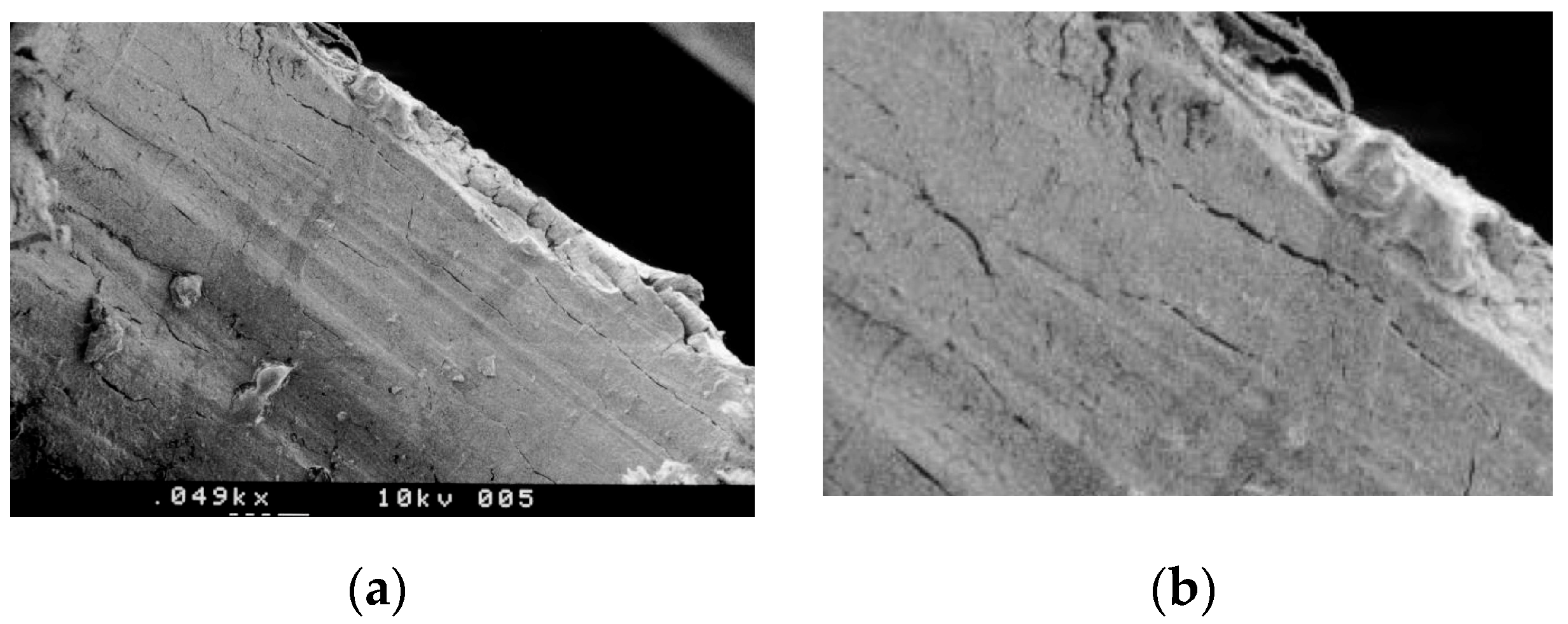
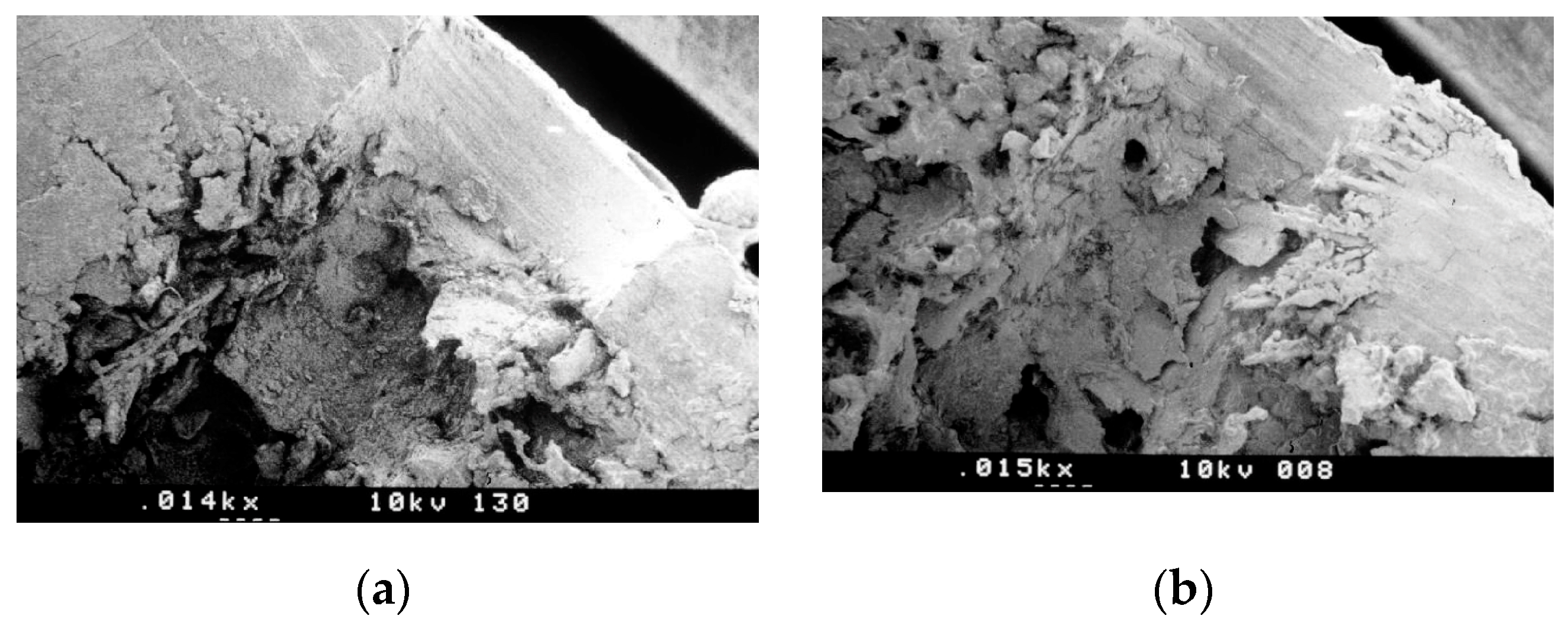
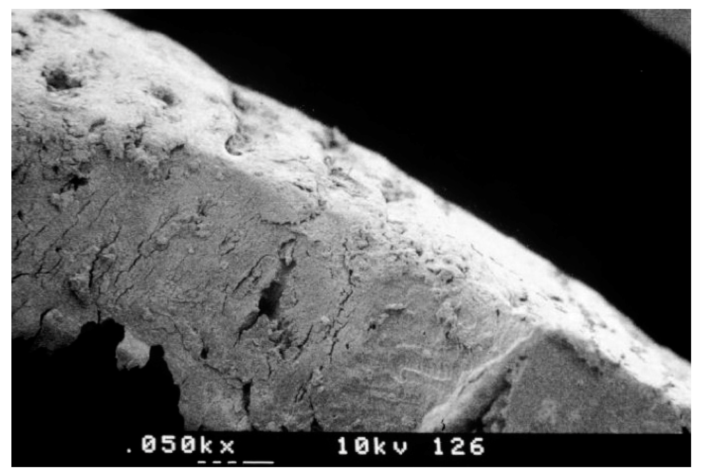
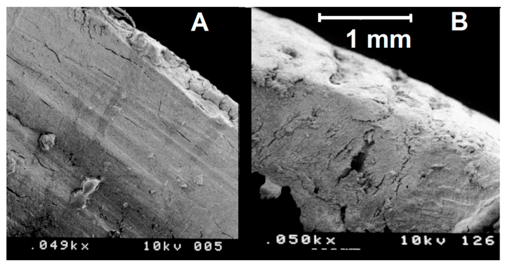
3.1. SEM Morphologic Analysis

The morphologic analysis showed differences between the implant sites prepared with a standard drill technique (group A) and UISP (group B).

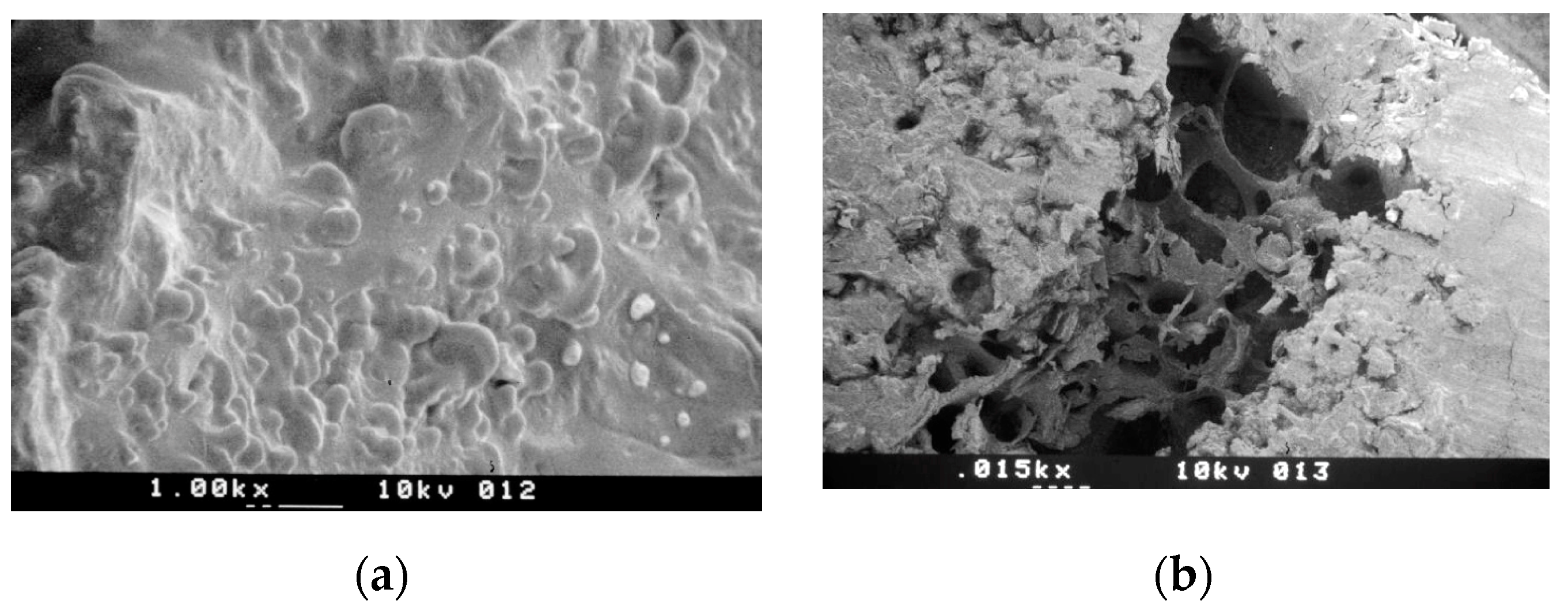
3.1.1. Group A—Drill Implant Site Preparation

In the cortical bone of group A, the bone walls appeared signed by several turning strips and showed several interconnected micro-cracks, exfoliating the lamellar bone surface in parallel layers, mostly perpendicular to the work axis of the implant drills (Figure 1a). Mineralized bone fragments and bone marrow particles crushed by the cutting edges of the drilling bit were spread over the bone walls, forming a dense smear layer (Figure 1b). This smear layer appeared stuffed at the bone walls, closing most of the bone vascular canals and filling the small medullary spaces of the cortical bone.
Signs of trauma as a result of drilling were also seen in cancellous bone where the trabeculae appeared extensively damaged. Large empty spaces, like bone lacunae, were visible at the implant site walls, probably due to a lack of trabeculae caused by the impact of the cutting blades (Figure 2a), so the shape of the bone preparation appaired irregular with several craters. Fragments of damaged trabeculae appaired packed into the available medullary spaces (Figure 2b).

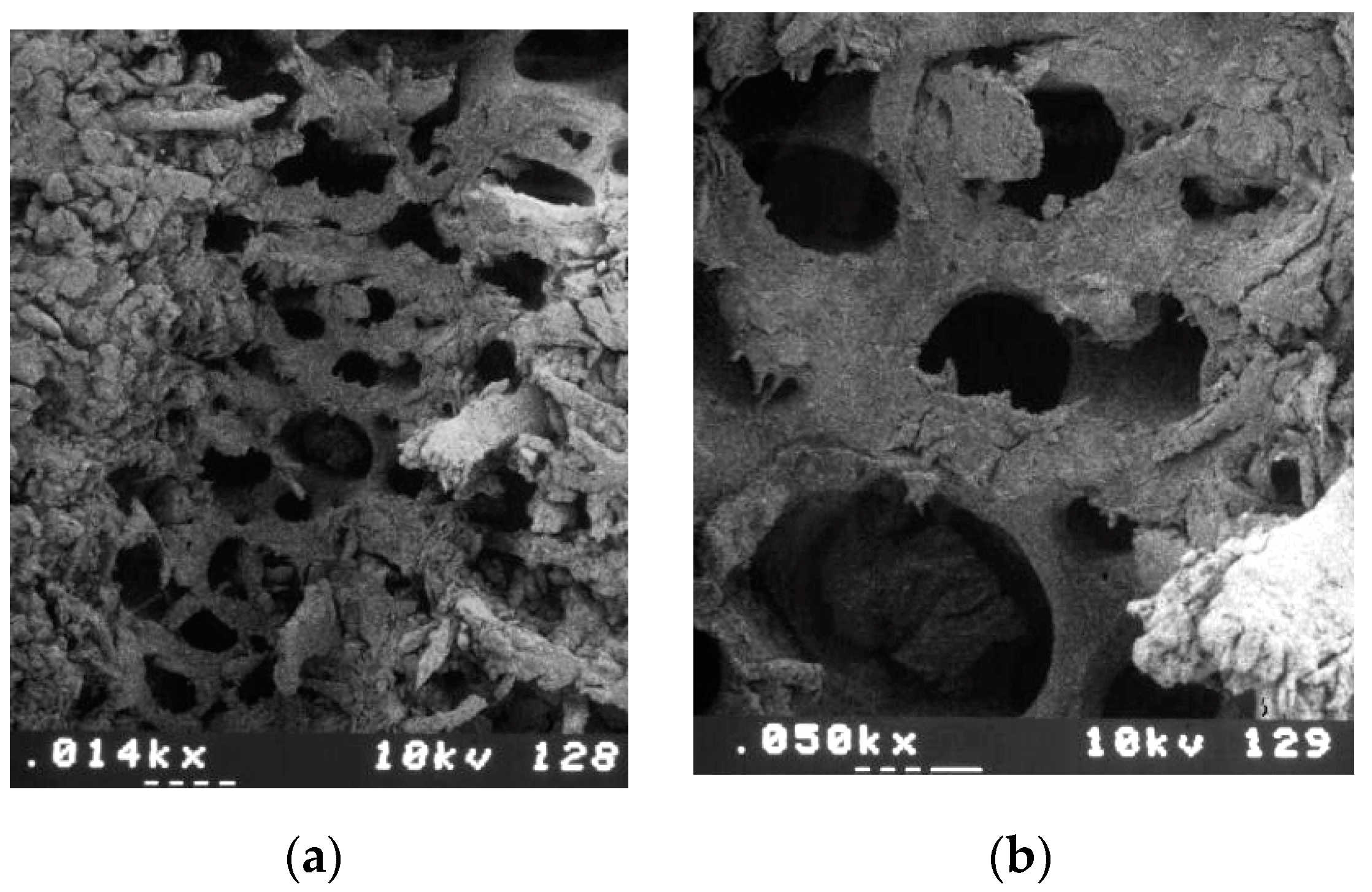
3.1.2. Group B—Ultrasonic Implant Site Preparation

In the cortical bone of group B, bone surfaces appeared clean, slightly roughened by piezoelectric action, and many open vascular canals were visible. No bone fragments or biologic material were seen over the bone walls, and the bone vascular canals appeared clean and open, including the haversian canals and the few small medullary spaces of the cortical bone (Figure 3).
Compared with group A, cortical bone appeared intact, showing more opened vascular canals and an evident reduction of damage. Bone cracks were very rare and never interconnected. No signs of delaminate bone layers were observed (Figure 4A,B).
At a higher magnification, hemispheres exposed by the vibrating action of piezoelectric implant site preparation were observed all over the cortical bone (Figure 5a). These hemispheres represented the exposed extremities of the collagen fibers and other bone proteins that constitute the architecture of the lamellar bone. Ultrasonic shock waves coupled with cavitation of the saline cooling solution disaggregated the peripheral calcium phosphate crystals from collagen fibers, releasing the proteins of the bone matrix. After UISP, the cancellous bone preparation showed cleaned walls, completely free of bone fragments (Figure 5b).
The architecture of the mineralized trabecular structure in the vicinity of the implant site was exposed to powerful ultrasonic cavitation of the cooling solution, which detached the bone marrow and the periosteum from the trabeculae. The trabecular bone, under SEM, revealed a significant reduction of fractures by the action of UISP when compared with implant drills (Figure 6a,b).

4. Discussion

In recent decades, many authors have sought to identify factors that minimize damage during implant site preparation [1,2,3,4,5,6,7,8,9,10]. An appropriate procedure should procure a thin layer of necrotic bone subsequently replaced by vital bone tissue [25,26,27,28]. All of the surgical factors that increase the thickness of the bone necrotic layer can negatively compromise implant osseointegration. Extension of the necrotic zone around the preparation site is considered proportional to the amount of heat generated during osteotomy, which can be related to different factors: (i) the operator (pressure, status, movement, speed, and duration of drilling); (ii) the manufacturer (design and sharpness of the drill and the irrigation system); (iii) the implant site (cortical thickness, bone density, and depth drilled); (iv) the patient’s age; (v) the surgical method (drilling, ultrasonic, and condensation [1,2,3,7,8,9]). The bone damage is related to the magnitude of the temperature elevation and the time the tissue is subjected to a damaging temperature, identified as 47 °C for 1 min [15,16,17,18,19,20,21,22]. Heat generation, as evidenced by Matthews and Hirsch [20], can also be influenced by the manufacturer’s precision (sharpness of the cutting tool) [15] and by the surface deformation and roughness shown by the worn burs that cause a more significant and continuous temperature rise than new burs [16,17]. Drill wear is another important factor in heat generation during bone drilling [1,9]. It is an unavoidable and irreversible process, which increases friction in the cutting zone [12]. As well as a negative thermal impact, wear causes higher cutting forces and drill vibrations, which can result in cutting edge breakage or complete drill breakage in the flute or shank zone [12]. The literature data reports that, for thermal impact, ultrasonic tips are safe when properly used [37]. Ultrasonic vibrations provide a “gentler action” involving less local delivery of mechanical energy than rotating techniques [8,37].
This study showed the capacity of UISP to better preserve the cortical and cancellous bone structure and vascularization in comparison with the conventional surgical drill technique. The SEM evaluation of this research evidenced that the use of surgical bone drills can be destructive, breaking off large fragments of trabeculae and weakening bone in the vicinity of the implant site, leaving bone fragments likely to occlude medullary spaces and the three-dimensional cortical bone vascular canal system. Bone vascular canal lesions may interfere with peri-implant bone nutrition and cortical bone damage may stimulate early osteoclastic activation [16]. In cancellous bone, the broken trabeculae can be removed by the drill and the cooling fluid, or will form the peri-implant bone structure [38]. Detached trabeculae stuffed in the surgical implant drill–bone interface may produce an intraoperative increase in the insertion torque levels, causing thermal bone damage; the presence of necrotic bone tissue in the bone–fixture interface may also cause macrophagic osteoclastic activity, reducing the primary stability of the implant and delaying the osseointegration process.
Cortical bone preparation with Piezosurgery® allows the preservation of the bone surface, considerably decreasing the presence of micro-fractures and a smear layer. The characteristic cleaned bone surface also avoids closure of bone vascular canals by the compression of bone debris between bone surfaces and the cutting device. In cancellous bone, Piezosurgery® assures a precise cutting of the trabeculae and a reduction of cracks weakening the bone structure and fragments compressed into the trabecular architecture. The thin fragments produced may also release bone osteoinductive proteins (BMPs) in the early phase of implant healing [39].
A less traumatic strategy for implant site preparation by Piezosurgery® compared with the standard surgical drill technique is also assured by reduced pressure during the osteotomy [40]. For effective cutting, the surgical implant drill technique requires a torque rotation of 15–50 N/cm and a pressure of approximately 2–6 kg exerted by the operator’s hand; this can reduce the surgical precision and can induce thermal bone damage. The cutting power of the current Piezosurgery® devices are assured by mechanical micro-vibrations of approximately 40 μm with a frequency ranging from 30 to 70 Hz and a pressure of 0.4 kg [41]. This allows the intraoperative regulation of forced oscillation in relation to the degree of bone density [42].
Another important aspect is the primary stability that the implant site preparation assures. Detached trabeculae stuffed in the surgical implant drill–bone interface may produce an intraoperative increase of the insertion torque levels favoring thermal bone damage; the presence of necrotic bone tissue in the bone–fixture interface may also activate macrophagic osteoclastic activity, thereby reducing the primary stability of the implant and delaying the osseointegration process [16]. Rebaudi et al. reported an impressive reduction in the stability and reverse-torque levels in the first weeks of healing after forced insertion of mini-implants in undersized sites [32]. Some authors have reported how the preparation of the implant site through a bone condensation method achieves only “the illusion” of stability at the implant insertion by compression of fractured bone fragments on the damaged bone walls of the implant site [43,44,45,46,47]. This process really occurs in well-vascularized sites and in the absence of traumatic bone damage that may cause inflammatory responses and an increase in macrophage activity [16]. The presence of compressed bone fragments and the absence of adequate vascularization may delay or inhibit peri-implant osseointegration [48]. Many authors have therefore suggested that implant site preparation using surgical drills, rather than bone compactors, results in more predictable peri-implant bone healing [45,47]. This study was only able to evidence the cutting precision and the minimizing of trauma to the bone structures during the cutting action, but no information about the assured implant primary stability could be reported. Some authors have reported how piezoelectric osteotomy compared with conventional methods requires a longer surgical time [8,37]. Traditional surgery therefore undoubtedly maintains the record for speed of execution, even if it is a method of implant site preparation that presents higher risks related to operative maneuvers and lower comfort for the patient.

5. Conclusions

SEM evaluation shows how ultrasonic implant site preparation is able to assure cleaner bone surfaces and lower bone trauma compared with the conventional surgical implant drill technique.

Author Contributions

Conceptualization, G.S.; methodology, F.B.; investigation, F.R.; supervision, G.M.; project administration, A.R. All authors have read and agree to the published version of the manuscript.

Funding

This research received no external funding.

Acknowledgments

The authors wish to acknowledge the support of BioCRA (Biomaterials Clinical-Histological Research Association) in carrying out this study. The authors also wish to thank Marco Sanscritto, Marco Capurro, and Giorgio Facco, Engineer, University of Genoa, Genoa (Italy) and Edoardo Martino DDS, Genova, Italy.

Conflicts of Interest

The authors declare no conflict of interest.

References

  1. Marenzi, G.; Impero, F.; Scherillo, F.; Sammartino, J.C.; Squillace, A.; Spagnuolo, G. Effect of different surface treatments on titanium dental implant micro-morphology. Materials 2019, 12, 733. [Google Scholar] [CrossRef] [PubMed] [Green Version]
  2. Abuhussein, H.; Pagni, G.; Rebaudi, A.; Wang, H.L. The effect of thread pattern upon implant osseointegration. Clin. Oral Implants Res. 2010, 21, 129–136. [Google Scholar] [CrossRef] [PubMed] [Green Version]
  3. Trisi, P.; Rao, W.; Rebaudi, A. A histometric comparison of smooth and rough titanium implants in human low-density jawbone. Int. J. Oral Maxillofac. Implants 1999, 14, 689–698. [Google Scholar] [PubMed]
  4. Lamazza, L.; Lollobrigida, M.; Vozza, I.; Palmieri, L.; Stacchi, C.; Lombardi, T.; De Biase, A. Piezoelectric implant site preparation: Influence of handpiece movements on temperature elevation. Materials 2020, 13, 4072. [Google Scholar] [CrossRef]
  5. Rebaudi, A.; Laffi, N.; Benedicenti, S.; Angiero, F.; Romanos, G.E. Microcomputed tomographic analysis of bone reaction at insertion of orthodontic mini-implants in sheep. Int. J. Oral Maxillofac. Implants 2011, 26, 1233–1240. [Google Scholar]
  6. Amghar-Maach, S.; Sánchez-Torres, A.; Camps-Font, O.; Gay-Escoda, C. Piezoelectric surgery versus conventional drilling for implant site preparation: A meta-analysis. J. Prosthodont. Res. 2018, 62, 391–396. [Google Scholar] [CrossRef]
  7. Bernabeu-Mira, J.C.; Pellicer-Chover, H.; Peñarrocha-Diago, M.; Peñarrocha-Oltra, D. In vitro study on bone heating during drilling of the implant site: Material, design and wear of the surgical drill. Materials 2020, 13, 1921. [Google Scholar] [CrossRef]
  8. Maglione, M.; Bevilacqua, L.; Dotto, F.; Costantinides, F.; Lorusso, F.; Scarano, A. Observational study on the preparation of the implant site with piezosurgery vs. drill: Comparison between the two methods in terms of postoperative pain, surgical times, and operational advantages. BioMed Res. Int. 2019, 2019, 8483658. [Google Scholar] [CrossRef] [Green Version]
  9. Marenzi, G.; Sammartino, J.C.; Scherillo, F.; Rengo, C.; De Rosa, A.; Graziano, V.; Spagnuolo, G. Comparative analysis of the chemical composition and microstructure conformation between different dental implant bone drills. Materials 2019, 12, 1866. [Google Scholar] [CrossRef] [Green Version]
  10. Tretto, P.H.W.; Fabris, V.; Cericato, G.O.; Sarkis-Onofre, R.; Bacchi, A. Does the instrument used for the implant site preparation influence the bone-implant interface? A systematic review of clinical and animal studies. Int. J. Oral Maxillofac. Surg. 2019, 48, 97–107. [Google Scholar] [CrossRef] [Green Version]
  11. Gürkan, A.; Tekdal, G.P.; Bostancı, N.; Belibasakis, G.N. Cytokine, chemokine, and growth factor levels in peri-implant sulcus during wound healing and osseointegration after piezosurgical versus conventional implant site preparation: Randomized, controlled, split-mouth trial. J. Periodontol. 2019, 90, 616–626. [Google Scholar] [CrossRef] [PubMed]
  12. Rashad, A.; Sadr-Eshkevari, P.; Weuster, M.; Schmitz, I.; Prochnow, N.; Maurer, P. Material attrition and bone micromorphology after conventional and ultrasonic implant site preparation. Clin. Oral Implants Res. 2013, 24 (Suppl. A100), 110–114. [Google Scholar] [CrossRef] [PubMed] [Green Version]
  13. Rashad, A.; Kaiser, A.; Prochnow, N.; Schmitz, I.; Hoffmann, E.; Maurer, P. Heat production during different ultrasonic and conventional osteotomy preparations for dental implants. Clin. Oral Implants Res. 2011, 22, 1361–1365. [Google Scholar] [CrossRef] [PubMed]
  14. Vercellotti, T. Piezoelectric surgery in implantology: A case report-a new piezoelectric ridge expansion technique. Int. J. Periodontics Restor. Dent. 2000, 20, 358–365. [Google Scholar]
  15. Pereira, C.C.; Gealh, W.C.; Meorin-Nogueira, L.; Garcia-Júnior, I.R.; Okamoto, R. Piezosurgery applied to implant dentistry: Clinical and biological aspects. J. Oral Implantol. 2014, 40, 401–408. [Google Scholar] [CrossRef]
  16. Canullo, L.; Peñarrocha, D.; Peñarrocha, M.; Rocio, A.G.; Penarrocha-Diago, M. Piezoelectric vs. conventional drilling in implant site preparation: Pilot controlled randomized clinical trial with crossover design. Clin. Oral Implants Res. 2014, 25, 1336–1343. [Google Scholar] [CrossRef]
  17. Fugito Junior, K.; Cortes, A.R.; de Carvalho Destro, R.; Yoshimoto, M. Comparative study on the cutting effectiveness and heat generation of rotary instruments versus piezoelectric surgery tips using scanning electron microscopy and thermal analysis. Int. J. Oral Maxillofac. Implants 2018, 33, 345–350. [Google Scholar] [CrossRef]
  18. Trisi, P.; Berardini, M.; Falco, A.; Vulpiani, M.P.; Masciotra, L. Effect of 50 to 60 C heating on osseointegration of dental implants in dense bone: An in vivo histological study. Implants Dent. 2014, 23, 516–521. [Google Scholar] [CrossRef] [Green Version]
  19. Lajolo, C.; Valente, N.A.; Romandini, W.G.; Petruzzi, M.; Verdugo, F.; D’Addona, A. Bone heat generated using conventional implant drills versus piezosurgery unit during apical cortical plate perforation. J. Periodontol. 2018, 89, 661–668. [Google Scholar] [CrossRef]
  20. Matthews, L.S.; Hirsch, C.J. Temperatures measured in human cortical bone when drilling. Bone Joint Surg. Am. 1972, 54, 297–308. [Google Scholar] [CrossRef]
  21. Trisi, P.; Berardini, M.; Falco, A.; Vulpiani, M.P. Effect of temperature on the dental implant osseointegration development in low-density bone: An in vivo histological evaluation. Implants Dent. 2015, 24, 96–100. [Google Scholar] [CrossRef] [PubMed]
  22. Quaranta, A.; Andreana, S.; Spazzafumo, L.; Piemontese, M. An in vitro evaluation of heat production during osteotomy preparation for dental implants with compressive osteotomes. Implants Dent. 2013, 22, 161–164. [Google Scholar] [CrossRef] [PubMed]
  23. Scarano, A.; Iezzi, G.; Perrotti, V.; Tetè, S.; Staiti, G.; Mortellaro, C.; Cappucci, C. Ultrasonic versus drills implant site preparation: A histologic analysis in bovine ribs. J. Craniofac. Surg. 2014, 25, 814–817. [Google Scholar] [CrossRef] [PubMed]
  24. Marenzi, G.; Sammartino, J.C.; Quaremba, G.; Graziano, V.; El Hassanin, A.; Qorri, M.E.; Sammartino, G.; Iorio-Siciliano, V. Clinical influence of micromorphological structure of dental implant bone drills. BioMed Res. Int. 2018, 2018, 8143962. [Google Scholar] [CrossRef] [Green Version]
  25. Di Fiore, A.; Sivolella, S.; Stocco, E.; Favero, V.; Stellini, E. Experimental analysis of temperature differences during implant site preparation: Continuous drilling technique versus intermittent drilling technique. J. Oral Implantol. 2018, 44, 46–50. [Google Scholar] [CrossRef]
  26. Scarano, A.; Carinci, F.; Quaranta, A.; Di Iorio, D.; Assenza, B.; Piattelli, A. Effects of bur wear during implant site preparation: An in vitro study. Int. J. Immunopathol. Pharmacol. 2007, 20 (Suppl. 1), 23–26. [Google Scholar] [CrossRef]
  27. Oliveira, N.; Alaejos-Algarra, F.; Mareque-Bueno, J.; Ferrés-Padró, E.; Hernández-Alfaro, F. Thermal changes and drill wear in bovine bone during implant site preparation. A comparative in vitro study: Twisted stainless steel and ceramic drills. Clin. Oral Implants Res. 2012, 23, 963–969. [Google Scholar] [CrossRef]
  28. Chacon, G.E.; Bower, D.L.; Larsen, P.E.; McGlumphy, E.A.; Beck, F.M. Heat production by 3 implant drill systems after repeated drilling and sterilization. J. Oral Maxillofac. Surg. 2006, 64, 265–269. [Google Scholar] [CrossRef]
  29. Mozzati, M.; Gallesio, G.; Pol, R.; Cipollina, A.; Giacalone, A.; Mucciacito, N.; Galli, M.; Karanxha, L.; Greco Lucchina, A.; Del Fabbro, M. Implant site preparation using ultrasonic technique vs. Conventional drilling: Three-year follow-up of a split-mouth study. J. Biol. Regul. Homeost. Agents 2019, 33 (Suppl. 2), 49–58. [Google Scholar]
  30. Semez, G.I.; Luca, R.E.; Cescato, A.; Faoro, V.; Mocuţa, D.E.; Todea, D.C. Effect of the laser beam on implant site preparation: A preliminary pilot study. Rom. J. Morphol. Embryol. 2018, 59, 861–867. [Google Scholar]
  31. Schwarz, F.; Olivier, W.; Herten, M.; Sager, M.; Chaker, A.; Becker, J. Influence of implant bed preparation using an Er:YAG laser on the osseointegration of titanium implants: A histomorphometrical study in dogs. J. Oral Rehabil. 2007, 34, 273–281. [Google Scholar] [CrossRef] [PubMed]
  32. Rebaudi, A.; Cella, R.; Cecchini, G.; Trisi, P. Pre-operative evaluation of bone quality/density evaluation using novel CT/microCT-based Hard-Normal-Soft classification system. Int. J. Oral Maxillofac. Implants 2010, 25, 31–41. [Google Scholar]
  33. Bosshardt, D.D.; Salvi, G.E.; Huynh-Ba, G.; Ivanovski, S.; Donos, N.; Lang, N.P. The role of bone debris in early healing adjacent to hydrophilic and hydrophobic implant surfaces in man. Clin. Oral Implants Res. 2011, 22, 357–364. [Google Scholar] [CrossRef] [PubMed]
  34. Paolantoni, G.; Marenzi, G.; Blasi, A.; Mignogna, J.; Sammartino, G. Findings of a four-year randomized controlled clinical trial comparing two-piece and one-piece zirconia abutments supporting single prosthetic restorations in maxillary anterior region. BioMed Res. Int. 2016, 2016, 8767845. [Google Scholar] [CrossRef] [PubMed] [Green Version]
  35. Coyac, B.R.; Salvi, G.; Leahy, B.; Li, Z.; Salmon, B.; Hoffmann, W.; Helms, J.A. A novel system exploits bone debris for implant osseointegration. J. Periodontol. 2020. [Google Scholar] [CrossRef] [PubMed]
  36. Noetzel, N.; Fienitz, T.; Kreppel, M.; Zirk, M.; Safi, A.F.; Rothamel, D. Osteotomy speed, heat development, and bone structure influence by various piezoelectric systems-an in vitro study. Clin. Oral Investig. 2019, 23, 4029–4041. [Google Scholar] [CrossRef]
  37. Lamazza, L.; Garreffa, G.; Laurito, D.; Lollobrigida, M.; Palmieri, L.; De Biase, A. Temperature values variability in piezoelectric implant site preparation: Differences between cortical and corticocancellous bovine bone. BioMed Res. Int. 2016, 2016, 6473680. [Google Scholar] [CrossRef] [Green Version]
  38. Vercellotti, T.; Stacchi, C.; Russo, C.; Rebaudi, A.; Vincenzi, G.; Pratella, U.; Baldi, D.; Mozzati, M.; Monagheddu, C.; Sentineri, R.; et al. Ultrasonic implant site preparation using piezosurgery: A multicenter case series study analyzing 3,579 implants with a 1- to 3-year follow-up. Int. J. Periodontics Restor. Dent. 2014, 34, 11–18. [Google Scholar] [CrossRef] [Green Version]
  39. Preti, G.; Martinasso, G.; Peirone, B.; Navone, R.; Manzella, C.; Muzio, G.; Russo, C.; Canuto, R.A.; Schierano, G. Cytokines and growth factors involved in the osseointegration of oral titanium implants positioned using piezoelectric bone surgery versus a drill technique: A pilot study in minipigs. J. Periodontol. 2007, 78, 716–722. [Google Scholar] [CrossRef] [Green Version]
  40. Sirolli, M.; Mafra, C.E.; Santos, R.A.; Saraiva, L.; Holzhausen, M.; César, J.B. Influence of piezosurgery on bone healing around titanium implants: A histological study in rats. Braz. Dent. J. 2016, 27, 278–283. [Google Scholar] [CrossRef]
  41. Rastelli, C.; Falisi, G.; Gatto, R.; Galli, M.; Saccone, E.; Severino, M.; DI Paolo, C. Implant stability in different techniques of surgical sites preparation: An in vitro study. Oral Implantol. 2014, 7, 33–39. [Google Scholar] [CrossRef] [PubMed]
  42. El-Kholey, K.E.; Elkomy, A. Does the drilling technique for implant site preparation enhance implant success in low-density bone? A systematic review. Implants Dent. 2019, 28, 500–509. [Google Scholar] [CrossRef] [PubMed]
  43. Marzook, H.A.M.; Yousef, E.A.; Denewar, M.; Farahat, M.R.L. In-vitro assessment of bone viability with different implant drill speeds. Br. J. Oral Maxillofac. Surg. 2020. [Google Scholar] [CrossRef] [PubMed]
  44. Kucukguven, M.B.; Topaloglu, G.; Isıkhan, S.Y.; Tosun, E.; Saysel, M.Y. In vitro evaluation of the primary stability of short implants in different surgical techniques. Int. J. Oral Maxillofac. Implants 2020, 35, 700–706. [Google Scholar] [CrossRef]
  45. Li, Z.; Arioka, M.; Liu, Y.; Aghvami, M.; Tulu, S.; Brunski, J.B.; Helms, J.A. Effects of condensation and compressive strain on implant primary stability: A longitudinal, in vivo, multiscale study in mice. Bone Jt. Res. 2020, 9, 60–70. [Google Scholar] [CrossRef]
  46. Falisi, G.; Severino, M.; Rastelli, C.; Bernardi, S.; Caruso, S.; Galli, M.; Lamazza, L.; Di Paolo, C. The effects of surgical preparation techniques and implant macro-geometry on primary stability: An in vitro study. Med. Oral Patol. Oral Cir. Bucal. 2017, 22, e201–e206. [Google Scholar] [CrossRef]
  47. Nóbrega, A.R.; Norton, A.; Silva, J.A.; Silva, J.P.; Branco, F.M.; Anitua, E. Osteotome versus conventional drilling technique for implant site preparation: A comparative study in the rabbit. Int. J. Periodontics Restor. Dent. 2012, 32, e109–e115. [Google Scholar]
  48. Möhlhenrich, S.C.; Kniha, K.; Heussen, N.; Hölzle, F.; Modabber, A. Effects on primary stability of three different techniques for implant site preparation in synthetic bone models of different densities. Br. J. Oral Maxillofac. Surg. 2016, 54, 980–986. [Google Scholar] [CrossRef]
Figure 1. Scanning electron microscopy (SEM) analysis of a drill implant site preparation: (a) The attrite of the sharp tip of the drill on cortical bone formed an implant site signed by several turning strips. Compressed bone fragments formed a smear layer covering most of the bone vascular canals. (b) SEM micrograph magnification shows the walls of the implant site preparation, which present micro-cracks exfoliating the cortical bone in parallel layers, perpendicular to the work axis of the drill.
Applsci 10 08909 g001
Figure 2. SEM analysis of a drill implant site preparation: (a) Drill group shows signs of trauma as a result of drilling, which were also seen in trabecular bone. Bone trabeculae seemed fractured and not precisely cut. (b) SEM micrograph magnification shows some large bone chips that had detached from the walls of the implant site during drill preparation; these were packed into the medullary spaces of the trabecular bone, often filling most of the available space.
Applsci 10 08909 g002
Figure 3. SEM analysis of ultrasonic implant site preparation. Bone surfaces appeared slightly roughened by piezoelectric preparation. No smear layer, bone fragments or biologic material were seen over bone walls, and the bone vascular canals appeared clean and open, including the haversian canals and the few small medullary spaces of the cortical bone.
Applsci 10 08909 g003
Figure 4. These SEM micrograph magnifications compare cortical bone preparations: (A) Performed with the drill; (B) performed with the ultrasonic technique. Compared with group A, the cortical bone of group B showed more opened vascular canals and an evident significant reduction of damage.
Applsci 10 08909 g004
Figure 5. SEM micrograph magnifications of an ultrasonic implant site preparation: (a) Textured appearance of the bone surface due to hemispheres, which represent the extremities of the collagen fibers. (b) The cancellous bone walls were cleaned, completely free of bone fragments. The architecture of the mineralized trabecular structure seemed well preserved and the bone trabeculae appeared precisely cut.
Applsci 10 08909 g005
Figure 6. SEM analysis of ultrasonic implant site preparation: (a) The spongious structure of the implant site reveals intact trabeculae and a reduction of fractures. (b) SEM micrograph magnification shows how the exposed spongious bone presented cleaned surfaces.
Applsci 10 08909 g006
Publisher’s Note: MDPI stays neutral with regard to jurisdictional claims in published maps and institutional affiliations.

