Article

Comparisons of the Anti-Inflammatory Activity of Dendropanax morbifera LEV Leaf Extract Contents Based on the Collection Season and Concentration of Ethanol as an Extraction Solvent

1
Department of Nano Bio Engineering, Seoul National University of Science and Technology, 232, Gongneung-ro, Nowon-gu, Seoul 01811, Korea
2
Department of Food Science and Technology, Seoul National University of Science and Technology, 232, Gongneung-ro, Nowon-gu, Seoul 01811, Korea
*
Author to whom correspondence should be addressed.
Appl. Sci. 2020, 10(23), 8756; https://doi.org/10.3390/app10238756
Submission received: 15 October 2020 / Revised: 30 November 2020 / Accepted: 2 December 2020 / Published: 7 December 2020

Abstract

:
This study aimed to compare the anti-inflammatory activity of 10-year-old Dendropanax morbifera LEV (DM) leaf extracts. The leaves were collected during different seasons (May, August, and November), and the extracts were prepared using different methods (hot water, 30% ethanol, or 60% ethanol). Lipopolysaccharide-stimulated RAW264.7 cells were treated with these extracts for 12 h. The anti-inflammatory effects were evaluated by measuring the production of nitrite; prostaglandin E2 (PGE2); and inflammatory cytokines such as interleukin (IL)-6, IL-1β, and tumor necrosis factor-alpha, in addition to the mRNA expression levels of inducible nitric oxide synthase (iNOS) and cyclooxygenase-2 and activation of the nuclear factor κB (NF-κB)/mitogen-activated protein kinase (MAPK) pathways. The amyrin and polyphenol compositions of the extracts were analyzed using a triple time-of-flight mass spectrometer and high-performance liquid chromatography. The 30% ethanol extract harvested in May and 60% ethanol extracts collected in August and November displayed the highest inhibitions of nitrite, PGE2, and inflammatory cytokines. The 60% ethanol extract harvested in August suppressed activation of the NF-κB and MAPK signaling pathways. The contents of amyrin and polyphenol compounds were highly dependent on the ethanol concentration used during each season. These results suggest that ethanol extracts of DM leaves may have the potential to regulate inflammatory responses.

Graphical Abstract

1. Introduction

Inflammation is a local reaction to cell damage, characterized by increased blood flow, white blood cell penetration, capillary dilation, and the local production of host chemical mediators that eliminate toxicants and initiate damaged tissue repair [1]. In particular, chronic low-grade inflammation is the principal cause of various chronic diseases, such as cardiovascular disease, type 2 diabetes mellitus, metabolic syndrome, non-alcoholic fatty liver disease, and various cancers [2,3]. During a chronic inflammatory state, nitric oxide and proinflammatory cytokines such as interleukin-6 (IL-6), IL-1β, and tumor necrosis factor-α (TNF-α) are secreted by activated macrophages [4,5]. Therefore, anti-inflammatory activity has been widely used as a screening method for identifying functional ingredients.
Dendropanax morbifera LEV (DM) is an evergreen broad-leaved tree mainly grown on Jeju Island, Korea. DM has been used in the production of golden varnish and is consumed as a tea. The leaves, roots, seeds, and stems of this plant have been used as folk medicine for infectious diseases, liver disease, skin diseases, and immunological enhancement, in addition to food supplementation [6,7,8]. DM ethanol extracts display xanthine oxidase inhibitory effects that can prevent diseases related to hyperuricemia [9]. Furthermore, DM water extracts exhibit lower mRNA and protein levels of fat-related genes and anti-obesity effects in 3T3-L1 cells [10,11]. DM leaves have been proven to contain chlorogenic acid, rutin, polyacetylenes, and oleifolioside A. Moreover, polyacetylenes from DM have been reported to display anti-complement activity [12,13]. Rutin isolated from DM has been shown to prolong the activated partial thromboplastin time, prothrombin time, and closure time [14]. Additionally, DM chloroform extracts have been reported to possess potent anti-inflammatory and analgesic effects [15]. In another study, DM leaf extracts reduced D-galactose-induced mouse senescence by reducing proinflammatory cytokines, such as IL-1β, IL-6, and TNF-α [16]. Oleifolioside A isolated from DM targeted both the nuclear factor κB (NF-κB) and mitogen-activated protein kinase (MAPK) signaling pathways in RAW264.7 cells [17]. Ahn et al. [18] reported that the DM chemical composition varied with seasonal changes in the sampling time. Most previous studies using DM extracts have involved methanol or chloroform. Our previous study also reported that the antioxidant capacity and major flavonoids such as chlorogenic acid and rutin exhibited seasonal variation [19]. The current study aimed to confirm seasonal and extract solvent variation in the anti-inflammatory activity on RAW264.7 mouse macrophage cells stimulated with lipopolysaccharide (LPS). In addition, major active chemicals, including terpenoid and polyphenolic compounds, were analyzed.

2. Materials and Methods

2.1. Plant Material

The leaves were harvested from DM grown on Jeju Island for 10 years. The harvest period was sampled three times in May, August, and November. The leaves were dried at 50 °C for 15 h. The fraction solvents were hot water, 30% ethanol, and 60% ethanol. Water extraction was conducted in a water bath at 85 °C for 3 h. In addition, the leaf extracts were prepared using 30% and 60% ethanol for 4 h at 75 and 70 °C, respectively. After concentration under reduced pressure, lyophilization was conducted. The extraction progress is displayed in Figure 1.

2.2. Cell Culture

RAW264.7 mouse macrophage cells were obtained from the Korean Cell Line Bank (Seoul, Korea). The conditions for cell growth were Dulbecco’s Modified Eagle Medium (Gibco, Grand Island, NY, USA) with fetal bovine serum (10%), penicillin-streptomycin (2%), and HEPES (2%) in an incubated atmosphere of 5% CO2 at 37 °C. The medium was changed at 48–72 h intervals. The cell dish was replaced when RAW264.7 exceeded 80% confluency.

2.3. Cytotoxicity Assay

DM cytotoxicity was measured using the 3-(4,5-Dimethylthiazol-2-yl)-2,5-Diphenyltetrazolium Bromide (MTT) assay. RAW264.7 cells were cultured in a 96-well plate for 24 h. The cells were treated with DM extracts of various concentrations (50, 100, 250, and 500 μg/mL) at 12 h. Following this, MTT solution was added to the cells for 2 h to produce formazan. The formazan was dissolved with dimethyl sulfoxide (Junsei Chemical, Tokyo, Japan). The absorbance was measured at 560 nm using a spectrophotometer (BioTekInstruments, Inc., Winooski, VT, USA).

2.4. Measurement of Cytokines

The prostaglandin E2 (PGE2) and proinflammatory cytokines (IL-6, IL-1β, and TNF-α) were produced by RAW264.7 cells stimulated with LPS (2 μM). Cytokine levels were measured using a Quantikine® enzyme-linked immunosorbent assay (ELISA) kit (R&D Systems Inc., Minneapolis, MN, USA).

2.5. Determination of Nitric Oxide (NO)

RAW264.7 cells were seeded at 1 × 10⁶ cells/well in a 96-well plate. After 24 h, the cells were incubated for 12 h in a 5% CO2 atmosphere at 37 °C with varying concentrations of DM extracts and treated with LPS (2 μg/mL). To verify NO activity, the azo product was measured using the Griess assay. Briefly, Griess reagent was prepared using sulfanilamide (5 mM), Naphthylethylenediamine dihydrochloride (0.5 mM), and H3PO4 (3.125%). This reagent was allowed to react with an equivalent volume of the cell culture supernatants or standard for 10 min in a 5% CO2 atmosphere at 37 °C. The absorbance was read at 540 nm.

2.6. RNA Isolation and Quantitative Real-Time Polymerase Chain Reaction (qRT-PCR)

To isolate RNA, the cells were lysed using TRIzol reagent (1 mL) (Life Technologies, Rockville, MD, USA), following the manufacturer’s protocol. After adding chloroform (0.5 mL) for 5 min, the supernatants were mixed with isopropanol (0.5 mL). cDNA was synthesized from RNA using a Transcriptor cDNA Synthesis Kit (Roche Molecular Systems, Inc., Pleasanton, CA, USA). The Universal Probe Library (UPL) method was used to quantify the expression of inducible nitric oxide synthase (iNOS), cyclooxygenase-2 (COX-2), and GAPDH in the cells using a Light Cycler 96 system (Hoffmann La Roche, Basel, Switzerland). The PCR thermal cycling procedure was as follows: Initial denaturing at 95 °C for 600 s; 85 cycles at 95 °C for 10 s; and 60 °C for 30 s. Relative mRNA expression levels were quantified using the comparative 2−∆∆Cq method and normalized to GAPDH. The nucleotide sequences used in the qRT-PCR analysis were as follows: Inducible nitric oxide synthase (iNOS) (sense, 5′-CTTTGCCACGGACGAGAC-3′; antisense, 5′-TCATTGTACTCTGAGGGCTGAC-3′); cyclooxygenase-2 (COX-2) (sense, 5′-GATGCTCTTCCGAGCTGTG-3′; antisense, 5′-GGATTGGAACAGCAAGGATTT-3′); and GAPDH (sense, 5′-AAGAGGGATGCTGCCCTTAC-3′; antisense, 5′- CCATTTTGTCTACGGGACGA-3′) [19].

2.7. Protein Assay and Western Blotting

Proteins were extracted using the PRO-PREPTM lysis buffer (iNtRON Biotechnology, Seongnam, Korea). The Bradford protein assay was used to quantify proteins (30 μg) [20]. The cell lysates were separated using 10% sodium dodecyl sulfate-polyacrylamide gel electrophoresis and transferred onto a polyvinylidene difluoride membrane (Bio-Rad Laboratories, Hercules, CA, USA) at 4 °C for 1 h. The membrane was blocked with 5% nonfat dry milk in Tris-buffered saline with Tween 20 (1%) (TBST) for 1 h at room temperature. After probing with the primary antibodies against nuclear factor kappa B (NF-κB) (#8242, Cell signaling), phosphorylated NF-κB (p-NF-κB) (#3033, Cell signaling, Danvers, MA, US), IκB kinase α (IKKα) (#2682, Cell Signaling), phosphorylated IKKα/β (#2697, Cell signaling), c-Jun N-terminal kinase (JNK) (#9252, Cell Signaling), phosphorylated JNK (p-JNK) (#4668, Cell Signaling), p38 MAPK (#8690, Cell Signaling), and phosphorylated p38 MAPK (p38 MAPK) (#9215, Cell Signaling) in 5% nonfat dry milk in TBST at 4 °C overnight, the membrane was washed with TBST three times. The blot was incubated with the secondary antibody (#7074, Cell Signaling) for 1 h. Finally, the membrane was soaked in electrochemiluminescence solution (Bio-Rad Laboratories) as a chemiluminescent substrate for horseradish peroxidase and visualized using the ChemiDoc MP Imaging system (Bio-Rad Laboratories).

2.8. Amyrin Analysis

This analysis used the Exion-AD high-performance liquid chromatography (HPLC) system (SCIEX, Concord, Canada) equipped with a Halo C8 column (2.0 × 75 mm; i.d., 2.7 µm) and a triple time-of-flight 5600+ mass spectrometer. The temperature of the column oven was maintained at 35 °C. The mobile phase for HPLC comprised 0.1% (v/v) formic acid in water (solvent A) and 0.1% (v/v) formic acid in acetonitrile (solvent B) and was applied as follows: 0–0.2 min, 10% (B); 0.2–4 min, 10–85% (B); 4–15 min, 85–95% (B); 15–16 min, 95% (B); and 16–16.2 min, 95–10% (B), followed by re-equilibration with 10% (B) for 16.2–20 min at a flow rate of 0.4 mL/min and an injection volume of 5 µL. The alpha and beta amyrins from DM were verified by matching their retention times with external standards. A stock mixed standard solution of the compounds was prepared in methanol. Standard calibration curves were constructed over the concentration range of 0.1–10 µg/mL. Lyophilized DM extracts were dissolved in methanol, filtered, and injected into the HPLC system. The compounds isolated from the DM extract were quantified using atmospheric pressure chemical ionization (APCI)-positive tandem mass spectrometry (MS).

2.9. Analysis for the Identification of Selected Polyphenol Compounds

This analysis used the Shiseido SI-2 HPLC system (Shiseido, Tokyo, Japan) equipped with the Capcell Pak C18 MG S-5 column (4.6 × 250 mm; i.d., 5 µm). The temperature of the column oven was maintained at 40 °C. The mobile phase for HPLC comprised 3% (v/v) acetic acid in water (solvent A) and methanol (solvent B) and was applied as follows: 0–8 min, 0–10% (B); 8–30 min, 10–55% (B); and 30–36 min, 55–0% (B), followed by re-equilibration with 0% (B) for 36–40 min at a flow rate of 1.0 mL/min and an injection volume of 10 µL. Compounds in the extracts of DM, such as chlorogenic acid, 4-methylcatechol, p-coumaric acid, and rutin, were verified by matching their retention times with external standards. A stock mixed standard solution of the compounds was prepared in methanol. Standard calibration curves were constructed over the concentration range of 0.1–10 µg/mL. The compounds were detected at a wavelength of 280 nm. Lyophilized DM extracts were dissolved in methanol, filtered, and injected into the HPLC system.

2.10. Statistical Analysis

Statistical analyses were performed using Dunnett’s test, one-way analysis of variance, and Pearson’s correlation analysis (SPSS Statistics20, SPSS Inc., Chicago, IL, USA). p-values < 0.05 were considered statistically significant. The data are presented as the mean ± standard error for triplicate determinations.

3. Results

3.1. Inhibitory Effect of DM Extracts on Nitrite and PGE2 Production

Before the quantification of proinflammatory compounds, the cytotoxicity of RAW264.7 cells treated with various DM extracts was tested (Figure 2). When DM extracts were treated at concentrations of 50, 100, 250, and 500 μg/mL for all extracts, cytotoxicity was not observed in 50 μg/mL of all extracts. The DM extracts harvested in May showed approximately 98% cell viability for all extracts. In DM extract groups harvested in August, the cell viability was high, in the order of the 30% ethanol extract > hot water extract >60% ethanol extract. The DM extracts harvested in November showed a higher cell viability in hot water and the 60% ethanol extract than in the 30% ethanol extract. Therefore, all DM extracts of 50 μg/mL were considered non-toxic, like the control group. Based on the cytotoxicity test results, optimal and physiological attainable concentrations of DM extracts (12.5, 25, and 50 μg/mL) were selected for further experiments. To confirm the anti-inflammatory effects, nitrite and PGE2 levels in RAW264.7 cells treated with various DM extracts were quantified. The DM extracts expectedly decreased the nitrite level in 50 μg/mL of extracts, regardless of solvents and seasons, except for hot water extracts obtained in May. Sometimes, a nitrite increase was observed in 12.5 μg/mL of the hot water extract harvested in November compared to the LPS-treated group (Figure 3a). The 30% ethanol extract harvested in May and August and the 60% ethanol extract harvested in August and November displayed dose-dependent inhibitory effects. Similarly, the PGE2 level also declined in all 50 μg/mL extracts except for the 30% ethanol extract harvested in August. Sometimes, 12.5 or/and 25 μg/mL of 30% ethanol extracts harvested in all seasons exhibited increased PGE2 production compared to the LPS-treated group (Figure 3b). Of note, all extracts obtained with hot water and 60% ethanol harvested in all seasons significantly inhibited inflammatory responses. Compared to control cells, 60% ethanol extracts harvested in August and November decreased both nitrite and PGE2 levels to normal control levels. When the concentration of the sample was set at 50 μg/mL, 60% ethanol extracts harvested in August and November showed the most prominent inhibitory activity. To prove a decreasing trend for nitrite and PGE2 levels, the mRNA expression of downstream elements was also quantified. When iNOS and COX-2 mRNA levels were analyzed in RAW264.7 cells treated with LPS and each extract at 50 μg/mL, the treated cells displayed significantly decreased mRNA expression levels of iNOS and COX-2 (p < 0.05 for all treated samples; Figure 3c,d). In a group treated with extracts harvested in May, the mRNA expression levels of iNOS and COX-2 were lower in the 30% ethanol extract group than in the 60% ethanol extract group. All DM extracts inhibited the mRNA levels of iNOS and COX-2 compared with the LPS-treated group. Among various extracts, 60% ethanol extracts harvested in August and November decreased the values to those of normal control cells and showed the most prominent decreasing capability.

3.2. Inhibitory Effect of DM Extracts on Proinflammatory Cytokine Production

Proinflammatory cytokines (IL-6, IL-1β, and TNF-α) that play an important role in the defense against pathogen invasion were measured. As shown in Figure 3, the concentration of cytokines was higher in the LPS groups than in the control group. The DM extracts (25 and 50 μg/mL) prepared with hot water and 30% ethanol harvested in May, 30% and 60% ethanol harvested in August, and all extracts harvested in November exhibited significant inhibitory activity for the production of IL-6 (p < 0.05) (Figure 4a). In addition, these extracts also displayed an anti-inflammatory effect at 12.5 μg/mL, except for the 60% ethanol extract harvested in November. However, several extracts, including the 12.5 or/and 25 μg/mL of hot water and 60% ethanol harvested in May and hot water extract (all concentrations) harvested in August, showed an increase in the IL-6 level compared to LPS-treated group. In terms of IL-1β production, 50 μg/mL of DM extracts, except for 60% ethanol harvested in May, hot water harvested in August, and 30% ethanol harvested in November, showed a significantly suppressed IL-1β level compared to the LPS-treated group (Figure 4b). The production of IL-1β was higher in hot water (12.5 and 25 μg/mL) and 60% ethanol (all concentrations) extracts harvested in May, hot water (all concentrations) and 30% ethanol (12.5 μg/mL) extracts harvested in August, and hot water (12.5 and 25 μg/mL) and 30% ethanol (all concentrations) extracts harvested in November than in LPS-treated cells. The TNF-α level decreased in 50 μg/mL of the 30% ethanol extract harvested in May, all extracts harvested in August, and hot water and 60% ethanol extracts harvested in November (Figure 4c). Partially, some extracts, such as hot water (all concentrations), 30% ethanol (12.5 μg/mL), and 60% ethanol (all concentrations) extracts harvested in May; hot water (12.5 and 25 μg/mL) and 30% ethanol (12.5 μg/mL) extracts harvested in August; and hot water (12.5 and 25 μg/mL) and 30% ethanol (all concentrations) extracts harvested in November, showed a considerable induction of TNF-α compared to that in the LPS-treated group. Among the extracts, the 60% ethanol extract harvested in August displayed the most prominent inhibitory activity. This extract inhibited proinflammatory cytokine production close to normal levels, even at the lowest concentrations (12.5 μg/mL).

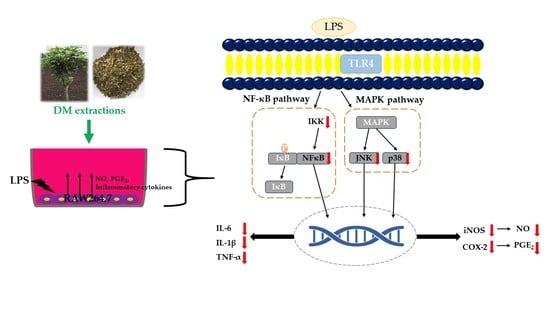
3.3. Effect of DM Extracts on the NF-κB and MAPK Signaling Pathway

The pretreatment with DM extracts significantly inhibited the production of nitrite and PGE2, mRNA expression level of iNOS and COX-2, and inflammatory cytokines level. To clarify the mechanism underlying the inhibition of inflammatory responses, regulation of the NF-κB and MAPK signaling pathway was demonstrated using the extracts showing the highest anti-inflammatory activities (Figure 5), namely the 30% ethanol extract in May and 60% ethanol extracts in August and November (Figure 3c,d). The NF-κB signaling pathway can be initiated by an external stimulus. The phosphorylation and successive proteasomal degradation of IκB are essential for classical activation [20]. The inhibitory κB kinase (IKK) complex phosphorylates and degrades IκB, resulting in the translocation of NF- κB to the nucleus. MAPK is involved in a wide range of signaling pathways, which are activated by various extracellular stimuli [21]. JNK, p38, and extracellular signal-regulated kinases (ERK) are included in the MAPK pathway. The LPS-treated group exhibited an increased phosphorylation of NF-κB, IKK, JNK, and p38. NF-κB phosphorylation was approximately 1.5, 1.8, and 1.5 times lower in the group treated with the extract from May, August, and November than in the group treated with LPS alone (p < 0.05). IKKα phosphorylation was approximately 1.5, 2.8, and 2 times lower in the group treated with the extract from May, August, and November than in the group treated with LPS alone (p < 0.05). JNK phosphorylation was approximately 1.7, 2.8, and 2.4 times lower in the group treated with the extract from May, August, and November than in the group treated with LPS alone (p < 0.05). The phosphorylation of p38 was approximately 2.2, 4.5, and 3.5 times lower in the group treated with the extract from May, August, and November than in the group treated with LPS alone (p < 0.05). All DM extracts significantly inhibited phosphorylation of the NF-κB signaling pathway-related protein. Among the extracts, the extract from August displayed the most prominent regulation ability.

3.4. Chemical Characteristics for Each DM Extract

The results confirming the biochemical components in each DM extract are presented in Table 1. The α-amyrin and β-amyrin were abundant in the order of the 60% ethanol extract from August > November > May. Commonly, the α- and β-amyrin contents of 100% hydrothermal extracts in DM collected in May and August could not be derived below the quantitative limit. The highest content of chlorogenic acid was found for the 60% ethanol extract from August. The 60% ethanol extract from August contained the greatest amount of 4-methylcatechol and p-coumaric acid. The rutin content was highest in the 60% ethanol extract from May, and the extracts collected in May displayed the highest values out of all monthly extracts. Moreover, the rutin content decreased as the months passed, and the rutin content of November was lower than in the others. In all groups, except for extracts harvested in November, the higher the ethanol content in the extraction solvent, the higher the flavonoid compound content observed.

3.5. Content Changes Based on Conditions

The correlation between the inflammation-related factors and major active components was shown in Figure 6. A negative correlation can be observed for the relationship between the two variables, in which one variable increases as the other decreases. The greater the correlation between X and Y, the closer the value of the Pearson´s correlation coefficient (r2) is to 1. Nitrite levels were negatively correlated with α-amyrin (r2 = 0.572, p = 0.018) and β-amyrin (r2 = 0.585, p = 0.016) with statistical significance. Furthermore, iNOS mRNA expression was negatively correlated with α-amyrin (r2 = 0.500, p = 0.033) and β-amyrin (r2 = 0.491, p = 0.035). COX-2 mRNA expression showed a negative correlation with α-amyrin (r2 = 0.523, p = 0.028) and β-amyrin (r2 = 0.519, p = 0.028). No other significant correlation was found (Figure S1 and Table S1). Both nitrite production and iNOS mRNA expression showed similar results of a decreased relationship with α-amyrin and β-amyrin. All relationships had more than a 50% negative correlation, except for that between β-amyrin and iNOS mRNA expression. It was revealed that as α-amyrin or β-amyrin contents increased, the contents or expression of inflammatory mediators diminished.

4. Discussion

Most chronic diseases are initiated by imbalances in major overarching processes, such as oxidative stress, metabolic stress, psychological stress, and inflammatory stress [22]. Plant food containing various secondary metabolites has been proposed to display anti-inflammatory responses [23]. Therefore, this study investigated the anti-inflammatory effect of various DM extracts according to seasonal variation and different ethanol concentrations as a solvent using the RAW264.7 murine macrophage cell line stimulated with LPS. DM leaves were harvested in May, August, and November, and each leaf sample was extracted using aqueous ethanol solutions (0, 30, and 60%). In the cytotoxicity assay, most extracts displayed cell viability close to that of the control group in 50 μg/mL, except for a few extracts. The hot water and 30% ethanol extracts harvested in August and all extracts harvested in November showed a higher cell viability than the control group. Previous studies have reported that plant extracts can enhance cell proliferation and increase the cell viability [24]. Likewise, hot water and 30% ethanol extracts harvested in August and all extracts harvested in November could have potential which increases cells. Except for a few extracts, most DM extracts showed lower inflammatory responses in LPS-stimulated RAW264.7 cells relative to LPS-treated cells. Among the extracts, the 30% ethanol extract harvested in May and 60% ethanol extracts harvested in August and November exhibited prominent inhibitory activity. This inhibition was almost close to the normal control cell level. Sometimes, the inflammatory reaction increased at a low concentration (12.5 μg/mL). Unlike human and in vivo studies, inflammation could be excessively induced in an in vitro study, as plants contain various phytochemicals and interact with each other [25,26,27]. Furthermore, this study was primarily based on cell viability data, and anti-inflammatory effects were revealed at a high concentration (50 μg/mL). Although identifying the principal bioactive components is a theoretically simple process to design, the actual discovery of the active compound may be considerably delayed, given that multiple metabolic pathways are involved in the plant physiology [23]. Therefore, further research should be conducted to determine what causes increased inflammation in a low concentration sample group compared to an LPS-treated group.
When inflammation is processed, NF-κB and MAPK pathways regulate the stimulation of macrophages as a trigger of events [17,28]. LPS-treated macrophages transcribe the NF-κB protein, which acts as a transcription factor by promoting inflammation [29,30]. In the cytoplasm, NK-κB, which is a heterodimer protein, maintains the complex inactivation state with the inhibitory protein IκBα. However, IκBα is phosphorylated and ubiquitinated when extracellular stimulation occurs, and NF-κB is translocated into the nucleus [31]. Likewise, MAPKs are activated to phosphorylate downstream subjects, including protein kinases, and transcription factors can transcribe the MAPK-regulated gene [32]. JNK and p38, which are part of the MAPK family and known to be directly involved in the cellular inflammatory response, are also phosphorylated when activated [29,33]. If proinflammatory cytokines are excessively produced, tissue damage and some proinflammatory responses occur, highlighting the significant role of proinflammatory cytokines as indicators in cell signaling and systematic inflammation [34].
The oxidation of L-arginine was catalyzed by nitric oxide synthase (NOS), releasing unstable and reactive NO. Among the isoforms of NOS, iNOS is activated by inflammatory stimulation and concerned with NO production [35]. A well-known inflammatory response in macrophages starts by combining LPS and TLR, which brings about a cascade of kinases [36]. COX has two isoenzyme forms: COX-1 and COX-2. COX-1 appears in various cells and mediates physiological activity, whereas COX-2, known as an inducible enzyme, is found in immune cells treated with LPS and inflammatory cytokines [37]. A previous study described that COX-2 is accountable for the synthesis of prostanoids, which are relevant in critical and chronic inflammatory conditions [38]. Therefore, COX-2 hyperactivation can trigger PGE2 production. Similarly, in this study, iNOS and COX-2 mRNA expression was upregulated in the LPS-treated group, and the resultant nitrite and PGE2 production were confirmed to be increased. In comparison, all DM extract groups significantly inhibited iNOS and COX-2 expression compared to the LPS-treated group. In terms of the nitrite and PGE2 level, among the extracts, the 60% ethanol extract harvested in August particularly suppressed both. Furthermore, DM aqueous ethanol extracts inhibited the phosphorylation of NF-κB, IKKα, JNK, and p38. Downregulation of these proteins was related to iNOS and COX-2 mRNA expression. Therefore, the downregulation of proinflammatory transcription factors reduced the production of proinflammatory cytokine (IL-6, IL-1β, and TNF-α) and cellular mediators (nitrite and PGE2).
DM leaves contain several polyphenolic compounds, such as chlorogenic acid, rutin, 4-methylcatechol, and p-coumaric acid and terpenoids, including α- and β-amyrin [12,13,19]. In the current study, these significant compounds were analyzed. Our results showed that terpenoids increased in the August and November extracts compared to extracts of May, and higher ethanol concentrations increased α- and β-amyrin. However, except for rutin, the major polyphenolic compounds were higher in the August extracts than in the May and November extracts. The rutin concentration was the highest in the May extract. Ethanol concentrations showed a similar pattern for extracting these active compounds. We identified the main DM active compounds through the literature review and analyzed them. However, given the preliminary nature of this study, we could not fully-characterize the active compounds from DM extracts. However, we estimated which components might contribute to suppressing inflammation by comparing the chief composition and anti-inflammatory activity. When the correlation between the concentrations of these compounds and anti-inflammatory activity was analyzed, α- and β-amyrin showed significant correlations with iNOS and COX-2 inhibition. The 60% ethanol extracts harvested in August and November displayed the strongest inhibitory activity, and the concentrations of α- and β-amyrin were the highest. Although the correlation between other compositions and inflammatory cytokines was also analyzed, there was no significant difference. This finding suggests a potential mechanism underlying the inhibition of inflammatory responses.
Organic solvent extraction methods, such as ethanol and methanol, are conventional and have been applied to extract phenolic compounds from plants. In this study, overall, as the ethanol concentration increased, both α- and β-amyrin in various extracts also increased. Among the well-known active chemicals in DM, α- and β-amyrin were the most active compounds, as evidenced by correlation analysis. Amyrin is a pentacyclic triterpene that displays biological activities, such as anti-inflammatory, antinociceptive, hepatoprotective, antipruritic, gastroprotective, antitumor, and anxiolytic activity [39,40]. Previous studies have reported that α- and β-amyrin can prevent NF-κB activation and that the inhibition of NO plays a prominent role in an L-arginine-induced pancreatitis model [41]. Furthermore, the release of TNF-α or its proinflammatory response was constrained by α- and β-amyrin [42]. Similar to reported findings, this study also confirmed the anti-inflammatory activity of amyrin in DM extracts by suppressing the cellular regulators iNOS and COX-2 and proinflammatory cytokines by preventing activation of the NF-κB and MAPK pathways in the 30 and 60% ethanol extract-treated groups. Although precise underlying mechanisms were not revealed in this study, the anti-inflammatory effects of DM extracts varied by season and extraction solvent used. Among the extracts, the 30% ethanol extract harvested in May and 60% ethanol extracts harvested in August and November exhibtied the highest anti-inflammatory effects in the RAW264.7 murine macrophage cell line stimulated with LPS. Unlike other seasons, in May, the 30% ethanol extract showed high anti-inflammatory activity. Therefore, we need to conduct further studies to identify unknown active components in the May harvest.
To the best of our knowledge, this is the first study to show that DM alleviates inflammatory responses through its active components of α- and β-amyrin. In this study, we found the optimal conditions, including the season and amount of ethanol for DM extracts, for maximizing the anti-inflammatory activity. We suggest that DM extracts could be developed as functional food ingredients for preventing inflammation-related diseases. This is because DM extracts significantly inhibited cellular mediators (nitrite and PGE2); the mRNA expression of iNOS and COX-2; and inflammatory cytokines, including IL-6, IL-1β, and TNF-α. In addition to this, the NF-κB/MAPK signaling pathway was downregulated by DM extracts. Nevertheless, although amyrins could be beneficial in standard compositions, this study has major limitations. As a preliminary screening study, we only analyzed the major compounds in extracts and investigated the correlation between the flavonoids and inflammatory cytokines. Although it was found that α-amyrin and β-amyrin were the first major starting points in anti-inflammatory effects, the roles of the active compounds and detailed mechanisms were not revealed in the current study. Therefore, additional studies on the profiling of various flavonoids and the physiological relevance of DM extracts using in vivo and human interventions should be conducted. This might produce the most proof for discovering detailed mechanisms underlying the anti-inflammatory activity of multi-compounds derived from DM extracts.

Supplementary Materials

The following are available online at https://www.mdpi.com/2076-3417/10/23/8756/s1, Figure S1: Relationship between α- (a) or β-amyrin (b) and PGE2 or proinflammatory cytokines. Correlations are presented as Pearson´s correlation coefficients (r2); Figure S2: Relationship between chlorogenic acid and inflammatory mediators (a) or proinflammatory cytokines (b). Correlations are presented as Pearson´s correlation coefficients (r2); Figure S3: Relationship between 4-methylcatechol and inflammatory mediators (a) or proinflammatory cytokines (b). Correlations are presented as Pearson´s correlation coefficients (r2); Figure S4: Relationship between p-coumaric acid and inflammatory mediators (a) or proinflammatory cytokines (b). Correlations are presented as Pearson´s correlation coefficients (r2); Figure S5: Relationship between Rutin and inflammatory mediators (a) or proinflammatory cytokines (b). Correlations are presented as Pearson´s correlation coefficients (r2); Table S1: The correlation r2 and p-values between flavonoid compounds and proinflammatory mediators or cytokines.

Author Contributions

Conceptualization, J.Y.K.; investigation, K.J.K., J.S.Y., and Y.-J.K.; data curation, K.J.K., J.S.Y., and Y.-J.K.; writing—original draft preparation, K.J.K. and J.Y.K.; writing—review and editing, K.J.K. and J.Y.K.; supervision, J.Y.K.; project administration, J.Y.K.; funding acquisition, J.Y.K. All authors have read and agreed to the published version of the manuscript.

Funding

This work was supported by the Advanced Research Project funded by the Seoul National University of Science and Technology (Project No. 2020-0297).

Acknowledgments

We express our sincere appreciation to the Agency for Jeju Plant Resources Dendropanax for providing the Dendropanax samples.

Conflicts of Interest

The authors declare no conflict of interest.

References

  1. Calder, P.C.; Ahluwalia, N.; Albers, R.; Bosco, N.; Bourdet-Sicard, R.; Haller, D.; Holgate, S.T.; Jönsson, L.S.; Latulippe, M.E.; Marcos, A.; et al. A Consideration of Biomarkers to be Used for Evaluation of Inflammation in Human Nutritional Studies. Br. J. Nutr. 2013, 109, S1–S34. [Google Scholar] [CrossRef] [PubMed] [Green Version]
  2. Hotamisligil, G.S. Inflammation and metabolic disorders. Nature 2006, 444, 860–867. [Google Scholar] [CrossRef] [PubMed]
  3. Libby, P. Inflammation in atherosclerosis. Nature 2002, 420, 868–874. [Google Scholar] [CrossRef]
  4. Ellulu, M.S. Obesity, cardiovascular disease, and role of vitamin C on inflammation: A review of facts and underlying mechanisms. Inflammopharmacology 2017, 25, 313–328. [Google Scholar] [CrossRef]
  5. Poffenberger, M.C.; Straka, N.; El Warry, N.; Fang, D.; Shanina, I.; Horwitz, M.S. Lack of IL-6 during Coxsackievirus Infection Heightens the Early Immune Response Resulting in Increased Severity of Chronic Autoimmune Myocarditis. PLoS ONE 2009, 4, e6207. [Google Scholar] [CrossRef] [PubMed]
  6. Hyun, T.K.; Kim, M.-O.; Lee, H.; Kim, Y.; Kim, E.; Kim, J.-S. Evaluation of anti-oxidant and anti-cancer properties of Dendropanax morbifera Leveille. Food Chem. 2013, 141, 1947–1955. [Google Scholar] [CrossRef] [PubMed]
  7. Kim, R.-W.; Lee, S.-Y.; Kim, S.-G.; Heo, Y.-R.; Son, A.M.-K. Antimicrobial, Antioxidant and Cytotoxic Activities of Dendropanax morbifera Leveille extract for mouthwash and denture cleaning solution. J. Adv. Prosthodont. 2016, 8, 172–180. [Google Scholar] [CrossRef] [Green Version]
  8. Kim, W.; Kim, D.W.; Yoo, D.-Y.; Jung, H.Y.; Nam, S.M.; Kim, J.W.; Hong, S.-M.; Kim, D.-W.; Choi, J.H.; Moon, S.M.; et al. Dendropanax morbifera Leveille extract facilitates cadmium excretion and prevents oxidative damage in the hippocampus by increasing antioxidant levels in cadmium-exposed rats. BMC Complement. Altern. Med. 2014, 14, 428. [Google Scholar] [CrossRef] [Green Version]
  9. Cho, S.S.; Song, S.-H.; Choi, C.Y.; Park, K.-M.; Shim, J.-H.; Park, D.-H. Optimization of the Extraction Conditions and Biological Evaluation of Dendropanax morbifera H. Lev as an Anti-Hyperuricemic Source. Molecules 2018, 23, 3313. [Google Scholar] [CrossRef] [Green Version]
  10. Song, J.-H.; Kang, H.-B.; Kim, J.H.; Kwak, S.; Sung, G.-J.; Park, S.-H.; Jeong, J.-H.; Kim, H.; Lee, J.; Jun, W.; et al. Antiobesity and Cholesterol-Lowering Effects of Dendropanax morbifera Water Extracts in Mouse 3T3-L1 Cells. J. Med. Food 2018, 21, 793–800. [Google Scholar] [CrossRef]
  11. Sun, S.; Kee, H.J.; Jin, L.; Ryu, Y.; Choi, S.Y.; Kim, G.R.; Jeong, M.H. Gentisic acid attenuates pressure overload-induced cardiac hypertrophy and fibrosis in mice through inhibition of the erk1/2 pathway. J. Cell. Mol. Med. 2018, 22, 5964–5977. [Google Scholar] [CrossRef] [PubMed]
  12. Chung, I.-M.; Kim, M.-Y.; Park, S.-D.; Park, W.-H.; Moon, H.-I. In vitro evaluation of the antiplasmodial activity of Dendropanax morbifera against chloroquine-sensitive strains of Plasmodium falciparum. Phytother. Res. 2009, 23, 1634–1637. [Google Scholar] [CrossRef] [PubMed]
  13. Chung, I.-M.; Song, H.-K.; Kim, S.-J.; Moon, H.-I. Anticomplement activity of polyacetylenes from leaves of Dendropanax morbiferaLeveille. Phytother. Res. 2010, 25, 784–786. [Google Scholar] [CrossRef]
  14. Choi, J.-H.; Kim, D.-W.; Park, S.-E.; Lee, H.-J.; Kim, K.-M.; Kim, K.-J.; Kim, M.-K.; Kim, S.-J.; Kim, S. Anti-thrombotic effect of rutin isolated from Dendropanax morbifera Leveille. J. Biosci. Bioeng. 2015, 120, 181–186. [Google Scholar] [CrossRef] [PubMed]
  15. Akram, M.; Kim, K.-A.; Kim, E.-S.; Syed, A.S.; Kim, C.Y.; Lee, J.S.; Bae, O.-N. Potent Anti-inflammatory and Analgesic Actions of the Chloroform Extract of Dendropanax morbifera Mediated by the Nrf2/HO-1 Pathway. Biol. Pharm. Bull. 2016, 39, 728–736. [Google Scholar] [CrossRef] [PubMed] [Green Version]
  16. Lee, K.Y.; Jung, H.Y.; Yoo, D.Y.; Kim, W.; Kim, J.W.; Kwon, H.J.; Kim, D.W.; Yoon, Y.S.; Hwang, I.K.; Choi, J.H. Dendropanax morbifera Leveille extract ameliorates D-galactose-induced memory deficits by decreasing inflammatory responses in the hippocampus. Lab. Anim. Res. 2017, 33, 283–290. [Google Scholar] [CrossRef] [PubMed] [Green Version]
  17. Yu, H.Y.; Kim, K.S.; Lee, Y.C.; Moon, H.I.; Lee, J.H. Oleifolioside a, a new active compound, attenuates lps-stimulated inos and cox-2 expression through the downregulation of nf-kappab and mapk activities in raw 264.7 macrophages. Evid. Based Complement. Alternat. Med. 2012, 2012, 637512. [Google Scholar] [CrossRef] [PubMed] [Green Version]
  18. Ahn, J.C.; Kim, S.H.; Kim, M.Y.; Kim, O.T.; Kim, K.S.; Hwang, B. Seasonal variations in yields of Hwangchil lacquer and major sesquiterpene compounds from selected superior individuals of Dendropanax morbifera Lev. J. Plant Biol. 2003, 46, 38–40. [Google Scholar] [CrossRef]
  19. Youn, J.S.; Kim, Y.-J.; Na, H.J.; Jung, H.R.; Song, C.K.; Kang, S.Y.; Kim, J.Y. Antioxidant activity and contents of leaf extracts obtained from Dendropanax morbifera LEV are dependent on the collecting season and extraction conditions. Food Sci. Biotechnol. 2018, 28, 201–207. [Google Scholar] [CrossRef]
  20. Shao, H.J.; Jeong, J.B.; Kim, K.J.; Lee, S.H. Anti-inflammatory activity of mushroom-derived hispidin through blocking of nf-kappab activation. J. Sci. Food Agric. 2015, 95, 2482–2486. [Google Scholar] [CrossRef]
  21. Sun, P.; Zhou, K.; Wang, S.; Li, P.; Chen, S.; Lin, G.; Zhao, Y.; Wang, T. Involvement of mapk/nf-kappab signaling in the activation of the cholinergic anti-inflammatory pathway in experimental colitis by chronic vagus nerve stimulation. PLoS ONE 2013, 8, e69424. [Google Scholar]
  22. Van Ommen, B.; Keijer, J.; Heil, S.G.; Kaput, J. Challenging homeostasis to define biomarkers for nutrition related health. Mol. Nutr. Food Res. 2009, 53, 795–804. [Google Scholar] [CrossRef] [PubMed]
  23. Kim, J.Y.; Kwon, O. Culinary plants and their potential impact on metabolic overload. Ann. N. Y. Acad. Sci. 2011, 1229, 133–139. [Google Scholar] [CrossRef] [PubMed]
  24. Moezizadeh, M.; Javand, F.; Tabatabaei, F.S. Effects of extracts of Salvadora persica on proliferation and viability of human dental pulp stem cells. J. Conserv. Dent. 2015, 18, 315–320. [Google Scholar] [CrossRef] [Green Version]
  25. Phan, M.A.T.; Paterson, J.; Bucknall, M.; Arcot, J. Interactions between phytochemicals from fruits and vegetables: Effects on bioactivities and bioavailability. Crit. Rev. Food Sci. Nutr. 2018, 58, 1310–1329. [Google Scholar] [CrossRef]
  26. Yeum, K.-J.; Russell, R.M. Biological functions of plant pigment phytochemicals in humans. In Systems Biology of Free Radicals and Antioxidants; Laher, I., Ed.; Springer: Berlin/Heidelberg, Germany, 2014; pp. 4023–4045. [Google Scholar]
  27. Mestas, J.; Hughes, C.C.W. Of Mice and Not Men: Differences between Mouse and Human Immunology. J. Immunol. 2004, 172, 2731–2738. [Google Scholar] [CrossRef] [Green Version]
  28. Saji, N.; Francis, N.; Schwarz, L.J.; Blanchard, C.L.; Santhakumar, A.B. The Antioxidant and Anti-Inflammatory Properties of Rice Bran Phenolic Extracts. Foods 2020, 9, 829. [Google Scholar] [CrossRef]
  29. Cho, B.O.; So, Y.; Jin, C.H.; Nam, B.M.; Yee, S.T.; Jeong, I.Y. 3-deoxysilybin exerts anti-inflammatory effects by suppressing nf-kappab activation in lipopolysaccharide-stimulated raw264.7 macrophages. Biosci. Biotechnol. Biochem. 2014, 78, 2051–2058. [Google Scholar] [CrossRef] [Green Version]
  30. Hobbs, S.; Reynoso, M.; Geddis, A.V.; Mitrophanov, A.Y.; Matheny, R.W., Jr. Lps-stimulated nf-kappab p65 dynamic response marks the initiation of tnf expression and transition to il-10 expression in raw 264.7 macrophages. Physiol. Rep. 2018, 6, e13914. [Google Scholar] [CrossRef] [Green Version]
  31. Oeckinghaus, A.; Ghosh, S. The nf-kappab family of transcription factors and its regulation. Cold Spring Harb. Perspect. Biol. 2009, 1, a000034. [Google Scholar] [CrossRef]
  32. Whitmarsh, A.J. Regulation of gene transcription by mitogen-activated protein kinase signaling pathways. Biochim. Biophys. Acta Bioenerg. 2007, 1773, 1285–1298. [Google Scholar] [CrossRef] [PubMed]
  33. Huang, G.; Shi, L.Z.; Chi, H. Regulation of JNK and p38 MAPK in the immune system: Signal integration, propagation and termination. Cytokine 2009, 48, 161–169. [Google Scholar] [CrossRef] [PubMed] [Green Version]
  34. Zhang, M.J.-M.; An, J. Cytokines, Inflammation, and Pain. Int. Anesthesiol. Clin. 2007, 45, 27–37. [Google Scholar] [CrossRef] [Green Version]
  35. Cheng, A.; Wan, F.; Jin, Z.; Wang, J.; Xu, X. Nitrite oxide and inducible nitric oxide synthase were regulated by polysaccharides isolated from Glycyrrhiza uralensis Fisch. J. Ethnopharmacol. 2008, 118, 59–64. [Google Scholar] [CrossRef] [PubMed]
  36. Poteser, M.; Wakabayashi, I. Serum albumin induces iNOS expression and NO production in RAW 267.4 macrophages. Br. J. Pharmacol. 2004, 143, 143–151. [Google Scholar] [CrossRef]
  37. Li, Y.; Zou, L.; Li, T.; Lai, D.; Wu, Y.; Qin, S. Mogroside V inhibits LPS-induced COX-2 expression/ROS production and overexpression of HO-1 by blocking phosphorylation of AKT1 in RAW264.7 cells. Acta Biochim. Biophys. Sin. 2019, 51, 365–374. [Google Scholar] [CrossRef]
  38. Chun, K.-S.; Surh, Y.-J. Signal transduction pathways regulating cyclooxygenase-2 expression: Potential molecular targets for chemoprevention. Biochem. Pharmacol. 2004, 68, 1089–1100. [Google Scholar] [CrossRef]
  39. Da Silva Júnior, W.F.; Pinheiro, J.G.D.O.; De Menezes, D.L.B.; E Silva, N.E.d.S.; De Almeida, P.D.O.; Lima, E.S.; Da Veiga Júnior, V.F.; De Azevedo, E.P.; De Lima, Á.A.N. Development, physicochemical characterization and in vitro anti-inflammatory activity of solid dispersions of α,β amyrin isolated from protium oilresin. Molecules 2017, 22, 1512. [Google Scholar] [CrossRef] [Green Version]
  40. Da Silva Júnior, W.F.; Bezerra de Menezes, D.L.; de Oliveira, L.C.; Koester, L.S.; de Almeida, P.D.O.; Lima, E.S.; de Azevedo, E.P.; da Veiga Junior, V.F.; de Lima, Á.A.N. Inclusion complexes of β and HPβ-cyclodextrin with α,β amyrin and in vitro anti-inflammatory activity. Biomolecules 2019, 9, 241. [Google Scholar] [CrossRef] [Green Version]
  41. Melo, C.M.; Morais, T.C.; Tome, A.R.; Brito, G.A.C.; Chaves, M.H.; Rao, V.S.; Santos, F.A. Anti-inflammatory effect of α,β-amyrin, a triterpene from Protium heptaphyllum, on cerulein-induced acute pancreatitis in mice. Inflamm. Res. 2011, 60, 673–681. [Google Scholar] [CrossRef]
  42. Pinto, S.A.H.; Pinto, L.M.S.; Cunha, G.M.A.; Chaves, M.H.; Santos, F.A.; Rao, V. Anti-inflammatory effect of α,β-amyrin, a pentacyclic triterpene from Protium heptaphyllum in rat model of acute periodontitis. Inflammopharmacology 2007, 16, 48–52. [Google Scholar] [CrossRef] [PubMed]
Figure 1. Preparation of Dendropanax morbifera extracts. W100, hot water extracts; E30, 30% ethanol extracts; and E60, 60% ethanol extracts.
Applsci 10 08756 g001
Figure 2. Cell viability of RAW264.7 macrophage cells treated with Dendropanax morbifera extracts. W100, hot water extracts; E30, 30% ethanol extracts; and E60, 60% ethanol extracts. Data are presented as the mean ± SEs of triplicate determinations of independent wells.
Applsci 10 08756 g002
Figure 3. Effects on the production of nitrite (a), prostaglandin E2 (b), mRNA expression levels of inducible nitric oxide synthase (c), and cyclooxygenase-2 (d) in the RAW264.7 cells stimulated with lipopolysaccharide. W100, hot water extracts; E30, 30% ethanol extracts; and E60, 60% ethanol extracts. Data are presented as the mean ± SEs of triplicate determinations of independent wells. Statistical analysis was performed using one-way ANOVA. *, p < 0.05 compared to the lipopolysaccharide (LPS)-treated group.
Applsci 10 08756 g003
Figure 4. Concentration profiles of interleukin (IL)-6 (a), IL-1β (b), and tumor necrosis factor-α (TNF-α) (c) in the RAW264.7 cells stimulated with lipopolysaccharide. W100, hot water extracts; E30, 30% ethanol extracts; and E60, 60% ethanol extracts. Data are presented as the mean ± SEs of triplicate determinations of independent wells. Statistical analysis was performed using one-way ANOVA. *, p < 0.05 compared to the LPS-treated group.
Applsci 10 08756 g004
Figure 5. The effect of Dendropanax morbifera extracts on the nuclear factor κB and mitogen-activated protein kinase signaling pathways. Western blotting bands for the phosphorylation of proteins (a) and quantification of the band intensity on nuclear factor κB (NF-κB) (b), IκB kinase α (IKKα) (c), c-Jun N-terminal kinase (JNK) (d), and phosphorylated p38 MAPK (p38) (e). W100, hot water extracts; E30, 30% ethanol extracts; and E60, 60% ethanol extracts. Data are presented as the mean ± SEs of triplicate determinations of independent experiments. Statistical analysis was performed using one-way ANOVA. *: p < 0.05 compared to the LPS-treated group.
Applsci 10 08756 g005
Figure 6. Relationship between amyrin and inflammation regulators. Correlations are presented as Pearson´s correlation coefficient (r2). The correlation between a-amyrin and nitrite (a), and the relative mRNA expression levels of inducible nitric oxide synthase (iNOS) (b) and cyclooxygenase-2 (COX-2) (c). The correlation between β-amyrin and nitrite (d), and the relative mRNA expression levels of iNOS (e) and COX-2 (f).
Applsci 10 08756 g006
Table 1. Profile and concentration of flavonoid compounds in Dendropanax morbifera extracts.
MonthEtOH (%)α-Amyrin
(mg/kg)
β-Amyrin
(mg/kg)
Chlorogenic Acid (mg/g)4-Methylcatechol (mg/g)p-Coumaric Acid (mg/g)Rutin (mg/g)
May0N.D. *N.D. *39.61±0.520.08±0.052.77±0.07127.90±0.02
3024.39±1.3137.86±1.1371.44±1.480.38±0.135.21±0.12167.00±3.05
60292.97±11.83433.76±11.2176.71±0.160.68±0.095.56±0.02169.36±0.61
August0N.D. *N.D. *60.40±0.070.21±0.045.38±0.02105.31±2.20
308.28±0.8811.82±0.2166.98±4.210.71±0.036.59±0.15119.35±2.47
60876.70±18.201508.87±20.2179.43±0.112.00±0.089.76±0.43134.54±0.51
November08.17±0.4712.30±0.6025.65±1.44N.D. *2.20±0.0043.45±0.22
30N.D. *4.94±0.5941.76±0.810.12±0.064.70±0.0647.88±0.22
60860.65±35.971399.34±59.3842.87±1.530.08±0.053.92±0.1172.03±2.53
*N.D., not detected (<limit of quantitation; α-amyrin, 0.01 mg/kg; β-amyrin, 0.07 mg/kg; and 4-methylcatechol, 0.01 mg/g).
Publisher’s Note: MDPI stays neutral with regard to jurisdictional claims in published maps and institutional affiliations.

