Review

Vitamin D and the NLRP3 Inflammasome

by
Matthew Tunbridge
1,2,* and
Pedro Henrique França Gois
1,2,3
1
Nephrology Department, Royal Brisbane and Women’s Hospital, Herston 4029, Australia
2
Faculty of Medicine, University of Queensland, Brisbane 4072, Australia
3
Conjoint Internal Medicine Laboratory—Kidney Research Group, Queensland Institute of Medical Research, Brisbane 4029, Australia
*
Author to whom correspondence should be addressed.
Appl. Sci. 2020, 10(23), 8462; https://doi.org/10.3390/app10238462
Submission received: 1 November 2020 / Revised: 18 November 2020 / Accepted: 24 November 2020 / Published: 27 November 2020
(This article belongs to the Special Issue Vitamin D in Human Health and Disease)

Abstract

:
Vitamin D (VD) is a steroid hormone classically known for its key role in maintaining calcium homeostasis in the body. VD also has important immunomodulatory functions. This review explores evidence for a role of VD in attenuating the activation of the nucleotide-binding domain-like receptor protein 3 (NLRP3) inflammasome. Dysregulated and inappropriate NLRP3 inflammasome activation occurs in a range of human diseases, including autoinflammatory disorders, metabolic disorders, and infections. VD appears to mediate its effects by binding of the VD receptor (VDR) to the sensor protein NLRP3, inhibiting deubiquitination and downstream inflammasome assembly. Some early clinical evidence suggests improved outcomes in inflammasome-mediated disorders when VD-deficient patients are treated with supplementation therapy.

1. Introduction

Vitamin D (VD) is a multifunctional secosteroid hormone precursor best known for its role in maintaining calcium homeostasis and bone health. However, VD has wide-ranging effects beyond this traditionally understood role, including the regulation of immune responses, inflammation and oxidative stress, and cancer prevention.
VD is a precursor of the active 1,25(OH)2-VD and is available endogenously in two major forms—ergocalciferol (VD2) and cholecalciferol (VD3). VD2 is available in the diet in a variety of vegetables and fortified foods, while VD3 is present in some animal-based foods. VD3 can also be synthesized in the skin by conversion from 7-dehydrocholesterol in a multi-step ultraviolet-B light-dependent pathway (Figure 1) [1,2].
VD in both forms is transported by the VD-binding protein (VDBP) in plasma to the liver, where it undergoes hydroxylation of the 25 carbon by 25-hydroxylase (a cytochrome P450 enzyme, CYP2R1) to become 25(OH)-VD2 or 25(OH)-VD3 [2,3]. 25(OH)-VD is the major circulating form of VD, the levels of which are measured as markers of overall VD status. A subsequent hydroxylation step on the 1 carbon occurs in the kidney, mediated by 1α-hydroxylase (CYP27B1) to create the active form 1,25(OH)2-VD (Figure 1) [2]. Nevertheless, both 25(OH)-VD and 1,25(OH)2-VD are constituently active and can bind to and activate the VD receptor (VDR) [2]. Hydroxylation of VD3 at alternative positions mediated by CYP11A1 can generate secosteroid metabolites with important actions [4]. Metabolites such as 20(OH)-VD3 and 20,23(OH)2-VD3 have activity at the VDR and other receptors, including the aryl hydrocarbon receptor (AhR) [5], retinoic acid orphan receptor (ROR)-α, and ROR-γ [6], producing novel effects including photoprotective and antiproliferative activity [7,8].
The genomic actions of VD are mediated by the VDR [9,10]. The VDR is a member of the steroid hormone nuclear receptor family that acts as a transcription factor when activated. VDR has three domains, a C-terminal ligand binding domain for VD binding, an N-terminal DNA binding domain that binds to VD response elements (VDREs) in the promoter region of VD-regulated genes, and a hinge region that binds these domains together [9,10]. The VDR generally interacts with a heterodimer partner retinoid-X receptor (RXR) and coactivators/coregulators, increasing translational activity at a range of target genes that allow for multiple and varied responses across different cell types [2,9,10].
1,25(OH)2-VD2 and VD3 also exert rapid non-genomic actions via multiple second messenger pathways [11,12]. These appear to be mediated by both the nuclear VDR, as well as a membrane-bound receptor known as membrane-associated rapid response steroid binding protein (MARRS) [12,13]. These non-genomic actions are of interest as they appear to be implicated in many of the immunomodulatory effects of VD.
Despite the critical role VD plays in health, VD deficiency (VDD) is a common problem worldwide. While definitions are controversial, VDD is generally defined as circulating levels of 25(OH)-VD below 50 nmol/L (<20 ng/mL) [14]. Reported frequencies in some populations are as high as 90% [15,16]. This is concerning given the wide range of physiological actions that appear to be influenced by VD, in particular our evolving understanding of the role of VD in moderating immune function.
VD has a complex role in immune response regulation, both enhancing and suppressing different responses [17]. Emerging evidence suggests a role for VD in modulating inflammation via the inflammasome cascade [18,19,20]. Part of the innate immune system, inflammasomes are large, multimeric protein complexes that assemble in the cell in response to danger signals [21,22]. While an important part of the immune response, inflammasome activation has been implicated in the pathogenesis of a wide range of human diseases [23]. Through its receptor, VD appears to be able to inhibit inflammasome assembly and attenuate over-zealous inflammatory responses in animal models of disease [18,20].
This review will focus on the specific role VD may play in moderating inflammasome activity, in particular from the nucleotide-binding domain-like receptor protein 3 (NLRP3) inflammasome, and examine the evidence for VD supplementation in conditions where the inflammasome is implicated.

2. The Nucleotide-Binding Domain-Like Receptor Protein 3 (NLRP3) Inflammasome

Innate immune function relies upon immune cell detection of pathogens by pattern-recognition receptors (PRRs). Activation of PRRs by highly conserved pathogen-associated molecular patterns (PAMPs) and danger-associated molecular patterns (DAMPs) then leads to downstream effects with production of the signalling proteins interferon-α and β, and pro-inflammatory cytokines such as interleukins [21].
Inflammasomes form a critical part of this signalling cascade. Inflammasomes are large, multimeric protein complexes that assemble in the cell cytosol after sensing PAMPs or DAMPs [21,22]. Although there are several described inflammasomes, the most well-studied of these is the NLRP3 inflammasome [21,22].
The NLRP3 inflammasome comprises the NLRP3 sensor protein, the apoptosis-associated speck-like (ASC) adaptor protein, and the pro-caspase-1 effector protein. The inflammasome can be activated through both canonical and non-canonical pathways that then lead to the processing and release of interleukin (IL)-1β and IL-18 [21,22].
Canonical activation of the NLRP3 inflammasome is a multi-step process that requires priming and then activation of the inflammasome [21,22]. The first signal comes when PRRs are activated by PAMPs and DAMPs. The toll-like receptor (TLR) family plays a significant role in downstream signalling, for instance, TLR4 activation, which occurs in response to the common Gram-negative bacterial cell wall molecule lipopolysaccharide (LPS) [24]. Transcriptional activation via the nuclear factor kappa-light-chain-enhancer of activated B cells (NF-ĸB) pathway leads to increased expression of NLRP3 and pro-interleukin 1β expression [21,22]. Deubiquitination of NLRP3 mediated by the BRCA1/BRCA2-containing complex, subunit 3 (BRCC-3), also appears to be a critical step in priming to allow for the oligomerisation of NLRP3 with the other subunits of the inflammasome [24,25], and a complex series of other regulatory steps involving the ubiquitination and phosphorylation of both NLRP3 and ASC is not yet fully understood [26].
After priming, inflammasome activation requires a second signal that may be triggered in response to a wide range of stimuli. A recent review proposed the term homeostasis-altering molecular processes (HAMPs) to describe a number of these signals that include altered membrane potential due to potassium efflux or calcium influx, extracellular adenosine triphosphate (ATP) and oxidised deoxyribonucleic acid (DNA), lysosomal degradation products, mitochondrial damage due to reactive oxygen species (ROS), altered mitochondrial membrane potential, and oxidised mitochondrial DNA [27].
Once the inflammasome is activated, the conformational changes induced by oligomerisation activate caspase 1, which then cleaves pro-IL 1β and 18 to release their active cytokine forms [21,22]. Caspase 1 is also able to cleave gasdermin D (GSDMD), the activated form of which translocates to the membrane creating pores that can mediate pyroptosis—a lytic and inflammatory type of programmed cell death—and allow for the release of IL-1β and 18 into the surrounding environment [28,29,30]. This process is outlined in Figure 2.
Noncanonical NLRP3 inflammasome activation can occur in the absence of a priming signal through the activation of murine caspase 11 (or the human equivalents caspase 4 or 5) [32]. Interferon signalling increases the expression of pro-caspase-11 that subsequently activates the NLRP3 inflammasome and pyroptosis. Gram-negative bacterial products such as lipopolysaccharide are strong activators of caspase 11 and in animal models, an absence of caspase 11 attenuates the inflammasome response to several Gram-negative bacteria such as Escherichia coli [21,22,32].
Disordered inflammation is implicated in the pathogenesis of an extraordinary range of human diseases, and as our understanding of the inflammasome continues to grow, it has become clear that the inflammasome plays a significant pathological role [21]. Mutations in NLRP3 can lead to a heterogeneous group of cyclical fever syndromes known as cryopyrin-associated periodic syndromes (CAPS) [33]. Inappropriate inflammasome activation may affect outcomes from infections including respiratory tract infections (such as the novel coronavirus, Mycobacterium tuberculosis, and bacterial pneumonias). Increasingly, inflammasomes are also being linked to autoinflammatory conditions, where the inflammasome may be the initiator or exaggerator of inflammatory responses. These include neurodegenerative conditions (multiple sclerosis, Alzheimer’s disease, and Parkinson’s disease), rheumatological conditions (gout, rheumatoid arthritis, psoriatic arthritis), and metabolic conditions (obesity, atherosclerotic cardiovascular disease, and diabetes mellitus) [21,23].

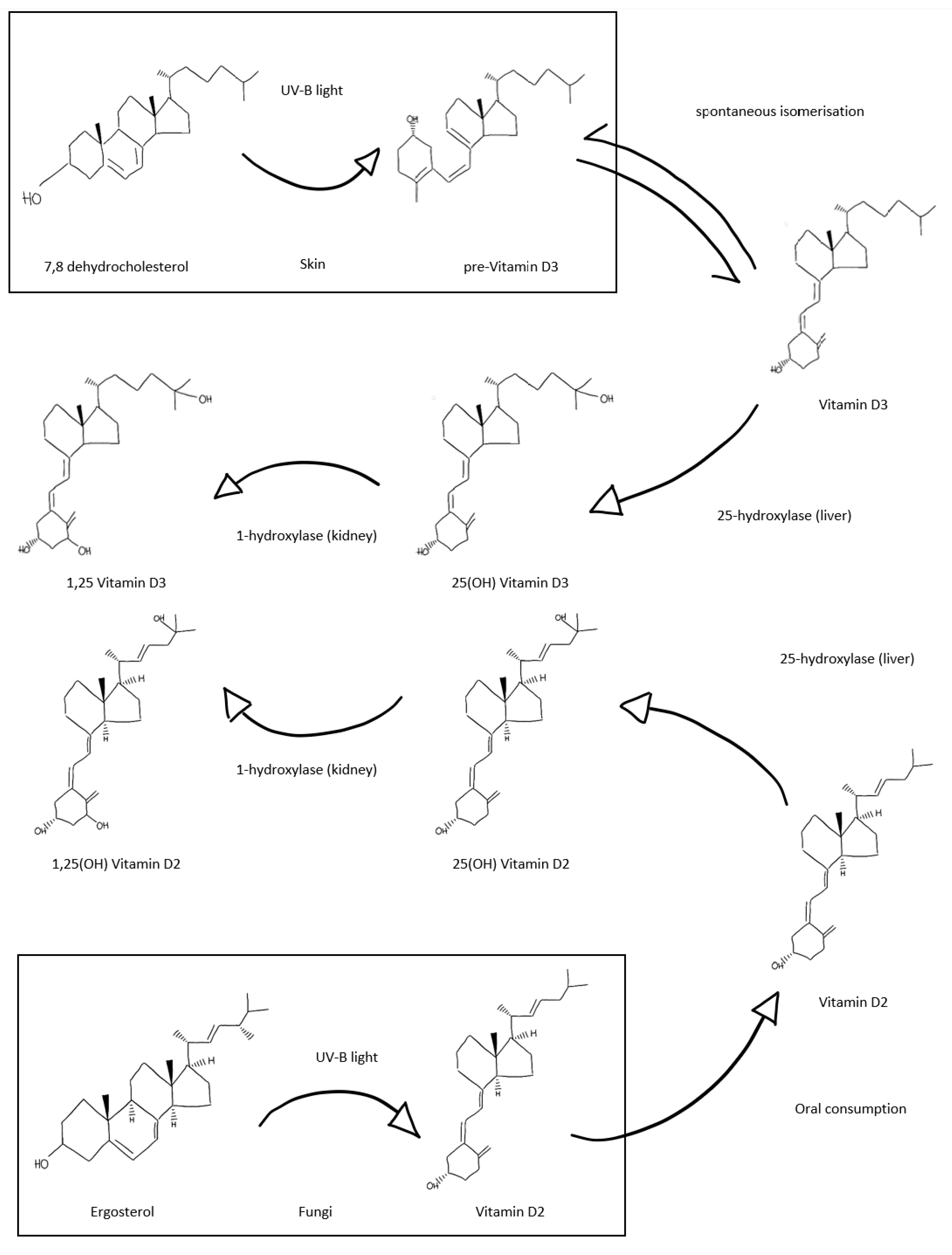
3. Vitamin D and the Emerging Role in Immunity

Innate and adaptive immune responses to infection involve complex and dynamic interactions between a wide array of immune cell types, cell-bound and free signalling molecules, chemokines, and hormones [34]. VD interacts with both the innate and adaptive immune responses in a complex manner, which can upregulate and enhance some responses, while attenuating others and encouraging the differentiation of regulatory T cells [17].
The innate immune system relies on a system of PRRs that respond to PAMPs [34]. TLRs are the major sensing molecule on the cell surface of innate immune cells, such as polymorphonuclear cells, monocytes, and macrophages [35]. This large family of transmembrane PRRs respond to a wide range of highly conserved PAMPs such as cell wall molecules (e.g., lipopolysaccharide, peptidoglycans) and RNA/DNA sequences [35]. Activation of TLRs leads to the downstream production of antimicrobial peptides (AMPs) that provide a rapid defence from pathogens [35].
1,25(OH)2-VD3 promotes the differentiation of monocytes into macrophages in both murine and human cells [36,37]. Furthermore, VD plays a major role in enhancing the mechanisms of pathogen elimination. Macrophages and monocytes possess an intracrine machinery for local VD activation, allowing intracellular VDR signalling and transcription of important AMPs such as cathelicidin [38] and beta-defensin-2 [39], implicated in host immune response to bacteria (e.g., Mycobacterium tuberculosis) and viruses [17]. On the other hand, 1,25(OH)2-VD3 was found to suppress the differentiation of monocytes into dendritic cells, even when activated by profound stimuli such as LPS, while also inhibiting production of the signalling molecules IL-10 and IL-12 that are important for dendritic cellular function. It is hypothesised that immature dendritic cells and low IL-12 levels can promote the development of regulatory T cells that suppress inflammation in a tolerogenic state [40].
VD may also act as an inhibitor or suppressor of the adaptive immune system. Adaptive immune responses require a complex series of interactions between antigen presenting cells (APCs), T lymphocytes, and B lymphocytes [34]. APCs in the form of dendritic cells and macrophages present processed antigen to T lymphocytes for recognition and clonal selection of appropriately responding cells [34]. These processes are heavily influenced by an array of signalling molecules that modulate T cell differentiation, B cell activation, and antibody formation [34]. The anti-inflammatory effect of 1,25(OH)2-VD3 is partially mediated by the reduction in cytosolic and nuclear NF-ĸB protein expression of T lymphocytes [41], as well as by the inhibition of the interferon-γ-mediated Th1 shift of T lymphocytes [42]. Moreover, recent studies have also highlighted the direct immunomodulatory role of 1,25(OH)2-VD3 in B cells, inhibiting plasma cell differentiation and activation [43].

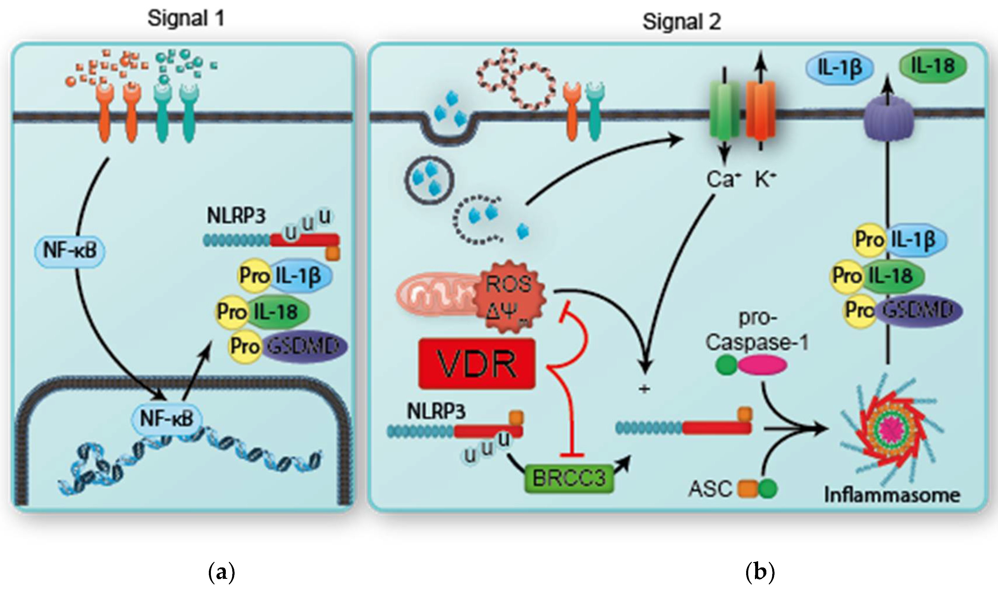
4. The Intersection of Vitamin D and the Inflammasome

Inflammasome pathways form an integral role in the innate immune system, and inappropriate inflammasome activity is strongly implicated in an array of human disease. Data from genetic studies suggested a role for the VDR affecting inflammasome activity. Al-Daghri et al. showed that a single nucleotide polymorphism (SNP) in the VDR gene is strongly associated with increased inflammasome activity and circulating levels of IL-1β and IL-18 [44].
This effect appears to be due to a direct interaction between VDR and NLRP3. Huang et al. performed co-immunoprecipitation of VDR and NLRP3 in ex vivo murine CD4+ T cells, showing VDR complexing with NLRP3 in nuclear extracts. These complexes could also be formed in vivo, and appeared to be functionally significant with chromatin precipitation, demonstrating higher NLRP3 and RNA polymerase II activity at the IL-4 locus in VDR knockout cells [19].
Rao et al. further investigated the interaction between VDR and the NLRP3 inflammasome. Notably, co-transfection of VDR with other components of the inflammasome did not find any interaction with either ASC or caspase-1. The interacting elements were found to be the ligand-binding domain of VDR with the amino-terminal pyrin domain of NLRP3 [20]. They further demonstrated that VDR knockout macrophages have increased IL-1β and IL-18 when activated by either the canonical or non-canonical pathways, without a concomitant increase in inflammasome-independent cytokines such as tumour necrosis factor α. These effects were then attenuated with the restoration of VDR expression [20].
Deubiquitination of NLRP3 is a critical step in inflammasome activation and the binding of NLRP3 to ASC. BRCC3 is a deubiquitinase that has a crucial role in posttranscriptional activation of NLRP3, the inhibition of which almost completely blocks NLRP3 activation [24]. Rao et al. further demonstrated that the VDR-binding of NLRP3 appears to attenuate BRCC3-mediated deubiquitination of the LRR domain of NLRP3 [20]. This increase in NLRP3 ubiquitination leads to increased protein degradation [18].
Independent of VDR-NLRP3 binding and inhibition, VDR can also increase levels of the mitochondrial membrane protein UCP2 (uncoupling protein-2). This protein directly prevents the production of ROS, and its activity is inhibited by the inflammasome. Cao et al. showed that UCP2 levels were significantly increased when in vitro 1,25(OH)-VD3 solution was added to canonically activated macrophage cell cultures [18]. These actions are summarised in Figure 2.
VDR agonists can induce these effects by their actions on the VDR to reduce inflammasome activity. Studies have shown that 25(OH)-VD3 and 1,25(OH)2-VD3 are both capable of inhibiting NLRP3 inflammasome activation and downstream IL-1β signalling [20,45,46].
These actions seem to be beneficial in multiple models of disease. In in vitro models of air pollution, 1,25(OH)2-VD3 attenuated the inflammatory response mediated by p38/NF-kB/NLRP3 [47]. In a murine model of liver injury, VDR agonist calcipotriol demonstrated NLRP3-attenuating effects that reduced the severity of liver injury and fibrosis [48]. Similarly, in a murine model of ulcerative colitis, 1,25(OH)2-VD3 treatment reduced colonic inflammation, likely due to its anti-inflammasome effects [18]. Meanwhile, a murine model of diabetic retinopathy demonstrated significant downregulation of the NLRP3 inflammasome response in the setting of hyperglycaemia, with a subsequent reduction in apoptosis and preservation of normal retinal structure [49].

5. Potential Roles for Vitamin D as a Therapeutic Option in Inflammasome Disorders

VDD is associated with a wide range of disorders, and there is overlap in the prevalence of VDD and a number of inflammasome-mediated conditions (Table 1). In general, the role of VDR activation therapy is limited to treating VDD. There is limited evidence in any population to suggest that supplementing VD to high or supraphysiological levels provides any clinical benefit. We discuss here the evidence for VD supplementation across a range of disorders that are associated with inflammasome activity.

5.1. Neurodegenerative Disease

Alzheimer’s disease, Parkinson’s syndrome, and Multiple Sclerosis (MS) have all been associated with inflammasome activity, and there is a clear association between VDD and higher risk of disease or disease progression in each [56,57,58].
In Alzheimer’s dementia, a randomised controlled trial (RCT) of 210 VDD patients compared supplemental VD3 800 IU/day for 12 months vs. a placebo. This trial did show that after 12 months of treatment, the VD-treated group had lower circulating disease markers than the placebo (plasma amyloid beta), higher cognitive function, and a higher intelligence quotient [59].
In Parkinson’s disease, only two small trials enrolling a total of 72 patients with durations of 6–12 months were analysed in a meta-analysis that found while 25(OH)-VD levels were increased in treatment groups at the end of the study period, there was no significant benefit in any outcome measures [58].
A Cochrane review of VD supplementation in MS identified 12 trials enrolling 933 patients for analysis, with significant risk of bias. There was no evidence of improved MS-related outcomes with VD supplementation in the meta-analysis, but the quality of the evidence is very low, and larger, well-conducted studies are underway [60]

5.2. Rheumatological Disease

Strong observational evidence links rheumatoid arthritis disease activity with VDD [61], but there are only a few studies examining the role of VD supplementation. In one interesting study of early disease in 39 patients, the addition of a 300,000 unit dose of VD3 did not change the T cell subtype or cytokine profile, although interferon and IL-17/23 were chiefly measured [62]. Two other small studies of daily supplementation showed significant pain relief after 3 months of therapy in 121 patients [63], and small but significant functional improvements after 6 months of therapy in 59 patients [64].
In gout, inflammation is mediated primarily through the NLRP3 inflammasome and IL-1β. However, there is limited evidence to suggest that gout is associated with VDD, and no trials of supplementation have been conducted.

5.3. Metabolic Disease

Observational evidence supports an association between low VD levels and metabolic conditions such as type 2 diabetes mellitus (T2DM) [65], atherosclerotic disease [66,67], and obesity [44,68].
Several large, well-conducted trials have been conducted examining a role for VD supplementation in T2DM. A large trial included 2423 patients who met the criteria for pre-diabetes but were not vitamin D-deficient. Patients received either 4000 IU/day of VD3 or a placebo, and while there was a trend towards reduced risk of developing overt T2DM, this did not reach statistical significance [69]. Similarly, in another large trial of 1312 diabetic patients not selected for VDD, supplementation of 2000 IU/day of VD3 versus a placebo did not attenuate the diabetes-associated decline in renal function over 5 years of therapy [70]. However, two meta-analyses have suggested the benefit of VD supplementation on measures of glycaemic control and insulin resistance, albeit with some substantial heterogeneity between studies [71,72].
There are just a few small and heterogeneous studies of VD3 or other VD analogue supplementation in patients with atherosclerosis, with mixed results ranging from no benefit all the way to significant attenuation of progressive atherosclerotic disease [73,74,75].
Similarly, only two small pilot studies examining VD supplementation in obesity have been conducted. A pilot study of 18 overweight patients examined the effect of combined low-calorie diet and 25,000 IU/week VD3 versus dieting alone. While there were no differences in weight loss, measures of insulin sensitivity did improve in those treated with VD3 [76]. Meanwhile, a placebo-controlled trial of high-dose 100,000 IU bolus of VD3 followed by 4000 IU/day over 4 months in 65 overweight patients showed no difference in functional outcomes, except in the subgroup of 20 patients with VDD who had a significant and clinically relevant improvement in back pain [77].

5.4. Infection

VD supplementation in respiratory tract infections has received significant attention and many trials have been conducted to evaluate the role of VD in respiratory infections.
VDD has been identified as an independent risk factor for active pulmonary tuberculosis infection, with the strongest association at levels below 25 nmol/L (<10 ng/mL) [78,79]. One very large trial of weekly bolus oral dosing of VD3 in 8851 VDD Mongolian children found no benefit to supplementation over 3 years as a preventative for developing pulmonary tuberculosis, although the event rate was very low (46/8851; 0.52%) [80]. Nine small- to medium-sized RCTs, including a total of 1868 patients, have examined the utility of 25(OH)-VD3 supplementation as an add-on therapy in patients who already have active pulmonary tuberculosis [81,82,83]. There was significant heterogeneity between dosing regimens among the trials (daily dosing, weekly/monthly bolus dosing, oral/intramuscular administration), while a number of trials did not test baseline or posttherapy 25(OH)-VD levels, making comparison and meta-analysis difficult. Four trials demonstrated improved sputum culture conversion [82,83,84,85], while three trials did not [81,86,87]. One trial found no difference in mortality, although posttherapy 25(OH)-VD levels remained low [88], while four measured tuberculosis scores, with only one of these showing positive results [83,85,87,89].
There is also evidence of a role for VDD in predisposition to influenza infections. Early evidence came from observations of the seasonal nature of influenza infections [90], and several large cohort studies [91,92] and one meta-analysis [93] have since found an inverse association between VD status and risk of respiratory tract infections. Trials with moderate heterogeneity and significantly varied dosing/administration protocols show possible benefit, the strongest with daily/weekly rather than bolus intermittent monthly dosing.
Given that VD has a wide range of modulating actions on immune activity, it is unclear whether an inflammasome-attenuating response is mediating the apparent effect in these conditions. In tuberculosis, an intact IL-1β signalling response is important for host defence in murine models, but this is independent of NLRP3 inflammasome activation [94], and it is possible that mycobacterial-induced inflammasome activation may promote persistent infection [95]. In respiratory tract infections such as influenza, the innate immune system inflammatory response is crucial to being able to mediate viral clearance and repair [96]. However, dysregulated or excessive inflammasome-mediated responses are associated with poor outcomes [96]. In patients with avian influenza, analysis of bronchoalveolar lavage and plasma samples found increased levels of IL-1β and IL-18 were poor prognostic markers [97,98].
It is possible to speculate that VD-mediated inhibition of inflammasome activation might be helpful in the most severe form of infection—sepsis. In sepsis, maladaptive inflammatory responses that would normally be protective become harmful and lead to organ dysfunction [99]. A hallmark of the septic response is high levels of the inflammasome-dependent IL-1β [99].
A medium-sized trial of a mixed intensive care unit (ICU) cohort that included septic patients randomised 475 VDD patients to receive either high-dose pulse VD3 monthly or a placebo. While the main outcomes were not different in the general population, pre-determined subgroup analysis of patients with 25(OH)-VD levels <30 nmol/L (<12 ng/mL) showed significant benefits in mortality at 28 days and lasting until up to 6 months [100].
In this context, there has recently been intense interest in VD supplementation in the setting of severe acute respiratory syndrome coronavirus 2 (SARS-CoV-2) infection, known as COVID-19 [101,102,103]. One small study has been conducted to date with 76 patients hospitalised with SARS-CoV-2 infection randomised to receive the best available therapy plus either oral 25(OH)-VD3 or no additional therapy until the time of discharge. Results were promising with reduced need for ICU treatment with 1/50 vs. 13/26 patients in each group admitted to the ICU (p < 0.001) [104].

6. Conclusions

It is increasingly clear that the NLRP3 inflammasome plays a significant role in human health and disease. VD is a key immune system regulator that appears to play a significant role in modulating NLRP3 inflammasome activity, inhibiting activation through the actions of the VDR. Randomised controlled trial evidence for VDR activating therapy in inflammasome-associated conditions remains scant and limited by small numbers, variable definitions with lack of selection for VDD, and heterogeneity between dosing protocols. It would be useful for trials to include outcomes that could be easily attributable to inflammasome activity attenuation, such as circulating levels of IL-1β or IL-18. While there does not appear to be a role for supraphysiologic VD supplementation, there is a role for supplementation in deficient patients as it is a well-tolerated and safe therapy.

Author Contributions

Conceptualization, P.H.F.G.; writing—original draft preparation, M.T.; writing—review and editing, M.T. and P.H.F.G.; supervision, P.H.F.G. All authors have read and agreed to the published version of the manuscript.

Funding

This research received no external funding.

Acknowledgments

The authors thank Kurt Giuliani for his assistance in adapting Figure 1.

Conflicts of Interest

The authors declare no conflict of interest.

References

  1. DeLuca, H.F. History of the discovery of vitamin D and its active metabolites. BoneKey Rep. 2014, 3, 479. [Google Scholar] [CrossRef] [PubMed] [Green Version]
  2. Bikle, D.D. Vitamin D metabolism, mechanism of action, and clinical applications. Chem. Biol. 2014, 21, 319–329. [Google Scholar] [CrossRef] [Green Version]
  3. Dusso, A.S.; Brown, A.J.; Slatopolsky, E. Vitamin D. Am. J. Physiol. Renal. Physiol. 2005, 289, F8–F28. [Google Scholar] [CrossRef]
  4. Slominski, A.T.; Kim, T.-K.; Li, W.; Yi, A.-K.; Postlethwaite, A.; Tuckey, R.C. The role of CYP11A1 in the production of vitamin D metabolites and their role in the regulation of epidermal functions. J. Steroid Biochem. Mol. Biol. 2013, 144, 28–39. [Google Scholar] [CrossRef] [PubMed] [Green Version]
  5. Slominski, A.T.; Kim, T.-K.; Janjetovic, Z.; Brożyna, A.A.; Żmijewski, M.A.; Xu, H.; Sutter, T.R.; Tuckey, R.C.; Jetten, A.M.; Crossman, D.K. Differential and Overlapping Effects of 20,23(OH)2D3 and 1,25(OH)2D3 on Gene Expression in Human Epidermal Keratinocytes: Identification of AhR as an Alternative Receptor for 20,23(OH)2D3. Int. J. Mol. Sci. 2018, 19, 3072. [Google Scholar] [CrossRef] [PubMed] [Green Version]
  6. Slominski, A.T.; Kim, T.K.; Takeda, Y.; Janjetovic, Z.; Brożyna, A.A.; Skobowiat, C.; Wang, J.; Postlethwaite, A.; Li, W.; Tuckey, R.C.; et al. RORα and ROR γ are expressed in human skin and serve as receptors for endogenously produced noncalcemic 20-hydroxy- and 20,23-dihydroxyvitamin D. FASEB J. 2014, 28, 2775–2789. [Google Scholar] [CrossRef] [PubMed] [Green Version]
  7. Slominski, A.; Kim, T.-K.; Li, W.; Postlethwaite, A.; Tieu, E.W.; Tang, E.K.Y.; Tuckey, R.C. Detection of novel CYP11A1-derived secosteroids in the human epidermis and serum and pig adrenal gland. Sci. Rep. 2015, 5, 14875. [Google Scholar] [CrossRef] [PubMed] [Green Version]
  8. Tongkao-On, W.; Carter, S.; Reeve, V.E.; Dixon, K.M.; Gordon-Thomson, C.; Halliday, G.M.; Tuckey, R.C.; Mason, R.S. CYP11A1 in skin: An alternative route to photoprotection by vitamin D compounds. J. Steroid Biochem. Mol. Biol. 2015, 148, 72–78. [Google Scholar] [CrossRef] [PubMed]
  9. Haussler, M.R.; Jurutka, P.W.; Mizwicki, M.; Norman, A.W. Vitamin D receptor (VDR)-mediated actions of 1α,25(OH)2vitamin D3: Genomic and non-genomic mechanisms. Best Pract. Res. Clin. Endocrinol. Metab. 2011, 25, 543–559. [Google Scholar] [CrossRef]
  10. Pike, J.W.; Meyer, M.B. The Vitamin D Receptor: New Paradigms for the Regulation of Gene Expression by 1,25-Dihydroxyvitamin D3. Endocrinol. Metab. Clin. N. Am. 2010, 39, 255–269. [Google Scholar] [CrossRef] [Green Version]
  11. Norman, A.W.; Okamura, W.H.; Hammond, M.W.; Bishop, J.E.; Dormanen, M.C.; Bouillon, R.; Van Baelen, H.; Ridall, A.L.; Daane, E.; Khoury, R.; et al. Comparison of 6-s-cis- and 6-s-trans-locked analogs of 1alpha,25-dihydroxyvitamin D3 indicates that the 6-s-cis conformation is preferred for rapid nongenomic biological responses and that neither 6-s-cis- nor 6-s-trans-locked analogs are preferred for genomic biological responses. Mol. Endocrinol. 1997, 11, 1518–1531. [Google Scholar] [PubMed] [Green Version]
  12. Sequeira, V.B.; Rybchyn, M.S.; Tongkao-On, W.; Gordon-Thomson, C.; Malloy, P.J.; Nemere, I.; Norman, A.W.; Reeve, V.E.; Halliday, G.M.; Feldman, D.; et al. The Role of the Vitamin D Receptor and ERp57 in Photoprotection by 1α,25-Dihydroxyvitamin D3. Mol. Endocrinol. 2012, 26, 574–582. [Google Scholar] [CrossRef] [PubMed] [Green Version]
  13. Nemere, I.; Farach-Carson, M.C.; Rohe, B.; Sterling, T.M.; Norman, A.W.; Boyan, B.D.; Safford, S.E. Ribozyme knockdown functionally links a 1,25(OH)2D3 membrane binding protein (1,25D3-MARRS) and phosphate uptake in intestinal cells. Proc. Natl. Acad. Sci. USA 2004, 101, 7392–7397. [Google Scholar] [CrossRef] [PubMed] [Green Version]
  14. Holick, M.F. Vitamin D status: Measurement, interpretation, and clinical application. Ann. Epidemiol. 2009, 19, 73–78. [Google Scholar] [CrossRef] [Green Version]
  15. Palacios, C.; Gonzalez, L. Is vitamin D deficiency a major global public health problem? J. Steroid Biochem. Mol. Biol. 2014, 144 Pt A, 138–145. [Google Scholar] [CrossRef] [Green Version]
  16. Van Schoor, N.M.; Lips, P. Global Overview of Vitamin D Status. Endocrinol. Metab. Clin. N. Am. 2017, 46, 845–870. [Google Scholar] [CrossRef] [PubMed]
  17. Gois, P.H.F.; Ferreira, D.; Olenski, S.; Seguro, A.C. Vitamin D and Infectious Diseases: Simple Bystander or Contributing Factor? Nutrients 2017, 9, 651. [Google Scholar] [CrossRef] [Green Version]
  18. Cao, R.; Ma, Y.; Li, S.; Shen, D.; Yang, S.; Wang, X.; Cao, Y.; Wang, Z.; Wei, Y.; Li, S.; et al. 1,25(OH) 2 D 3 alleviates DSS-induced ulcerative colitis via inhibiting NLRP3 inflammasome activation. J. Leukoc. Biol. 2020, 108, 283–295. [Google Scholar] [CrossRef] [PubMed]
  19. Huang, H.; Hong, J.-Y.; Wu, Y.-J.; Wang, E.-Y.; Liu, Z.; Cheng, B.-H.; Mei, L.; Liu, Z.-G.; Yang, P.-C.; Zheng, P. Vitamin D receptor interacts with NLRP3 to restrict the allergic response. Clin. Exp. Immunol. 2018, 194, 17–26. [Google Scholar] [CrossRef] [Green Version]
  20. Rao, Z.; Chen, X.; Wu, J.; Xiao, M.; Zhang, J.; Wang, B.; Fang, L.; Zhang, H.; Wang, X.; Yang, S.; et al. Vitamin D Receptor Inhibits NLRP3 Activation by Impeding Its BRCC3-Mediated Deubiquitination. Front. Immunol. 2019, 10, 2783. [Google Scholar] [CrossRef]
  21. Guo, H.; Callaway, J.B.; Ting, J.P. Inflammasomes: Mechanism of action, role in disease, and therapeutics. Nat. Med. 2015, 21, 677–687. [Google Scholar] [CrossRef] [PubMed] [Green Version]
  22. He, Y.; Hara, H.; Núñez, G. Mechanism and Regulation of NLRP3 Inflammasome Activation. Trends Biochem. Sci. 2016, 41, 1012–1021. [Google Scholar] [CrossRef] [PubMed] [Green Version]
  23. Fusco, R.; Siracusa, R.; Genovese, T.; Cuzzocrea, S.; Di Paola, R. Focus on the Role of NLRP3 Inflammasome in Diseases. Int. J. Mol. Sci. 2020, 21, 4223. [Google Scholar] [CrossRef] [PubMed]
  24. Juliana, C.; Fernandes-Alnemri, T.; Kang, S.; Farias, A.; Qin, F.; Alnemri, E.S. Non-transcriptional Priming and Deubiquitination Regulate NLRP3 Inflammasome Activation. J. Biol. Chem. 2012, 287, 36617–36622. [Google Scholar] [CrossRef] [Green Version]
  25. Py, B.F.; Kim, M.-S.; Vakifahmetoglu-Norberg, H.; Yuan, J. Deubiquitination of NLRP3 by BRCC3 Critically Regulates Inflammasome Activity. Mol. Cell 2013, 49, 331–338. [Google Scholar] [CrossRef] [Green Version]
  26. Moretti, J.; Blander, J.M. Increasing complexity of NLRP3 inflammasome regulation. J. Leukoc. Biol. 2020. [Google Scholar] [CrossRef]
  27. Liston, A.; Masters, S.L. Homeostasis-altering molecular processes as mechanisms of inflammasome activation. Nat. Rev. Immunol. 2017, 17, 208–214. [Google Scholar] [CrossRef]
  28. Monteleone, M.; Stanley, A.C.; Chen, K.W.; Brown, D.L.; Bezbradica, J.S.; Von Pein, J.B.; Holley, C.L.; Boucher, D.; Shakespear, M.; Kapetanovic, R.; et al. Interleukin-1β Maturation Triggers Its Relocation to the Plasma Membrane for Gasdermin-D-Dependent and -Independent Secretion. Cell Rep. 2018, 24, 1425–1433. [Google Scholar] [CrossRef] [Green Version]
  29. Sborgi, L.; Rühl, S.; Mulvihill, E.; Pipercevic, J.; Heilig, R.; Stahlberg, H.; Farady, C.J.; Müller, D.J.; Broz, P.; Hiller, S. GSDMD membrane pore formation constitutes the mechanism of pyroptotic cell death. EMBO J. 2016, 35, 1766–1778. [Google Scholar] [CrossRef]
  30. Liu, X.; Zhang, Z.; Ruan, J.; Pan, Y.; Magupalli, V.G.; Wu, J.R.V.G.M.H.; Lieberman, J. Inflammasome-activated gasdermin D causes pyroptosis by forming membrane pores. Nature 2016, 535, 153–158. [Google Scholar] [CrossRef] [Green Version]
  31. Giuliani, K.; Kassianos, A.J.; Healy, H.G.; Gois, P.H.F. Pigment Nephropathy: Novel Insights into Inflammasome-Mediated Pathogenesis. Int. J. Mol. Sci. 2019, 20, 1997. [Google Scholar] [CrossRef] [PubMed] [Green Version]
  32. Pellegrini, C.; Antonioli, L.; Lopez-Castejon, G.; Blandizzi, C.; Fornai, M. Canonical and Non-Canonical Activation of NLRP3 Inflammasome at the Crossroad between Immune Tolerance and Intestinal Inflammation. Front. Immunol. 2017, 8, 36. [Google Scholar] [CrossRef] [PubMed] [Green Version]
  33. Booshehri, L.M.; Hoffman, H.M. CAPS and NLRP3. J. Clin. Immunol. 2019, 39, 277–286. [Google Scholar] [CrossRef] [PubMed]
  34. Parkin, J.; Cohen, B. An overview of the immune system. Lancet 2001, 357, 1777–1789. [Google Scholar] [CrossRef]
  35. Takeda, K.; Akira, S. Toll-like receptors. Curr. Protoc. Immunol. 2015, 109, 14.12.1–14.12.10. [Google Scholar] [CrossRef]
  36. Orikasa, M.; Kawase, T.; Suzuki, A. Induction of macrophagic and granulocytic differentiation of murine bone marrow progenitor cells by 1,25-dihydroxyvitamin D3. Calcif. Tissue Int. 1993, 53, 193–200. [Google Scholar] [CrossRef]
  37. Provvedini, D.M.; Deftos, L.J.; Manolagas, S.C. 1,25-Dihydroxyvitamin D3 promotes in vitro morphologic and enzymatic changes in normal human monocytes consistent with their differentiation into macrophages. Bone 1986, 7, 23–28. [Google Scholar] [CrossRef]
  38. Gombart, A.F.; Borregaard, N.; Koeffler, H.P. Human cathelicidin antimicrobial peptide (CAMP) gene is a direct target of the vitamin D receptor and is strongly up-regulated in myeloid cells by 1,25-dihydroxyvitamin D 3. FASEB J. 2005, 19, 1067–1077. [Google Scholar] [CrossRef] [Green Version]
  39. Liu, P.T.; Schenk, M.; Walker, V.P.; Dempsey, P.W.; Kanchanapoomi, M.; Wheelwright, M.; Vazirnia, A.; Zhang, X.; Steinmeyer, A.; Zügel, U.; et al. Convergence of IL-1β and VDR Activation Pathways in Human TLR2/1-Induced Antimicrobial Responses. PLoS ONE 2009, 4, e5810. [Google Scholar] [CrossRef] [Green Version]
  40. Adorini, L.; Penna, G. Dendritic cell tolerogenicity: A key mechanism in immunomodulation by vitamin D receptor agonists. Hum. Immunol. 2009, 70, 345–352. [Google Scholar] [CrossRef]
  41. Yu, X.P.; Bellido, T.; Manolagas, S.C. Down-regulation of NF-kappa B protein levels in activated human lymphocytes by 1,25-dihydroxyvitamin D3. Proc. Natl. Acad. Sci. USA 1995, 92, 10990–10994. [Google Scholar] [CrossRef] [Green Version]
  42. Hewison, M. Vitamin D and the Immune System: New Perspectives on an Old Theme. Endocrinol. Metab. Clin. N. Am. 2010, 39, 365–379. [Google Scholar] [CrossRef] [PubMed] [Green Version]
  43. Chen, S.; Sims, G.P.; Chen, X.X.; Gu, Y.Y.; Chen, S.; Lipsky, P.E. Modulatory effects of 1,25-dihydroxyvitamin D3 on human B cell differentiation. J. Immunol. 2007, 179, 1634–1647. [Google Scholar] [CrossRef] [PubMed] [Green Version]
  44. Al-Daghri, N.; Guerini, F.R.; Al-Attas, O.S.; Alokail, M.S.; Alkharfy, K.M.; Draz, H.M.; Agliardi, C.; Costa, A.S.; Saulle, I.; Mohammed, A.K.; et al. Vitamin D Receptor Gene Polymorphisms Are Associated with Obesity and Inflammosome Activity. PLoS ONE 2014, 9, e102141. [Google Scholar] [CrossRef]
  45. Dai, Y.; Zhang, J.; Xiang, J.; Li, Y.; Wu, D.; Xu, J. Calcitriol inhibits ROS-NLRP3-IL-1β signaling axis via activation of Nrf2-antioxidant signaling in hyperosmotic stress stimulated human corneal epithelial cells. Redox Biol. 2019, 21, 101093. [Google Scholar] [CrossRef] [PubMed]
  46. Tulk, S.E.; Liao, K.-C.; Muruve, D.A.; Li, Y.; Beck, P.L.; Macdonald, J.A. Vitamin D 3 Metabolites Enhance the NLRP3-Dependent Secretion of IL-1? From Human THP-1 Monocytic Cells. J. Cell. Biochem. 2015, 116, 711–720. [Google Scholar] [CrossRef] [PubMed]
  47. Xin, L.; Che, B.; Zhai, B.; Luo, Q.; Zhang, C.; Wang, J.; Wang, S.; Fan, G.; Liu, Z.; Feng, J.; et al. 1,25-Dihydroxy Vitamin D(3) Attenuates the Oxidative Stress-Mediated Inflammation Induced by PM(2.5)via the p38/NF-κB/NLRP3 Pathway. Inflammation 2019, 42, 702–713. [Google Scholar] [CrossRef] [PubMed]
  48. Wang, X.; Wang, G.; Qu, J.; Yuan, Z.; Pan, R.; Li, K. Calcipotriol Inhibits NLRP3 Signal Through YAP1 Activation to Alleviate Cholestatic Liver Injury and Fibrosis. Front. Pharmacol. 2020, 11, 200. [Google Scholar] [CrossRef]
  49. Lu, L.; Lu, Q.; Chen, W.; Li, J.; Li, C.; Zheng, Z. Vitamin D3 Protects against Diabetic Retinopathy by Inhibiting High-Glucose-Induced Activation of the ROS/TXNIP/NLRP3 Inflammasome Pathway. J. Diabetes Res. 2018, 2018, 8193523. [Google Scholar] [CrossRef] [Green Version]
  50. Wallin, M.T.; Culpepper, W.J.; Nichols, E.; Bhutta, Z.A.; Gebrehiwot, T.T.; Hay, S.I.; Khalil, I.A.; Krohn, K.J.; Liang, X.; Naghavi, M.; et al. Global, regional, and national burden of multiple sclerosis 1990–2016: A systematic analysis for the Global Burden of Disease Study 2016. Lancet Neurol. 2019, 18, 269–285. [Google Scholar] [CrossRef] [Green Version]
  51. Nichols, E.; Szoeke, C.E.; Vollset, S.E.; Abbasi, N.; Abd-Allah, F.; Abdela, J.; Aichour, M.T.E.; Akinyemi, R.O.; Alahdab, F.; Asgedom, S.W.; et al. Global, regional, and national burden of Alzheimer’s disease and other dementias, 1990-2016: A systematic analysis for the Global Burden of Disease Study 2016. Lancet Neurol. 2019, 18, 88–106. [Google Scholar] [CrossRef] [Green Version]
  52. Dorsey, E.R.; Elbaz, A.; Nichols, E.; Abd-Allah, F.; Abdelalim, A.; Adsuar, J.C.; Ansha, M.G.; Brayne, C.; Choi, J.Y.J.; Collado-Mateo, D.; et al. Global, regional, and national burden of Parkinson’s disease, 1990-2016: A systematic analysis for the Global Burden of Disease Study 2016. Lancet Neurol. 2018, 17, 939–953. [Google Scholar] [CrossRef] [Green Version]
  53. Saeedi, P.; Petersohn, I.; Salpea, P.; Malanda, B.; Karuranga, S.; Unwin, N.; Colagiuri, S.; Guariguata, L.; Motala, A.A.; Ogurtsova, K.; et al. Global and regional diabetes prevalence estimates for 2019 and projections for 2030 and 2045: Results from the International Diabetes Federation Diabetes Atlas, 9th edition. Diabetes Res. Clin. Pr. 2019, 157, 107843. [Google Scholar] [CrossRef] [PubMed] [Green Version]
  54. Global Health Observatory, World Health Organisation. Prevalence of Obesity among Adults, BMI ≥ 30 (Age-Standardized Estimate) (%) 2017. Available online: https://www.who.int/data/gho/data/indicators/indicator-details/GHO/prevalence-of-obesity-among-adults-bmi-=-30-(age-standardized-estimate)-(-) (accessed on 7 October 2020).
  55. Population Reference Bureau. 2016 World Population Data Sheet. Available online: https://www.prb.org/2016-world-population-data-sheet/ (accessed on 7 October 2020).
  56. Alfredsson, L.; Armstrong, B.K.; Butterfield, D.A.; Chowdhury, R.; De Gruijl, F.R.; Feelisch, M.; Garland, C.F.; Hart, P.H.; Hoel, D.G.; Jacobsen, R.; et al. Insufficient Sun Exposure Has Become a Real Public Health Problem. Int. J. Environ. Res. Public Health 2020, 17, 5014. [Google Scholar] [CrossRef] [PubMed]
  57. Fitzgerald, K.C.; Munger, K.; Köchert, K.; Arnason, B.G.W.; Comi, G.; Cook, S.; Goodin, D.S.; Filippi, M.; Hartung, H.-P.; Jeffery, D.R.; et al. Association of Vitamin D Levels With Multiple Sclerosis Activity and Progression in Patients Receiving Interferon Beta-1b. JAMA Neurol. 2015, 72, 1458–1465. [Google Scholar] [CrossRef] [Green Version]
  58. Zhou, Z.; Zhou, R.; Zhang, Z.; Li, K. The Association Between Vitamin D Status, Vitamin D Supplementation, Sunlight Exposure, and Parkinson’s Disease: A Systematic Review and Meta-Analysis. Med. Sci. Monit. 2019, 25, 666–674. [Google Scholar] [CrossRef]
  59. Jia, J.; Hu, J.; Huo, X.; Miao, R.; Zhang, Y.; Ma, F. Effects of vitamin D supplementation on cognitive function and blood Aβ-related biomarkers in older adults with Alzheimer’s disease: A randomised, double-blind, placebo-controlled trial. J. Neurol. Neurosurg. Psychiatry 2019, 90, 1347–1352. [Google Scholar] [CrossRef]
  60. Jagannath, V.A.; Filippini, G.; Di Pietrantonj, C.; Asokan, G.V.; Robak, E.W.; Whamond, L.; Robinson, S.A. Vitamin D for the management of multiple sclerosis. Cochrane Database Syst. Rev. 2018, 9, Cd008422. [Google Scholar] [CrossRef]
  61. Lee, Y.H.; Bae, S.-C. Vitamin D level in rheumatoid arthritis and its correlation with the disease activity: A meta-analysis. Clin. Exp. Rheumatol. 2016, 34, 827–833. [Google Scholar]
  62. Buondonno, I.; Rovera, G.; Sassi, F.; Rigoni, M.M.; Lomater, C.; Parisi, S.; Pellerito, R.; Isaia, G.C.; D’Amelio, P. Vitamin D and immunomodulation in early rheumatoid arthritis: A randomized double-blind placebo-controlled study. PLoS ONE 2017, 12, e0178463. [Google Scholar] [CrossRef] [Green Version]
  63. Gopinath, K.; Danda, D. Supplementation of 1,25 dihydroxy vitamin D3 in patients with treatment naive early rheumatoid arthritis: A randomised controlled trial. Int. J. Rheum. Dis. 2011, 14, 332–339. [Google Scholar] [CrossRef] [PubMed]
  64. Soubrier, M.; Lambert, C.; Combe, B.; Gaudin, P.; Thomas, T.; Sibilia, J.; Dougados, M.; Dubost, J.-J. A randomised, double-blind, placebo-controlled study assessing the efficacy of high doses of vitamin D on functional disability in patients with rheumatoid arthritis. Clin. Exp. Rheumatol. 2018, 36, 1056–1060. [Google Scholar] [PubMed]
  65. Song, Y.; Wang, L.; Pittas, A.G.; Del Gobbo, L.C.; Zhang, C.; Manson, J.E.; Hu, F.B. Blood 25-Hydroxy Vitamin D Levels and Incident Type 2 Diabetes: A meta-analysis of prospective studies. Diabetes Care 2013, 36, 1422–1428. [Google Scholar] [CrossRef] [PubMed] [Green Version]
  66. Elamin, M.B.; Abu Elnour, N.O.; Elamin, K.B.; Fatourechi, M.M.; Alkatib, A.A.; Almandoz, J.P.; Liu, H.; Lane, M.A.; Mullan, R.J.; Hazem, A.; et al. Vitamin D and Cardiovascular Outcomes: A Systematic Review and Meta-Analysis. J. Clin. Endocrinol. Metab. 2011, 96, 1931–1942. [Google Scholar] [CrossRef] [Green Version]
  67. Nsengiyumva, V.; Fernando, M.E.; Moxon, J.V.; Krishna, S.M.; Pinchbeck, J.; Omer, S.M.; Morris, D.R.; Jones, R.E.; Moran, C.S.; Seto, S.W.; et al. The association of circulating 25-hydroxyvitamin D concentration with peripheral arterial disease: A meta-analysis of observational studies. Atherosclerosis 2015, 243, 645–651. [Google Scholar] [CrossRef]
  68. Pereira-Santos, M.; Costa, P.R.F.; Assis, A.M.O.; Santos, C.A.S.T.; Santos, D.B. Obesity and vitamin D deficiency: A systematic review and meta-analysis. Obes. Rev. 2015, 16, 341–349. [Google Scholar] [CrossRef]
  69. Pittas, A.G.; Dawson-Hughes, B.; Sheehan, P.; Ware, J.H.; Knowler, W.C.; Aroda, V.R.; Brodsky, I.; Ceglia, L.; Chadha, C.; Chatterjee, R.; et al. Vitamin D Supplementation and Prevention of Type 2 Diabetes. N. Engl. J. Med. 2019, 381, 520–530. [Google Scholar] [CrossRef] [Green Version]
  70. De Boer, I.H.; Zelnick, L.R.; Ruzinski, J.; Friedenberg, G.; Duszlak, J.; Bubes, V.Y.; Hoofnagle, A.N.; Thadhani, R.; Glynn, R.J.; Buring, J.E.; et al. Effect of Vitamin D and Omega-3 Fatty Acid Supplementation on Kidney Function in Patients With Type 2 Diabetes: A Randomized Clinical Trial. JAMA 2019, 322, 1899–1909. [Google Scholar] [CrossRef]
  71. Lee, C.J.; Iyer, G.; Liu, Y.; Kalyani, R.R.; Bamba, N.; Ligon, C.B.; Varma, S.; Mathioudakis, N. The effect of vitamin D supplementation on glucose metabolism in type 2 diabetes mellitus: A systematic review and meta-analysis of intervention studies. J. Diabetes Complicat. 2017, 31, 1115–1126. [Google Scholar] [CrossRef]
  72. Li, X.; Liu, Y.; Zheng, Y.; Wang, P.; Zhang, Y. The Effect of Vitamin D Supplementation on Glycemic Control in Type 2 Diabetes Patients: A Systematic Review and Meta-Analysis. Nutrients 2018, 10, 375. [Google Scholar] [CrossRef] [Green Version]
  73. Wu, Z.; Wang, T.; Zhu, S.; Li, L. Effects of vitamin D supplementation as an adjuvant therapy in coronary artery disease patients. Scand. Cardiovasc. J. 2016, 50, 9–16. [Google Scholar] [CrossRef] [PubMed]
  74. Farrokhian, A.; Raygan, F.; Bahmani, F.; Talari, H.R.; Esfandiari, R.; Esmaillzadeh, A.; Asemi, Z. Long-Term Vitamin D Supplementation Affects Metabolic Status in Vitamin D–Deficient Type 2 Diabetic Patients with Coronary Artery Disease. J. Nutr. 2017, 147, 384–389. [Google Scholar] [CrossRef] [PubMed] [Green Version]
  75. Gepner, A.D.; Haller, I.V.; Krueger, D.C.; Korcarz, C.E.; Binkley, N.; Stein, J.H. A randomized controlled trial of the effects of vitamin D supplementation on arterial stiffness and aortic blood pressure in Native American women. Atherosclerosis 2015, 240, 526–528. [Google Scholar] [CrossRef] [PubMed] [Green Version]
  76. Cefalo, C.M.; Conte, C.; Sorice, G.P.; Moffa, S.; Sun, V.A.; Cinti, F.; Salomone, E.; Muscogiuri, G.; Brocchi, A.A.; Pontecorvi, A.; et al. Effect of Vitamin D Supplementation on Obesity-Induced Insulin Resistance: A Double-Blind, Randomized, Placebo-Controlled Trial. Obesity 2018, 26, 651–657. [Google Scholar] [CrossRef] [PubMed] [Green Version]
  77. Brady, S.R.; Naderpoor, N.; De Courten, M.P.; Scragg, R.; Cicuttini, F.; Mousa, A.; De Courten, B. Vitamin D supplementation may improve back pain disability in vitamin D deficient and overweight or obese adults. J. Steroid Biochem. Mol. Biol. 2019, 185, 212–217. [Google Scholar] [CrossRef] [PubMed]
  78. Nnoaham, K.E.; Clarke, A. Low serum vitamin D levels and tuberculosis: A systematic review and meta-analysis. Int. J. Epidemiol. 2008, 37, 113–119. [Google Scholar] [CrossRef]
  79. Zeng, J.; Wu, G.; Yang, W.; Gu, X.; Liang, W.; Yao, Y.; Song, Y. A serum vitamin D level <25nmol/L pose high tuberculosis risk: A meta-analysis. PLoS ONE 2015, 10, e0126014. [Google Scholar]
  80. Ganmaa, D.; Uyanga, B.; Zhou, X.; Gantsetseg, G.; Delgerekh, B.; Enkhmaa, D.; Khulan, D.; Ariunzaya, S.; Sumiya, E.; Bolortuya, B.; et al. Vitamin D Supplements for Prevention of Tuberculosis Infection and Disease. N. Engl. J. Med. 2020, 383, 359–368. [Google Scholar] [CrossRef]
  81. Daley, P.; Jagannathan, V.; John, K.R.; Sarojini, J.; Latha, A.; Vieth, R.; Suzana, S.; Jeyaseelan, L.; Christopher, D.J.; Smieja, M.; et al. Adjunctive vitamin D for treatment of active tuberculosis in India: A randomised, double-blind, placebo-controlled trial. Lancet Infect. Dis. 2015, 15, 528–534. [Google Scholar] [CrossRef]
  82. Martineau, A.R.; Timms, P.M.; Bothamley, G.H.; Hanifa, Y.; Islam, K.; Claxton, A.P.; Packe, G.E.; Moore-Gillon, J.C.; Darmalingam, M.; Davidson, R.N.; et al. High-dose vitamin D3 during intensive-phase antimicrobial treatment of pulmonary tuberculosis: A double-blind randomised controlled trial. Lancet 2011, 377, 242–250. [Google Scholar] [CrossRef] [Green Version]
  83. Mily, A.; Rekha, R.S.; Kamal, S.M.M.; Arifuzzaman, A.S.M.; Rahim, Z.; Khan, L.; Haq, A.; Zaman, K.; Bergman, P.; Brighenti, S.; et al. Significant Effects of Oral Phenylbutyrate and Vitamin D3 Adjunctive Therapy in Pulmonary Tuberculosis: A Randomized Controlled Trial. PLoS ONE 2015, 10, e0138340. [Google Scholar] [CrossRef] [PubMed]
  84. Nursyam, E.W.; Amin, Z.; Rumende, C.M. The effect of vitamin D as supplementary treatment in patients with moderately advanced pulmonary tuberculous lesion. Acta Med. Indones 2006, 38, 3–5. [Google Scholar] [PubMed]
  85. Coussens, A.K.; Wilkinson, R.J.; Hanifa, Y.; Nikolayevskyy, V.; Elkington, P.T.; Islam, K.; Timms, P.M.; Venton, T.R.; Bothamley, G.H.; Packe, G.E.; et al. Vitamin D accelerates resolution of inflammatory responses during tuberculosis treatment. Proc. Natl. Acad. Sci. USA 2012, 109, 15449–15454. [Google Scholar] [CrossRef] [PubMed] [Green Version]
  86. Ralph, A.P.; Waramori, G.; Pontororing, G.J.; Kenangalem, E.; Wiguna, A.; Tjitra, E.; Lolong, D.B.; Yeo, T.W.; Chatfield, M.D.; Soemanto, R.K.; et al. L-arginine and Vitamin D Adjunctive Therapies in Pulmonary Tuberculosis: A Randomised, Double-Blind, Placebo-Controlled Trial. PLoS ONE 2013, 8, e70032. [Google Scholar] [CrossRef] [Green Version]
  87. Tukvadze, N.; Sanikidze, E.; Kipiani, M.; Hebbar, G.; Easley, K.A.; Shenvi, N.; Kempker, R.R.; Frediani, J.K.; Mirtskhulava, V.; Alvarez, J.A.; et al. High-dose vitamin D3 in adults with pulmonary tuberculosis: A double-blind randomized controlled trial. Am. J. Clin. Nutr. 2015, 102, 1059–1069. [Google Scholar] [CrossRef] [Green Version]
  88. McHugh, T.; Honeyborne, I. Vitamin D as supplementary treatment for tuberculosis: A double-blind, randomized, placebo-controlled trial. Am. J. Respir. Crit. Care Med. 2009, 179, 843–850. [Google Scholar] [CrossRef]
  89. Salahuddin, N.; Ali, F.; Hasan, Z.; Rao, N.; Aqeel, M.; Mahmood, S.F. Vitamin D accelerates clinical recovery from tuberculosis: Results of the SUCCINCT Study [Supplementary Cholecalciferol in recovery from tuberculosis]. A randomized, placebo-controlled, clinical trial of vitamin D supplementation in patients with pulmonary tuberculosis’. BMC Infect. Dis. 2013, 13, 22. [Google Scholar] [CrossRef] [Green Version]
  90. Hope-Simpson, R.E. The role of season in the epidemiology of influenza. J. Hyg. 1981, 86, 35–47. [Google Scholar] [CrossRef] [Green Version]
  91. Ginde, A.A.; Mansbach, J.M.; Camargo, C.A., Jr. Association between serum 25-hydroxyvitamin D level and upper respiratory tract infection in the Third National Health and Nutrition Examination Survey. Arch. Intern. Med. 2009, 169, 384–390. [Google Scholar] [CrossRef] [Green Version]
  92. Berry, D.J.; Hesketh, K.; Power, C.; Hyppönen, E. Vitamin D status has a linear association with seasonal infections and lung function in British adults. Br. J. Nutr. 2011, 106, 1433–1440. [Google Scholar] [CrossRef] [Green Version]
  93. Martineau, A.R.; Jolliffe, D.A.; Greenberg, L.; Aloia, J.F.; Bergman, P.; Dubnov-Raz, G.; Esposito, S.; Ganmaa, D.; Ginde, A.A.; Goodall, E.C.; et al. Vitamin D supplementation to prevent acute respiratory infections: Individual participant data meta-analysis. Health Technol. Assess. 2019, 23, 1–44. [Google Scholar] [CrossRef] [PubMed]
  94. Dorhoi, A.; Nouailles, G.; Jörg, S.; Hagens, K.; Heinemann, E.; Pradl, L.; Oberbeck-Müller, D.; Duque-Correa, M.A.; Reece, S.T.; Ruland, J.; et al. Activation of the NLRP3 inflammasome by Mycobacterium tuberculosis is uncoupled from susceptibility to active tuberculosis. Eur. J. Immunol. 2012, 42, 374–384. [Google Scholar] [CrossRef] [PubMed]
  95. Liu, C.H.; Liu, H.; Ge, B. Innate immunity in tuberculosis: Host defense vs pathogen evasion. Cell. Mol. Immunol. 2017, 14, 963–975. [Google Scholar] [CrossRef] [PubMed]
  96. Tate, M.D.; Mansell, A. An update on the NLRP3 inflammasome and influenza: The road to redemption or perdition? Curr. Opin. Immunol. 2018, 54, 80–85. [Google Scholar] [CrossRef] [PubMed]
  97. Wang, Z.; Zhang, A.; Wan, Y.; Liu, X.; Qiu, C.; Xi, X.; Ren, Y.; Wang, J.; Dong, Y.; Bao, M.; et al. Early hypercytokinemia is associated with interferon-induced transmembrane protein-3 dysfunction and predictive of fatal H7N9 infection. Proc. Natl. Acad. Sci. USA 2014, 111, 769–774. [Google Scholar] [CrossRef] [Green Version]
  98. Guo, J.; Huang, F.; Liu, J.; Chen, Y.; Wang, W.; Cao, B.; Zou, Z.; Liu, S.; Pan, J.; Bao, C.; et al. The Serum Profile of Hypercytokinemia Factors Identified in H7N9-Infected Patients can Predict Fatal Outcomes. Sci. Rep. 2015, 5, 10942. [Google Scholar] [CrossRef]
  99. Skirecki, T.; Cavaillon, J.-M. Inner sensors of endotoxin—implications for sepsis research and therapy. FEMS Microbiol. Rev. 2019, 43, 239–256. [Google Scholar] [CrossRef]
  100. Amrein, K.; Schnedl, C.; Holl, A.; Riedl, R.; Christopher, K.B.; Pachler, C.; Purkart, T.U.; Waltensdorfer, A.; Münch, A.; Warnkross, H.; et al. Effect of High-Dose Vitamin D3 on Hospital Length of Stay in Critically Ill Patients With Vitamin D Deficiency: The VITdAL-ICU Randomized Clinical Trial. JAMA 2016, 312, 1520–1530. [Google Scholar] [CrossRef] [Green Version]
  101. Grant, W.B.; Lahore, H.; McDonnell, S.L.; Baggerly, C.A.; French, C.B.; Aliano, J.L.; Bhattoa, H.P. Evidence that Vitamin D Supplementation Could Reduce Risk of Influenza and COVID-19 Infections and Deaths. Nutrients 2020, 12, 988. [Google Scholar] [CrossRef] [Green Version]
  102. Ali, N. Role of vitamin D in preventing of COVID-19 infection, progression and severity. J. Infect. Public Health 2020, 13, 1373–1380. [Google Scholar] [CrossRef]
  103. Meltzer, D.O.; Best, T.J.; Zhang, H.; Vokes, T.; Arora, V.; Solway, J. Association of Vitamin D Deficiency and Treatment with COVID-19 Incidence. medRxiv 2020. [Google Scholar] [CrossRef]
  104. Castillo, M.E.; Costa, L.M.E.; Barrios, J.M.V.; Alcalá-Díaz, J.F.; Lopez-Miranda, J.; Bouillon, R.; Gomez, J.M.Q. Effect of calcifediol treatment and best available therapy versus best available therapy on intensive care unit admission and mortality among patients hospitalized for COVID-19: A pilot randomized clinical study. J. Steroid Biochem. Mol. Biol. 2020, 203, 105751. [Google Scholar] [CrossRef] [PubMed]
Figure 1. The process of Vitamin D2 and D3 production and activation. 7 dehydrocholesterol is converted to pre-Vitamin D3 in a UV-B-dependent manner in the skin. Pre-Vitamin D3 spontaneously isomerises to Vitamin D3, which is then converted to 25(OH)-Vitamin D3 by 25-hydroxylase (primarily CYP2R1) in the liver. The final activation step occurs in the kidney, where 1-hydroxylase (CYP27B1, CYP27A1) creates 1,25(OH)-Vitamin D3. Vitamin D2 is created from ergosterol in a UV-B-dependent manner in fungi. Following oral consumption, Vitamin D2 undergoes similar activating processes by 25-hydroxylase (CYP2R1) and 1-hydroxylase (CYP27B1). UV-B: ultraviolet B light. CYP—cytochrome P450 enzyme.
Applsci 10 08462 g001
Figure 2. The canonical inflammasome activation pathway. (a) Signal 1 involves the activation of cell-surface PRRs by PAMPs and DAMPs. This activates downstream signalling with translocation of NF-ĸB to the nucleus and upregulation of the expression of NLRP3, pro-IL-1β, pro-IL-18, and pro-GSDMD. (b) Signal 2 involves activation of the primed components by a range of PAMPs/DAMPs/HAMPs that include extracellular oxidised DNA, extracellular ATP, lysosomal degradation products, calcium influx and potassium efflux, mitochondrial damage with altered membrane potential, and the release of oxidised mitochondrial DNA. BRCC-3-mediated deubiquitination of NLRP3 allows for the activation and oligomerisation of NLRP3 with ASC and pro-caspase-1. After oligomerisation, conformational change activates caspase-1, allowing cleavage and release of IL-1β, IL-18, and GSDMD. GSDMD translocates to the cell membrane where it allows for rapid release of IL-1β and IL-18 into the external environment and mediates pyroptosis. PRR—pattern-recognition receptor. PAMP—pathogen-associated molecular pattern. DAMP—danger-associated molecular pattern. HAMP—homeostasis-altering molecular process. NF-ĸB—nuclear factor kappa-light-chain-enhancer of activated B cells. NLRP3—nucleotide-binding domain-like receptor protein 3. IL—interleukin. GSDMD—gasdermin D. ATP—adenosine triphosphate. DNA—deoxyribonucleic acid. BRCC-3—BRCA1/BRCA2-containing complex, subunit 3. ASC—apoptosis-associated speck-like protein. Adapted from Giuliani et al. with permission from publisher [31].
Applsci 10 08462 g002
Table 1. Global prevalence of vitamin D deficiency and other disorders.
RegionVDDADPDMSObesityT2DM
South Africa7–20%0.26%0.03%0.01%28.30%4.50%
South America20–59%0.47%0.09%0.02%23.79%5.79%
Australia31%0.88%0.17%0.09%29.00%5.70%
USA36%1.24%0.22%0.16%36.20%10.30%
Mexico38%0.52%0.05%0.01%28.90%10.80%
Russian Federation43–64%0.71%0.17%0.07%23.10%7.60%
New Zealand56%0.86%0.13%0.56%30.80%5.20%
Europe57–64%1.37%0.19%0.09%23.22%6.61%
North Africa60%0.44%0.05%0.03%25.77%8.93%
Canada61%0.88%0.29%0.22%29.40%9.20%
Brazil77%0.82%0.06%0.01%22.10%6.40%
Asia78–98%0.93%0.11%0.01%6.02%4.42%
Middle East90%0.48%0.05%0.04%26.81%9.35%
Northern Europe92%1.27%0.19%0.16%21.83%5.52%
VDD—vitamin D deficiency. AD—Alzheimer’s disease. PD—Parkinson’s disease. MS—Multiple Sclerosis. T2DM—type 2 diabetes mellitus. Table data sourced from [16,17,50,51,52,53,54,55,56]. The colour gradient shows increasing prevalence from low (green) to high (red).
Publisher’s Note: MDPI stays neutral with regard to jurisdictional claims in published maps and institutional affiliations.

