Article

In Vitro Comparison of Surgical Implant Placement Accuracy Using Guides Fabricated by Three Different Additive Technologies

by
Chuchai Anunmana
1,
Chananchida Ueawitthayasuporn
2,
Sirichai Kiattavorncharoen
3 and
Prakan Thanasrisuebwong
4,*
1
Department of Prosthodontics, Faculty of Dentistry, Mahidol University, Bangkok 10400, Thailand
2
Master of Science Program in Implant Dentistry, Faculty of Dentistry, Mahidol University, Bangkok 10400, Thailand
3
Department of Oral and Maxillofacial Surgery, Faculty of Dentistry, Mahidol University, Bangkok 10400, Thailand
4
Dental Implant Center, Dental Hospital, Faculty of Dentistry, Mahidol University, Bangkok 10400, Thailand
*
Author to whom correspondence should be addressed.
Appl. Sci. 2020, 10(21), 7791; https://doi.org/10.3390/app10217791
Submission received: 22 September 2020 / Revised: 16 October 2020 / Accepted: 20 October 2020 / Published: 3 November 2020
(This article belongs to the Special Issue Current Trends and Applications of Digital Technology in Dentistry)

Abstract

:
Various three-dimensional (3D) printing technologies are commercially available on the market, but the influence of different technologies on the accuracy of implant-guided surgery is unclear. Thus, three printing technologies: Stereolithographic (SLA), Digital light processing (DLP), and Polyjet were evaluated in this study. An entire 30 polyurethane models replicated the clinical situation. Ten surgical guides were printed by SLA, DLP, and PolyJet. Then, implant-guided surgery was performed, and their accuracy outcomes were measured concerning angular deviation, 3D deviation at the entry point, and apex. On top of that, the total processing time was also compared. For the angular deviation, the mean deviation was not statistically significant among all technologies. For the 3D deviation, PolyJet was statistically more accurate than DLP (p = 0.002) and SLA (p = 0.017) at the entry point. PolyJet was also statistically more accurate than DLP (p = 0.007) in regards to 3D deviation at the apex. Within the limitation of this study, the deviations from the experiment showed that PolyJet had the best outcome regarding the 3D deviations at the entry point and at the apex, meanwhile, the DLP printer had the shortest processing time.

1. Introduction

Over the last few decades, computer-aided design (CAD) and computer-aided manufacturing (CAM) has been improved significantly for dental applications [1]. Consequently, the development of CAD/CAM innovates the contemporary approach called “3D printing”, which is considered state-of-the-art technology for manufacturing surgical guides as it reduces production time and operating costs significantly while possessing superior capabilities of fabricating complex structural parts compared with traditional approaches [2,3].
3D printing technology can be classified into two major types: additive manufacturing and subtractive manufacturing. Both types are well-known for the fabrication of implant surgical guides. Compared to subtractive manufacturing, additive manufacturing offers lower costs and has a higher capability in terms of complexity and precision. Thereby, additive manufacturing is gaining popularity rapidly in dentistry. The production process of additive techniques have well-established steps and are as follows: to begin, the 3D virtual model of surgical guides is designed with CAD software. Afterwards, the 3D virtual model is converted into slices with the uniform thickness as a so-called layer height. In the subsequent stage, the 3D model is printed physically from the bottom-up, slice by slice. Finally, post-processing of the 3D printed model is conducted to ensure that the products had the desired quality with the steps such as curing and polishing [4]. There are various additive technologies available in the market such as SLA, DLP, and Polyjet. The production processes of all technologies are very similar in principle; however, all of them can produce different results significantly. Typically, SLA technology uses UV laser beams to polymerize a liquid resin into a solid part. The model is immersed in a chemical bath for cleaning after printing. Unlike SLA, the PolyJet printer uses an inkjet-like print head jetting photopolymer droplets, and UV light is subsequently used to solidify the polymer to form a 3D model. Lastly, the DLP technology, which is similar to the SLA technology, uses a digital projector instead of UV laser beams to solidify a liquid resin [5].
Several studies measured the accuracy of each 3D printing technology, concerning the capability to reconstruct the virtual 3D models into the exact physical surgical guides [6,7,8,9]. In the dental field, the common measurement to evaluate the accuracy includes the deviation of angle, entry point, and apex, which are influenced by layer thickness, material quality, cast orientation, 3D model shape, and software and hardware capabilities. Although the effect of each printing factor currently remains unclear, especially in dentistry, many of them can be controlled by the user to ensure the finest quality of a printed model.
For guided implant surgery, the introduction of the desktop 3D printer enables the clinicians to fabricate surgical guides in an office [10]. A clinician could produce a surgical guide using a 3D printer, but the technique needs to be improved further to enhance the guide’s stability during the surgery. However, those shortcomings have later been solved. By increasing the accuracy of the 3D-printed guide, studies proved that surgical guides were suitable for clinical use.
Even though static guided implant surgery increases efficiency, it imposes several uncertainties, especially the risk of inaccuracies occurred during each step [11]. The precision of guided implant surgery is defined as the measured deviation of the superimposition between the virtual and actual implant positions [12]. This deviation can be caused by various factors, including data collection, data manipulation, surgical guide fabrication, and inaccurate surgical guide position as well as errors in the drilling [13,14]. For instance, the data collection of guided surgery may have an error inaccuracy from the superimposition method. The post-surgical CBCT, if not superimposed properly, can lead to measurement error. These data are often further analyzed statistically in different plans and can be subjected to multiple comparisons. Different guide fabrication methods, 3D printing or milling, along with guide positioning intramurally and experience of surgeons in the placement of drills, can also affect the accuracy of implant placement. These factors may affect the angulation, depth, and diameter of the implant drilling, or the placement of an implant that converts into the 3D deviation of the implant position [15,16].
However, there have been limited studies that show the impact of static surgical guides fabricated with different 3D printing technologies explicitly according to the accuracy of the implant position. Clinicians currently have no idea if these printers would produce the same accuracy for their implant guides and often choose a printer without knowing the accuracy of printed products. Thus, the study aimed to evaluate the deviation of implant position among the SLA, DLP, and PolyJet 3D printer as well as the total processing time of these 3D printers. The outcome would assist in 3D printer selection among these common printing technologies. The null hypothesis was that there were no differences in the accuracy of implant position among the static surgical guides produced by SLA, DLP, and PolyJet.

2. Materials and Methods

2.1. Model Preparation

The sample size was chosen based on a previous study [17] that used 10 samples per study group. For this study, the entire 30 samples were made by using a standard maxillary dentoform as a template. First, the maxillary dentoform was scanned in Standard Tessellation Language (STL) file format by a dental laboratory 3D scanner (Straumann® CARES® CADCAM; Institute Straumann AG). Next, the STL file was exported to the 3D software program “Meshmixer™” to design the right maxillary molar edentulous ridge as the simulated alveolar ridge to mock up a real clinical situation. The alveolar ridge morphology was designed as 14 mm in depth from the coronal to the apical region and 7 mm in bucco-lingual width. The uppermost coronal position of the alveolar ridge was placed at 3 mm below the gingival margin of the right maxillary first molar. Then, the STL file of the previously designed template model was printed with a laboratory 3D printer (Straumann® CARES®, P30, Institute Straumann AG). These template models were coated with lacquer and then duplicated in a silicone mold using drillable polyurethane (Axson, Germany) for 30 models, 10 models per group. All of these models were quality controlled by caliper measurements at the base of the individual model to confirm the correctness of duplicated models’ dimensions. All models were weighed by a digital analytical balance (Sartorius, Germany). Then, all models with various weights were distributed equally into each group to ensure that every group had a similar distribution of the model in terms of weight.

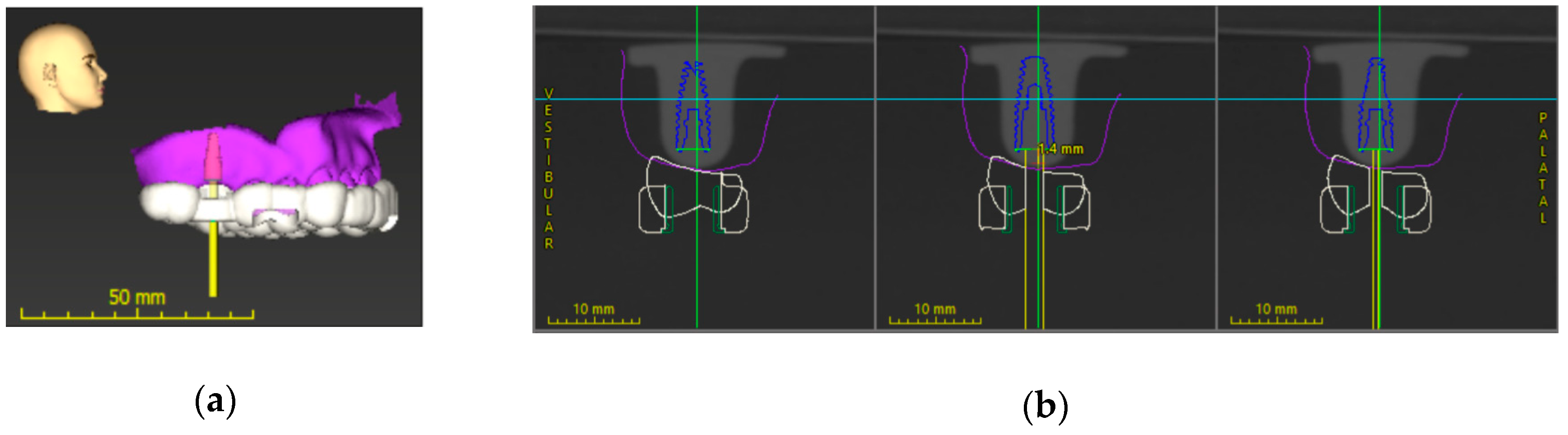
2.2. Implant Position Planning Procedure and Surgical Guide Fabrication

A duplicated model was randomly selected and scanned with an intraoral scanner (Straumann® Virtuo Vivo™, Institute Straumann AG) and a Cone-Beam Computed Tomography (CBCT) machine (J.Morita Inc.) that generate STL and DICOM files, respectively. Afterwards, STL and DICOM files were merged with coDiagnostiX™ software. Then, the digitally diagnostic wax-up was obtained by an STL file of a standard maxillary dentoform. The 4.1 × 10 mm Straumann® BLT implant was virtually planned in the desired position (Figure 1).
The full-arch tooth-supported surgical guides with a 5-mm-diameter Straumann® T-sleeve were designed with 2.5-mm thickness and 0.20-mm offset. Two inspecting windows were used to ensure that surgical guides fit well on the models, which were located at the buccal of the maxillary first premolar and the mesiobuccal cusp of the maxillary second molar (Figure 1). This experiment divided samples equally into 3 groups, each containing 10 samples, and the 4.1 × 10 mm Straumann® BLT was placed with 3 surgical guides fabricated by different 3D printers in each group. To fabricate the static surgical guides, each type of 3D printer was calibrated at the manufacturer’s recommendation. Finally, all surgical guides were printed with 3 different 3D additive manufacturing printers following the manufacturer’s instructions including DLP, SLA, and PolyJet (Figure 2). The overview of 3D printers was shown in Table 1.

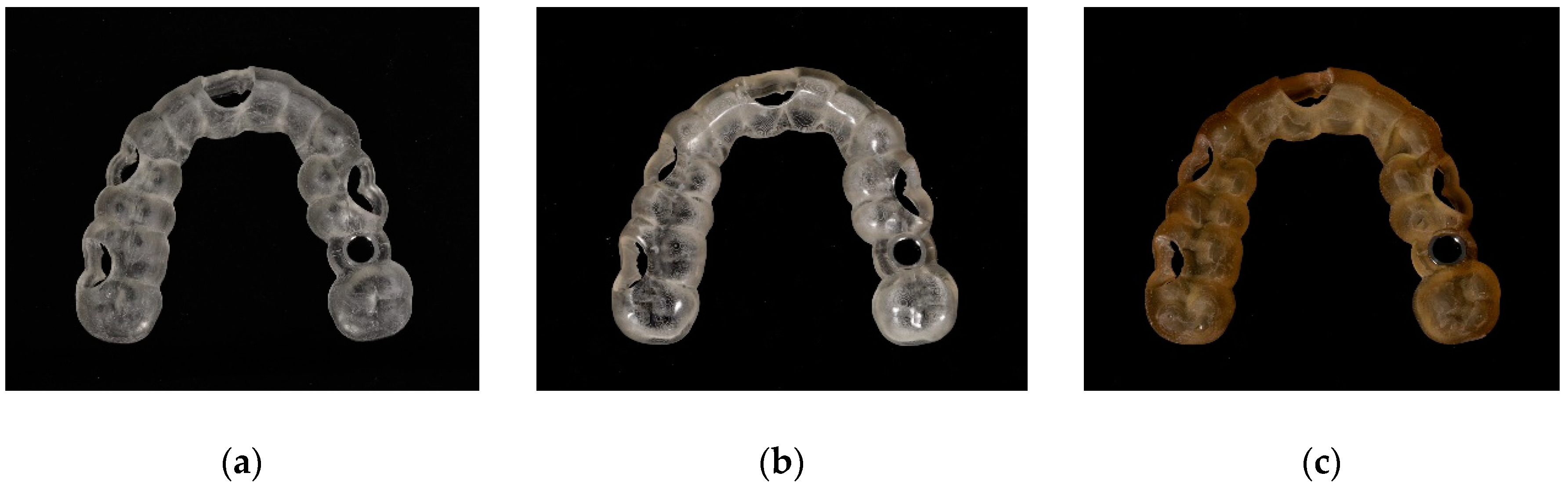
2.3. Post-Processing

Different 3D printing technologies require different post-processing. For the SLA printer, the post-processing of surgical guides included 6 steps: removing printed parts from the platform, rinsing with Isopropyl alcohol (IPA) to clean residual resin, air-drying to blow IPA away, post-printing curing at the temperature of 60 °C, and detaching and polishing the surgical guides. Unlike the SLA printer, the post-processing of PolyJet involved 5 steps: printed parts removal, cutting supports using a wax carver, rinsing with water, air-drying to clean supports, and post-curing at the temperature of 30 °C. Finally, the DLP technology required the 4 steps of post-processing: printed parts removal, washing with Straumann P wash, post-curing by Straumann P Cure, and smoothing the surface of surgical guides.

2.4. Implant Placement Procedure

Prior to the implant placement, each static surgical guide was placed on the duplicated model while performing a fit check via inspecting windows. Implant-guided surgery was carried out by one experienced surgeon. The surgical guides were divided into a group of three, and each group was assigned to perform surgery in a random sequence to prevent familiarity. Then, based on the Straumann® guided surgery system protocols, Straumann® BLT dummy implants were positioned by using guided instruments. (Guided Surgical Kit; Institute Straumann AG).

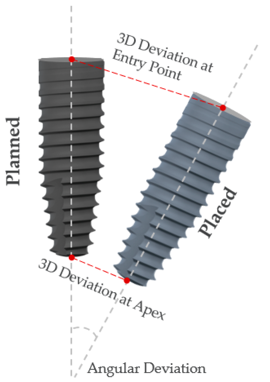
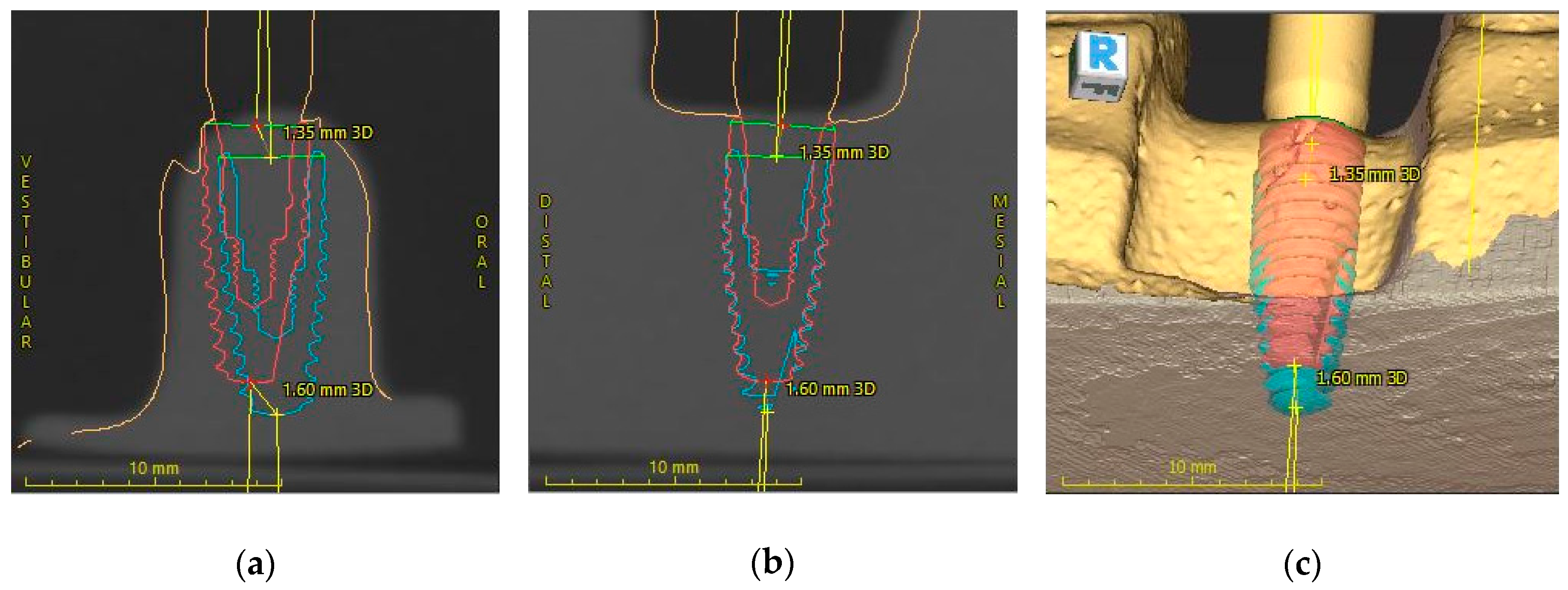
2.5. Implant Position Accuracy Measurement

After the implant placement procedure, a scan body (Straumann® CARES® Mono) was tightened onto each implant using a screwdriver. Each model with a scan body was captured by the intraoral scanner (Straumann® Virtuo Vivo™), and exported into an STL file to superimpose on the originally planned model. The 3D measurements, including 3D deviation at the entry point (mm), 3D deviation at the apex (mm), and angular deviation (°) were calculated automatically via the coDiagnostiX™ software version 9.7 (Figure 3 and Figure 4).

2.6. Statistical Analysis
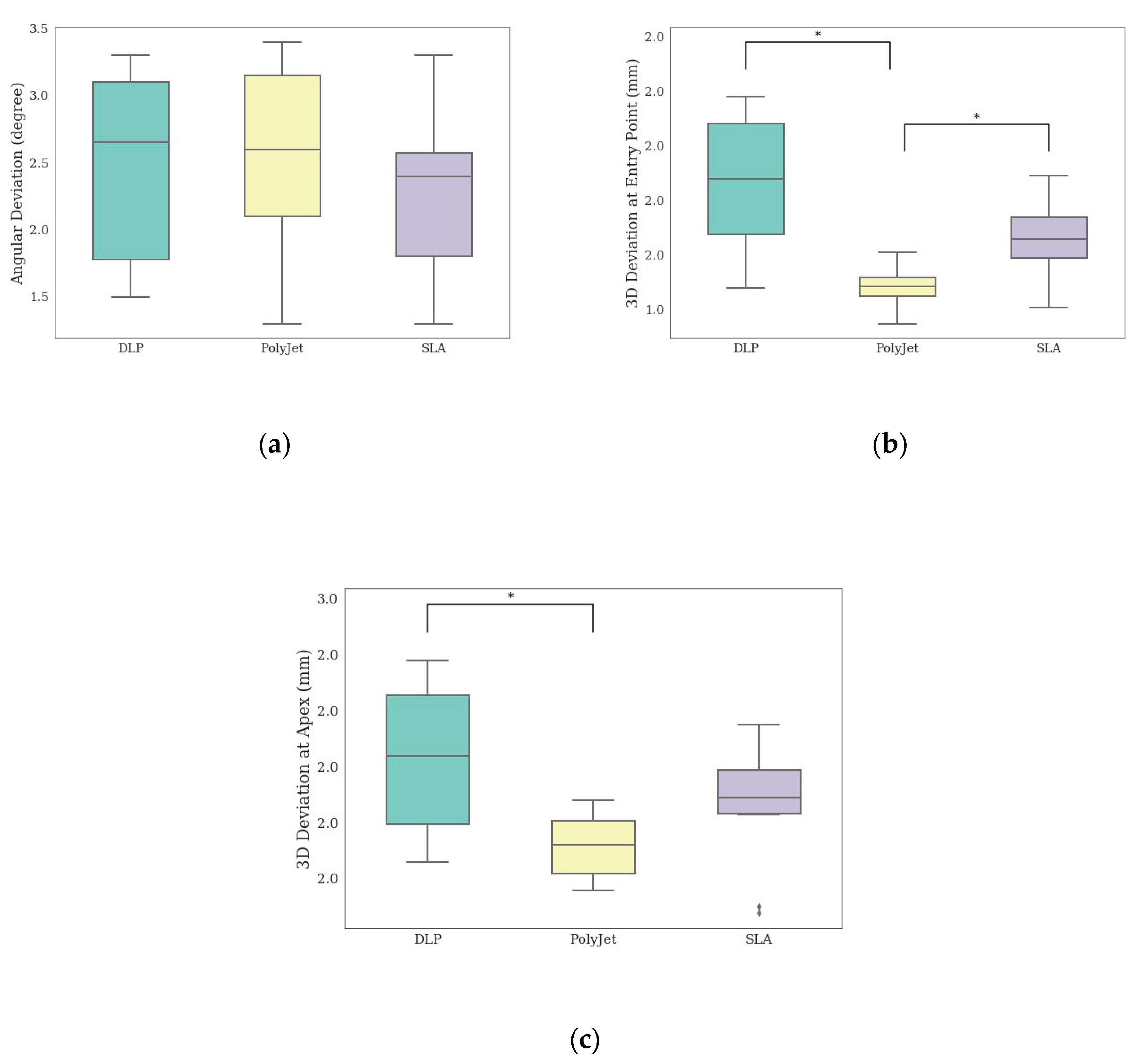
The primary measurements were analyzed by IBM SPSS Statistics software version 18 (SPSS Inc.). The data normality was estimated by the Shapiro–Wilk test. The one-way ANOVA was used to determine whether the differences of 3D deviations were statically significant (p < 0.05). The distribution of measurements was illustrated by using boxplots as shown in Figure 5.

3. Results

The mean, SD, and p-Value of the 3D deviation of the measurements were shown in Table 2. For the angular deviation, the mean deviation was not statistically significant among all technologies. Considering the 3D deviation at the entry point, PolyJet achieved a lower mean of the deviation than that of the other groups. Lastly, the lowest deviation of the 3D offset tip was produced by PolyJet. There was a statistically significant difference between PolyJet and DLP (p = 0.007), but no significant difference between PolyJet and SLA was found.
In terms of manufacturing speed, DLP clearly demonstrated the fastest printing time of 22 min, followed by PolyJet and SLA with the printing time of 248 and 337 min, respectively. When accounting for the post-processing time, the overall processing time rankings remained unchanged (Table 3).

4. Discussion

This study investigated whether the overall accuracy of implant positions was affected by three different additive printing technologies. The accuracy was determined by three measurements: angular deviation, 3D deviation at the entry point, and 3D deviation at the apex. Therefore, the study concluded that there were statistically significant differences among all additive printing technologies, and the null hypothesis was rejected.
According to the experiment, based on the implant position, Polyjet provided the best outcome, while DLP and SLA reported diminished accuracy. For the entry point and apical deviations, the result did not align with Herschdorfer’s study [18], which claimed that there was no statistically significant difference among all 3D printers. The reason for this might be due to the different types of 3D printing technologies, which were SLA, Polyjet, and Multijet. Although the mean deviation of the angle was not different between all technologies, the result was consistent with a recent study [18]. The finding also suggested that the angular deviation might be less sensitive to printing technologies. The angular deviations from PolyJet, DLP, and SLA were within the range of clinical acceptability, based on the recent systematic review [19,20,21].
Overall, these findings are in accordance with a recently published study, which investigated the trueness and precision of surgical guides between PolyJet, DLP, SLA, and FFF (fused filament fabrication). The trueness of PolyJet outperformed that of SLA and DLP and in the aspect of precision, the performance of PolyJet surpassed SLA [3]. On the contrary, the processing time of DLP technology, which is another critical factor in choosing 3D printers, was by far the shortest, compared to PolyJet and SLA.
In general, every step in the workflow of implant-guided surgery carried a margin of inaccuracy [15]. Several factors influence the accuracy of the static guided implant surgery such as study design, surgical guide design, type of guided surgery, implant characteristics, operator’s experience, and printing technique. This study, therefore, minimized the overall effect of confounding factors as much as possible through various techniques. To minimize confounding factors, we utilized the same guide design, drill protocol, surgeon, and type of surgical models. The implant placement using different printed guides was also done at random to reduce the drilling memorization of the surgeon. Considering the study design, this study was performed in vitro aspect. This method appeared to be the most reliable compared to in vivo and cadaver studies because the models provided clinicians with better accessibility and visualization. Besides, the advantage of an in vitro study compared to an in vivo one was the absence of patient movement, saliva, or blood [19]. Additionally, for surgical guide design, this experiment used tooth-support surgical guides, which showed less deviation of implant position compared with those supported by the bone and by mucosa, in all aspects of the entry point, apex point, depth, and angulation [19,22,23,24,25,26,27,28]. Next, another vital factor is that the type of guided surgery can give rise to the deviation. When there are sufficient bone and keratinized mucosa, fully guided surgery is the most precise approach for flapless surgery, which can decrease chair time, compared to a free-handed approach [11,29,30] or pilot-drill, half-guided surgery [11,29,30,31,32]. Additionally, these studies performed fully guided surgery to maximize accuracy. Moreover, implant characteristic is an important factor influencing the accuracy. El Kholy et al. investigated the influence of macrodesign on precision. The tapered macrodesign (BLT) offered greater accuracy compared to parallel-walled macro designs (TL,BL), owing to the geometric insertion, the drill design, and the thread design [33]. As a consequence, an implant with a tapered design (BLT) was used in this research to minimize deviation. Another related factor is the experience of an operator. A study reported that the difference between angular and linear deviations was not statistically significant between experienced and inexperienced surgeons, yet it is better to experiment with the experienced surgeon to avoid the confounding factor for this study [34]. In another study, they evaluated whether the deviation of the static surgical guide was affected by the type and number of teeth supporting the surgical guide. The accuracy of surgical guides, supported by four teeth which can decrease cost and processing time, was not different to that of full-arch-supported guides. However, to lessen the error from the external factor, this study used the full-arch-supported guides [35]. Lastly, regarding the printing technique, there was no statistically significant difference in implant position between a surgical guide manufactured by the additive technique “3D printing” and the subtractive technique “milling”. Nevertheless, while producing comparable accuracy for implant guided surgery, the surgical guide fabricated by 3D printers could be a substitution due to the ease of production and the diminution of laboratory time and materials, consequently increasing cost-effectiveness [36].
However, the deviation of the implant position can be affected by the surgical procedure. To begin with, the surgical guides were not designed for an individual model but rather were designed based on a randomly selected model. Apart from the surgical procedure, due to the roughness surface of the sleeve or resin interface as well as the offset setting, the placing of the guide sleeve into the surgical guide probably contributed to another aspect affecting the implant deviation. Moreover, differences in post-processing recommendations and instruments may have an influence on the surface of the surgical guide [37]. Although, with quality control such as caliper and weight measurements, the models may not perfectly replicate bone properties including strength, elasticity, hardness, and density. Additionally, the print quality can be enhanced to improve the correctness of the study. A 3D printer resolution is an important indicator to determine the printing quality and is directly associated with the layer thickness. Specifically, the layer of thickness can affect 3D smoothness significantly. The thinner the layer is, the smoother 3D prints become. With the greater thickness of the layer, it was observed that the surface of a printed layer was rough, as each layer was easily noticeable. Furthermore, the recent study also suggested that the effect of layer thickness can result in the deviation of the intaglio dimension of the surgical guide [37]. Ultimately, an in vitro study might not truly reflect the error of implant position performed in a clinical trial. The deviation in an in vitro study might be relatively marginal, and accordingly can be neglected in a clinical trial. Thus, there should be a further study conducting a clinical trial to illustrate the true potential of each 3D printer. Lastly, human errors should also be taken into consideration.
On the whole, to make the final decision on choosing a 3D printer for fabricating surgical guides, apart from the printer’s accuracy and processing time, additional factors such as price, operating cost, maintenance cost, and after-sales service should all be taken into account.

5. Conclusions

Within the limitation of this study, the deviations from the experiment proved that the PolyJet was the most accurate 3D printer regarding the 3D deviations at the entry point and apex; however, the DLP printer had the shortest processing time.

Author Contributions

Conceptualization, P.T.; methodology, P.T. and C.A.; software, C.U.; validation, P.T. and C.A.; formal analysis, C.A. and C.U.; investigation, P.T. and C.A.; resources, P.T.; data curation, C.A. and C.U.; writing—original draft preparation, P.T., C.A., and C.U.; writing—review and editing, P.T., C.A., and S.K.; visualization, C.A. and S.K.; supervision, P.T.; project administration, P.T. All authors have read and agreed to the published version of the manuscript.

Funding

This research received no external funding.

Acknowledgments

The authors gratefully acknowledge the faculty and staff at the dental implant center of Mahidol University for the support of equipment and facilities.

Conflicts of Interest

The authors declare no conflict of interest.

References

  1. Kernen, F.; Kramer, J.; Wanner, L.; Wismeijer, D.; Nelson, K.; Flügge, T. A review of virtual planning software for guided implant surgery-data import and visualization, drill guide design and manufacturing. BMC Oral Health 2020, 20, 251. [Google Scholar] [CrossRef]
  2. Kessler, A.; Hickel, R.; Reymus, M. 3D Printing in Dentistry-State of the Art. Oper Dent 2020, 45, 30–40. [Google Scholar] [CrossRef]
  3. Kim, S.Y.; Shin, Y.S.; Jung, H.D.; Hwang, C.J.; Baik, H.S.; Cha, J.Y. Precision and trueness of dental models manufactured with different 3-dimensional printing techniques. Am. J. Orthod. Dentofac. Orthop. 2018, 153, 144–153. [Google Scholar] [CrossRef] [Green Version]
  4. Oberoi, G.; Nitsch, S.; Edelmayer, M.; Janjić, K.; Müller, A.S.; Agis, H. 3D Printing—Encompassing the Facets of Dentistry. Front. Bioeng. Biotechnol. 2018, 6. [Google Scholar] [CrossRef] [Green Version]
  5. Rungrojwittayakul, O.; Kan, J.Y.; Shiozaki, K.; Swamidass, R.S.; Goodacre, B.J.; Goodacre, C.J.; Lozada, J.L. Accuracy of 3D Printed Models Created by Two Technologies of Printers with Different Designs of Model Base. J. Prosthodont. 2020, 29, 124–128. [Google Scholar] [CrossRef]
  6. Brown, G.B.; Currier, G.F.; Kadioglu, O.; Kierl, J.P. Accuracy of 3-dimensional printed dental models reconstructed from digital intraoral impressions. Am. J. Orthod. Dentofac. Orthop. 2018, 154, 733–739. [Google Scholar] [CrossRef] [Green Version]
  7. Favero, C.S.; English, J.D.; Cozad, B.E.; Wirthlin, J.O.; Short, M.M.; Kasper, F.K. Effect of print layer height and printer type on the accuracy of 3-dimensional printed orthodontic models. Am. J. Orthod. Dentofac. Orthop. 2017, 152, 557–565. [Google Scholar] [CrossRef] [Green Version]
  8. Park, M.-E.; Shin, S.-Y. Three-dimensional comparative study on the accuracy and reproducibility of dental casts fabricated by 3D printers. J. Prosthet. Dent. 2018, 119, 861.e1–861.e7. [Google Scholar] [CrossRef]
  9. Sun, Y.; Ding, Q.; Tang, L.; Zhang, L.; Sun, Y.; Xie, Q. Accuracy of a chairside fused deposition modeling 3D-printed single-tooth surgical template for implant placement: An in vitro comparison with a light cured template. J. Cranio-Maxillofac. Surg. 2019, 47, 1216–1221. [Google Scholar] [CrossRef]
  10. Di Giacomo, G.A.; Cury, P.R.; de Araujo, N.S.; Sendyk, W.R.; Sendyk, C.L. Clinical application of stereolithographic surgical guides for implant placement: Preliminary results. J. Periodontol. 2005, 76, 503–507. [Google Scholar] [CrossRef]
  11. Arisan, V.; Karabuda, C.Z.; Mumcu, E.; Özdemir, T. Implant positioning errors in freehand and computer-aided placement methods: A single-blind clinical comparative study. Int. J. Oral Maxillofac. Implants 2013, 28, 190–204. [Google Scholar] [CrossRef] [Green Version]
  12. Vercruyssen, M.; Hultin, M.; Van Assche, N.; Svensson, K.; Naert, I.; Quirynen, M. Guided surgery: Accuracy and efficacy. Periodontology 2000 2014, 66, 228–246. [Google Scholar] [CrossRef] [PubMed]
  13. Cassetta, M.; Di Mambro, A.; Giansanti, M.; Stefanelli, L.V.; Barbato, E. Is it possible to improve the accuracy of implants inserted with a stereolithographic surgical guide by reducing the tolerance between mechanical components? Int. J. Oral Maxillofac. Surg. 2013, 42, 887–890. [Google Scholar] [CrossRef] [PubMed]
  14. Zhou, W.; Liu, Z.; Song, L.; Kuo, C.-L.; Shafer, D. Clinical Factors Affecting the Accuracy of Guided Implant Surgery—A Systematic Review and Meta-analysis. J. Evid. Based Dent. Pract. 2017, 18. [Google Scholar] [CrossRef]
  15. Al Yafi, F.; Camenisch, B.; Al-Sabbagh, M. Is Digital Guided Implant Surgery Accurate and Reliable? Dent. Clin. N. Am. 2019, 63, 381–397. [Google Scholar] [CrossRef] [PubMed]
  16. El Kholy, K.; Janner, S.F.M.; Schimmel, M.; Buser, D. The influence of guided sleeve height, drilling distance, and drilling key length on the accuracy of static Computer-Assisted Implant Surgery. Clin. Implant Dent. Relat. Res. 2019, 21, 101–107. [Google Scholar] [CrossRef] [Green Version]
  17. Deeb, G.R.; Allen, R.K.; Hall, V.P.; Whitley, D., 3rd; Laskin, D.M.; Bencharit, S. How Accurate Are Implant Surgical Guides Produced with Desktop Stereolithographic 3-Dimentional Printers? J. Oral Maxillofac. Surg. 2017, 75, 2559.e1–2559.e8. [Google Scholar] [CrossRef]
  18. Herschdorfer, L.; Negreiros, W.M.; Gallucci, G.O.; Hamilton, A. Comparison of the accuracy of implants placed with CAD-CAM surgical templates manufactured with various 3D printers: An in vitro study. J. Prosthet. Dent. 2020. [Google Scholar] [CrossRef]
  19. Bover-Ramos, F.; Viña-Almunia, J.; Cervera-Ballester, J.; Peñarrocha-Diago, M.; García-Mira, B. Accuracy of Implant Placement with Computer-Guided Surgery: A Systematic Review and Meta-Analysis Comparing Cadaver, Clinical, and In Vitro Studies. Int. J. Oral Maxillofac. Implants 2018, 33, 101–115. [Google Scholar] [CrossRef]
  20. Sigcho López, D.A.; García, I.; Da Silva Salomao, G.; Cruz Laganá, D. Potential Deviation Factors Affecting Stereolithographic Surgical Guides: A Systematic Review. Implant Dent. 2019, 28, 68–73. [Google Scholar] [CrossRef]
  21. Tahmaseb, A.; Wu, V.; Wismeijer, D.; Coucke, W.; Evans, C. The accuracy of static computer-aided implant surgery: A systematic review and meta-analysis. Clin. Oral Implants Res. 2018, 29, 416–435. [Google Scholar] [CrossRef] [Green Version]
  22. Behneke, A.; Burwinkel, M.; Behneke, N. Factors influencing transfer accuracy of cone beam CT-derived template-based implant placement. Clin. Oral Implants Res. 2012, 23, 416–423. [Google Scholar] [CrossRef]
  23. Ersoy, A.E.; Turkyilmaz, I.; Ozan, O.; McGlumphy, E.A. Reliability of implant placement with stereolithographic surgical guides generated from computed tomography: Clinical data from 94 implants. J. Periodontol. 2008, 79, 1339–1345. [Google Scholar] [CrossRef]
  24. Ozan, O.; Turkyilmaz, I.; Yilmaz, B. A preliminary report of patients treated with early loaded implants using computerized tomography-guided surgical stents: Flapless versus conventional flapped surgery. J. Oral Rehabil. 2007, 34, 835–840. [Google Scholar] [CrossRef]
  25. Pozzi, A.; Polizzi, G.; Moy, P.K. Guided surgery with tooth-supported templates for single missing teeth: A critical review. Eur. J. Oral Implantol. 2016, 9 (Suppl. S1), S135–S153. [Google Scholar]
  26. Tahmaseb, A.; Wismeijer, D.; Coucke, W.; Derksen, W. Computer technology applications in surgical implant dentistry: A systematic review. Int. J. Oral Maxillofac. Implants 2014, 29, 25–42. [Google Scholar] [CrossRef] [Green Version]
  27. Younes, F.; Cosyn, J.; De Bruyckere, T.; Cleymaet, R.; Bouckaert, E.; Eghbali, A. A randomized controlled study on the accuracy of free-handed, pilot-drill guided and fully guided implant surgery in partially edentulous patients. J. Clin. Periodontol. 2018, 45, 721–732. [Google Scholar] [CrossRef]
  28. Derksen, W.; Wismeijer, D.; Flügge, T.; Hassan, B.; Tahmaseb, A. The accuracy of computer-guided implant surgery with tooth-supported, digitally designed drill guides based on CBCT and intraoral scanning. A prospective cohort study. Clin. Oral Implants Res. 2019, 30, 1005–1015. [Google Scholar] [CrossRef]
  29. Pozzi, A.; Tallarico, M.; Marchetti, M.; Scarfò, B.; Esposito, M. Computer-guided versus free-hand placement of immediately loaded dental implants: 1-year post-loading results of a multicentre randomised controlled trial. Eur. J. Oral Implantol. 2014, 7, 229–242. [Google Scholar]
  30. Vercruyssen, M.; Cox, C.; Coucke, W.; Naert, I.; Jacobs, R.; Quirynen, M. A randomized clinical trial comparing guided implant surgery (bone- or mucosa-supported) with mental navigation or the use of a pilot-drill template. J. Clin. Periodontol. 2014, 41, 717–723. [Google Scholar] [CrossRef]
  31. Amorfini, L.; Migliorati, M.; Drago, S.; Silvestrini-Biavati, A. Immediately Loaded Implants in Rehabilitation of the Maxilla: A Two-Year Randomized Clinical Trial of Guided Surgery versus Standard Procedure. Clin. Implant Dent. Relat. Res. 2017, 19, 280–295. [Google Scholar] [CrossRef]
  32. Bencharit, S.; Staffen, A.; Yeung, M.; Whitley, D., 3rd; Laskin, D.M.; Deeb, G.R. In Vivo Tooth-Supported Implant Surgical Guides Fabricated With Desktop Stereolithographic Printers: Fully Guided Surgery Is More Accurate Than Partially Guided Surgery. J. Oral Maxillofac. Surg. 2018, 76, 1431–1439. [Google Scholar] [CrossRef]
  33. El Kholy, K.; Ebenezer, S.; Wittneben, J.G.; Lazarin, R.; Rousson, D.; Buser, D. Influence of implant macrodesign and insertion connection technology on the accuracy of static computer-assisted implant surgery. Clin. Implant Dent. Relat. Res. 2019, 21, 1073–1079. [Google Scholar] [CrossRef] [PubMed]
  34. Rungcharassaeng, K.; Caruso, J.M.; Kan, J.Y.; Schutyser, F.; Boumans, T. Accuracy of computer-guided surgery: A comparison of operator experience. J. Prosthet. Dent. 2015, 114, 407–413. [Google Scholar] [CrossRef] [PubMed] [Green Version]
  35. El Kholy, K.; Lazarin, R.; Janner, S.F.M.; Faerber, K.; Buser, R.; Buser, D. Influence of surgical guide support and implant site location on accuracy of static Computer-Assisted Implant Surgery. Clin. Oral Implants Res. 2019, 30, 1067–1075. [Google Scholar] [CrossRef] [PubMed]
  36. Henprasert, P.; Dawson, D.V.; El-Kerdani, T.; Song, X.; Couso-Queiruga, E.; Holloway, J.A. Comparison of the Accuracy of Implant Position Using Surgical Guides Fabricated by Additive and Subtractive Techniques. J. Prosthodont. 2020, 29, 534–541. [Google Scholar] [CrossRef] [Green Version]
  37. Dalal, N.; Ammoun, R.; Abdulmajeed, A.A.; Deeb, G.R.; Bencharit, S. Intaglio Surface Dimension and Guide Tube Deviations of Implant Surgical Guides Influenced by Printing Layer Thickness and Angulation Setting. J. Prosthodont. 2020, 29, 161–165. [Google Scholar] [CrossRef]
Figure 1. (a) 3D and (b) 2D cross-sectional illustration of tooth 16 implant position and surgical guide planning on a model.
Applsci 10 07791 g001
Figure 2. Implant surgical guides fabricated by (a) DLP, (b) PolyJet, and (c) SLA printers.
Applsci 10 07791 g002
Figure 3. The illustration of 3D measurements represents the implant deviation between the preoperatively planned and the postoperatively placed implant position.
Applsci 10 07791 g003
Figure 4. The 3D measurement of implant deviation in coDiagnostiX™ software. Post-operative implant position (blue) merged with pre-operative implant position (red); (a) Cross-sectional; (b) Tangential view; (c) 3D view.
Applsci 10 07791 g004
Figure 5. Boxplots of mean 3D deviation: (a) Angular deviation; (b) 3D deviation at entry point; and (c) 3D deviation at apex. The asterisk represents whether the difference of a measurement between any 2 groups is statistically significant (p < 0.05).
Applsci 10 07791 g005
Table 1. Brief description of different 3D printers.
PrinterDLPPolyJet SLA
ModelP30+
(Straumann)
Objet® Eden260VS (Stratasys)Form2
(Formlabs)
Layer thickness100 µm16.5 µm50 µm
MaterialP Pro Surgical
Guide Clear
MED 610Dental SG Resin
Table 2. The comparison of Mean ± SD of the 3D deviation among DLP, PolyJet, and SLA 3D printers.
DLP
(n = 10)
PolyJet
(n = 10)
SLA
(n = 10)
MeasurementMean ± SD *p-Value
Angular Deviation (degree)2.47 ± 0.722.54 ± 0.702.3 ± 0.610.72
Deviation at entry point (mm)1.87 ± 0.25 a1.48 ± 0.07 b1.66 ± 0.15 a<0.001 **
Deviation at apex (mm)2.03 ± 0.26 a1.72 ± 0.12 b1.86 ± 0.22 a, b0.01 **
* Different superscript between 2 groups within the same measurement means that the difference is statistically significant. (p < 0.05). ** p-Value < 0.05.
Table 3. The comparison of processing and post-processing time among DLP, PolyJet, and SLA 3D printers.
PrinterDLPPolyJetSLA
Printing time (min)22248337
Post-processing time (min)23716
Total (min)45255353
Publisher’s Note: MDPI stays neutral with regard to jurisdictional claims in published maps and institutional affiliations.

