Article

Characterization of Gait and Postural Regulation in Late-Onset Pompe Disease

1
Clinic St. Georg, Delitzscher Str. 141, 04129 Leipzig, Germany
2
Department of Neurology, Martin-Luther-University Halle-Wittenberg, Ernst-Grube-Str. 40, 06120 Halle (Saale), Germany
3
Department of Orthopaedic and Trauma Surgery, Martin-Luther-University Halle-Wittenberg, Ernst-Grube-Str. 40, 06120 Halle (Saale), Germany
4
Department of Health Sciences, University of Colorado, Colorado Springs, CO 80918, USA
5
University Clinic and Policlinic of Radiology, Martin-Luther-University Halle-Wittenberg, Ernst-Grube-Str. 40, 06120 Halle (Saale), Germany
*
Author to whom correspondence should be addressed.
Appl. Sci. 2020, 10(19), 7001; https://doi.org/10.3390/app10197001
Submission received: 11 September 2020 / Revised: 29 September 2020 / Accepted: 7 October 2020 / Published: 8 October 2020

Abstract

:

Featured Application

Pompe disease is a neurological disease with significant impacts on gait and balance. Therefore, it is important to measure these characteristics in a functional, valid and reliable manner. This investigation used a cross-sectional study design utilizing gait analysis and posturography to quantify the difference between patients with late-onset Pompe disease (LOPD) and matched asymptomatic subjects. These results may be useful to develop more specific and efficient rehabilitation programs depending on the individual abilities of the patients.

Abstract

Pompe disease is a multisystemic disorder with the hallmark of progressive skeletal muscle weakness that often results in difficulties in walking and balance. However, detailed characterization of gait and postural regulation with this disease is lacking. The objective of this investigation was to determine if differences exist between the gait and postural regulation of LOPD patients and a matched control group. The gaits of 16 patients with LOPD were assessed using a gait analysis mobile system (RehaGait) and a dynamometric treadmill (FDM-T 1.8). The Interactive Balance System (IBS) was used to evaluate postural regulation and stability. All measures were compared to individual reference data. Demographic (age, gender), morphological (body height, body mass) and clinical data (muscle strength according to the Medical Research Council Scale (MRC Scale), as well as the 6-min walking test and a 10-m fast walk) were also recorded. Compared to individual reference data, LOPD patients presented with reduced gait velocity, cadence and time in single stand. A total of 87% of LOPD patients had abnormalities during posturographic analysis presenting with differences in postural subsystems. This study provides objective data demonstrating impaired gait and posture in LOPD patients. For follow-up analysis and as outcome measurements during medical or physiotherapeutic interventions, the findings of this investigation may be useful.

1. Introduction

Pompe disease (Online Mendelian Inheritance in Man (OMIM) 606800) is an autosomal recessive inherited orphan disease caused by mutations in the glucosidase alpha acid (GAA) gene. Resulting in deficient activity of the enzyme alpha-1,4-glucosidase that is located in cellular lysosoms involved in the degradation of glycogen. Consequently, glycogen accumulation occurs not only in skeletal muscle, but also in cardiac and smooth muscles [1].
The entrapment of lysosomal glycogen results in muscle damage by a number of pathogenic mechanisms, such as defective autophagy, calcium homeostasis, oxidative stress, and mitochondrial abnormalities [2]. Recently, metabolic abnormalities and energy deficits have also been shown to contribute to this pathogenic cascade [3]. The severity of clinical manifestations, tissue impairment and age of onset correlate with the residual enzymatic activity and can be classified into two forms: the Infantile Onset Pompe Disease (IOPD) or classical form has no or very low enzymatic activity levels, leading to severe general muscular weakness with floppy infant syndrome. Cardiomyopathy and respiratory failure usually lead to death within the first year of life. The late-onset Pompe disease (LOPD) or non-classical form has a higher residual enzyme activity. This disease progresses slowly and may start at any age after the first year of life. A common sign of LOPD is weakness of the proximal limbs, predominantly affecting the hip girdle. Axial muscles as well as respiratory muscles may also be impaired due to myopathy [4]. Patients often present clinically with back pain and exercise intolerance and dyspnea [5]. Recently, the systemic characteristics of this primary muscular disorder have been discovered: alteration of smooth muscles of the intestine and vessels, especially ectasia of vertebrobasial arteries, and the involvement of the nervous system (white matter lesions and small fiber neuropathy) widening the Pompe disease phenotype [6].
Since 2006, a causal treatment is available with enzyme replacement therapy (ERT) using recombinant human α-glucosidase. This treatment has been shown to improve patient survival and muscle strength to a variable extent [7]. It can also improve the quality of life of LOPD patients due to better mobility and participation in activities of daily life [8]. However, the actual ERT is not a cure for the disease and further therapeutic approaches are necessary. A detailed understanding of LOPD patients’ gait patterns and balance may help improve physiotherapeutic strategies in the prevention of falls and preserving the patient’s ambulatory status. Additionally, analysis of these features might be a tool for following up on future therapeutic strategies (i.e., next level ERT order gene therapy) [9]. There is a paucity in this research, with one study reporting reduced spatio-temporal parameters during gait in 22 LOPD patients on ERT [10] and another showing deficits in standing postural stability among five LOPD patients [11].
The aim of this study was to provide a systematic analysis of both gait parameters and postural abilities, as well as their interaction in patients with LOPD. These data were then compared to a group of matched healthy controls.

2. Materials and Methods

2.1. Subjects

All subjects provided written informed consent prior to data collection. This study protocol was approved by the local ethics committee (approval number: 2019-164). Sixteen ambulatory LOPD patients with a genetically confirmed diagnosis were included (9 women; mean age: 54.2 ± 15.3 years, range: 19–82 years, Table 1).
Fifteen of sixteen patients (94%) were on ERT at the standard dose of 20 mg/kg biweekly (range: 9–135 months; mean: 67.4 months). The duration of the disease ranged from 2 years to 40 years (mean: 15.9 years). One female was tested before ERT was started. A total of 44% of the patients required temporarily non-invasive ventilation and 50% of the patients used walking aids. All LOPD patients were individually matched with healthy controls based on the relevant selection criteria sex, age, and body height to guarantee a valid comparison with asymptomatic subjects and to avoid the recruitment of an asymptomatic control group [12,13]. The reference data were obtained from 1860 subjects for mobile gait analysis [14], from 141 subjects for treadmill analysis [15] and from 1724 subjects for the posturographic parameters [16].

2.2. Clinical Analysis

Synopsis of the study protocol for each patient is depicted in Figure 1. All LOPD patients were examined clinically using MRC scale similar to previous trials with LOPD patients [11,17]. For lower limb assessment, the bilateral strength of the hip flexors and extensors, hip abductors and adductors, knee flexors and extensors, and ankle dorsiflexors and plantarflexors were assessed. For correlation analysis, the bilateral sum of the MRC scale was used for each individual strength variable, as well as the bilateral sum of all examined muscle groups were obtained. Furthermore, all LOPD patients completed a 6-min walking test and a 10-m fast walk [4]. Concomitant peripheral neuropathy was examined via a clinical screening for symptoms of polyneuropathy (e.g., reduction of distal sensibility and tendon reflexes, pain of distal legs) and confirmation by nerve conduction analyses.

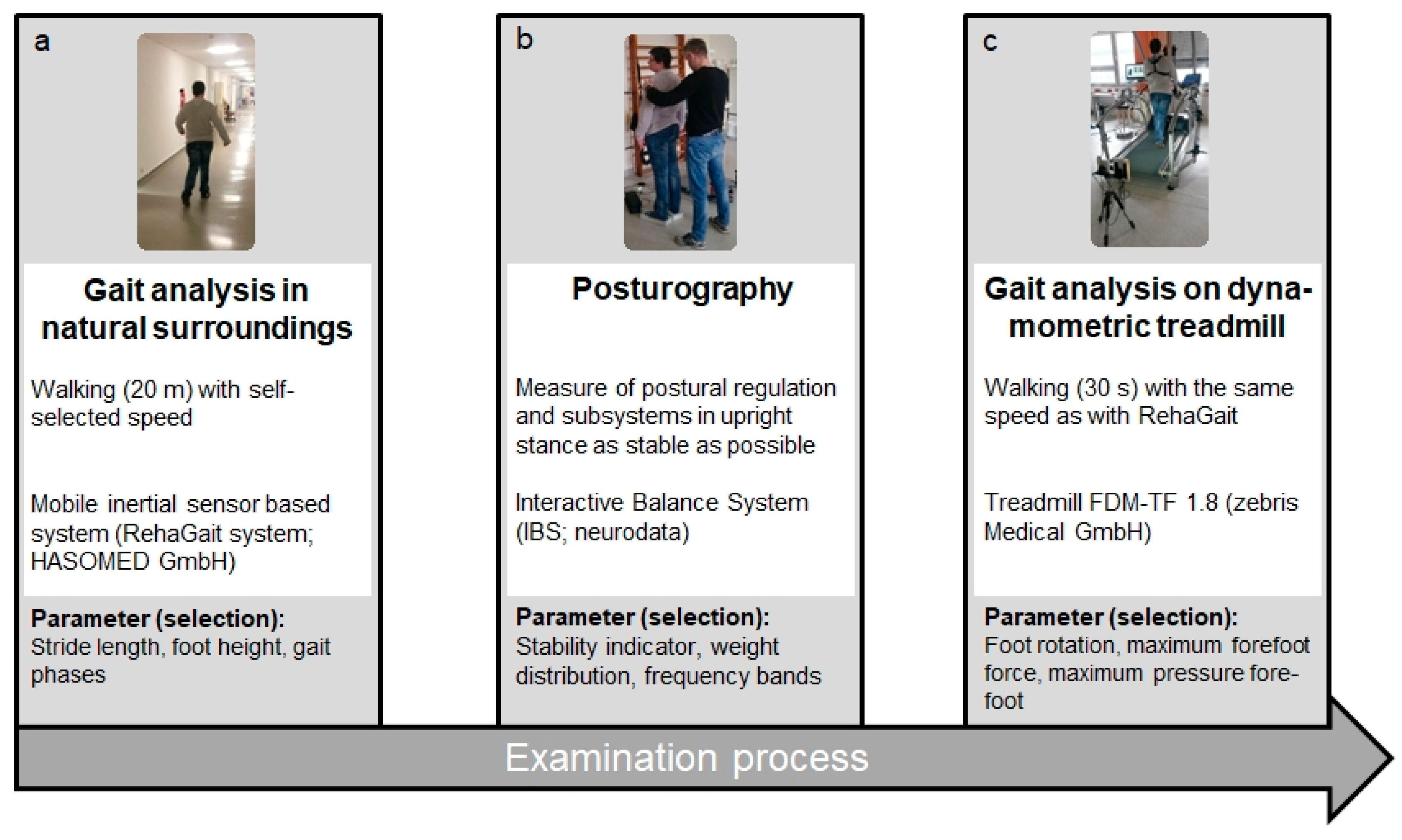
2.3. Mobile Gait Analysis Using an Inertial Sensor Based System

Participants were initially equipped with a mobile inertial sensor-based system (RehaGait® HASOMED GmbH, Magdeburg, Germany) (Figure 1a). This system has been reported to have a high intraobserver reliability (intraclass correlation coefficients (ICCs) range: 0.691–0.959) [18]. Donath et al. [19] also reported good to excellent intraobserver reliability by testing 22 healthy subjects on two separate days (time interval: 7 days). Additionally, these authors confirmed the validity of this system by comparing it to the FDM-T system (zebris medical GmbH, Isny, Germany), which was also used in the present study. These authors captured spatio-temporal gait data simultaneously using both gait analysis systems and with the exception of speed and stride length at a slow speed (15% below habitual walking speed), both systems showed a high level of accordance. In line with the study of Donath et al. [19], each sensor (dimensions: 60 × 15 × 35 mm) contained a 3-axis accelerometer (±8 g), a 3-axis gyroscope (±1000 °/s) and a 3-axis compass (±1.3 Gs). The sensors were attached to the lateral aspect of the shoe using special straps to measure linear acceleration, angular velocity and the magnetic field of the foot (sampling rate: 500 Hz). Heel-strike was used to determine each gait cycle. All other gait events (full contact, heel-off, toe-off) were identified relative to heel-strike. These gait phases were then used to derive orientation and position and spatio-temporal gait parameters.
Since self-selected speed has been suggested to provide the most functionally relevant data [20], each subject was instructed to walk through a 20-m common hospital corridor (without any obstacles) at a self-preferred speed. LOPD patients wore their own personal walking shoes and the first walking trial was used for subjects to adjust to the test conditions. Data from the second trial were used for analysis. Each gait parameter for all recorded steps was analyzed.

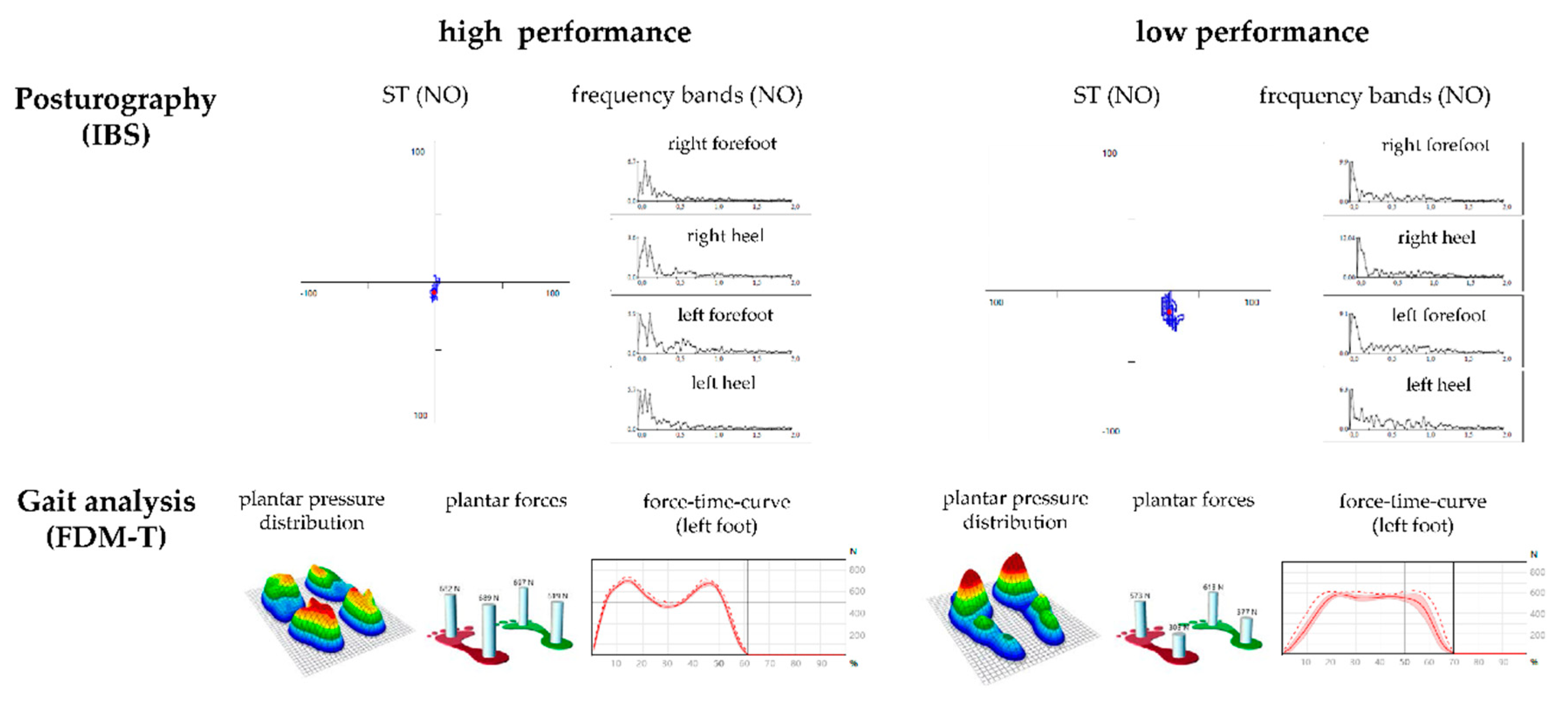
2.4. Balance Measurement Using Posturography

Posturographic assessment was established with the IBS (neurodata GmbH, Vienna, Austria) (Figure 1b). The IBS consists of four independent force plates used to measure postural regulation at a sampling rate of 32 Hz. Sway intensities at different frequency ranges were determined using Fast Fourier Transformation (FFT). The raw signal (force-time signal) was subtracted from the mean value and then subjected to a FFT with a rectangular window. On the ordinate, the amplitude of the frequency components was exposed and, consequently, the ordinate was dimensionless in that the results of the FFT are proportional to the output signal [21]. Different functional frequency bands were used to delineate the postural subsystems (F1, F2–4, F5–6, F7–8) [22,23,24]:
  • F1: Frequency band 1 (0.01–0.03 Hz)—visual and nigrostriatal system;
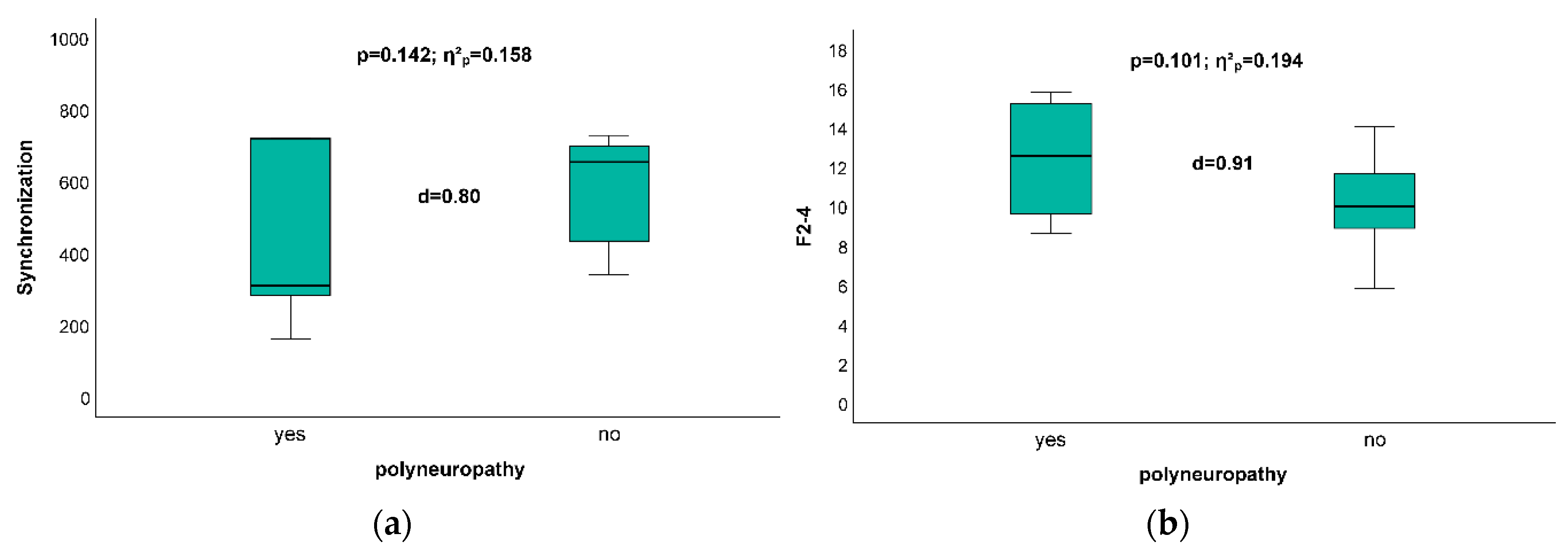
  • F2–4: Frequency band 2–4 (0.03–0.5 Hz)—peripheral-vestibular system;
  • F5–6: Frequency band 5–6 (0.5–1.0 Hz)—somatosensory system;
  • F7–8: Frequency band 7–8 (>1.0 Hz)—cerebellar system.
Additionally, motor output was determined as:
  • Stability indicator (ST): The root mean square of successive differences in pressure signals. Greater instability is indicated by a greater ST.
  • Weight distribution index (WDI): Standard deviation of the weight distribution score.
  • Synchronization (Synch): Six values that describe the relationship of vibration patterns between plates calculated as a scalar product: 1000—complete coactivity; −1000—complete compensation; 0—no coactivity or compensation.
  • Forefoot–hindfoot ratio (Heel): Percentage of load distribution between the forefoot and hindfoot with an emphasis on heel loading.
  • Left–right ratio (Left): Percentage of load distribution between the left and right feet with an emphasis on left side loading.
Subjects were tested barefoot using a single trial (32 s) for each of the following test conditions:
  • Head straight, eyes open, without foam pads (NO);
  • Head straight, eyes closed, without foam pads (NC);
  • Head straight, eyes open, on foam pads (PO);
  • Head straight, eyes closed, on foam pads (PC);
  • Head rotated 45° to the right, eyes closed, without foam pads (HR);
  • Head rotated 45° to the left, eyes closed, without foam pads (HL);
  • Head up (dorsiflexed), eyes closed, without foam pads (HB);
  • Head up (plantarflexed), eyes closed, without foam pads (HF).
Initially, subjects were asked to stand upright, with their weight evenly distributed on the two force plates while focusing on a fixed target (placed at each subject’s respective body height). In this starting position, subjects were then asked to stand freely and as still as possible. Positions, reliability, frequency bands, and parameters of motor output used in the IBS have been previously described in detail [12]. For example, the intraobserver reliability has been confirmed using both asymptomatic subjects [25] and patients [26,27]. ICCs for every parameter and all test positions have been reported from 0.71 to 0.95 [25,26,27]. In support of these previous reliability investigations, which showed that the reliability averaged over eight positions were clearly higher than from a single position, we also used the mean values captured in the eight test positions for all parameters.

2.5. Gait Analysis Using a Dynamometric Treadmill

Gait trials were performed on a dynamometric treadmill with a fall protector (h/p/cosmos/quasar, FDM-T, zebris medical GmbH, Isny, Germany, Figure 1c) [15]. This instrumented treadmill (length: 1.5 m; width: 0.5 m) contained an integrated pressure sensor mat comprising a matrix of high-quality capacitive force sensors (range, 1–120 N/cm2; precision, 1–120 N/cm2 ± 5 %; sensor area: 135.5 × 54.1 cm; resolution: 1.4 sensors per cm2; total number of sensors: 10.240) and analysis software [19]. The assessment captures the dynamic pressure distribution under the feet while walking on the treadmill at a sampling rate of 300 Hz. Two mobile camera modules were positioned behind and beside the treadmill to confirm correct foot placement on the treadmill. Prior to data collection, the force plate was set to zero in order to calibrate the entire measurement system [28]. Spatio-temporal gait parameters were calculated automatically from the pressure data within the FDM-T software for the heel, midfoot, and forefoot. Based on the manufacturer’s specifications, heel-strike was defined as initial contact (threshold: 1 N/cm2), while toe-off was the final data frame before all foot pressures were sub-threshold. Furthermore, stride length was defined as the distance between two consecutive heel contact points (alternate sides), stride time was the time between two consecutive heel-strikes (same foot) and cadence is the number of steps taken per minute [19]. Each subject walked (30 s duration) at their self-selected gait speed, while wearing their own personal shoes.

2.6. Statistics

The balance and gait analysis results of the LOPD patients were defined as conspicuous when outside a reference range between the 10th percentile (P10) and the 90th percentile (P90) obtained from the matched control group.
Relationships between clinical predictors and the test parameters were calculated using Pearson bivariate two-sided product moment correlations, because the measures obtained were normally distributed. Correlation (r) was graded as: < 0.1, trivial; 0.1–0.3, small; 0.3–0.5, moderate; 0.5–0.7, large; 0.7–0.9, very large; and 0.9–1.0, nearly perfect [29].
Differences between groups (patients with and without polyneuropathy (PN)) were tested using a one-factor (group) univariate general linear model. Differences between means (group effect) were considered statistically significant if p-values were <0.05 or partial eta-squared (ηp2) values were greater than 0.15. Due to the relatively small number of cases in each group (n < 10), decisions on significance were based on both statistical values.
All statistical analysis was performed using SPSS version 25.0 for Windows (IBM, Armonk, NY, USA).

3. Results

3.1. Gait Analysis with Mobile Device (Rehagait) and Dynamometric Treadmill Analysis

LOPD patients showed gait abnormalities in cadence (75%), velocity (69%) and percentage of time in double limb support (56%) compared to the matched references (Table 2 and Table 3).
Maximum foot height was reduced in 6% of LOPD patients, stride length in 25% and step widths in 36% (Table 3). Foot rotation and lateral displacement were conspicuous in 71% of the patients. In most patients (71%), pressure distribution across the heel, midfoot and forefoot regions were outside the inter-percentile range in one or more items (Table 2 and Table 3).
There was a moderate relationship between overall hip muscle strength and the percentage of single limb support during the gait cycle (rRehaGait = 0.525, p = 0.037). Step widths showed a moderate correlation with strength of hip muscles (rtreadmill = −0.579, p = 0.030) and total strength of the lower limbs (r = −0.613, p = 0.020). Performance on the 10-m fast walk was highly correlated with walking speed (rRehaGait = −0.919, p < 0.001; rtreadmill = −0.750, p = 0.002) and stride length (rRehaGait = −0.779, p = 0.001). There was also a moderate correlation between 10-m fast walk performance and the percentage of mid stance phase (rRehaGait = −0.589, p = 0.027). Stride length correlated with the percentage of double support (rRehaGait = −0.749, p = 0.002) and single support (rRehaGait = −0.907, p < 0.001), as well as with forefoot pressure (rtreadmill = 0.578, p = 0.031), and heel pressure (rtreadmill = 0.851, p < 0.001).
Cadence was significantly correlated with hip flexion strength (rRehaGait = 0.575, p = 0.020; rtreadmill = 0.539, p = 0.047) and hip adduction strength (rRehaGait = 0.623, p = 0.010; rtreadmill = 0.620, p = 0.018). Furthermore, cadence was also highly correlated with performance during the 6-min walking test (rRehaGait = 0.742, p = 0.002; rtreadmill = 0.658, p = 0.014).

3.2. Posturographic Analysis

Most LOPD patients (87%) showed abnormal results (Table 4) for balance and postural regulation. One patient (no. 15) was unable to perform the test. Frequency band analysis revealed abnormal results in 53% of the patients: regulations in F1 and F2–4 were most often affected in this cohort. Three patients presented with isolated abnormalities in F1 that detects for visual [22,24] and nigrostriatal [30] contribution to balance regulation. Postural stability (parameter: ST) and forefoot–heel coordination (parameter: Synch) were affected in 60% and 53%, respectively. Patient no. 5 had the lowest level of balance and postural regulation (Table 4) with abnormal results for all frequency bands combined with a disturbed gait pattern with the mobile gait analysis. This patient was not able to perform the gait treadmill test (Table 3). The muscle strength of the knee and forefoot–heel ratio correlated moderately. Results in F5–6 also correlated with performance in the 6-min walking test (r = −0.517, p = 0.049) and with ST (r = 0.983, p < 0.001).
Figure 2 presents two examples for low and high gait and postural performances. Postural stability, measured by a stability indicator, and postural regulation, measured by frequency bands, are presented for posturographic analysis. Plantar pressure distribution and plantar forces regarding heel, midfoot and forefoot (left and right) and force–time curves depict different gait patterns.

3.3. Impact of Existence of Polyneuropathy on Posturographic Results

All posturographic parameters were sub-analyzed according to the presence of PN. Patients with PN had significantly more abnormalities in the synchronization (p = 0.142, ηp2 = 0.158; Figure 3a) and in F2–4 (p = 0.101, ηp2 = 0.194; Figure 3b) than those without PN.
No relevant correlations were detected between disease duration or ERT duration and any performance parameter (e.g., 6-min walking test, 10-m fast walk, walking speed, postural stability measured by a stability indicator). A total of 69% (n = 11) of the LOPD patients were classified with mild axial weakness (dimension: strength of the axial musculature).

4. Discussion

Gait abnormalities in LOPD patients included reduced gait velocity, reduced stride length and a shift from time in single stance phase towards double limb support, which support the previous findings of McIntosh et al. [10]. These abnormalities most likely result from proximal lower limb weakness common with this disease [31,32]. However, in the current study, the only strength characteristic that correlated with gait was for the association between hip strength with time in single leg support stance and step widths.
Postural stability and regulation were reduced in most patients in this study, similar to those of previous research [11]. This is believed to result from reduced muscle strength. However, in the present study, there was no significant correlation between muscle weakness and postural parameters except for knee-joint force and forefoot–heel coordination. There were no relevant correlations between gait performance and posturographic results. In contrast, two patients with marked gait impairment performed better than the reference cohort in isolated parameters of posturography. Therefore, other muscular determinants of gait and balance must exist.
It has previously been reported that postural regulation in LOPD patients is impaired when their eyes are closed and believed to result from sensory deficits [10]. Consistently in this study, LOPD patients with clinical PN had significantly more abnormalities in the postural synchronization than those without. However, no differences in sway between LOPD patients with or without PN were observed in F5–6 (averaged over all test positions), which has been shown to be affected in patients with diabetic polyneuropathy [23]. It is possible that, in LOPD patients, additional effects on the muscle spindles [33] or spinal cord [34] may contribute to alterations in sway rather than PN. A differentiated calculation (F5–6NO + PO = open eyes vs. F5–6NC + PC + HR + HL + HB + HF = closed eyes) based on an effect size (d) provided a significantly larger difference with open eyes (with PN: 4.26 ± 2.43 vs. without PN: 3.04 ± 0.88; d = 0.74) than with eyes closed (with PN: 6.29 ± 3.44 vs. without PN: 5.14 ± 1.55; d = 0.47). This finding may be explained by the neuroplasticity of biological systems and the model of selective compensatory optimization. The alteration of afferent sensory (proprioceptive) information, potentially caused by mechanoreceptor damage (PN, Anterior Cruciate Ligament (ACL) surgery, High Tibial Osteotomy (HTO) surgery), may contribute to disturbances of postural regulation [12,13]. In support of Brehme et al. [13] and in contrast to Bartels et al. [12], we showed hyperactivity of the somatosensory system in the presence of PN. This hyperactivity could also be observed in the cerebellar system (four patients had abnormal posturographic findings; Table 4). This may partially explain the close relationship between the somatosensory system and the spinocerebellum system as an important part of the cerebellum, which is responsible for processing afferent (somatosensory) information. Obviously, most of the PN patients developed hyperactivity of the somatosensory (2/3) and cerebellar (3/4) system, which should be interpreted as an ineffective and excessive function of these postural subsystems. Only one patient (ID 7; Table 4) developed suppression in both subsystems.
LOPD patients of this study also revealed balance deficiencies in visual and nigrostriatal regulation, cerebellar regulation, and in the vestibular subsystem with various interactions between the subsystems. This reflects the multi-systemic nature of Pompe disease since there are reports about cerebral [35,36] and vestibulocochlear affections [37] in LOPD patients. However, visual function has not yet been found when analyzed by evoked potentials [38]. Therefore, future research is necessary in this area.
When looking at the weak correlations between muscle strength, gait performance and balance in the LOPD patients of this study, the impact of factors other than those tested is possible. High-quality and frequent physiotherapy and individual training, as well as personal motivation, may result in better compensation of gait disturbances and balance. Furthermore, it has to be considered that each patient received physiotherapy in a non-standardized manner. Research has previously shown that the application of whole body vibration training with an oscillating platform was beneficial in LOPD patients for improving general muscle strength [39]. These authors speculated that this effect results from the stimulation of muscle spindles that might lead to reflex contraction of extrafusal muscle fibers [40]. However, influences of vibration on gait and posture have been shown to reduce the risk of falls by a more multimodal approach (e.g., ankle joint motion, sensation of foot plantar surface and fear) in older adults [41]. Finally, Corrado et al. [42] revealed that there is no research supporting the effectiveness of rehabilitation protocols among LOPD patients. Corrado et al. [42] performed a systematic review investigating current rehabilitation protocols for LOPD patients and concluded that studies with larger sample sizes and higher quality are necessary to reduce the lack of evidence surrounding rehabilitative treatments. The extremely large gait performance differences within LOPD patients (e.g., range of walking speed: 0.25–1.58 m/s; Table 2) and postural stability (e.g., range of ST: 11.7–57.5; Table 4) in our study may have also been a result of the small size of our cohort. At the same time, these large performance differences within LOPD patients are reasons for individualized physiotherapy. The gait and posturographic performances in some patients suggest that individualized physiotherapy has the potential to improve gait and balance performance in LOPD patients and may help prevent falls. As such, our study could be used as reference for future investigations using a lager patient cohort with a standardized physiotherapeutic program that would ideally be tested using a controlled randomized trial.
Another limitation of this study was that a lot of the LOPD patients did not have any experience walking on a treadmill. Consequently, this fact may be responsible for any deviating gait analyses. For this reason, we also conducted the mobile gait analysis in a more function environment in order to ensure a valid gait analysis. Based on our experience, the self-selected speed on the treadmill is typically slower than the speed used in a natural environment, because subjects feel more unstable when walking on a treadmill. Anxiety or depression could have caused a negative influence on the patient’s performance. These data were not obtained in the present study and should be analyzed in further investigations.

5. Conclusions

This study found relevant impairment of gait and balance parameters in LOPD patients that showed a wide variability between patients. Therefore, these results can only be partially explained by reduced muscle strength as result of the underlying myopathy. Yet, there must be additional regulatory systems that might be affected in the context of the multisystemic character of Pompe disease and individual factors that were not analyzed in this study. The assessment of gait and posture should be used for designing individual rehabilitation programs to improve the patient´s mobility. These findings also allow for detailed follow-up analysis and as outcome measurements for future medical and physiotherapeutic trials.

Author Contributions

Conceptualization, I.S., R.S., and S.Z.; methodology, I.S., R.S., and R.B.; formal analysis, K.S.D., R.S. and K.G.L.; investigation, S.S., I.S. and R.S.; resources, S.Z.; data curation, R.S.; writing—original draft preparation, I.S., R.S., R.B. and K.G.L.; writing—review and editing, I.S., R.S., K.G.L. and K.-S.D.; visualization, S.S.; supervision, S.Z. and I.S.; project administration, I.S., S.Z. and R.S. All authors have read and agreed to the published version of the manuscript.

Funding

This research received no external funding.

Acknowledgments

We are grateful to all the patients who participated after HTO surgery and made this study possible.

Conflicts of Interest

The authors declare no conflict of interest.

Ethical standards

All procedures performed in studies involving human participants or on human tissue were in accordance with the ethical standards of the institutional and/or national research committee and with the 1975 Helsinki declaration and its later amendments or comparable ethical standards. Informed consent was obtained from all individual participants included in the study.

References

  1. Hobson-Webb, L.D.; Proia, A.D.; Thurberg, B.L.; Banugaria, S.; Prater, S.N.; Kishnani, P.S. Autopsy findings in late-onset Pompe disease: A case report and systematic review of the literature. Mol. Genet. Metab. 2012, 106, 462–469. [Google Scholar] [CrossRef]
  2. Kohler, L.; Puertollano, R.; Raben, N. Pompe Disease: From Basic Science to Therapy. Neurotherapeutics 2018, 15, 928–942. [Google Scholar] [CrossRef] [Green Version]
  3. Meena, N.K.; Ralston, E.; Raben, N.; Puertollano, R. Enzyme Replacement Therapy Can Reverse Pathogenic Cascade in Pompe Disease. Mol. Ther. Methods Clin. Dev. 2020, 18, 199–214. [Google Scholar] [CrossRef]
  4. Cupler, E.J.; Berger, K.I.; Leshner, R.T.; Wolfe, G.I.; Han, J.J.; Barohn, R.J.; Kissel, J.T. AANEM Consensus Committee on Late-onset Pompe Disease. Consensus treatment recommendations for late-onset Pompe disease. Muscle Nerve 2012, 45, 319–333. [Google Scholar] [CrossRef] [PubMed] [Green Version]
  5. Taverna, S.; Cammarata, G.; Colomba, P.; Sciarrino, S.; Zizzo, C.; Francofonte, D.; Zora, M.; Scalia, S.; Brando, C.; Lo Curto, A.; et al. Pompe disease: Pathogenesis, molecular genetics and diagnosis. Aging (Albany N. Y.) 2020, 12, 15856–15874. [Google Scholar] [CrossRef]
  6. Toscano, A.; Rodolico, C.; Musumeci, O. Multisystem late onset Pompe disease (LOPD): An update on clinical aspects. Ann. Transl. Med. 2019, 7, 284. [Google Scholar] [CrossRef] [PubMed]
  7. Schoser, B.; Stewart, A.; Kanters, S.; Hamed, A.; Jansen, J.; Chan, K.; Karamouzian, M.; Toscano, A. Survival and long-term outcomes in late-onset Pompe disease following alglucosidase alfa treatment: A systematic review and meta-analysis. J. Neurol. 2017, 264, 621–630. [Google Scholar] [CrossRef] [PubMed]
  8. Gungor, D.; Kruijshaar, M.E.; Plug, I.; Rizopoulos, D.; Kanters, T.A.; Wens, S.C.; Reuser, A.J.J.; van Doorn, P.A.; van der Ploeg, A.T. Quality of life and participation in daily life of adults with Pompe disease receiving enzyme replacement therapy: 10 years of international follow-up. J. Inherit. Metab. Dis. 2016, 39, 253–260. [Google Scholar] [CrossRef] [PubMed] [Green Version]
  9. Ronzitti, G.; Collaud, F.; Laforet, P.; Mingozzi, F. Progress and challenges of gene therapy for Pompe disease. Ann. Transl. Med. 2019, 13, 287. [Google Scholar] [CrossRef] [PubMed]
  10. McIntosh, P.T.; Case, L.E.; Chan, J.M.; Austin, S.L.; Kishnani, P. Characterization of gait in late onset Pompe disease. Mol. Genet. Metab. 2015, 116, 152–156. [Google Scholar] [CrossRef] [PubMed]
  11. Valle, M.S.; Casabona, A.; Fiumara, A.; Castiglione, D.; Sorge, G.; Cioni, M. Quantitative analysis of upright standing in adults with late-onset Pompe disease. Sci. Rep. 2016, 6, 37040. [Google Scholar] [CrossRef] [PubMed] [Green Version]
  12. Bartels, T.; Brehme, K.; Pyschik, M.; Pollak, R.; Schaffrath, N.; Schulze, S.; Delank, K.S.; Laudner, K.G.; Schwesig, R. Postural stability and regulation before and after anterior cruciate ligament reconstruction—A two-years longitudinal study. Phys. Ther. Sport 2019, 38, 49–58. [Google Scholar] [CrossRef] [PubMed]
  13. Brehme, K.; Bartels, T.; Pyschik, M.; Jenz, M.; Delank, K.S.; Laudner, K.G.; Schwesig, R. Postural stability and regulation before and after high tibial osteotomy and rehabilitation. Appl. Sci. 2020, 10, 6517. [Google Scholar] [CrossRef]
  14. Schwesig, R.; Leuchte, S.; Fischer, D.; Ullmann, R.; Kluttig, A. Inertial sensor based reference gait data for healthy subjects. Gait Posture 2011, 33, 673–678. [Google Scholar] [CrossRef]
  15. Lauenroth, A.; Laudner, K.G.; Schulze, S.; Delank, K.S.; Fieseler, G.; Schwesig, R. Treadmill-based gait reference data for healthy subjects. Dependence on functional and morphologic parameters. Man. Med. 2018, 56, 182–187. [Google Scholar] [CrossRef]
  16. Schwesig, R.; Fischer, D.; Kluttig, A. Are there changes in postural regulation across the lifespan? Somatosens. Mot. Res. 2013, 30, 167–174. [Google Scholar] [CrossRef]
  17. Regnery, C.; Kornblum, C.; Hanisch, F.; Vielhaber, S.; Strigl-Pill, N.; Grunert, B.; Müller-Felber, W.; Glocker, F.X.; Spranger, M.; Deschauer, M.; et al. 36 months observational clinical study of 38 adult Pompe disease patients under alglucosidase alfa enzyme replacement therapy. J. Inherit. Metab. Dis. 2012, 35, 837–845. [Google Scholar] [CrossRef]
  18. Schwesig, R.; Kauert, R.; Wust, S.; Becker, S.; Leuchte, S. Reliability of the novel gait analysis system RehaWatch. Biomed. Tech. (Berl) 2010, 55, 109–115. [Google Scholar] [CrossRef]
  19. Donath, L.; Faude, O.; Lichtenstein, E.; Nüesch, C.; Mündermann, A. Validity and reliability of a portable gait analysis system for measuring spatiotemporal gait characteristics: Comparison to an instrumented treadmill. J. Neuroeng. Rehabil. 2016, 13, 6. [Google Scholar] [CrossRef] [Green Version]
  20. Auvinet, B.; Berrut, G.; Touzard, C.; Moutel, L.; Collet, N.; Chaleil, D.; Barrey, E. Reference data for normal subjects obtained with an accelerometric device. Gait Posture 2002, 16, 124–134. [Google Scholar] [CrossRef]
  21. Reinhardt, L.; Heilmann, F.; Teicher, M.; Wollny, R.; Lauenroth, A.; Delank, K.S.; Schwesig, R.; Kurz, E. Comparison of posturographic outcomes between two different de-vices. J. Biomech. 2019, 86, 218–224. [Google Scholar] [CrossRef] [PubMed]
  22. Friedrich, M.; Grein, H.J.; Wicher, C.; Schuetze, J.; Mueller, A.; Lauenroth, A.; Hottenrott, K.; Schwesig, R. Influence of pathologic and simulated visual dysfunctions on the postural system. Exp. Brain Res. 2008, 186, 305–314. [Google Scholar] [CrossRef] [PubMed]
  23. Oppenheim, U.; Kohen-Raz, R.; Alex, D.; Kohen-Raz, A.; Azarya, M. Postural characteristics of diabetic neuropathy. Diabetes Care 1999, 22, 328–332. [Google Scholar] [CrossRef] [PubMed]
  24. Schwesig, R.; Goldich, Y.; Hahn, A.; Muller, A.; Kohen-Raz, R.; Kluttig, A.; Morad, Y. Postural control in subjects with visual impairment. Eur. J. Ophthalmol. 2011, 21, 303–309. [Google Scholar] [CrossRef]
  25. Schwesig, R.; Becker, S.; Fischer, D. Intraobserver reliability of posturography in healthy subjects. Somatosens. Mot. Res. 2014, 31, 16–22. [Google Scholar] [CrossRef]
  26. Schwesig, R.; Fischer, D.; Becker, S.; Lauenroth, A. Intraobserver reliability of posturography in patients with vestibular neuritis. Somatosens. Mot. Res. 2014, 31, 28–34. [Google Scholar] [CrossRef]
  27. Schwesig, R.; Hollstein, L.; Plontke, S.K.; Delank, K.S.; Fieseler, G.; Rahne, T. Comparison of intraobserver single-task reliabilities of the Interactive Balance System (IBS) and Vertiguard in asymptomatic subjects. Somatosens. Mot. Res. 2017, 34, 9–14. [Google Scholar] [CrossRef]
  28. Schulze, S.; Schwesig, R.; Edel, M.; Fieseler, G.; Delank, K.S.; Hermassi, S.; Laudner, K.G. Treadmill based reference running data for healthy subjects is dependent on speed and morphological parameters. Hum. Mov. Sci. 2017, 55, 269–275. [Google Scholar] [CrossRef]
  29. Hopkins, W.G. Measures of reliability in sports medicine and science. Sports Med. 2000, 30, 1–15. [Google Scholar] [CrossRef] [Green Version]
  30. Schwesig, R.; Becker, S.; Lauenroth, A.; Kluttig, A.; Leuchte, S.; Esperer, H.D. A novel posturographic method to differentiate sway patterns of patients with Parkinson’s disease from patients with cerebellar ataxia. Biomed. Tech. (Berl.) 2009, 54, 347–356. [Google Scholar] [CrossRef]
  31. Chan, J.; Desai, A.K.; Kazi, Z.B.; Corey, K.; Austin, S.; Hobson-Webb, L.D.; Case, L.E.; Jones, H.N.; Kishnani, P.S. The emerging phenotype of late-onset Pompe disease: A systematic literature review. Mol. Genet. Metab. 2017, 120, 163–172. [Google Scholar] [CrossRef] [PubMed]
  32. Schneider, I.; Zierz, S. Profile of alglucosidase alfa in the treatment of Pompe disease: Safety, efficacy, and patient acceptability. Res. Rep. Endocr. Disord. 2015, 6, 1–9. [Google Scholar] [CrossRef] [Green Version]
  33. van der Walt, J.D.; Swash, M.; Leake, J.; Cox, E.L. The pattern of involvement of adult-onset acid maltase deficiency at autopsy. Muscle Nerve 1987, 10, 272–281. [Google Scholar] [CrossRef]
  34. DeRuisseau, L.R.; Fuller, D.D.; Qiu, K.; DeRuisseau, K.C.; Donnelly, W.H., Jr.; Mah, C.; Reier, P.J.; Byrne, B.J. Neural deficits contribute to respiratory insufficiency in Pompe disease. Proc. Natl. Acad. Sci. USA 2009, 106, 9419–9424. [Google Scholar] [CrossRef] [PubMed] [Green Version]
  35. Hensel, O.; Schneider, I.; Wieprecht, M.; Kraya, T.; Zierz, S. Decreased outlet angle of the superior cerebellar artery as indicator for dolichoectasia in late onset Pompe disease. Orphanet J. Rare Dis. 2018, 13, 57. [Google Scholar] [CrossRef] [PubMed] [Green Version]
  36. Musumeci, O.; Marino, S.; Granata, F.; Morabito, R.; Bonanno, L.; Brizzi, T.; Lo Buono, V.; Corallo, F.; Longo, M.; Toscano, A. Central nervous system involvement in late-onset Pompe disease: Clues from neuroimaging and neuropsychological analysis. Eur. J. Neurol. 2019, 26, 442-e35. [Google Scholar] [CrossRef] [PubMed]
  37. Hanisch, F.; Rahne, T.; Plontke, S.K. Prevalence of hearing loss in patients with late-onset Pompe disease: Audiological and otological consequences. Int. J. Audiol. 2013, 52, 816–823. [Google Scholar] [CrossRef]
  38. Wirsching, A.; Muller-Felber, W.; Schoser, B. Are evoked potentials in patients with adult-onset pompe disease indicative of clinically relevant central nervous system involvement? J. Clin. Neurophysiol. 2014, 31, 362–366. [Google Scholar] [CrossRef]
  39. Montagnese, F.; Thiele, S.; Wenninger, S.; Schoser, B. Long-term whole-body vibration training in two late-onset Pompe disease patients. Neurol. Sci. 2016, 37, 1357–1360. [Google Scholar] [CrossRef]
  40. Vry, J.; Schubert, I.J.; Semler, O.; Haug, V.; Schonau, E.; Kirschner, J. Whole-body vibration training in children with Duchenne muscular dystrophy and spinal muscular atrophy. Eur. J. Paediatr. Neurol. 2014, 18, 140–149. [Google Scholar] [CrossRef]
  41. Yang, F.; King, G.A.; Dillon, L.; Su, X. Controlled whole-body vibration training reduces risk of falls among community-dwelling older adults. J. Biomech. 2015, 48, 3206–3212. [Google Scholar] [CrossRef] [PubMed]
  42. Corrado, B.; Ciardi, G.; Iammarrone, C.S. Rehabilitation management of Pompe disease, from childhood trough adulthood: A systematic review of the literature. Neurol. Int. 2019, 11, 7983. [Google Scholar] [CrossRef] [PubMed] [Green Version]
Figure 1. (ac) Examination protocol.
Applsci 10 07001 g001
Figure 2. Examples for high and low performances concerning gait and posture stability and regulation.
Applsci 10 07001 g002
Figure 3. (a,b) Synchronization (a) and F2–4 (b) depending on polyneuropathy (yes: n = 6; no: n = 9).
Applsci 10 07001 g003
Table 1. Characteristics of late-onset Pompe disease (LOPD) patients. IVS1—first intervening sequence (of glucosidase alpha acid (GAA) gene).
Subject ID (No.)SexGAA-GenotypeAge
(y)
Disease Duration
(y)
Duration ERT
(mo)
NIVUse of Ambulatory Aids
1MIVS1 (−13T > G) p.C103G5814135YesNone
2FIVS1 (−13T > G) c.925G > A6229NoNone
3MIVS1 (−13T > G) p.G309R82637NoRolling walker
4FIVS1 (−13T > G) p.L552P531799YesE-wheelchair
5MIVS1 (−13T > G) IVS1 (−13T > G)621329YesRolling walker
6MIVS1 (−13T > G) p.P493L511563NoWalking sticks
7FIVS1 (−13T > G) p.P493L612163NoNone
8FIVS1 (−13T > G)IVS9 G > C)261567NoNone
9FIVS1 (−13T > G)c307 T > G5023119YesRolling walker
10MIVS1 (−13T > G) c.832delC46617YesNone
11FIVS1 (−13T > G) c.2481 + 102_2646 + 31del541948NoNone
12FIVS1 (−13T > G) c.2481 + 102_2646 + 31del571844NoRolling walker
13MIVS1 (−13T > G)c.525delT1919132NoNone
14FIVS1 (−13T > G) c.525delT66733YesRolling walker
15MIVS1 (−13T > G)c.2136-7delGT5040116YesNone
16FIVS1 (−13T > G)c.1019T > C70200NoRolling walker
M—Male; F—Female; ERT—Enzyme replacement therapy; NIV—Non-invasive ventilation; y—years; mo—months.
Table 2. Gait parameters of LOPD patients compared to reference data from matched controls. LOPD patients (ID: 1–16) with polyneuropathy (PN) are marked in bold.
Subject ID NumberStride Length (m)Walking Speed (m/s)Cadence (steps/min)Stance Phase (%)Single Support (%)Maximum Foot Height (m)
RRValueRRValueRRValueRRValueRRValueRRValue
10.98–1.341.061.18–1.600.72 *96–11080 *57–6465 *36–4232 *0.10–0.240.18
21.25–1.531.411.15–1.591.30108–12410857–635737–43400.09–0.240.17
31.11–1.451.180.80–1.430.98102–11698 *57–646136–42360.10–0.240.15
40.78–1.160.70 *1.21–1.610.34 *89–10055 *58–6579 *35–4122 *0.09–0.240.16
51.13–1.471.11 *1.15–1.591.04 *103–11711057–6365 *36–42370.10–0.240.17
61.23–1.531.411.21–1.611.19 *105–12199 *57–635736–43420.11–0.240.14
71.26–1.581.421.16–1.591.29108–124104 *57–6364 *37–43380.10–0.250.20
81.28–1.611.601.14–1.551.34107–12599 *56–635937–43400.10–0.250.18
90.85–1.200.911.22–1.610.49 *92–10463 *58–656435–4132 *0.09–0.240.12
101.32–1.641.511.22–1.611.41109–12711256–625937–43400.10–0.250.20
111.20–1.501.411.20–1.611.18 *106–121100 *57–636037–43370.09–0.240.13
120.70–1.090.38 *1.19–1.610.19 *86–9757 *58–6682 *35–4118 *0.09–0.240.07 *
131.41–1.721.611.09–1.501.58 *111–13111756–625837–44420.10–0.250.20
140.82–1.100.67 *1.10–1.560.45 *92–10479 *58–6566 *36–4129 *0.08–0.210.10
151.02–1.381.061.04–1.540.80 *97–11291 *57–646336–4232 *0.10–0.250.13
160.73–1.100.40 *1.04–1.540.25 *88–9874 *58–6576 *35–4127 *0.09–0.230.09
∑ * (n/%)5/31%11/69%12/75%7/44%7/44%1/6%
RR—reference range; *—outside of reference data; percentage over 10% marked in bold.
Table 3. Gait analysis and clinical parameters of LOPD patients.
Subject ID13108116723116155914412
Mobile gait parameters using RehaGait
Stride length (m)- --------X---XXX
Walking speed (m/s)- - - XX- - - XXXXXXXX
Cadence (steps/min)- -XXXXXXXXX- XXXX
Stance (%)- -- - - X - -XX -X -XXX
Single support (%)- - -- - -- -XXX -XXXX
Double support (%)- -- - - X -- XXXXXXXX
Maximum foot high (m)- -- - - ----------X
Treadmill gait parameters using FDM-T system
Foot rotation (°)XXXX- - XXXXXNA -- NAX
Step width (m)-XX- - XX- - - XNA -- NA-
Initial stance (%)- XXXXX- --- -NAX -NAX
Mid stance (%)XXX-XXX-X- -NA -XNAX
Terminal stance (%)XXX- XXX-X- -NA - -NAX
Lateral displacement of Gait line (m)X - --XXXXXXXNAXXNA-
Clinical data
Muscle strength hip -XXXXXXXX -XXXXXX
Muscle strength knee- -XXXXXXXXXXXX -X
Muscle strength ankle- --XXX- X- - -- -- - -
Polyneuropathy- -- X -X-XX- -X -X--
6MWT (m)800590470445420370323318315265254200125114105 NA
Time 10-m fast walk (s)55.36.26.79.610.69.41212.7241712.717.4292825
- indicates normal results (inside the reference range of matched controls). X indicates abnormal results (outside the reference range of matched controls). Gray highlight indicates results superior to those of references. Abnormal muscle force values were defined as any result < 5 according to MRC grading for a single measurement. 6MWT—6-min walking test. NA—not available because patients were not able to perform the test.
Table 4. Posturograpic analysis of LOPD patients compared to matched controls. Values are the means of eight test positions. LOPD patients (ID: 1–16) with PN are marked in bold.
Para-MeterF1F2–4F5–6F7–8STSynchHeel (%)Left (%)
IDRR Value RR Value RR Value RR Value RR Value RR Value RR Value RR Value
110.7–25.820.06.25–14.815.2 *2.92–6.816.620.51–1.081.33 *13.8–29.336.6 *379–80772036.5–67.449.046.0–55.146.7
29.48–21.022.7 *6.86–13.010.02.94–6.964.290.53–1.290.7713.4–32.824.6426–76266436.8–57.435.4 *43.6–55.358.0 *
312.2–22.916.17.98–15.111.33.08–8.324.590.54–2.560.8615.1–40.125.5341–776161 *39.1–57.025.4 *42.6–56.452.6
410.6–22.713.16.98–12.48.92.75–6.735.910.48–1.171.0213.5–30.431.0 *528–84065442.2–64.846.844.3–54.349.0
510.4–23.426.0 *6.44–12.415.8 *3.00–6.839.36 *0.51–1.341.99 *14.7–30.957.5 *518–770314 *36.9–56.636.1 *47.3–53.050.2
610.7–25.810.5 *6.25–14.810.32.92–6.814.950.51–1.080.9313.8–29.329.8 *379–807341 *36.5–67.418.0 *46.0–55.160.1 *
79.48–21.015.86.86–13.09.72.94–6.962.85 *0.53–1.290.47 *13.4–32.815.8426–762308 *36.8–57.443.243.6–55.343.9
89.59–20.319.55.84–10.211.7 *2.40–4.644.94 *0.44–1.011.06 *11.7–21.230.2 *515–79370038.2–56.547.146.1–53.954.8 *
910.6–22.725.6 *6.98–12.414.1 *2.75–6.736.75 *0.48–1.170.9713.5–30.433.4 *528–84070442.2–64.839.1 *44.3–54.347.5
1011.2–22.510.5 *6.25–10.98.92.86–4.984.900.50.–1.151.0413.3–23.430.0 *415–78743538.7–54.232.9 *46.2–53.952.7
1110.6–22.713.56.98–12.48.72.75–6.732.780.48–1.170.5113.5–30.416.7528–84072242.2–64.829.2 *44.3–54.350.0
1210.6–22.78.5 *6.98–12.45.9 *2.75–6.733.620.48–1.170.6713.5–30.421.2528–840500 *42.2–64.830.8 *44.3–54.352.1
1312.6–24.912.67.10–12.46.5 *3.16–5.762.20 *0.58–1.190.38 *15.0–28.111.7 *229–664729 *36.3–59.847.645.1–55.650.2
149.48–21.020.56.86–13.013.9 *2.94–6.968.51 *0.53–1.291.52 *13.4–32.846.8 *426–762284 *36.8–57.442.443.6–55.335.9 *
169.73–23.715.76.92–14.712.23.41–8.713.970.66–1.610.6616.4–39.522.7423–78743438.1–58.549.243.9–56.850.4
∑ * (n/%)6/40%7/47%5/33%6/40%9/60%7/44%9/53%4/27%
RR—reference range. Gray highlighted—results superior to those of references. *—outside of reference data.

