Article

Follow-Up Study Evaluating the Long Term Outcome of ChondroMimetic in the Treatment of Osteochondral Defects in the Knee

1
Department of Orthopaedics and Traumatology, Uzsoki Hospital, 1145 Budapest, Hungary
2
MS2 INSIGHT Consulting, LLC, Denver, CO 80220, USA
3
Fluid Analytics Cambridge, Cambridge CB1 8DH, UK
4
Fowler Kennedy Sport Medicine Clinic, University of Western Ontario, London, ON N6A 3K7, Canada
5
Qmetrics Technologies Pittsford, New York, NY 14534, USA
6
Collagen Solutions Plc Eden Prairie, Eden Prairie, MN 55344, USA
7
Nuclear Medicine Uzsoki Hospital, 1145 Budapest, Hungary
8
Department of Traumatology, Semmelweis University, 1085 Budapest, Hungary
*
Author to whom correspondence should be addressed.
Co-First Authors.
Appl. Sci. 2020, 10(16), 5642; https://doi.org/10.3390/app10165642
Submission received: 7 June 2020 / Revised: 25 July 2020 / Accepted: 11 August 2020 / Published: 14 August 2020

Abstract

:
Scaffolds are thought to be a key element needed for successful cartilage repair treatments, and this prospective extension study aimed to evaluate long-term structural and clinical outcomes following osteochondral defect treatment with a cell-free biphasic scaffold. Structural outcomes were assessed using quantitative 3-D magnetic resonance imaging (MRI) and morphological segmentation to determine the percentage of defect filling and repair cartilage T2 relaxation times, and clinical outcomes were determined with the modified Cincinnati Rating System, and the Knee Injury and Osteoarthritis Outcome Score (KOOS). Seventeen subjects with osteochondral defects in the knee were treated with ChondroMimetic scaffolds, from which 15 returned for long-term evaluation at a mean follow-up of 7.9 ± 0.3 years. The defects treated were trochlear donor sites for mosaicplasty in 13 subjects, and medial femoral condyle defects in 2 subjects. MRI analysis of scaffold-treated defects found a mean total defect filling of 95.2 ± 3.6%, and a tissue mean T2 relaxation time of 52.5 ± 4.8 ms, which was identical to the T2 of ipsilateral control cartilage (52.3 ± 9.2 ms). The overall modified Cincinnati Rating System score was statistically significant from baseline (p = 0.0065), and KOOS subscales were equivalent to other cartilage repair techniques. ChondroMimetic treatment resulted in a consistently high degree of osteochondral defect filling with durable, cartilage-like repair tissue at 7.9 years, potentially associated with clinical improvement.

Graphical Abstract

1. Introduction

Articular cartilage damage continues to present a therapeutic challenge, and despite multiple and differing approaches ranging from bone marrow stimulation [1], autologous chondrocyte implantation (ACI) [2], and various grafting procedures incorporating allografts and autografts [3,4], none are recognized as sufficient. Recent reviews demonstrate that clinical research has failed to substantiate the superiority of one technique or product, in large part because of heterogeneous patient demographics, the differing etiologies of defects, and the varying study designs and outcome measures [5,6,7]. The lack of longer term (5–10 years) clinical evidence in cartilage repair further limits evidence-based treatment algorithms. Nonetheless, the fundamental goal of any cartilage repair treatment should be to avoid the progression to secondary osteoarthritis [8,9], by achieving structural repair that is comparable to native hyaline cartilage, and thereby assuring long-term durability, joint function, and pain relief.
ChondroMimetic (Collagen Solutions, Plc) was developed to be a cell-free, osteochondral scaffold for use in cartilage repair. The biphasic implant comprises a 2 mm, unmineralized, type I bovine collagen and chondroitin-6-sulphate glycosaminoglycan (collagen/GAG) chondral layer, coupled to a 6mm, mineralized, subchondral bone layer, containing collagen/GAG and calcium phosphate brushite. These two distinct layers are separated by a continuous or ‘soft’ interface that closely mimics the compositional transition between mineralized bone and unmineralized cartilage at the tide mark [10]. As an open-cell foam, the ChondroMimetic scaffold which allows compression during surgical handling for press-fit implantation, followed by auto-expansion to fit the defect shape, bridging any gaps or irregularities [11]. In a defect with stable margins, ChondroMimetic offers structural and mechanical protection for the marrow-derived blood clot, similarly to mechanisms claimed for other techniques (e.g., autologous matrix-induced chondrogenesis (AMIC) [12], BST-CarGel [13]). Large animal preclinical studies with ChondroMimetic demonstrated a consistency in osteochondral repair characterized by a high degree of cartilage defect filling with a hyaline-like repair tissue in both medial femoral condyle and trochlear defect sites [14,15,16].
Since long term data (>5 years post treatment) is fast becoming a required component for clinical decision-making in cartilage repair, the aim of this prospective extension study was to determine the long-term effectiveness of ChondroMimetic treatment of osteochondral defects in subjects enrolled in a previous short-term study. Using 3D quantitative magnetic resonance imaging (MRI) techniques, a consistently high degree of osteochondral defect filling with durable, cartilage-like repair tissue was found at 7.9 years and potential clinical benefit.

2. Methods

2.1. Study Design and Participants

This study was an extension of a prospective, interventional study which enrolled 17 subjects between February 2009 and March 2010 at a single clinical site (formerly unpublished). The 17 eligible subjects (8 males, 9 females) were between 18 and 50 years of age with at least one osteochondral defect measuring <12 mm in the knee. Subjects with a body mass index >40 kg/m2 or a previous cartilage repair treatment in the index knee were excluded. All subjects who participated in the original interventional study were eligible to be enrolled in the extension study, which was designed to provide long-term follow up at a single time point which included a clinic visit and a single MRI scan. All subjects were asked to provide written informed consent prior to extension study activities, which were approved by national and local ethics review boards. Both studies were performed in accordance with guidelines for Good Clinical Practice. This extension study was registered with ClinicalTrials.gov (NCT03385642).

2.2. Interventional Details

During the original interventional study, following osteochondral defect preparation, the pre-loaded implant was hydrated with saline through the hydration portal in the delivery device, which was then positioned perpendicular to the defect with the tip inserted to the to the full defect depth. Simultaneous advancement of the implant and withdrawal of the device tip left the implant to expand into the defect volume. ChondroMimetic scaffolds were used to treat mosaicplasty donor sites in the trochlea in 15/17 subjects concurrent with mosaicplasty treatment of 12 femoral condyle defects and 3 ankle defects. ChondroMimetic scaffolds were also used to treat primary femoral condyle defects in 2/17 subjects. Ultimately, scaffolds chosen for use ranged from 6.5–8.5 mm in diameter. None of the subjects included in the study had tibial articular defects.

2.3. Outcome Measures

Neither the subjects nor the investigator were blinded to treatment in this study, but the administration of identical validated questionnaires, standardized acquisition of MRI scans, and subsequent semi-automated, quantitative MRI analysis of repair tissue structure by an independent 3rd party minimized method and analysis bias.

2.3.1. Structural Repair

Repair tissue structure, defined as the quantity (i.e., degree of defect filling (%)) and quality (T2 relaxation time) of new cartilage tissue, was assessed by three-dimensional (3D) quantitative MRI conducted at an imaging core lab (Qmetrics Technologies, Rochester, NY, USA) using, validated segmentation and 3D reconstruction techniques [17,18] which have been applied in Phase 3 clinical trials of cartilage repair and multiple osteoarthritis studies [19,20,21]. Standardized MRI scans were acquired for each subject upon enrolment into the extension study with a Siemens 3T MAGNETOM® Verio, A Tim+Dot System and dedicated Tx-Rx CP extremity coil at a single, prequalified and trained MRI clinic. Technician training and scanner magnet qualification were conducted prior to trial activities. A uniformity and linearity phantom scan was performed for quality control. Imaging phantoms within the field of view served as internal controls.
The imaging protocol was designed to provide very high spatial resolution with high in-plane resolution and thin slices for all sequences. The study images were acquired using a fat-suppressed 3D spoiled gradient-echo (FS SPGR) sequence in sagittal and axial planes (slice thickness: 2 mm), and a 2D dual-echo fat-suppressed fast spin-echo (FS dual echo) sequence in the sagittal plane (slice thickness 1.2 mm). For morphological analyses of cartilage, cartilage defects, bone and bone defects, sagittal and axial FS SPGR series were used. Sagittal FS dual-echo series were used for T2 analyses. All scans were sent to an imaging core lab for centralized scan quality review, storage, and quantitative analysis. The sagittal 3D FS SPGR series and the sagittal FS dual echo series were segmented for bone and cartilage using a semi-automated atlas-based segmentation method [17]. The segmentations were reviewed and edited, if needed, by a musculoskeletal radiologist with expertise in cartilage repair. Original cartilage and bone defect boundaries for each subject were manually established on FS SPGR and FS dual echo segmentations using edited segmentation of MRI scans (sagittal dual echo) obtained 10 days post-operatively in the original interventional ChondroMimetic study.
The segmented 3D volume of new repair tissue at follow-up was then used to quantitatively determine the degree of defect filling (%) and the mean T2 relaxation time of cartilage repair tissue. Filling was calculated as a ratio of new repair tissue (bone and cartilage) to the original osteochondral defect volume. In cases where multiple donor sites in the same knee were treated, confluence of defects was observed in the trabecular bone resulting in a single defect volume and a single fill ratio. A radiologist-selected region of interest (ROI) of cartilage on an untouched area of the trochlea in the same knee was segmented and analyzed for each subject as a native T2 control.

2.3.2. Clinical Benefit

Clinical outcomes at the extension study timepoint were evaluated using two subject self-reported questionnaires. The modified Cincinnati Rating System has 8 components in the score, assessing pain, swelling, giving way, overall activity levels and walking, stairs, running activity, and jumping/twisting. With this instrument, a higher score is a better result and a lower score implies greater disability. Grading of the scores followed that proposed by Bentley et al. [22], where a score of <30 as “poor”, 30–54 as “fair”, 55–79 as “good” and >80 as “excellent”.
The Knee injury and Osteoarthritis Outcome Score (KOOS) [23] in the Likert format has 5 subscales: Pain, other Symptoms, Function in daily living, Function in Sport and Recreation and knee-related Quality of Life. Scores are attributed by dimension, ranging from 0 to 100 (high score better outcome). The KOOS score was not administered in the original interventional study, and therefore baseline scores were not available.

2.4. Statistical Considerations

All statistical analyses were conducted using SAS 9.4 or above (SAS Institute Inc., Cary, NC, USA). All graphs were produced in Microsoft Excel 2016 (Microsoft Inc., Redmond, WA, USA). For statistical analysis purposes, baseline was defined for clinical outcome data as data collected pre-operatively, and for MRI analyses, 10 day post-operatively. Quantitative structural repair outcomes underwent univariate and descriptive analyses, investigating the scores at the single study time point for the degree of defect filling (total volume), and for repair tissue quality (T2). Clinical outcome data underwent univariate and descriptive analyses, for both modified Cincinnati Rating System and KOOS subscales. Statistical comparisons used Student’s t-tests.

3. Results

3.1. Enrolment and Subject Characteristics

Enrolment into the extension study targeted 8-year follow-up for the 17 subjects in the original clinical study. Ultimately, 15 of the original 17 subjects completed the single extension study visit, and their demographic characteristics are shown in Table 1. The study population was balanced by gender, and subjects received from 1 to 5 ChondroMimetic scaffolds, per knee, to treat trochlear donor sites for mosaicplasty in 13 subjects, and medial femoral condyle defects in 2 subjects. A total of 14 additional procedures since enrolment into the original interventional study were recorded by 12 individual subjects. The majority of these interventions involved either arthroscopy of knees or ankles or intra-articular injections. No interventions were specifically attributed to the ChondroMimetic treatment by the Investigator.
There were no withdrawals from the extension study. The only missing data in the study was MRI analysis from 1 subject who did not yield usable scans due to movement.

3.2. Repair Outcomes

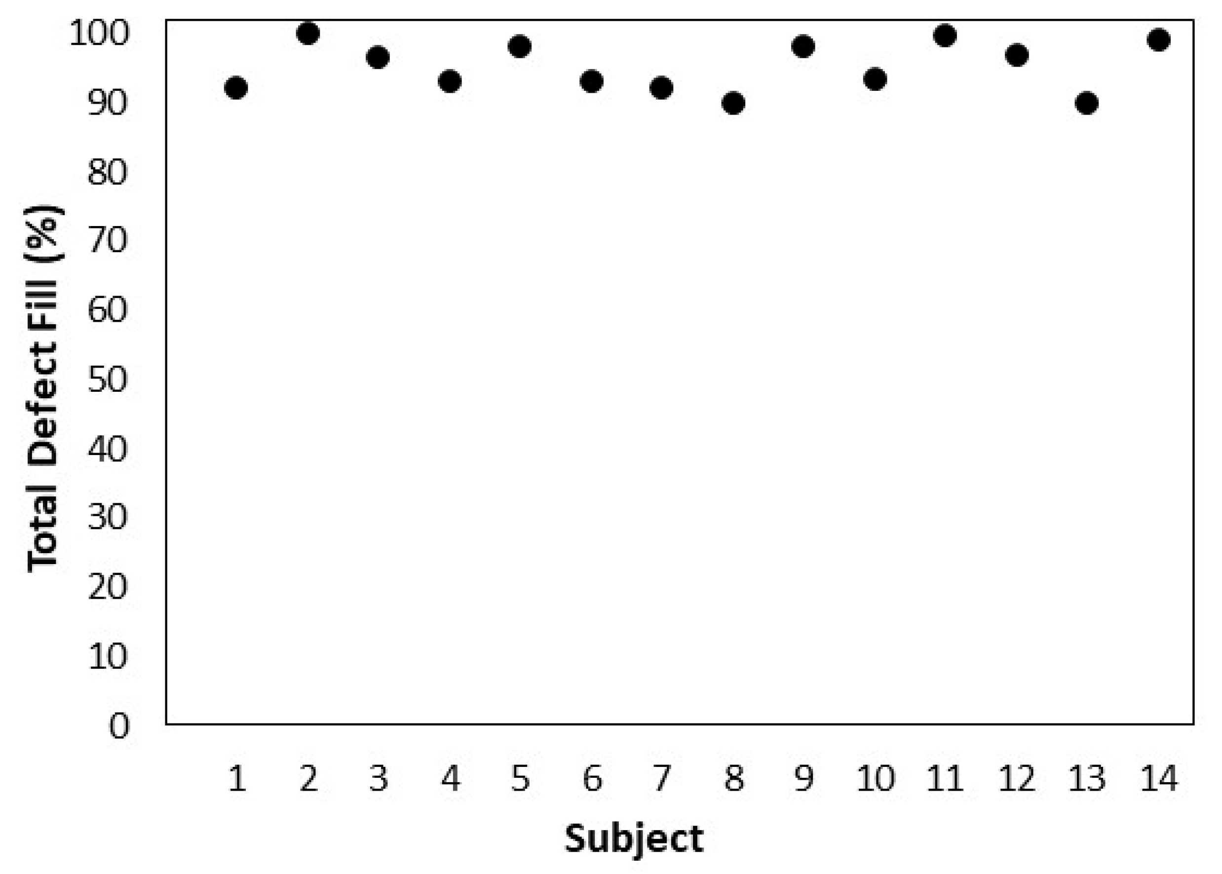
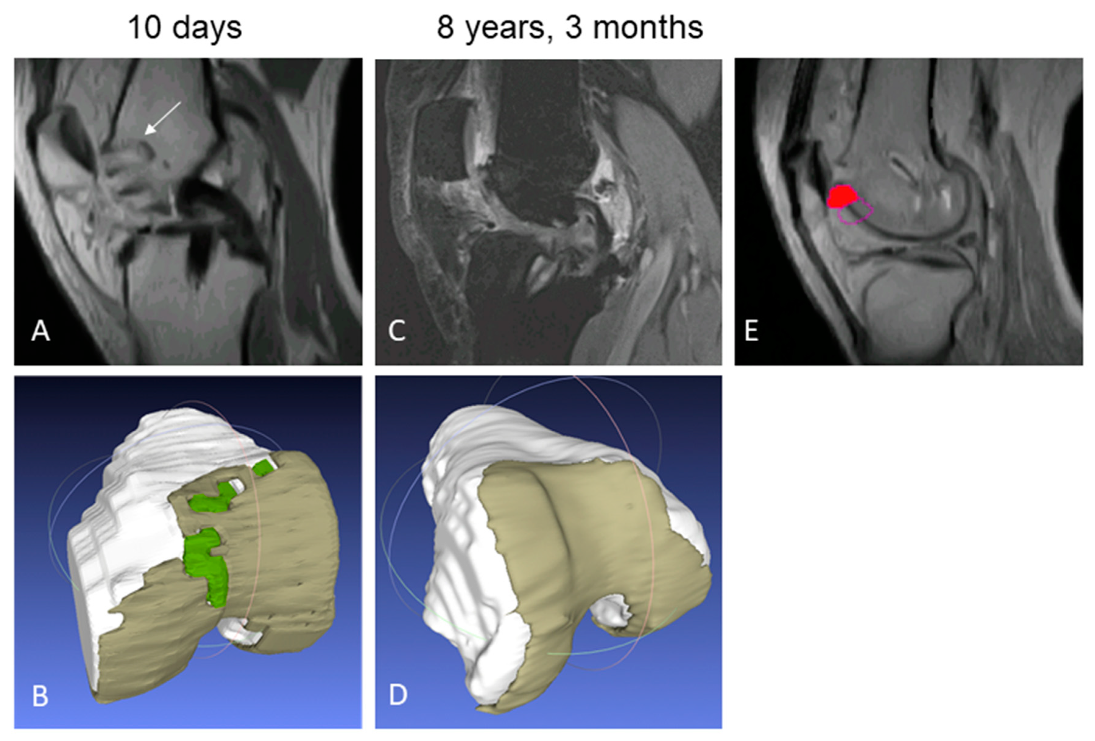
3.2.1. Structural Repair

The quantity and quality of ChondroMimetic repair tissues was determined for 14 subjects (the MRI from the fifteenth subject was unusable due to movement; see Table 2). ChondroMimetic-treated osteochondral defects at 7.9 years post-treatment demonstrated a substantial degree of defect filling of 95.2 ± 3.6%. Figure 1 shows the Total Defect Fill (%) of individual subjects and illustrates a low variability and consistently high degree of defect filling following ChondroMimetic treatment. In addition, subjects treated with ChondroMimetic had a mean T2 relaxation time of 52.5 ± 4.8 ms for the cartilage repair tissue ROI, which was nearly equal to the T2 relaxation time of the ipsilateral native control cartilage regions (52.3 ± 9.2 ms (p = 0.94)), and suggestive of a very similar tissue biochemical make-up and structure. The T2 standard deviations calculated for each repair tissue volume were comparable to native cartilage (17.3 ± 3.3 vs. 13.1 ± 5.3 ms), with a variance that was statistically equal to that of native cartilage (p = 0.11 by F-test).
Using each scan series, full 3D reconstructions were generated of treated knees at the 10-day post-operative baseline, and at follow-up, to assist in visualizing the morphological impact of ChondroMimetic repair. Figure 2 illustrates a representative reconstruction conducted for a 29-year-old female subject who received 5 ChondroMimetic scaffolds in the trochlea, which are clearly visible filling the newly treated osteochondral defects in Figure 2A by 2D MRI, and are similarly visible in the 3D rendering (Figure 2B). At the follow-up after 8 years and 3 months, both the subchondral bone and cartilage aspects of the original defects appear as near-normal in both Figure 2C,D, compared with ipsilateral control regions in Figure 2E.

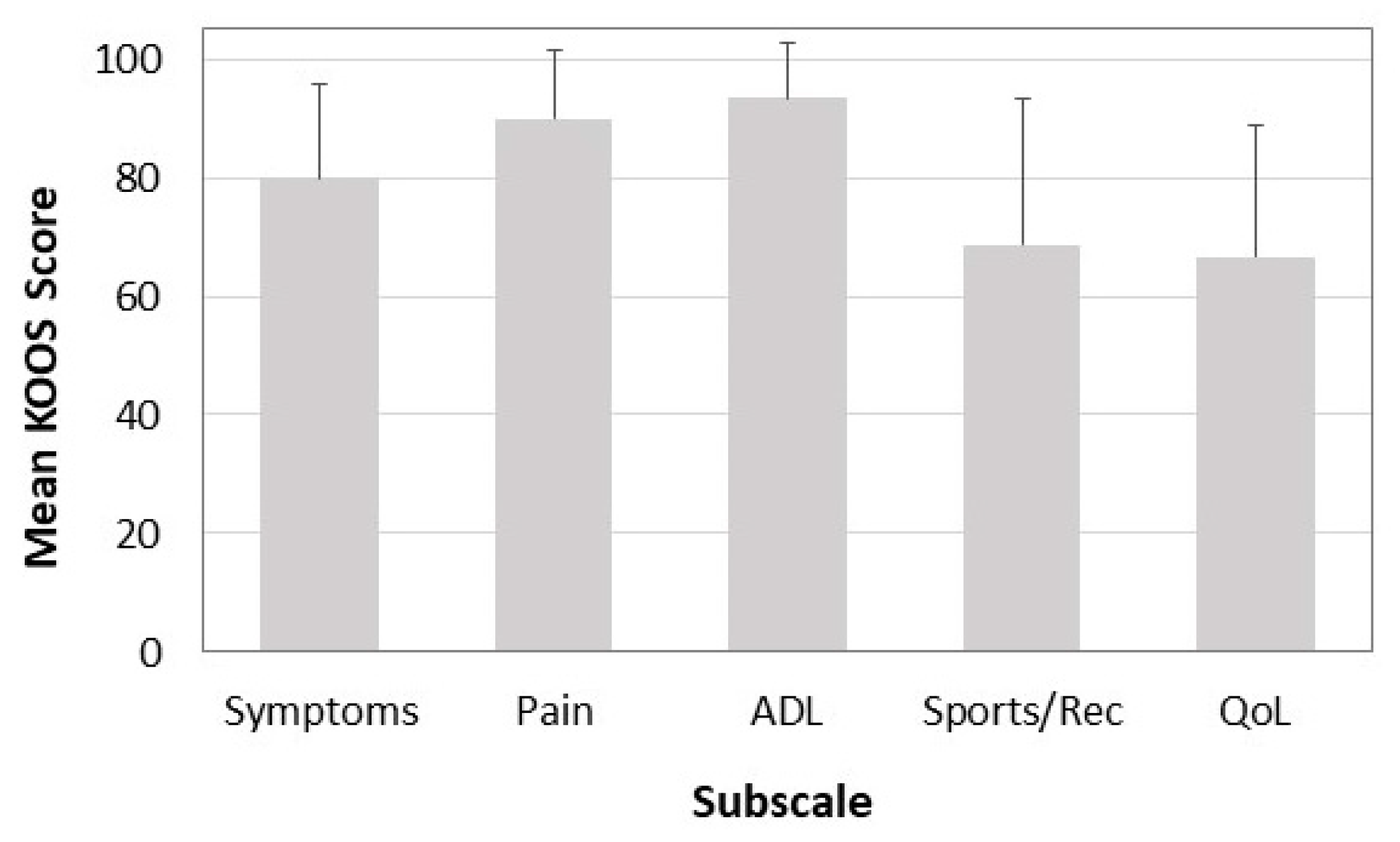
3.2.2. Clinical Benefit

The modified Cincinnati Rating System overall score at the study timepoint was 80.9 ± 15.5, which was statistically significant from baseline (p = 0.0065) and represents a grading of ‘excellent’.
The KOOS subscale data are presented in Figure 3. The mean pain score was 90.0 ± 11.4. The mean symptom score was 80.0 ± 15.8. The mean function in daily living (ADL) score was 93.4 ± 9.1. The mean function in sports and recreational activities (Sports/Rec) score was 68.7 ± 24.7. The knee related quality of life (QOL) mean score was 66.7 ± 22.2.

4. Discussion

The main finding of the current study is that voluminous, high quality repair cartilage results from ChondroMimetic treatment of osteochondral defects after 7.9 years, supporting the hypothesis that ChondroMimetic brings consistent and durable long-term structural repair with an appropriate safety profile. Improvements in clinical outcomes were highly significant over the baseline (p < 0.0065) for the 7.9-year follow-up, which suggests a benefit of ChondroMimetic treatment when used as backfill for mosaicplasty donor sites. The use of validated, three-dimensional quantitative MRI [17,18], which assessed the repair tissue quantity and quality, is a particularly valuable aspect of this study, with a high level of standardization and precision and conducted under Good Clinical Practice guidelines.
The high degree of defect filling by new bone and repair cartilage seen in ChondroMimetic defects at 7.9 years (95.2%) is a critical outcome, as any joint resurfacing should aim to reestablish mechanical homeostasis and anatomical shape with an integrated surface, particularly since chondrocyte-mediated biosynthesis, remodeling, and either tissue repair or degradation over time is dependent on the mechanical loading conditions of cartilage [24]. Normal surface morphology and joint articulation would be expected to improve biomechanical conditions and inhibit the degeneration of defect and peri-defect tissues and slow the progression of secondary osteoarthritis [8,9]. This particular outcome was not fully unexpected, since critical size osteochondral defects in goat knees treated with ChondroMimetic scaffolds yielded similarly striking filling [14]. Furthermore, this level of defect filling is greater than that resulting from another off-the-shelf biological scaffold, BST-CarGel, at 5 years post-treatment, which produced %Fill of 93.79%, as determined by very similar quantitative MRI methods [13].
Quantitative MRI also identified durable, high quality cartilage repair tissue by T2 relaxation time for ChondroMimetic-treated defects. T2 relaxation time is well known to be sensitive to, and highly dependent on, the extracellular cartilage matrix and particularly the collagen network structure, orientation, as well as macromolecular concentration, and tissue hydration [25,26,27,28,29]. When acquired under standardized conditions coupled with three-dimensional segmentation of regions of interest, T2 relaxation time has been an effective, quantitative method for comparing repair tissue to native articular cartilage, and/or to repair tissue resulting from another treatment [13,30,31]. Here, the determination of the quality of repair is evidenced by the closeness of measured T2 values to that found from the ipsilateral normal articular cartilage (52.3 vs. 52.5 ms, p = 0.94), indicating a near-normal level of tissue quality after 7.9 years. The overall level of repair tissue organization achieved is further evidenced by the T2 standard deviations calculated for each repair tissue volume, which were comparable to native cartilage with a variance that was statistically equal to that of native cartilage. In contrast, a tissue with regions of differing organization, or of mixed hyaline-fibrocartilage tissue, would be expected to have a more widespread variance than normal cartilage. When comparing to T2 outcomes resulting from MRI studies with other scaffolds, ChondroMimetic repair tissue demonstrated a similar closeness to native control cartilage as a study with Hyalograft C [32] in the knee, but was improved compared to the MaioRegen scaffold in the ankle [30], and BST-CarGel in the knee [13], neither of which resulted in T2 relaxation times similarly close to control cartilage. This finding also confirms consistency of repair in improved collagen-based organization and content, which are necessary components for long-term durability of cartilage since collagen breakdown is considered to be a critical step in the progression of osteoarthritis [33,34].
It may be considered a limitation of the study that ChondroMimetic was used as back-fill treatment for the majority of osteochondral defects (13/15) in the study, particularly since it is generally believed that mosaicplasty donor sites are: (1) not exposed to weight bearing forces; and (2) ultimately filled with a fibrocartilage through self-repair processes and are asymptomatic [35]. To the contrary, donor sites experience high loads with daily activities, depending on their size and location, from 1.3 times up to 7.8 times body weight [36]. Furthermore, Andrade et al. [37] reviewed 21 original mosaicplasty studies comprising 1726 patients and found that donor site morbidity reportedly occurs in 5.9–16.9% of cases and manifests through crepitation and knee stiffness, instability during activities of daily living, and persistent pain. The fact that in this study no adverse events or symptoms were reported to be related to donor sites or ChondroMimetic implants over the 7.9 year follow-up, and modified Cincinnati and KOOS scores demonstrated a high level of satisfaction, demonstrates that ChondroMimetic treatment was safe and may suggest some clinical benefit, further supported by the MRI-assessed high quality cartilage repair. Furthermore, the regeneration of bone and cartilage to near-native levels of fill and biochemical structure suggest that Chondromimetic may fit into an algorithm for treatment of full-thickness, primary cartilage lesions. Indeed, two full weight bearing primary defects on the medial femoral condyles were treated in this study with ChondroMimetic and demonstrated similarly high degrees of total defect fill (mean: 95.85%) and high quality repair tissue with T2 relaxation times comparable to native cartilage (means: 46.7 ms vs. 47.7 ms, respectively), in addition to satisfactory clinical benefit.
A true study limitation was the lack of an active control group, such as microfracture, which has been suggested by current regulatory guidance [38]. But use of an ipsilateral native cartilage control ROI for quantitative structural outcomes provided an appropriate internal control for making a determination of quality and durability of cartilage repair. Also, the small sample size of the study could limit the interpretation of the results. However, the low variance in both clinical and structural quantitative MRI outcomes nonetheless supports a consistency in repair that would likely be maintained with a larger sample size. Long-term follow-up studies can also present challenges that limit interpretation of study outcomes. For example, here, 14 procedures were conducted in the index knee of 12 subjects over the 8-year follow-up period. However, it can be concluded that it is highly unlikely that these procedures had any effect on the assessments conducted for this study. In 2 cases, the procedures were not knee related (e.g., lung biopsy). The other 12 included: (1) hyaluronic acid injections in 4 subjects performed at least 12 months prior to the extension study visit, and therefore would be expected to have lost any therapeutic effect; (2) microfracture of new primary lesions in 3 subjects, although this would represent a worst case scenario since the procedure could worsen the pain at the time of the extension study visit, or simply decrease the new pain associated with the new lesion back to baseline levels; and (3) second-look arthroscopy in 5 subjects, conducted 1–2 years post-operative by the investigator and unrelated to the study (e.g., to remove bone material from an HTO; to remove malleolar screws from ankles).
The ChondroMimetic scaffold was designed to provide a cell-free cartilage repair alternative, with biological components and architecture, conducive to the re-establishment of the subchondral bone whilst supporting regeneration of articular cartilage. With time and experience, it has been generally recognized that scaffolds are essential in supporting the processes of cartilage repair and indeed, almost all current approaches have incorporated some type of scaffold. So-called “augmented bone marrow stimulation” involves concurrent implantation of an exogenous scaffold (e.g., AMIC [12], BST-CarGel [13], Biocartilage [39]), 3rd generation ACI techniques incorporate membranes to support cell delivery and residency (Hyalograft C [40], MACI [41]), and grafting techniques, by their nature, involve implantation of natural osteochondral scaffolds [42]. The need to have cells, at all, is being questioned when scaffolds alone, with their low cost and simplicity, bring equivalent results in a more cost-effective manner [43].

5. Conclusions

Treatment of osteochondral defects in the knee with the ChondroMimetic scaffold was associated with a consistently high degree of filling with a cartilage-like repair tissue over a long-term (7.9 year) follow-up period, improved clinical scores, and noted lack of donor site morbidity, underscoring potential benefits of a cell-free, single step scaffold from both a clinical and a health economic standpoint.

Author Contributions

Conceptualization, A.B., M.S.S., G.B. and J.H.; Formal analysis, M.S.S. and S.T.; Funding acquisition, G.B. and J.H.; Investigation, A.B., G.V., I.K. and L.H.; Methodology, A.B. and M.S.S.; Project administration, A.B. and G.B.; Supervision, M.S.S. and L.H.; Visualization, M.S.S.; Writing—original draft, M.S.S.; Writing—review & editing, A.B., M.S.S., A.K.L., A.G., S.T., G.B., J.H. and L.H. All authors read and approved the final manuscript.

Funding

Collagen Solutions (study sponsor) was the sole funding source and was responsible for study design, data interpretation, and manuscript publication.

Conflicts of Interest

G.B. and J.H. were employees of the study sponsor at the time of the study. M.S.S., A.K.L. and A.G. receive consultancy fees from the study sponsor. A.K.L. holds equity in the study sponsor. A.G. receives consultancy fees and research support from Smith & Nephew and Ossur. A.B., S.T., G.V., I.K. and L.H. have no potential conflict of interest to declare.

Ethical Approval

Ethics approval was obtained from the National Scientific and Ethical Committee of the Medical Research Council of Hungary (ETT TUKEB) (Reference: 58813-/2016/EKU) and the local Research Ethics Committee of Uzsoki Hospital (Uzsoki IKEB) (Reference: 223-IK/2016).

List of Abbreviations

2Dtwo dimensional
3Dthree dimensional
ACIAutologous chondrocyte implantation
AMICautologous matrix-induced chondrogenesis
CCIcharacterized chondrocyte implantation
FS SPGRfat-suppressed 3D spoiled gradient-echo
GAGglycosaminoglycan
kgkilogram
KOOSKnee Injury and Osteoarthritis Outcome Score
mmeter
mmmillimeter
msmillisecond
MRIMagnetic Resonance Imaging
PRPPlatelet rich plasma
QoLQuality of life
ROIregion-of-interest
T2Transverse relaxation time

References

  1. Steadman, J.R.; Rodkey, W.G.; Briggs, K.K. Microfracture: Its History and Experience of the Developing Surgeon. Cartilage 2010, 1, 78–86. [Google Scholar] [CrossRef] [PubMed] [Green Version]
  2. Peterson, L.; Vasiliadis, H.S.; Brittberg, M.; Lindahl, A. Autologous chondrocyte implantation: A long-term follow-up. Am. J. Sports Med. 2010, 38, 1117–1124. [Google Scholar] [CrossRef] [PubMed]
  3. Bugbee, W.D.; Pallante-Kichura, A.L.; Gortz, S.; Amiel, D.; Sah, R. Osteochondral allograft transplantation in cartilage repair: Graft storage paradigm, translational models, and clinical applications. J. Orthop. Res. Off. Publ. Orthop. Res. Soc. 2016, 34, 31–38. [Google Scholar] [CrossRef] [PubMed] [Green Version]
  4. Hangody, L.; Duska, Z.; Karpati, Z. Autologous Osteochondral Mosaicplasty. Tech. Knee Surg. 2002, 1, 13–22. [Google Scholar] [CrossRef]
  5. Riboh, J.C.; Cvetanovich, G.L.; Cole, B.J.; Yanke, A.B. Comparative efficacy of cartilage repair procedures in the knee: A network meta-analysis. Knee Surg. sports Traumatol. Arthrosc. Off. J. ESSKA 2017, 25, 3786–3799. [Google Scholar] [CrossRef]
  6. Mundi, R.; Bedi, A.; Chow, L.; Crouch, S.; Simunovic, N.; Sibilsky Enselman, E.; Ayeni, O.R. Cartilage Restoration of the Knee: A Systematic Review and Meta-analysis of Level 1 Studies. Am. J. Sports Med. 2016, 44, 1888–1895. [Google Scholar] [CrossRef]
  7. Richter, D.L.; Schenck, R.C., Jr.; Wascher, D.C.; Treme, G. Knee Articular Cartilage Repair and Restoration Techniques: A Review of the Literature. Sports Health 2016, 8, 153–160. [Google Scholar] [CrossRef] [Green Version]
  8. Buckwalter, J.A.; Mankin, H.J. Articular Cartilage Part II: Degeneration and osteoarthrosis, repair, regeneration and transplantation. J. Bone Jt. Surg. 1997, 79, 612–632. [Google Scholar] [CrossRef]
  9. Davies-Tuck, M.L.; Wluka, A.E.; Wang, Y.; Teichtahl, A.J.; Jones, G.; Ding, C.; Cicuttini, F.M. The natural history of cartilage defects in people with knee osteoarthritis. OARS 2008, 16, 337–342. [Google Scholar] [CrossRef] [Green Version]
  10. Harley, B.A.; Lynn, A.K.; Wissner-Gross, Z.; Bonfield, W.; Yannas, I.V.; Gibson, L.J. Design of a multiphase osteochondral scaffold III: Fabrication of layered scaffolds with continuous interfaces. J. Biomed. Mater. Res. 2010, 92, 1078–1093. [Google Scholar] [CrossRef]
  11. Harley, B.A.; Lynn, A.K.; Wissner-Gross, Z.; Bonfield, W.; Yannas, I.V.; Gibson, L.J. Design of a multiphase osteochondral scaffold. II. Fabrication of a mineralized collagen-glycosaminoglycan scaffold. J. Biomed. Mater. Res. 2010, 92, 1066–1077. [Google Scholar] [CrossRef] [PubMed]
  12. Gille, J.; Behrens, P.; Volpi, P.; de Girolamo, L.; Reiss, E.; Zoch, W.; Anders, S. Outcome of Autologous Matrix Induced Chondrogenesis (AMIC) in cartilage knee surgery: Data of the AMIC Registry. Arch. Orthop. Trauma Surg. 2013, 133, 87–93. [Google Scholar] [CrossRef] [PubMed] [Green Version]
  13. Shive, M.S.; Stanish, W.D.; McCormack, R.; Forriol, F.; Mohtadi, N.; Pelet, S.; Desnoyers, J.; Methot, S.; Vehik, K.; Restrepo, A. BST-CarGel(R) Treatment Maintains Cartilage Repair Superiority over Microfracture at 5 Years in a Multicenter Randomized Controlled Trial. Cartilage 2015, 6, 62–72. [Google Scholar] [CrossRef] [PubMed]
  14. Getgood, A.M.; Kew, S.J.; Brooks, R.; Aberman, H.; Simon, T.; Lynn, A.K.; Rushton, N. Evaluation of early-stage osteochondral defect repair using a biphasic scaffold based on a collagen-glycosaminoglycan biopolymer in a caprine model. Knee 2012, 19, 422–430. [Google Scholar] [CrossRef] [PubMed]
  15. Getgood, A.; Henson, F.; Skelton, C.; Herrera, E.; Brooks, R.; Fortier, L.A.; Rushton, N. The Augmentation of a Collagen/Glycosaminoglycan Biphasic Osteochondral Scaffold with Platelet-Rich Plasma and Concentrated Bone Marrow Aspirate for Osteochondral Defect Repair in Sheep: A Pilot Study. Cartilage 2012, 3, 351–363. [Google Scholar] [CrossRef] [PubMed] [Green Version]
  16. Getgood, A.; Henson, F.; Skelton, C.; Brooks, R.; Guehring, H.; Fortier, L.A.; Rushton, N. Osteochondral tissue engineering using a biphasic collagen/GAG scaffold containing rhFGF18 or BMP-7 in an ovine model. J. Exp. Orthop. 2014, 1, 13. [Google Scholar] [CrossRef] [Green Version]
  17. Tamez-Pena, J.G.; Farber, J.; Gonzalez, P.C.; Schreyer, E.; Schneider, E.; Totterman, S. Unsupervised segmentation and quantification of anatomical knee features: Data from the Osteoarthritis Initiative. IEEE Trans. Biomed. Eng. 2012, 59, 1177–1186. [Google Scholar] [CrossRef]
  18. Tamez-Peña, J.; Shive, M.; Restrepo, A.; Gonzalez, P.; Schreyer, E.; Totterman, S. Validation of an Objective, Analyst-Independent, Non-Invasive Method for Assessing Effectiveness of Cartilage Repair Therapies in Multicenter RCTs. In Proceedings of the 6th International Workshop on Osteoarthritis Imaging, Hilton Head, SC, USA, 12–14 July 2012. [Google Scholar]
  19. Stanish, W.D.; McCormack, R.; Forriol, F.; Mohtadi, N.; Pelet, S.; Desnoyers, J.; Restrepo, A.; Shive, M.S. Novel scaffold-based BST-CarGel treatment results in superior cartilage repair compared with microfracture in a randomized controlled trial. J. Bone Jt. Surg. Am. Vol. 2013, 95, 1640–1650. [Google Scholar] [CrossRef] [Green Version]
  20. Gatti, A.A.; Noseworthy, M.D.; Stratford, P.W.; Brenneman, E.C.; Totterman, S.; Tamez-Pena, J.; Maly, M.R. Acute changes in knee cartilage transverse relaxation time after running and bicycling. J. Biomech. 2017, 53, 171–177. [Google Scholar] [CrossRef]
  21. Farber, J.M.; Totterman, S.M.; Martinez-Torteya, A.; Tamez-Pena, J.G. Scan-rescan precision of subchondral bone curvature maps from routine 3D DESS water excitation sequences: Data from the Osteoarthritis Initiative. Comput. Biol. Med. 2016, 69, 83–91. [Google Scholar] [CrossRef]
  22. Bentley, G.; Biant, L.C.; Carrington, R.W.; Akmal, M.; Goldberg, A.; Williams, A.M.; Skinner, J.A.; Pringle, J. A prospective, randomised comparison of autologous chondrocyte implantation versus mosaicplasty for osteochondral defects in the knee. J. Bone Jt. Surg. Br. Vol. 2003, 85, 223–230. [Google Scholar] [CrossRef] [PubMed] [Green Version]
  23. Bekkers, J.E.; de Windt, T.S.; Raijmakers, N.J.; Dhert, W.J.; Saris, D.B. Validation of the Knee Injury and Osteoarthritis Outcome Score (KOOS) for the treatment of focal cartilage lesions. OARS 2009, 17, 1434–1439. [Google Scholar] [CrossRef] [PubMed] [Green Version]
  24. Grodzinsky, A.J.; Levenston, M.E.; Jin, M.; Frank, E.H. Cartilage tissue remodeling in response to mechanical forces. Annu. Rev. Biomed. Eng. 2000, 2, 691–713. [Google Scholar] [CrossRef] [PubMed]
  25. Mosher, T.J.; Dardzinski, B.J. Cartilage MRI T2 relaxation time mapping: Overview and applications. Semin. Musculoskelet. Radiol. 2004, 8, 355–368. [Google Scholar] [CrossRef] [PubMed] [Green Version]
  26. Nieminen, M.T.; Rieppo, J.; Toyras, J.; Hakumaki, J.M.; Silvennoinen, J.; Hyttinen, M.M.; Helminen, H.J.; Jurvelin, J.S. T2 relaxation reveals spatial collagen architecture in articular cartilage: A comparative quantitative MRI and polarized light microscopic study. Magn. Reson. Med. Off. J. Soc. Magn. Reson. Med. 2001, 46, 487–493. [Google Scholar] [CrossRef]
  27. Trattnig, S.; Mamisch, T.C.; Welsch, G.; Glaser, C. Quantitative T2 Mapping of Matrix-Associated Autologous Chondrocyte Transplantation at 3 Tesla: An In Vivo Cross-Sectional Study. Investig. Radiol. 2007, 42, 442–448. [Google Scholar] [CrossRef]
  28. Xia, Y.; Moody, J.B.; Alhadlaq, H. Orientational dependence of T2 relaxation in articular cartilage: A microscopic MRI (microMRI) study. Soc. Magn. Reson. Med. 2002, 48, 460–469. [Google Scholar] [CrossRef]
  29. White, L.M.; Sussman, M.S.; Hurtig, M.; Probyn, L.; Tomlinson, G.; Kandel, R. Cartilage T2 assessment: Differentiation of normal hyaline cartilage and reparative tissue after arthroscopic cartilage repair in equine subjects. Radiology 2006, 241, 407–414. [Google Scholar] [CrossRef]
  30. Kaipel, M.; Schreiner, M.; Kellner, R.; Klikovits, J.; Apprich, S.; Brix, M.; Boszotta, H.; Domayer, S.; Trattnig, S. Beneficial clinical effects but limited tissue quality following osteochondral repair with a cell-free multilayered nano-composite scaffold in the talus. Foot Ankle Surg. 2017, 23, 302–306. [Google Scholar] [CrossRef]
  31. Lansdown, D.A.; Wang, K.; Cotter, E.; Davey, A.; Cole, B.J. Relationship Between Quantitative MRI Biomarkers and Patient-Reported Outcome Measures After Cartilage Repair Surgery: A Systematic Review. Orthop. J. Sports Med. 2018, 6, 2325967118765448. [Google Scholar] [CrossRef] [Green Version]
  32. Welsch, G.H.; Mamisch, T.C.; Domayer, S.E.; Dorotka, R.; Kutscha-Lissberg, F. Cartilage T2 Assessment at 3-T MR Imaging: In Vivo Differentiation of Normal Hyaline Cartilage from Reparative Tissue after Two Cartilage Repair Procedures—Initial Experience1. Radiology 2008, 247, 154–161. [Google Scholar] [CrossRef]
  33. Eyre, D. Collagen of articular cartilage. Arthritis Res. 2002, 4, 30–35. [Google Scholar] [CrossRef] [PubMed] [Green Version]
  34. Wu, J.P.; Kirk, T.B.; Zheng, M.H. Study of the collagen structure in the superficial zone and physiological state of articular cartilage using a 3D confocal imaging technique. J. Orthop. Surg. Res. 2008, 3, 29. [Google Scholar] [CrossRef] [PubMed] [Green Version]
  35. Hangody, L.; Feczko, P.; Bartha, L.; Bodo, G.; Kish, G. Mosaicplasty for the treatment of articular defects of the knee and ankle. Clin. Orthop. Relat. Res. 2001, 391, 328–336. [Google Scholar] [CrossRef] [PubMed]
  36. Flynn, T.W.; Soutas-Little, R.W. Patellofemoral joint compressive forces in forward and backward running. J. Orthop. Sports Phys. Ther. 1995, 21, 277–282. [Google Scholar] [CrossRef] [Green Version]
  37. Andrade, R.; Vasta, S.; Pereira, R.; Pereira, H.; Papalia, R.; Karahan, M.; Oliveira, J.M.; Reis, R.L.; Espregueira-Mendes, J. Knee donor-site morbidity after mosaicplasty—A systematic review. J. Exp. Orthop. 2016, 3, 31. [Google Scholar] [CrossRef] [Green Version]
  38. US Food and Drug Administration. Guidance for Industry: Preparation of IDEs and INDs for Products Intended to Repair or Replace Knee Cartilage; US Food and Drug Administration: Washington, DC, USA, 2011.
  39. Abrams, G.; Mall, N.A.; Fortier, L.; Roller, B.L.; Cole, B.J. BioCartilage: Background and Operative Technique. Oper. Tech. Sports Med. 2013, 21, 116–124. [Google Scholar] [CrossRef] [Green Version]
  40. Brix, M.O.; Stelzeneder, D.; Chiari, C.; Koller, U.; Nehrer, S.; Dorotka, R.; Windhager, R.; Domayer, S.E. Treatment of Full-Thickness Chondral Defects With Hyalograft C in the Knee: Long-term Results. Am. J. Sports Med. 2014, 42, 1426–1432. [Google Scholar] [CrossRef]
  41. Brittberg, M.; Recker, D.; Ilgenfritz, J.; Saris, D.B.F.; Group, S.E.S. Matrix-Applied Characterized Autologous Cultured Chondrocytes Versus Microfracture: Five-Year Follow-up of a Prospective Randomized Trial. Am. J. Sports Med. 2018, 46, 1343–1351. [Google Scholar] [CrossRef]
  42. Frenkel, S.R.; Di Cesare, P.E. Scaffolds for articular cartilage repair. Ann. Biomed. Eng. 2004, 32, 26–34. [Google Scholar] [CrossRef]
  43. Kon, E.; Roffi, A.; Filardo, G.; Tesei, G.; Marcacci, M. Scaffold-based cartilage treatments: With or without cells? A systematic review of preclinical and clinical evidence. Arthrosc. J. Arthrosc. Relat. Surg. 2015, 31, 767–775. [Google Scholar] [CrossRef] [PubMed]
Figure 1. Total Defect Fill (%) by quantitative MRI for individual subjects at a mean followup of 7.9 years after ChondroMimetic treatment. Total Defect Fill (%) was determined using 3D quantitative magnetic resonance imaging by calculating the ratio of the new repair tissue (bone and cartilage) volume at followup, to original defect (bone and cartilage) volume acquired from 10 days post-operative scans (baseline). (n = 14).
Applsci 10 05642 g001
Figure 2. Longitudinal MRI reconstruction of a ChondroMimetic-treated subject knee at 10 days (A,B) and 8 years, 3 months (C,D). (A) By 2D MRI, 5 ChondroMimetic implants can be observed with hyper-intense peri-implant boundaries, and lack of cartilage-like signal (white arrow). (B) 3D rendering of knee yields ChondroMimetic implants (green) surrounded by cartilage (brown) by automated software based on MRI signal. (C) At the follow-up after 8 years and 3 months, treated bone defects are filled with bony repair tissue and the treated cartilage defect is filled with cartilaginous repair tissue. (D) Automated rendering demonstrates cartilaginous tissue in repaired defects congruous with surrounding cartilage. (E) The trochlear control region of interest (filled red circle) of native cartilage used for T2 relaxation time comparison. The open purple circle shows an unused, alternate control region of interest.
Applsci 10 05642 g002
Figure 3. Knee Injury and Osteoarthritis Outcome Scores (KOOS) for ChondroMimetic subjects at a mean follow-up of 7.9 years. Data represents mean + standard deviation (n = 15).
Applsci 10 05642 g003
Table 1. Follow-up Characteristics of Trial Subjects .
CharacteristicChondromimetic (n = 15)
Consented subjects from original study, n (%)15 (88)
Follow-up, years7.9 ± 0.3
Age, years32.7 ± 9.3
Gender, n (%)
Male7 (46.7)
Female8 (53.3)
Body Mass Index, kg/m225.3 ± 4.2
Smokers, n (%)2 (13.3)
Index defects and implants
  Mosaicplasty donor site(s), n (%)13 (86.7)
  Medial femoral condyle defect, n (%)2 (13.3)
  Defect volume including missing bone, mm3 ¶1573 ± 1233
ChondroMimetic Implants/knee, median (range)2 (1–5)
Follow-up Pain, n (%) *
  No knee pain11 (73.3)
  Mild knee pain2 (13.3)
  Moderate knee pain2 (13.3)
Follow-up Activity Level, n (%) *
  Unlimited8 (53)
  Slightly limited 4 (27)
  Moderately limited 3 (20)
Additional interventions since treatment
  Index knee related procedures ^12
  Non-knee related procedures2
Plus-minus values are means ± standard deviation unless otherwise indicated. Determined using quantitative 3D magnetic resonance imaging (MRI) of 10 days postoperative scans. ^ Hyaluronic acid injection (4); microfracture of new primary lesion (3); second-look arthroscopy (5). * From physical examination and standardized interview during the 8-year study visit.
Table 2. Structural repair outcomes by 3 dimensional MRI at 7.9 years follow-up (n = 14).
VariableOutcome
Total defect fill (%)95.2 ± 3.6 (89.8, 99.9)
T2 relaxation time (ms)
Cartilage repair tissue52.5 ± 4.8 (44.4, 58.5) +
Ipsilateral native cartilage52.3 ± 9.2 (39.9, 78.5)
Values are presented as means ± standard deviation (min, max). T2 = transverse relaxation time; MRI = magnetic resonance imaging. + p = 0.94 by Student’s t-test compared to native cartilage.

