Article

Phase Transformation, Optical and Emission Performance of Zinc Silicate Glass-Ceramics Phosphor Derived from the ZnO–B2O3–SLS Glass System

by
Muhammad Faris Syazwan Mohd Shofri
1,
Mohd Hafiz Mohd Zaid
1,2,*,
Khamirul Amin Matori
1,2,
Yap Wing Fen
1,
Yazid Yaakob
1,
Suhail Huzaifa Jaafar
1,
Siti Aisyah Abdul Wahab
2 and
Yuji Iwamoto
3
1
Department of Physics, Faculty of Science, Universiti Putra Malaysia, Serdang 43400 UPM, Selangor, Malaysia
2
Materials Synthesis and Characterization Laboratory, Institute of Advanced Technology, Universiti Putra Malaysia, Serdang 43400 UPM, Selangor, Malaysia
3
Department of Life Science and Applied Chemistry, Nagoya Institute of Technology, Gokisocho, Showaku, Nagoya 466-8555, Japan
*
Author to whom correspondence should be addressed.
Appl. Sci. 2020, 10(14), 4940; https://doi.org/10.3390/app10144940
Submission received: 19 May 2020 / Revised: 13 June 2020 / Accepted: 16 June 2020 / Published: 18 July 2020
(This article belongs to the Section Materials Science and Engineering)

Abstract

:
A new transparent zinc silicate glass-ceramic was derived from the 55ZnO–5B2O3–40SLS glass system via a controlled heat-treatment method. The precursor glass sample was placed through the heat-treatment process at different temperatures to study the progress in phase transformation, optical performance and emission intensity of the zinc silicate glass-ceramics. For this project, material characterization was measured through several tests using densimeter and linear shrinkage measurement, X-ray diffraction (XRD), Fourier transform infrared reflection (FTIR), ultraviolet–visible (UV–Vis) and photoluminescence (PL) spectroscopy. The density and linear shrinkage measurements show that the density of the particular glass-ceramic samples increases with the progression of heating temperature. The XRD analysis displays the result in which the zinc silicate crystal starts to grow after the sample was treated at 700 °C. In addition, the FTIR spectra indicated that the crystallization of the zinc silicate phase occurred with the appearance of SiO4, ZnO4 and Si-O-Zn bands. UV–visible exhibited the small changes when the value for the optical band gap decreased from 3.867 to 3.423 eV, influenced by the temperature applied to the sample. Furthermore, the PL spectroscopy showed an enhancement of broad green emission at 534 nm upon the increased heat-treatment temperature. Thus, it can be concluded there is the progression of crystal growth as the heat-treatment temperature increased; three emission peaks appeared at 529, 570 and 682 nm for the green, yellow and red emissions, respectively.

1. Introduction

Recently, transition metal-based silicate glass and glass-ceramics have attracted significant interest in optoelectronic applications, such as phosphor host for light-emitting diodes, cathode ray tubes, plasma display panels and other electronic devices [1]. Silicate glasses with high ZnO content are of interest for various applications from technical glasses and glass ceramics to high-performance optical glasses. Zinc silicate (Zn2SiO4), also known as willemite, is a silicate-based mineral with a complex nesosilicate phenakite structure. In this phenakite structural arrangement, all of the atoms (Zn, Si and O) are generally in the overall position in the zinc silicate structure. This crystal structure is composed of a complex tetrahedral framework. This type of rigid lattice structure (noncentrosymmetric cationic sites) causes zinc silicate to have excellent optical properties [2]. Therefore, it is very important and commonly used in lighting devices and optical and optoelectronic applications, such as a phosphor [3].
Several methods are used to synthesize zinc silicate phosphor, including hydrothermal [4], polymer sonochemical [5], sol–gel [6], spray pyrolysis [7], and simple thermal treatment [8]. However, a long preparation period and complicated synthesis steps make these methods expensive. Several traditional methods, for example, the solid-state sintering method, have many positive aspects, such as fabrication and synthesization in a short time for laboratory preparation and large-scale production [9]. In addition, the use of silica (SiO2) sources in the chemical process is very expensive and disadvantageous; this chemical process is time consuming, with complex experimental methods and small production quantities [10]. Therefore, another alternative source such as glass waste as recycled raw material can be used. As an example, soda–lime–silica (SLS) glass is a conventional glass that is used daily. In Malaysia, widespread disposal of glass caused this material to become a natural pollutant. To address the problem, many researchers use this waste glass as a source of silica. This waste-to-wealth process can minimize manufacturing costs because it is free. Besides that, the addition of SLS glass waste also can lower the melting point, improve the clarity and enhance thermal stability, solubility and chemical stability of the final product [11,12,13]. Boron oxide (B2O3) is one of the most interesting oxides to study because it has a significant effect as a glass former or a modifier. Previous work proved that the addition of a small amount of B2O3 could act as a nucleation agent by supplying crystallization positions, thus resulting in structural changes and glass stability disorders, which are caused by the ion substitution in the glass system [14,15,16]. Thus, B2O3 is introduced into the glass system to lower the melting temperature. This is due to the uniqueness of B2O3 as a flux, whereby it weakens the bonds between the [BO]n basis units and reacts as network former, which can give a high chemical resistance for the glasses, reduce the viscosity, change the thermal expansion coefficient, lower the melting point and increase the heat resistance and breakage index [17].
Based on the facts and findings of the previous studies mentioned above, the main objective of this work is to study the effect of heat-treatment temperature on the phase transformation and optical and emission performance of new zinc silicate glass-ceramics derived from the ZnO–B2O3–SLS glass system. Another aim of this project is to evaluate the optimum preparation of the zinc silicate glass-ceramics as a robust phosphorus host in light-emitting diode applications.

2. Materials and Methods

ZnO, B2O3 and SLS are the main source of raw material for this preparation. The SLS glass waste was crushed to produce glass powder and ground into 45 µm powder. Then, the SLS glass powder was mixed with ZnO and B2O3 powder by following the composition mixture of 55ZnO–5B2O3–40SLS in weight percent. A mixed sample of 30 g was poured into a high purity alumina crucible (controltherm Sdn. Bdh., Malaysia) with dimensions 45 mm top diameter × 50 mm height, and then heated through the melting process at 1350 °C for 3 h. The melted sample was then poured immediately into a container filled with water in order to produce glass frits. The glass frits sample obtained after the fast quenching process is highly transparent with no bubbles. The crushing, grinding and sieving processes were repeated on the glass frits to form a fine glass sample powder with a particle size of less than 45 µm for the pelleting process. The powder was mixed with polyvinyl alcohol (PVA) as a binder and pressed into a specific shape using a hydraulic press machine to form 13 mm diameter glass pellets. The solid pellets were then subjected to the heat-treatment process at 600, 700, 800 and 900 °C for 2 h with a 10 °C/min constant heating rate. Then, the sample was characterized by using densimeter and vernier caliper measurements, and X-ray diffraction (XRD) (PW 3040/60 model, Philips, San Jose, CA, USA), Fourier transform infrared reflection (FTIR) (Spectrum 100 model, Perkin Elmer; Waltham, Massachusetts, USA), ultraviolet–visible (UV–Vis) (UV-3600 model, Shimadzu, Kyoto, JAPAN) and photoluminescence (PL) (LS 55 model; Perkin Elmer; Waltham, Massachusetts, USA) spectroscopic techniques.
In the electronic densimeter, the conventional Archimedes’ principle was applied to measure the density of the pallet sample. In this measurement, 50 mL of plain distilled water was utilized as the buoyancy liquid. In addition, the XRD measurement (PANalytical X’pert PRO, PW3040 model) in the range of 20° to 80° of 2θ was used to confirm the amorphous phase of the precursor glass and identify the crystal growth in the glass-ceramic sample. The FTIR absorption spectra of glass and glass-ceramic sample were measured in the range 400–1400 cm−1 using a Perkin Elmer Spectrum, 100 series model spectrometer, equipped with universal attenuated total reflectance (ATR). The UV-3600 UV–Vis–NIR spectrophotometer was exploited to identify the optical absorption and reflectance. These data were used to estimate the optical band gap of the sample in the wavelength range 400–800 nm using Kubelka–Munk (K–M) theory. Lastly, the photoluminescence excitation and emission spectra of the glass and glass-ceramic sample were measured using a Perkin Elmer, LS 55 model fluorescence spectrometer in the range of 450 to 600 nm.

3. Results

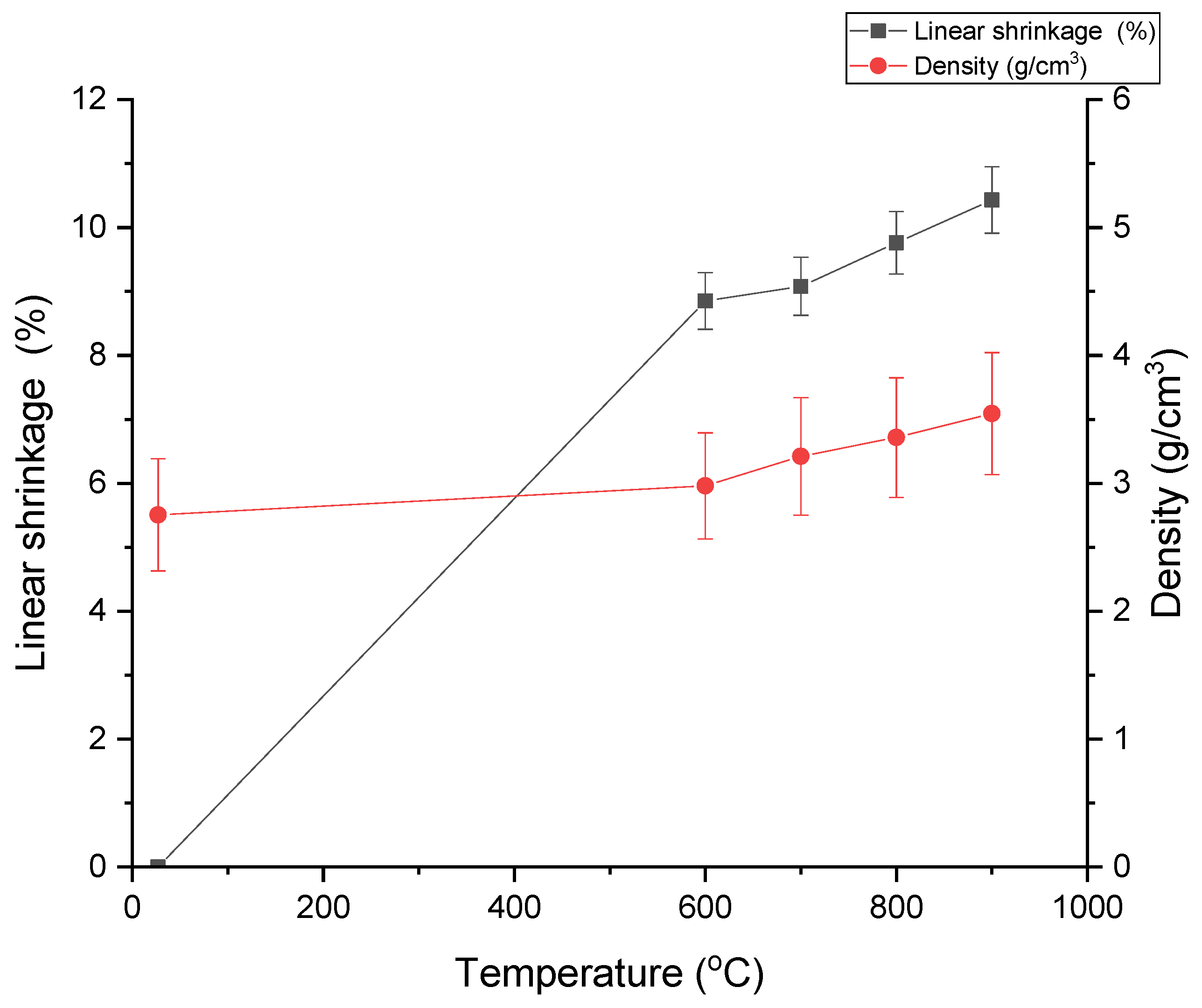
3.1. Density and Linear Shrinkage Analysis

Physical properties of a material are one of the most important features in the field of glass and ceramic research. Table 1 displays the variation of density evaluation and linear shrinkage measurement for precursor glass sample 55ZnO–5B2O3–40SLS, which was heat-treated at temperatures of 600, 700, 800 and 900 °C for 2 h. The successful transparent ZnO–B2O3–SLS precursor glass frit sample appeared light yellowish due to the high concentration of ZnO in the glass. The electronic densimeter measurement found that the density of the ZnO–B2O3–SLS glass sample was 2.754 g/cm3. This high-density value is attributed to ZnO molecules having a very high relative weight compared to other oxides contained in the glass sample. The increase in the heat-treatment temperature caused the sample density to increase. The density of the materials depends on the densities of its individual constituents. However, in the case of glass ceramics, many other factors also marginally influence the density of the final product. Some of these factors include the thermal history of the sample, measurement temperature, crystallization, creation of non-bridging oxygen’s (NBOs) and change in field strength of the modifier.
As shown in Figure 1, the density of the sample increased linearly from 2.980 to 3.545 g/cm3 with progression of heating temperature. This increase was due to the structural change and compactness of the sample structure after the heat-treatment process at higher temperatures [18]. The increasing density of the glass-ceramic samples was also caused by the decrease in the total fractional porosity of the sample. In general, density increases while the porosity of the material decreases monotonically with the depth. This is expected, because differential pressures usually increase with depth. As the heat-treatment temperature increases, the grains of the sample may shift to reach a more dense packing. At that moment, additional force will be imposed on the surface of the grain contacts. Figure 1 shows the effect of increasing the heat-treatment temperature on the linear shrinkage of the sample. According to the diagram, the shrinkage increased linearly with the rise in heating temperature. The linear shrinkage increased from 8.85 to 10.43% with the progression of heating temperature. This increasing trend is due to the compactness of the glass-ceramic structure where the porosity in the precursor glass structure disappeared. The densification of the glass-ceramic structure also indirectly caused the density of the ceramic glass sample to increase.

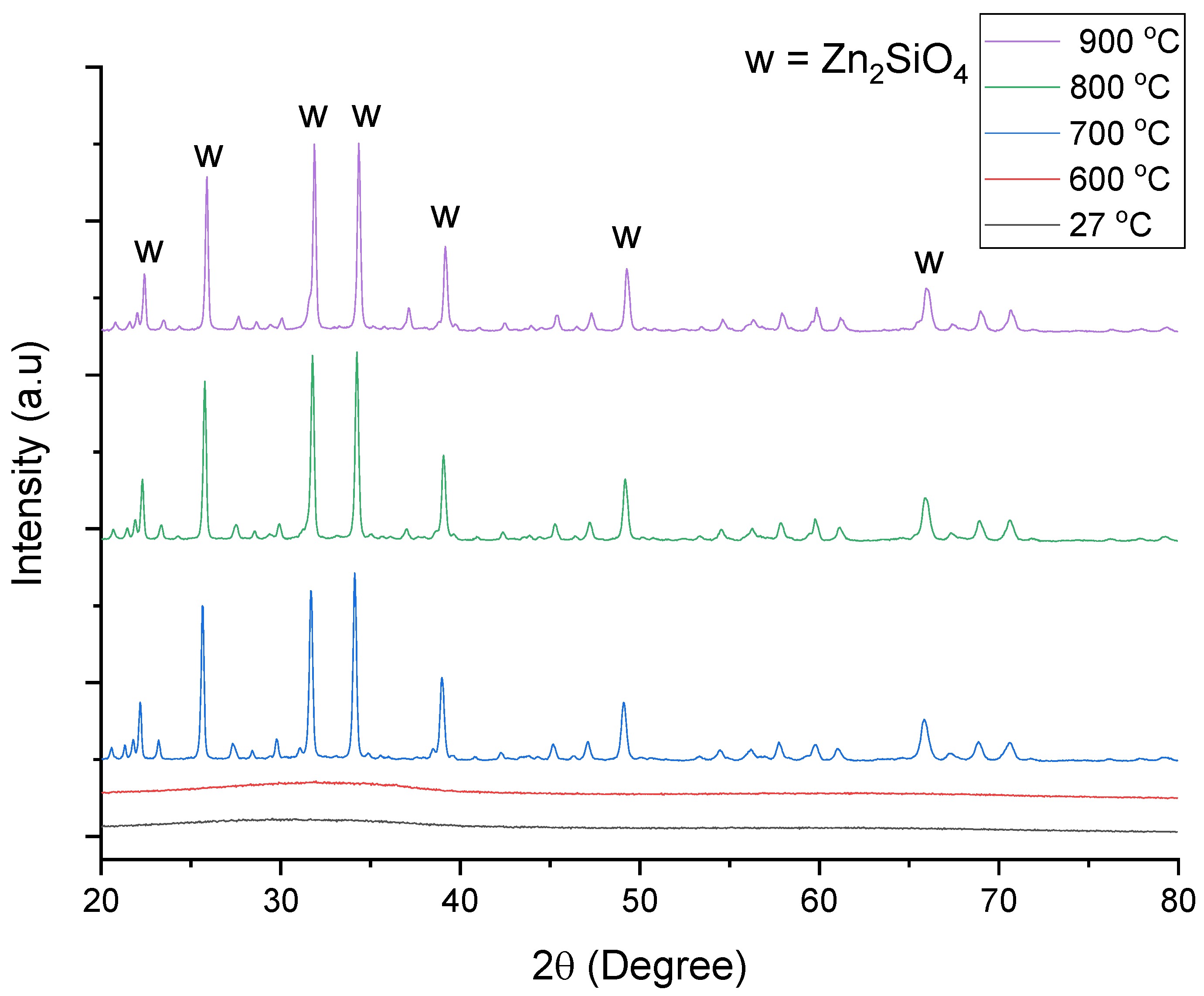
3.2. X-ray Diffraction (XRD) Analysis

Figure 2 presents the XRD structure of the precursor glass sample. Referring to Figure 2, the XRD measurement at 27 °C showed that the atomic structure in the glass sample was amorphous in nature, where the arrangement of atoms in the precursor glass sample was unstructured and randomly organized. However, the amorphous structure of the precursor glass changed when it was subjected to the heat-treatment process. Figure 2 displays the XRD pattern of the glass and glass-ceramic sample against heat-treatment temperature. When the glass sample was treated at 600 °C, the arrangement of atoms in the glass structure remained unorganized. This is evidenced by the presence of the amorphous phase through the XRD test. At the earlier stage of the heat-treatment process, the energy supplied to the glass sample was still insufficient to alter the arrangement of the atoms and the structure of the sample [19]. However, the structure change started to take place when the heat-treatment temperature was increased to 700 °C. Figure 2 shows that crystallization started to occur at the higher temperature of 700 °C. At 700 °C, several diffraction peaks started to appear, indicating the formation of zinc silicate crystals. Diffraction indicated the crystalline phase existed inside the glass structure [20]. There were seven major zinc silicate peaks formed at 2θ = 22.75°, 25.62°, 31.63°, 34.72°, 38.92°, 49.07° and 65.87°, which were proven by standard diffraction pattern of zinc silicate and α-Zn2SiO4 indicated by ICDD cards No. 37-1485 [21]. In terms of the different heat-treatment temperature applied, the increased of heat-treatment temperature caused higher diffusion of ions inside the sample, thus increasing the atomic mobility, which then accelerated the rate of crystal growth [22]. Furthermore, the full width at half maximum (FWHM) of the XRD decreased as the heat-treatment temperature increased. This indicates that the higher the heat-treatment temperature, the better the crystallization phase formed in the glass matrix [23]. The increase of NBOs promotes the replacement of oxide that leads to a greater formation of crystal, thus a higher composition of ZnO leads to a better crystallization of the zinc silicate phase [24].

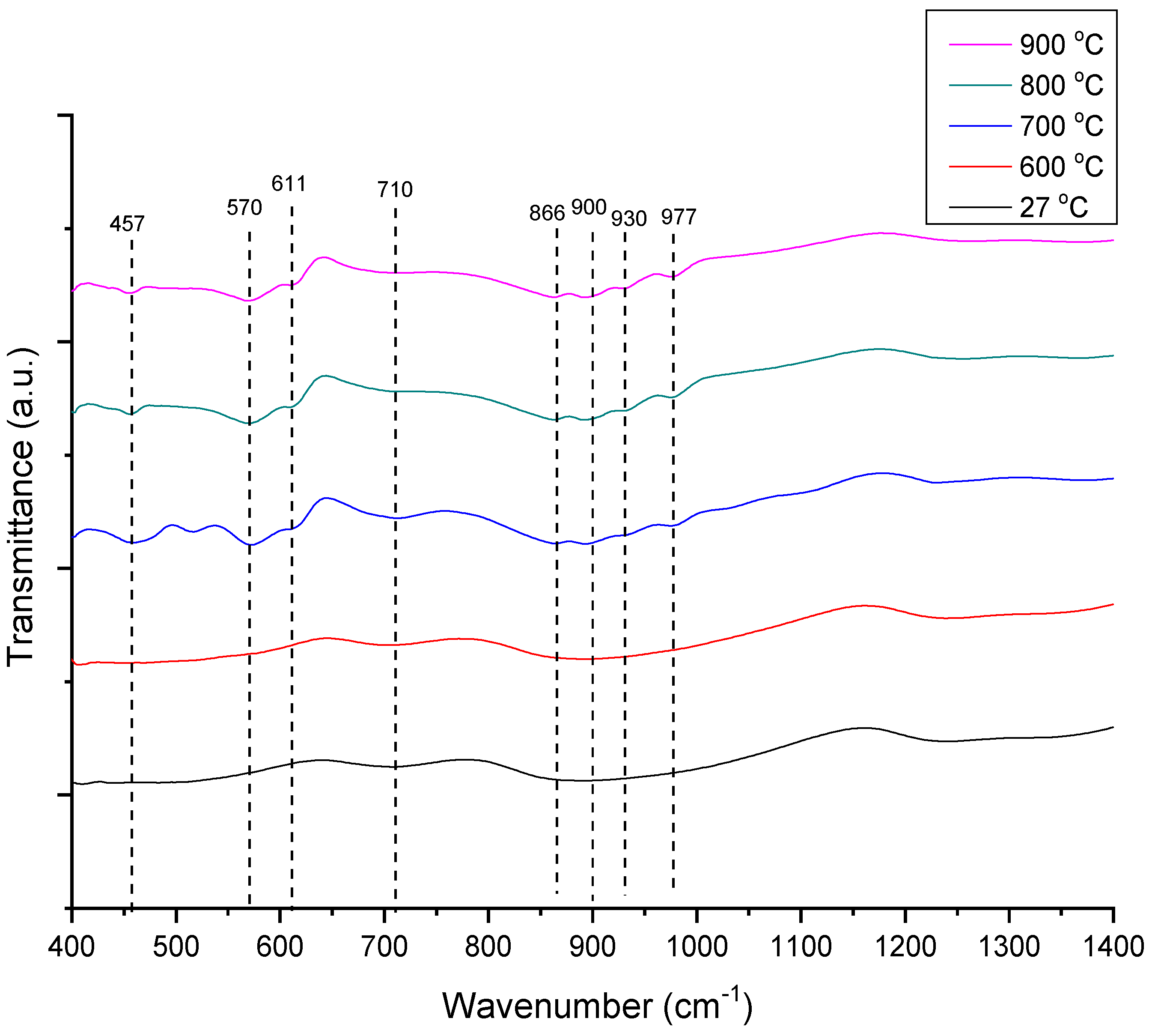
3.3. Fourier Transform Infrared Radiation (FTIR) Analysis

The FTIR measurement of glass and glass-ceramic samples was carried out for the sample precursor glass sample in the frequency of 400–1400 cm−1 to investigate the vibration modes of the sample, as shown in Figure 3. The information on functional groups of glass and glass-ceramic samples can be studied using infrared spectra measurement. The related vitreous and crystalline compounds of previous research results were compared to the infrared spectra obtained [25,26,27,28]. Figure 3 shows the small changes that occurred in bonding between atoms in the sample when heated to 600 °C. Based on the graph, it was found that the Si-O band shifted to a lower frequency, when compared to room temperature. These small changes prove that there were structural modifications to the different structural units in the sample. After the heating temperature increased, the IR spectrum of the glass-ceramics started to appear related to the IR spectra of the crystallized zinc silicate phase, which is at 457, 570, 611 and 710 cm−1 and associated as asymmetric deformation of SiO4, symmetric stretching of ZnO4, asymmetric stretching of ZnO4, and Si-O bond vibration, respectively. Additional IR spectra at 866 cm−1 are attributed to symmetric stretching of SiO4, and 900, 930 and 977 cm−1 as asymmetric stretching of SiO4 as shown in Table 2. Overall, the presence of SiO4 and ZnO4 related vibrations in the IR spectrum indicates the creation of the Zn2SiO4 linkages inside the samples [28,29]. Increased heating temperature may result in increased FTIR band intensities. Based on the analysis of Kullberg et al., IR bands corresponding to asymmetric stretching vibration Zn-O of ZnO4 units are found at 500–550 cm−1 and 650–730 cm−1, while for symmetric stretching vibration of Si-O-B bending, there was asymmetric stretching vibration of Si-O-B that overlapped with the B-O stretching modes of the BO4 tetrahedral at 900–950 cm−1 [30].

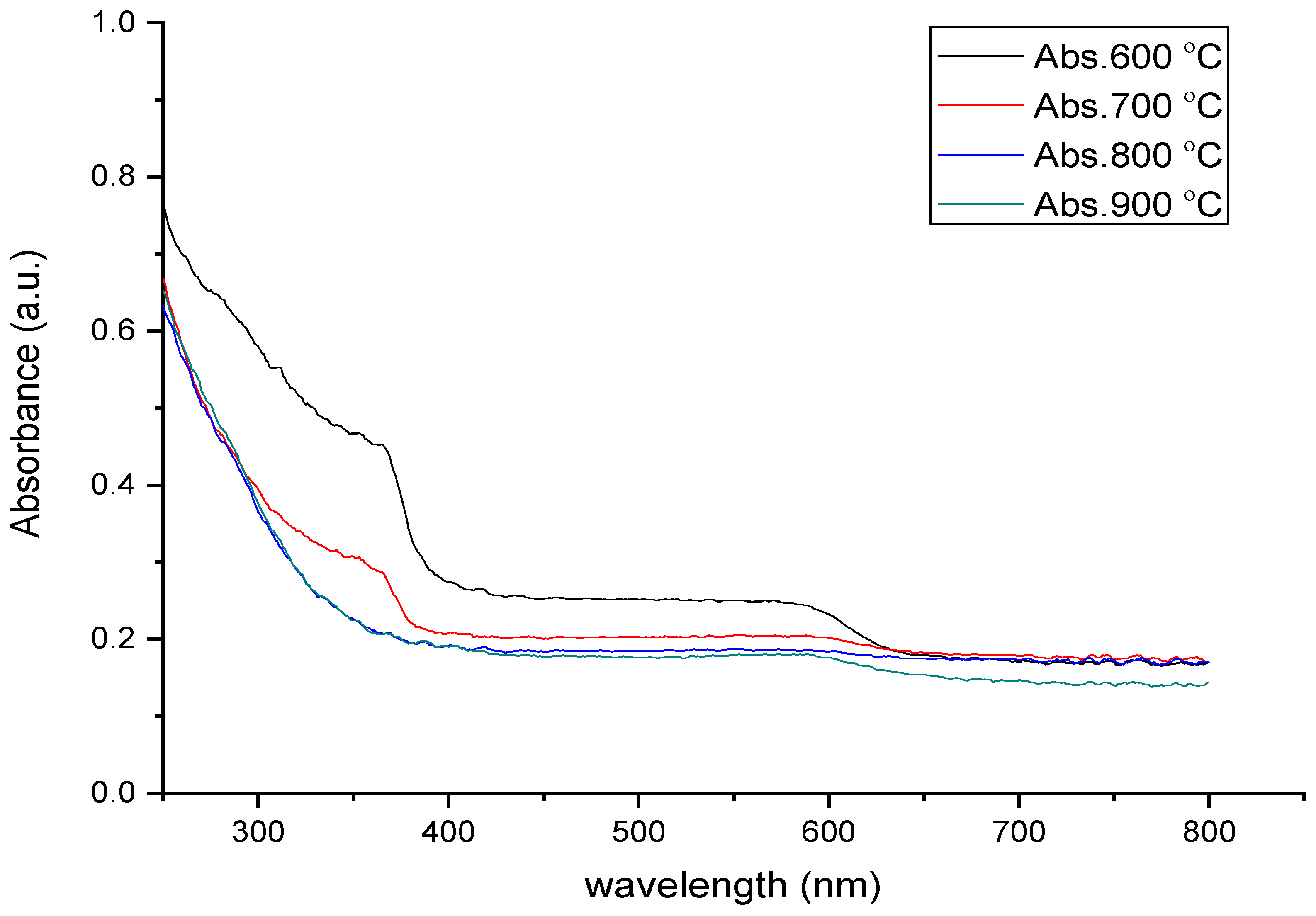
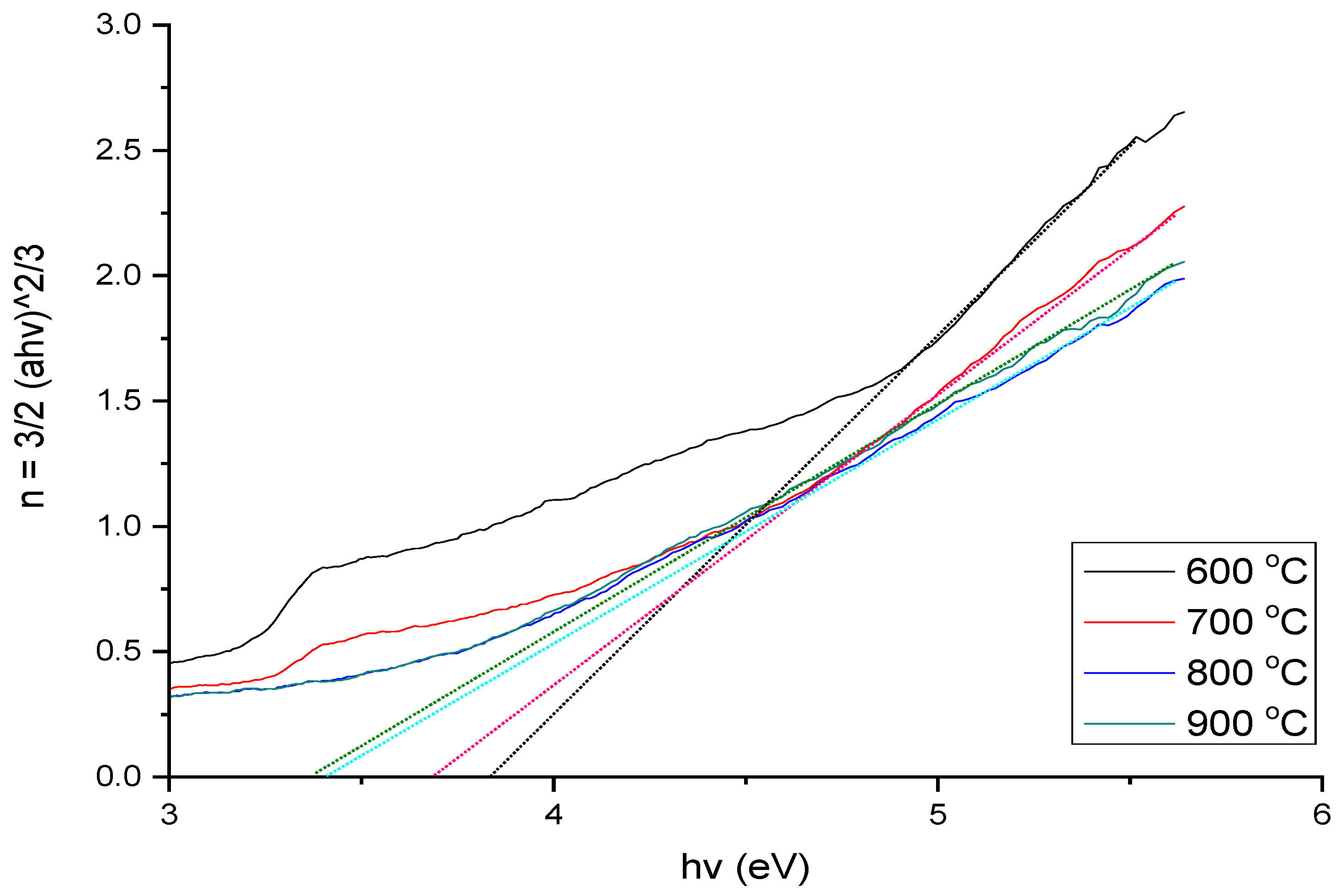
3.4. UV–Vis Analysis

Ultraviolet range studies are very important, as they provide scientific information to understand a specific material’s electronic band gap. In the higher wavelength region, optical absorbance is small, a comparison to the lower wavelength region will produce higher absorbance intensity due to ultraviolet “cut off” that emerges as a term to identify the factor for progress in the absorption energy [31,32]. From previous studies, it is found that a clear intensive absorption generated within the range 250–400 nm is mostly from its absorption curve in the glass and glass-ceramic systems [33]. Figure 4 displays the optical absorption UV–Vis spectra of the sample at different heat-treatment temperatures. Rapid rises in the absorbance toward lower wavelengths are present in the optical absorption spectrum of glass material. This rapid rise of absorption coefficient can be referred to as the fundamental absorption edge known as UV cut off. It was clear that there was no sharp absorption edge after the glass sample was heat-treated at 600 °C, which corresponds to the characteristic of the glassy state observed [34]. There was also a sharp absorption edge at 700 °C, and it disappeared after the sample was heated to a higher temperature (800 °C). This indicates that as the formation of zinc silicate crystal occurred, a few ZnO particles were left in the sample at 700 °C, but at 800 and 900 °C, all ZnO was used for the formation of zinc silicate crystal. This is consistent with the previous research [35,36]. Moreover, the heat-treated sample intensities decreased from 600 to 900 °C because of the fall of the hexagonal ZnO structure and the formation of a zinc silicate structure [37]. Meanwhile, as heat-treatment temperature increased, the absorption edge shifted back to a shorter wavelength. This can be due to the high crystallinity induced by the phase formation of zinc silicate crystal. To obtain the value of the energy band gap, the graph of (αhv)1/n vs. hv was plotted and the slope intercept of the line obtained is equal to the optical transition of band gap energy. This n can have various values such as 1/2, 1/3, 2 and 3; these values correspond to direct allowed, direct forbidden transitions, indirect allowed, and indirect forbidden transitions, respectively. The study of Zaid et al. (2016) observed that the optical band gap decreases due to the direct forbidden transitions [38]. Thus, in this experiment n = 2/3 (direct forbidden transition) was used to find the value of the band gap energy of the sample. Figure 5 and Table 3 show the optical band gap (Eopt) values for glass and glass-ceramic samples heat treated at various temperatures. It is found that with the rise in temperature of heat treatment, the value of Eopt of the glass-ceramic sample decreased from 3.867 to 3.423 units. This decline in the optical band gap resulted from shorter wavelength light having been scattered by the zinc silicate crystal as the crystallinity increased at higher heat-treatment temperature [39]. Furthermore, the progress in NBOs formation led to the defects in the bands, which is another potential reason for the decline in the optical band gap [40].

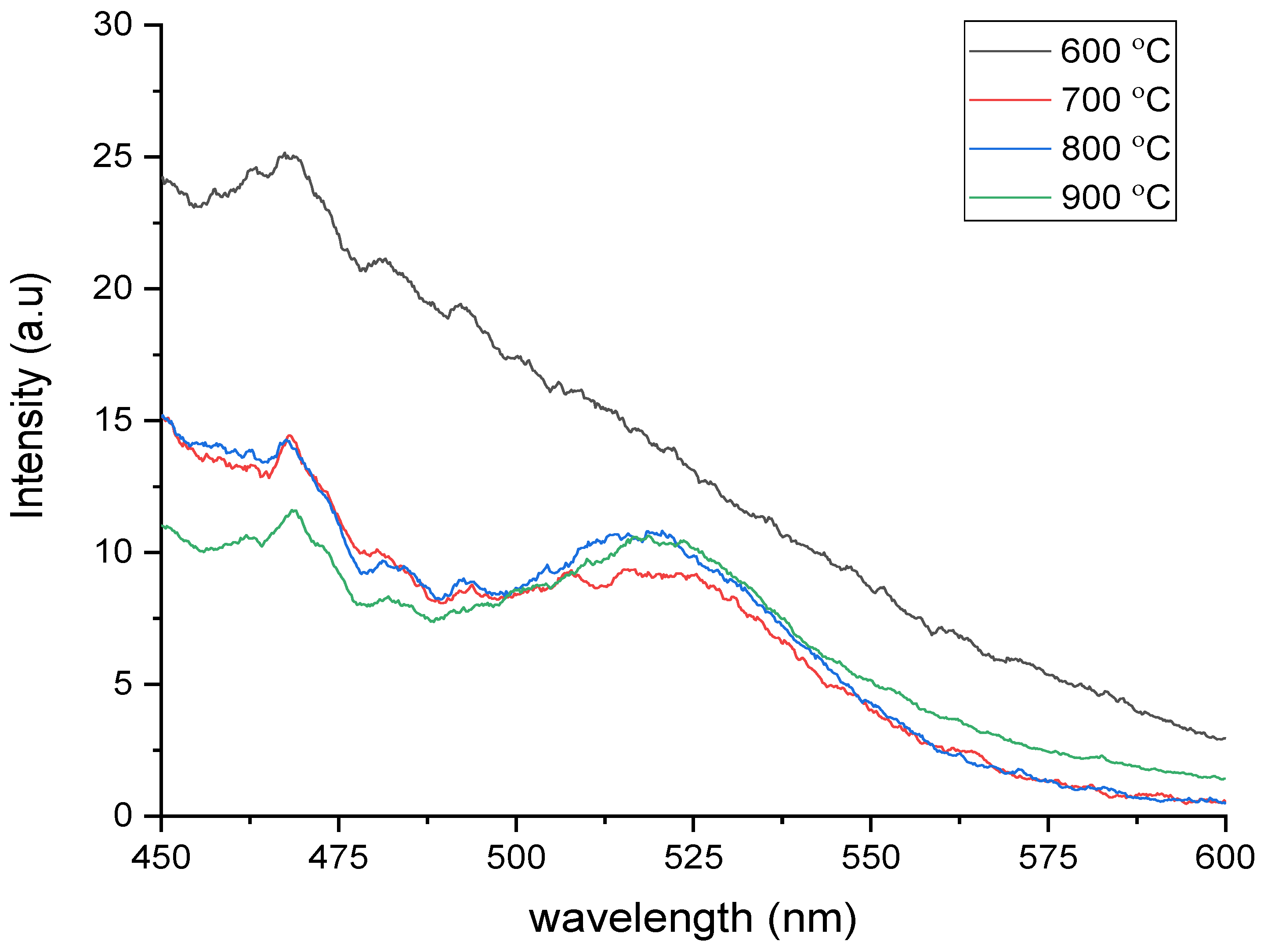
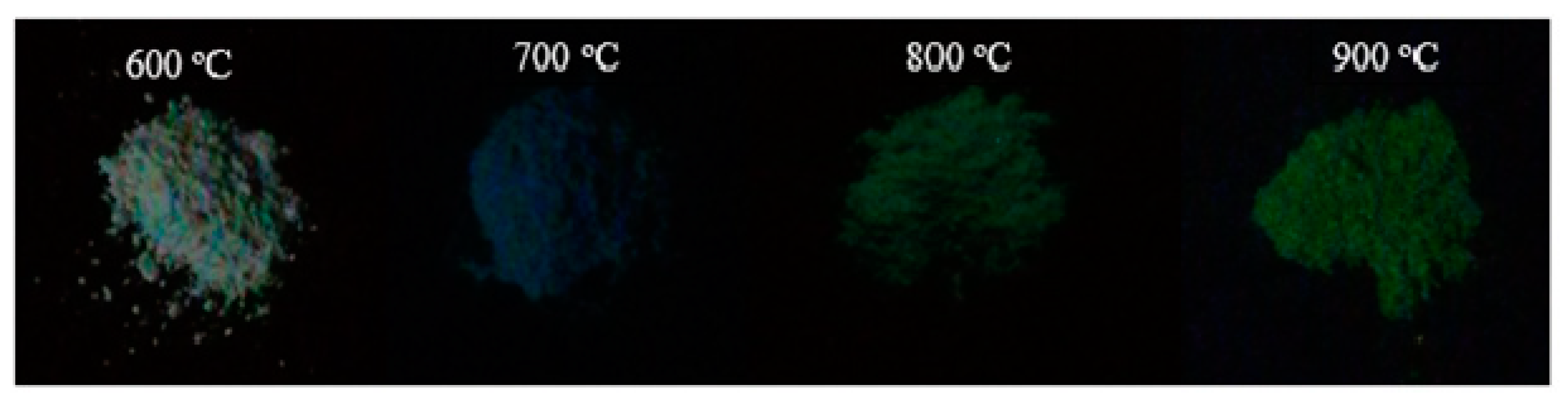
3.5. Photoluminescence Analysis

Figure 6 indicates the energy emission spectra of the glass and glass-ceramic sample excited at 254 nm. This graph shows the emission spectra of the sample that was excited at 254 nm and produced an intense green luminescence at 524 nm. The zinc silicate crystal luminescent properties are common, with the emission of 585 and 525 nm for a glass-ceramic sample showing a typical low and broad excitation at about 260 nm [38]. At a heat-treatment temperature of 600 °C, there are no emission spectra because at 600 °C the zinc silicate crystal does not form, as shown in the XRD. The emission spectra emitted at heat treatments of 700, 800 and 900 °C show that the intensity of the emission is increased with the increase of heat-treatment temperatures. This is because of the improvement in crystallinity inside the sample as the heat-treatment temperature progresses [41]. Moreover, the intensity variation depends on some aspects, which include the structural modification of glass matrix, the amount of the luminescent ions and the wavelength of the excitation source used [42]. In this case, variations of intensity on the glass matrix are probably due to structural modifications because of increased presence of Zn2SiO4 in the glasses. Figure 7 shows the sample 55ZnO–5B2O3–40SLS at a various heat-treatment temperatures under a UV lamp short wavelength of 254 nm. It was observed that at 600 °C there was no emission because the powder sample showed a white color under the UV lamp. This supports the result of photoluminescence measurement; there is no emission at 600 °C because zinc silicate crystal does not form. Moreover, the sample had the highest intensity at 900 °C, which is also the same as the photoluminescence measurement.

4. Conclusions

The phase transformation, optical and luminescence performance of new zinc silicate derived from the ZnO–B2O3–SLS glass system was successfully investigated. The result shows that the density and linear shrinkage of the sample increased with the progression of heating temperature. In addition, the XRD characterization shows that the samples start with an amorphous glassy state at 600 °C, but when sintered at 700, 800 and 900 °C, the samples show a sharp peak indicating presence of the crystal inside the samples. The IR spectrum of FTIR shows the presence of structurally associated SiO4 and ZnO4, which indicate the formation of a zinc silicate crystal phase. The optical band gap decreased due to short-wavelength light being scattered by the zinc silicate crystal as the crystallinity increased at higher heat-treatment temperature. Lastly, photoluminescence spectra excited at 254 nm resulted in emission spectrum emitted with a green color at 524 nm. Based on this research, ZnO reacts as network modifier or former because it is an intermediate oxide corresponding to the mixture of the glass matrix, and behaves to alter the structural glass system. Finally, B2O3 works as one of the most important glass formers and flux materials, which forms a connected backbone glass network.

Author Contributions

Conceptualization, M.F.S.M.S., M.H.M.Z. and K.A.M.; formal analysis, Y.W.F., Y.Y.; resources, S.H.J. and Y.I.; writing—original draft preparation, M.F.S.M.S.; writing—review and editing, S.A.A.W.; supervision, M.H.M.Z., K.A.M. and Y.W.F. All authors have read and agreed to the published version of the manuscript.

Funding

Highest appreciation to Universiti Putra Malaysia (UPM) and Ministry of Higher Education Malaysia for financial support via Fundamental Research Grant Scheme (FRGS) Vote 5540268 for this research work.

Conflicts of Interest

The authors declare no conflict of interest.

References

  1. Ehrt, D.; Flügel, S. Properties of zinc silicate glasses and melts. Mat. Sci. Eng. A 2011, 1, 312. [Google Scholar]
  2. Tarafder, A.; Molla, A.R.; Mukhopadhyay, S.; Karmakar, B. Fabrication and enhanced photoluminescence properties of Sm3+-doped ZnO–Al2O3–B2O3–SiO2 glass derived willemite glass-ceramic nanocomposites. Opt. Mater. 2014, 36, 1463–1470. [Google Scholar] [CrossRef]
  3. Xu, G.Q.; Xu, H.T.; Zheng, Z.X.; Wu, Y.C. Preparation and characterization of Zn2SiO4: Mn phosphors with hydrothermal methods. J. Lumin. 2010, 130, 1717–1720. [Google Scholar] [CrossRef]
  4. An, J.S.; Noh, J.H.; Cho, I.S.; Roh, H.S.; Kim, J.Y.; Han, H.S.; Hong, K.S. Tailoring the morphology and structure of nanosized Zn2SiO4: Mn2+ phosphors using the hydrothermal method and their luminescence properties. J. Phys. Chem. C 2010, 114, 10330–10335. [Google Scholar] [CrossRef]
  5. Masjedi-Arani, M.; Salavati-Niasari, M. A simple sonochemical approach for synthesis and characterization of Zn2SiO4 nanostructures. Ultrason. Sonochem. 2016, 29, 226–235. [Google Scholar] [CrossRef]
  6. Babu, B.C.; Buddhudu, S. Dielectric Properties of Willemite Zn2SiO4 Nano Powders by Sol-gel Method. Phys. Procedia 2013, 49, 128–136. [Google Scholar] [CrossRef] [Green Version]
  7. Kang, Y.C.; Park, S. Zn2SiO4: Mn phosphor particles prepared by spray pyrolysis using a filter expansion aerosol generator. Mater. Res. Bull. 2000, 35, 1143–1151. [Google Scholar] [CrossRef]
  8. Su, K.; Tilley, T.D.; Sailor, M.J. Molecular and polymer precursor routes to manganese-doped zinc orthosilicate phosphors. J. Am. Chem. Soc. 1996, 118, 3459–3468. [Google Scholar] [CrossRef]
  9. Tarafder, A.; Molla, A.R.; Dey, C.; Karmakar, B. Thermal, Structural, and Enhanced Photoluminescence Properties of Eu3+-doped Transparent Willemite Glass–Ceramic Nanocomposites. J. Am. Ceram. Soc. 2013, 96, 2424–2431. [Google Scholar] [CrossRef]
  10. Zaid, M.H.M.; Matori, K.A.; Ab Aziz, S.H.; Kamari, H.M.; Wahab, Z.A.; Effendy, N.; Alibe, I.M. Comprehensive study on compositional dependence of optical band gap in zinc soda lime silica glass system for optoelectronic applications. J. Non-Cryst. Solids 2016, 449, 107–112. [Google Scholar] [CrossRef]
  11. Zaid, M.H.M.; Matori, K.A.; Wah, L.C.; Sidek, H.A.A.; Halimah, M.K.; Wahab, Z.A.; Azmi, B.Z. Elastic moduli prediction and correlation in soda lime silicate glasses containing ZnO. Int. J. Phys. Sci. 2011, 6, 1404–1410. [Google Scholar]
  12. Yanbo, Q.; Ning, D.; Mingying, P.; Lüyun, Y.; Danping, C.; Jianrong, Q.; Chongshan, Z.; Akai, T. Spectroscopic properties of Nd3+-doped high silica glass prepared by sintering porous glass. J. Rare Earth. 2006, 24, 765–770. [Google Scholar] [CrossRef]
  13. Chimalawong, P.; Kirdsiri, K.; Kaewkhao, J.; Limsuwan, P. Investigation on the physical and optical properties of Dy3+ doped soda-lime-silicate glasses. Procedia Eng. 2012, 32, 690–698. [Google Scholar] [CrossRef] [Green Version]
  14. Hamnabard, Z.; Khalkhali, Z.; Qazvini, S.S.A.; Baghshahi, S.; Maghsoudipour, A. Preparation, heat treatment and photoluminescence properties of V-doped ZnO–SiO2–B2O3 glasses. J. Lumin. 2012, 132, 1126–1132. [Google Scholar] [CrossRef]
  15. Qazvini, S.S.A.; Hamnabard, Z.; Khalkhali, Z.; Baghshahi, S.; Maghsoudipour, A. Photoluminescence and microstructural properties of SiO2–ZnO–B2O3 system containing TiO2 and V2O5. Ceram. Int. 2012, 38, 1663–1670. [Google Scholar] [CrossRef]
  16. Li, H.C.; Wang, D.G.; Hu, J.H.; Chen, C.Z. Effect of the partial substitution of K2O, MgO, B2O3 for CaO on crystallization, structure and properties of Na2O–CaO–SiO2–P2O5 system glass-ceramics. Mater. Lett. 2013, 106, 373–376. [Google Scholar] [CrossRef]
  17. Zachariasen, W.H. The atomic arrangement in glass. J. Am. Chem. Soc. 1932, 54, 3841–3851. [Google Scholar] [CrossRef]
  18. Azuraida, A.; Halimah, M.K.; Sidek, A.A.; Azurahanim, C.A.C.; Iskandar, S.M.; Ishak, M.; Nurazlin, A. Comparative studies of bismuth and barium boro-tellurite glass system: Structural and optical properties. Chalcogenide Lett. 2015, 12, 497–503. [Google Scholar]
  19. Lakshminarayana, G.; Yang, R.; Mao, M.; Zhang, Y.; Qiu, J. Spectral analysis of optical centres formed in Bi, Bi/Yb, Pb, Pb/Yb, Sb, Sb/Yb and Sn, Sn/Yb-co-doped germanate glasses. J. Phys. D Appl. Phys. 2009, 42, 145108. [Google Scholar] [CrossRef]
  20. Loiko, P.; Dymshits, O.; Volokitina, A.; Alekseeva, I.; Shemchuk, D.; Tsenter, M.; Bachina, A.; Khubetsov, A.; Vilejshikova, E.; Petrov, P.; et al. Structural transformations and optical properties of glass-ceramics based on ZnO, β- and α-Zn2SiO4 nanocrystals and doped with Er2O3 and Yb2O3: Part, I. The role of heat-treatment. J. Lumin. 2018, 202, 47–56. [Google Scholar] [CrossRef]
  21. Chen, Q.; Ferraris, M.; Milanese, D.; Menke, Y.; Monchiero, E.; Perrone, G. Novel Er-doped PbO and B2O3 based glasses: Investigation of quantum efficiency and non-radiative transition probability for 1.5 μm broadband emission fluorescence. J. Non-Cryst. Solids 2003, 324, 12–20. [Google Scholar] [CrossRef]
  22. Rashid, S.S.A.; Ab Aziz, S.H.; Matori, K.A.; Zaid, M.H.M.; Mohamed, N. Comprehensive study on effect of sintering temperature on the physical, structural and optical properties of Er3+ doped ZnO-GSLS glasses. Results Phys. 2017, 7, 2224–2231. [Google Scholar] [CrossRef]
  23. Kullberg, A.T.G.; Lopes, A.A.S.; Veiga, J.P.B.; Lima, M.M.R.A.; Monteiro, R.C.C. Formation and crystallization of zinc borosilicate glasses: Influence of the ZnO/B2O3 ratio. J. Non-Cryst. Solids 2016, 441, 79–85. [Google Scholar] [CrossRef]
  24. Khalkhali, Z.; Hamnabard, Z.; Qazvini, S.S.A.; Baghshahi, S.; Maghsoudipour, A. Preparation phase formation and photoluminescence properties of ZnO–SiO2–B2O3 glasses with different ZnO/B2O3 ratios. Opt. Mater. 2012, 34, 850–855. [Google Scholar] [CrossRef]
  25. Iordanova, R.; Dimitrov, V.; Dimitriev, Y.; Klissurski, D. Glass formation and structure of glasses in the V2O5 MoO3 Bi2O system. J. Non-Cryst. Solids 1994, 180, 58–65. [Google Scholar] [CrossRef]
  26. Torres, F.J.; de Sola, E.R.; Alarcón, J. Mechanism of crystallization of fast fired mullite-based glass–ceramic glazes for floor-tiles. J. Non-Cryst. Solids 2006, 352, 2159–2165. [Google Scholar] [CrossRef]
  27. Effendy, N.; Abdul Wahab, Z.; Kamari, H.M.; Matori, K.A.; Aziz, S.H.A.; Zaid, M.H.M. Structural and optical properties of Er3+-doped willemite glass-ceramics from waste materials. Optik 2016, 127, 11698–11705. [Google Scholar] [CrossRef]
  28. Lukic, S.R.; Petrovic, D.M.; Dramicanin, M.D.; Mitric, M.; Dacanin, L. Optical and structural properties of Zn2SiO4: Mn2+ green phosphor nanoparticles obtained by a polymer-assisted sol-gel method. Scripta Mater. 2008, 58, 655–658. [Google Scholar] [CrossRef]
  29. Chao, P.T.; Shen, P.; Lin, C.C. Thermal cycle etching of willemite (0001): Effects of surface premelting, dislocation outcrops and polygonization. Mater. Sci. Eng. A 2002, 335, 191–197. [Google Scholar] [CrossRef]
  30. Kullberg, A.T.; Lopes, A.A.; Veiga, J.P.; Monteiro, R.C. Crystal growth in zinc borosilicate glasses. J. Cryst. Growth 2017, 457, 239–243. [Google Scholar] [CrossRef]
  31. Sabri, M.G.M.; Azmi, B.Z.; Rizwan, Z.; Halimah, M.K.; Hashim, M.; Zaid, M.H.M. Effect of temperature treatment on the optical characterization of ZnO-Bi2O3-TiO2 varistor ceramics. Int. J. Phys. Sci. 2011, 6, 1388–1394. [Google Scholar]
  32. Lee, C.S.; Matori, K.A.; Aziz, S.H.A.; Kamari, H.M.; Ismail, I.; Zaid, M.H.M. Influence of zinc oxide on the physical, structural and optical band gap of zinc silicate glass system from waste rice husk ash. Optik 2017, 136, 129–135. [Google Scholar] [CrossRef]
  33. Omri, K.; El Ghoul, J.; Alyamani, A.; Barthou, C.; El Mir, L. Luminescence properties of green emission of SiO2/Zn2SiO4: Mn nanocomposite prepared by sol-gel method. Physica E 2013, 53, 48–54. [Google Scholar] [CrossRef]
  34. Giancaterini, L.; Cantalini, C.; Cittadini, M.; Sturaro, M.; Guglielmi, M.; Martucci, A.; Resmini, A.; Anselmi-Tamburini, U. Au and Pt nanoparticles effects on the optical and electrical gas sensing properties of sol-gel-based ZnO thin-film sensors. IEEE Sens. J. 2015, 15, 1068–1076. [Google Scholar] [CrossRef]
  35. Ghoul, J.E.; Barthou, C.; Mir, L.E. Synthesis, structural and optical properties of nanocrystalline vanadium doped zinc oxide aerogel. Physica E 2012, 44, 1910–1915. [Google Scholar] [CrossRef]
  36. Omar, N.A.S.; Fen, Y.W.; Matori, K.A.; Zaid, M.H.M.; Norhafizah, M.R.; Nurzilla, M.; Zamratul, M.I.M. Synthesis and optical properties of europium doped zinc silicate prepared using low cost solid state reaction method. J. Mater. Sci.-Mater. Electron. 2016, 27, 1092–1099. [Google Scholar] [CrossRef]
  37. Syamimi, N.F.; Matori, K.A.; Lim, W.F.; Sidek, H.A.A.; Zaid, M.H.M. Effect of sintering temperature on structural and morphological properties of europium (III) oxide doped willemite. J. Spectroscopy 2014, 2014, 328931. [Google Scholar] [CrossRef]
  38. Zaid, M.H.M.; Matori, K.A.; Aziz, S.H.A.; Kamari, H.M.; Wahab, Z.A.; Fen, Y.W.; Alibe, I.M. Synthesis and characterization of low cost willemite based glass–ceramic for opto-electronic applications. J. Mater. Sci.-Mater. Electron. 2016, 27, 11158–11167. [Google Scholar] [CrossRef]
  39. Li, Z.Y.; Zhang, Z.Q. Fragility of photonic band gaps in inverse-opal photonic crystals. Phys. Rev. B 2000, 62, 1516. [Google Scholar] [CrossRef] [Green Version]
  40. Iskandar, S.M.; Halimah, M.K.; Daud, W.M.; Sidek, H.A.A.; Zaman, M.K. Optical and structural properties of PbO-B2O3-TeO2 glasses. J. Solid State Sci. Technol. 2010, 18, 84–90. [Google Scholar]
  41. Li, J.; Kuwabara, M. Preparation and luminescent properties of Eu-doped BaTiO3 thin films by sol–gel process. Sci. Technol. Adv. Mat. 2003, 4, 143–148. [Google Scholar] [CrossRef]
  42. Kam, C.H.; Buddhudu, S. Luminescence enhancement in (Nd3++Ce3+) doped SiO2: Al2O3 sol–gel glasses. Opt. Laser Eng. 2001, 35, 11–17. [Google Scholar] [CrossRef]
Figure 1. The linear shrinkage percentage and the density of the 55ZnO–5B2O3–40SLS glass and glass-ceramic sample at a various heat-treatment temperatures.
Applsci 10 04940 g001
Figure 2. X-ray diffraction (XRD) pattern of 55ZnO–5B2O3–40SLS glass and glass-ceramic sintered at various temperatures.
Applsci 10 04940 g002
Figure 3. Fourier transform infrared reflection (FTIR) pattern of 55ZnO–5B2O3–40SLS glass and glass-ceramic sample at various heat-treatment temperatures.
Applsci 10 04940 g003
Figure 4. Absorption spectra for 55ZnO–5B2O3–40SLS based glass and glass-ceramic sintered at different temperatures.
Applsci 10 04940 g004
Figure 5. A plot of the optical band gap of 55ZnO–5B2O3–40SLS glass and glass-ceramic samples at various heat-treatment temperatures.
Applsci 10 04940 g005
Figure 6. Photoluminescence (PL) spectra of 55ZnO–5B2O3–40SLS sample sintered at various heat-treatment temperatures.
Applsci 10 04940 g006
Figure 7. Sample of 55ZnO–5B2O3–40SLS treated at a various heat-treatment temperatures under ultraviolet–visible (UV) lamp of 254 nm.
Applsci 10 04940 g007
Table 1. The variation of bulk density and linear shrinkage of the 55ZnO–5B2O3–40SLS glass and glass-ceramic sample at various heat-treatment temperatures.
Temperature (°C)Density (g/cm3)Linear Shrinkage (%)
272.7540.00
6002.9808.85
7003.2119.08
8003.3589.76
9003.54510.43
Table 2. FTIR transmittance bands associated with assigned vibration modes.
Wavenumber (cm−1)Assigned of Vibration Modes
457Asymmetric deformation of SiO4
570Symmetric stretching of ZnO4
611Asymmetric stretching of ZnO4
710Si-O bond vibration
866Symmetric stretching of SiO4
900, 930, 977Asymmetric stretching of SiO4
Table 3. Variation of optical band gap against heat-treatment temperature.
Temperature (°C)Band Gap Energy (eV)
6003.867
7003.743
8003.484
9003.423

