The nanotechnology revolution embodies an interdisciplinary approach and is bringing scientists together from nearly every area of scientific study. This worldwide collaboration is significantly enhancing the quality of numerous areas such as electronics, renewable energy, transportation, consumer products, agriculture, healthcare, and medicinal drug discovery. The NanoFlorida Conference, coordinated for over a decade by leaders from Florida’s academic institutions, unites the state’s nanoscience community around the latest nanotechnology discoveries, with particular emphasis on the training, education, and development of students. Annual NanoFlorida conferences have addressed critical needs of research students and faculty within the State of Florida. Most importantly, these conferences provide a forum for academic and industrial researchers to share their recent discoveries, exchange new ideas, and develop professional relationships that strengthen the state’s nanotechnology research community.
The 2019 NanoFlorida International Conference extended this initiative with a global focus and an aim to advance the nanotechnology and nanoscience research in Florida, the US and globally. The conference focused on the theme of “Advances in Translational Nanotechnology,” with 259 attendees, 15 plenary speakers, 36 student oral presentations, and 137 poster presentations. The symposia included presentations in the following areas:
The conference fostered fruitful interaction among nanotechnology researchers, students, and leaders with the promise of ongoing impact by spotlighting emerging frontiers of nanoscience and attracting the next generation of the best and brightest to the field.
We would like to acknowledge the financial support of this conference by the National Science Foundation (grant #1933179), University of South Florida Research & Innovation (ResearchOne), University of Florida, University of Florida Research Service Center, Florida International University, University of Central Florida, FloridaMakes, Florida A&M University, National Institute for Standards and Technology, Florida High Tech Corridor, University of Miami, and the Florida Association for Nanotechnology, Inc. We would also like to acknowledge the assistance of Christen Bouchard, Danielle Gamboni, Jamie Martin, and Bianca Tolve with conference planning and execution.
Shyam S. Mohapatra, PhD, MBA, FNAI, FAIMBE, FAAAS Conference Chair
1. Nanomaterials and Devices
1.1. Thin-Piezo on Silicon Resonators Using a Novel Microfabrication Process
Abdulrahman Alsolami,
Adnan Zaman
and
Jing Wang
University of South Florida, Tampa, FL 33620, USA
Radio frequency microelectromechanical systems (RF MEMS) are widely viewed as a potential enabling technology for the multi-standard monolithic transceivers on a single chip with high reliability, high performance, and very low (virtually zero) DC power consumption. Among various RF MEMS device components, resonators offer unique benefits because of their very high quality factor, which enables the implementation of advanced functions such as low insertion loss filters, mixer-filters, ultra-low phase noise oscillators, and even RF front-end channel selection. Thin-piezo on silicon (TPoS) resonators, in particular, are of interest in building very high performance resonators. A new technique to build a TPoS resonator using a silicon single crystal wafer is developed. The thin-film piezoelectric on single-crystal silicon reactive etched (TPoSCRE) technique presented allows batch fabrication of TPoS resonators without using silicon-on-insulator (SOI) wafers, while also retaining the same number of photolithography steps as a typical SOI based MEMS TPoS resonators. Typically, building TPoS resonators requires expensive SOI wafers. This method helps to reduce the cost of the thin-film piezoelectric on silicon MEMS devices. The isotropic release process in this method utilized a thin film of aluminum oxide deposited by atomic layer deposition (ALD). The ALD film protects the sidewall of the Si device structure from being etched.
Figure 1 shows scan electron microscopy (SEM) images of the fabricated devices. The results of these devices are shown in
Figure 2.
Figure 1.
SEM view of TPoSCRE fabricated TPoS RF MEMS resonator devices.
Figure 1.
SEM view of TPoSCRE fabricated TPoS RF MEMS resonator devices.
Figure 2.
Measured frequency characteristics of two designs of thin-film piezoelectric on silicon (TPoS) resonators by newly developed TPoSCRE process.
Figure 2.
Measured frequency characteristics of two designs of thin-film piezoelectric on silicon (TPoS) resonators by newly developed TPoSCRE process.
1.2. Large Surface Area Nanowall-Structured Electrodes for Wearable Supercapacitors
Jayesh Cherusseri 1,
Kowsik Sambath Kumar 1,2
and
Jayan Thomas 1,2,3
1
NanoScience Technology Center, University of Central Florida, Orlando, Florida 32826, USA
2
Department of Materials Science and Engineering, University of Central Florida, Orlando, Florida 32816, USA
3
CREOL, College of Optics and Photonics, University of Central Florida, Orlando, Florida 32816, USA
The fast-growing development of flexible and wearable electronic devices necessitate flexible energy storage devices to supply them power. Energy storage devices such as supercapacitors have received great attention due to their environmental friendliness, light weight, high specific capacitance, high power density, etc. Metal-oxide based supercapacitors exhibit superior charge-storage capabilities due to their redox-type charge storage. The present work discusses the preparation of large surface area nanowall-structured electrodes for application in flexible supercapacitors. Manganese-oxide nanowalls (MO-NWs) are grown on an electrically conducting carbon fiber (CF) substrate via the electrochemical deposition method. Here, the CF serves not only the substrate for the growth of MO-NWs, but also the current collector for the supercapacitor. The morphology of the MO-NWs is characterized by scanning electron microscopy, atomic force microscopy, and the structural details are obtained from Raman spectroscopy and X-ray diffraction analysis. The supercapacitor electrodes and the symmetric supercapacitor fabricated using MO-NWs electrodes are characterized by electrochemical impedance spectroscopy, cyclic voltammetry, and galvanostatic charge/discharge measurement. The symmetric MO-NWs supercapacitor exhibits high specific capacitance and it is highly bendable, and hence a potential candidate for next generation wearable supercapacitors.
1.3. Multifunctional Magnetic Nanostructures for Enhanced Hyperthermia Therapy
Raja Das
Faculty of Materials Science and Engineering and Phenikaa Institute for Advanced Study (PIAS), Phenikaa University, Hanoi 10000, Vietnam
Phenikaa Research and Technology Institute (PRATI), A&A Green Phoenix Group, 167 Hoang Ngan, Hanoi 10000, Vietnam
Despite their great potential in magnetic hyperthermia therapy for cancer treatment, spherical magnetite (Fe3O4) or maghemite (γ-Fe2O3) nanoparticles, which are the most commonly employed and only FDA approved materials, yield limited heating capacity. Therefore, there is an increasing need for new strategies to improve the heating efficiency (evaluated by the specific absorption rate (SAR)) of these nanostructures. By utilizing the proven advantages of one dimensional (1D) nanostructures over their spherical and cubic counterparts, such as larger surface area, multisegmented capabilities, enhanced blood circulation time, and prolonged retention in tumors, we have demonstrated that the SAR of iron oxide nanostructures can be manipulated by altering their aspect ratio [1]. Calorimetric and AC magnetometry experiments performed on highly crystalline Fe3O4 nanorods consistently show large SAR values, which are superior to their spherical and cubic nanoparticles of similar volume. In this line of interest, we have advanced to synthesize 1D hollow magnetic nanotubes of Fe3O4 for dual-purpose applications in hyperthermia and drug delivery due to their high effective anisotropy and enhanced surface areas (inner and outer surfaces) [2]. Even though the 1D nanostructures show superior heating efficiency at the high field region (>400 Oe) due to their enhanced effective anisotropy, they do possess relatively poor heating efficiency at the low field region (<400 Oe, Brezovich safe limit). To overcome this, we have proposed a novel approach with which a synergistic exploitation of the magnetic and photothermal properties of the plasmonic metals and magnetic iron oxide, such as Ag(core)/Fe3O4(shell) nanoflowers, can reduce the magnetic field and laser intensities that are required in the case that both external stimuli are applied separately [3]. Our study establishes a key step towards optimizing the hyperthermia therapy through a combined multifunctional magnetic and photothermal treatment and improving our understanding of the therapeutic process to specific applications. These results pave a new pathway for the design of multifunctional magnetic nanostructures for advanced hyperthermia.
References
R. Das, J. Alonso, Z. N. Porshokouh, V. Kalappattil, D. Torres, M. H. Phan, E. Garaio, J. Á. García, J. L. S. Llamazares, and H. Srikanth J. Phys. Chem. C 120, 10086 (2016).
R. Das, J. A. Cardarelli, M. H. Phan, and H. Srikanth J. Alloy Compd. 48, 1461 (2019).
R. Das, N. R. Montes, J. Alonso, Z. Amghouz, P. Gorria, J.A. Blanco, M. H. Phan, and H. Srikanth ACS Appl. Mater. Interfaces 8, 25162 (2016).
1.4. Development of “Dynamic” 3D Microelectrodes Using Optimized, 3D Printed Microserpentines
Charles Didier 1,2,
Avra Kundu 1
and
Swaminathan Rajaraman 1,2,3,4
1
NanoScience Technology Center, University of Central Florida, 4353 Scorpius Street, Research I, Suite 231, Orlando, FL 32816, USA
2
Burnett School of Biomedical Sciences, University of Central Florida, 6900 Lake Nona Blvd, Orlando, FL 32827, USA
3
Department of Materials Science and Engineering, University of Central Florida, 12760 Pegasus Drive, Engineering I, Suite 207, Orlando, FL, 32816, USA
4
Department of Electrical and Computer Engineering, University of Central Florida, 4238 Scorpius Street, Orlando, FL 32816, USA
We explore the capabilities and limitations of 3D printed microserpentines (µserpentines) and utilize these structures to develop “dynamic” 3D microelectrodes for potential applications in in vitro, wearable, and implantable microelectrode arrays (MEAs). The device incorporates optimized 3D printed µserpentine designs with out-of-plane microelectrode structures, integrated on to a flexible Kapton® package with micromolded PDMS insulation. The flexibility of the optimized, printed µserpentine design was calculated through effective stiffness and effective strain equations, so as to allow for analysis of various designs for enhanced flexibility. The optimized, down selected µserpentine design was further sputter coated with 7–70 nm thick gold and the performance of these coatings was studied for maintenance of conductivity during uniaxial strain application. “Dynamic” three-dimensional MEAs were built on top of these µserpentines by combining them with a flexible Kapton® package and drop casted PDMS insulation. Bending/conforming analysis of the final devices (3D MEAs with a Kapton® package and PDMS insulation) were performed to qualitatively assess the robustness of the finished device toward dynamic MEA applications. Moreover, 3D microelectrode impedance measurements varied from 4.2 kΩ to 5.2 kΩ during the bending process demonstrating a small, acceptable change. Lastly, an example application with an artificial agarose skin composite model to assess the feasibility for basic transdermal electrical recording was further demonstrated.
1.5. Percolation Conductivity and Critical Exponents in Nanowire Networks: Role of Junction-To-Nanowire Resistance Ratio
Nicholas Fata,
Shreshtha Mishra,
Ying Xue,
Jeremy Hicks
and
Ant Ural
Department of Electrical and Computer Engineering and Nanoscience Institute for Medical and Engineering Technology (NIMET), University of Florida, Gainesville, FL 32611, USA
Random networks of one-dimensional (1D) nanoelements, such as carbon nanotubes, graphene nanoribbons, and metal nanowires, have recently attracted significant research interest for next-generation transparent conductors. Notably, silver and copper nanowire networks exhibit high optical transmittance, low sheet resistance, mechanical flexibility, and fast deposition, making them promising candidates to replace indium tin oxide (ITO), which suffers from brittleness, scarcity, high cost, and slow deposition. At high optical transmittance values required for transparent conductors, percolation transport governs the conductivity of metal nanowire networks. Thus, we utilize Monte Carlo simulations to theoretically calculate, predict, and optimize the conductivity of metal nanowire networks. The overall conductivity of the network is determined by two types of resistance elements, namely the nanowire–nanowire junction resistance and the resistance of the nanowire itself. In most Monte Carlo simulations, it is assumed that junction resistance far exceeds the nanowire resistance, as is the case with carbon nanotubes. However, junction resistance can be significantly lowered for metal nanowire networks, becoming comparable to, or far smaller than the nanowire resistance. In that case, the network conductance becomes nanowire-dominated, which is the highest conductance limit. In this work, we perform Monte Carlo simulations to study the effect of the junction-to-nanowire resistance ratio on nanowire network conductivity at different values of nanowire and device parameters, namely nanowire density, nanowire length, device length, device width, nanowire alignment, and nanowire curviness. Next, we investigate the effect of the resistance ratio on the percolation critical exponents, which characterize the power-law dependence of conductivity on the nanowire and device parameters near the percolation threshold. Our results demonstrate how the junction-to-nanowire resistance ratio affects the macroscopic conductivity of a network and its percolation critical exponents. They also show how Monte Carlo simulations are essential for providing insight into the percolation transport in transparent, conductive nanowire networks.
1.6. Atomic Layer Semiconductor and Heterostructure Nano-Devices and Systems for Classical and Quantum Information Processing
Philip Feng
Department of Electrical & Computer Engineering, University of Florida
Atomically thin crystals (including semiconducting, metallic & insulating layers) and their heterostructures offer compelling new platforms for 2D electronic, photonic devices and nanoelectromechanical systems (NEMS), where their unconventional and unique properties can be harnessed for engineering both classical signal processing and quantum transduction schemes. In this presentation, I will describe some of my research group’s latest effort on advancing device physics and engineering of 2D heterostructures and NEMS. In the classical domain, I will first show atomically thin radio frequency (RF) NEMS resonators with excellent electrical tunability and remarkably broad dynamic range. I will then demonstrate how the unusually strong and efficient coupling effects have led to ultra-broad resonance tuning of van der Waals heterostructure resonators, as well as stable, robust graphene NEMS operating at glowing temperatures with simultaneous light emission. Finally, toward quantum engineering, atomic defects in wide-bandgap semiconductors and emerging 2D crystals such as hexagonal boron nitride (h-BN) support quantum emitters with promises for enabling desirable qubits at room temperature. Built upon our prior work on SiC photonics and the first h-BN resonators, I will describe the development of such structures and devices as building blocks for enabling and interfacing with quantum emitters, toward realizing quantum transduction and information processing on chip-scale integrated platforms.
![Applsci 10 04851 i001]()
Relevant References
- [1]
Islam, van den Akker, Feng, “Polarization Sensitive Black Phosphorus…”, Optical Materials Express 9, 526-535 (2019).
- [2]
Islam, van den Akker, Feng, “Anisotropic Thermal Conductivity of Suspended…”, Nano Letters 18, 7683-7691 (2018).
- [3]
Islam, Lee, Feng, “Atomic Layer GaSe/MoS2 van der Waals Heterostructure…”, ACS Photonics 5, 2693-2700 (2018).
- [4]
Lee, Wang, Feng, et al., “Electrically Tunable Single & Few-Layer MoS2…”, Science Advances 4, eaao6653 (2018).
- [5]
Ye, Lee, Feng, “Electrothermally Tunable Graphene Resonators Operating…”, Nano Letters 18, 1678-1685 (2018).
- [6]
Yang, Lee, Feng, et al., “Tuning Optical Signature of Single- and Few-Layer…”, Nano Letters 17, 4568-4575 (2017).
- [7]
Wang, Jia, Feng, et al., “Resolving and Tuning Mechanical Anisotropy…”, Nano Letters 16, 5394-5400 (2016).
- [8]
Wang, Lee, Feng, “Spatial Mapping of Multimode Brownian Motions…”, Nature Communications 5, 5158 (2014).
- [9]
Falk, Feng, Awschalom, “Polytype Control of Spin Qubits in Silicon Carbide (SiC)”, Nature Communications 4, 1819 (2013).
- [10]
Lee, Wang, Feng, et al., “High Frequency MoS2 Nanomechanical Resonators”, ACS Nano 7, 6086-6091 (2013).
1.7. The Development of a Novel Continuous Flow Methodology to Achieve a Constant Deposition Rate during Electrophoretic Deposition
Noah D. Ferson,
Prabal Tiwari
and
Jennifer S. Andrew
Department of Materials Science and Engineering, University of Florida, 1698 Gale Lemerand Dr, Gainesville, FL 32611, USA
Abstract:
Electrophoretic deposition (EPD) is widely utilized in the assembly of colloids into porous structures [1]. EPD has been gaining attention as an effective assembly technique for biomaterials [2] in particular, EPD can be utilized to fabricate bioactive coatings for increased biocompatibility, biosensors, and for the deposition of biological entities. A current limitation in fabricating surfaces via EPD is the decrease in deposition rate as EPD persists. This is thought to occur as the particle mobility, electric field, and particle concentration decrease with time. In the current work, the parameters presented in Hamaker’s equation (Equation (1)) [3] are isolated to determine the dominant factor that affects the deposition rate. Hamaker’s equation states that particle mobility (μ), particle concentration (C), electric field (E), and electrode area (S) are what dictate the yield (Y) of the deposit.
Experimental evidence has been gathered by the authors to show that the decrease in particle mobility, caused from the increase in pH of the solution is the dominant factor for the decrease in deposition rate as time persists. In order to combat this decrease in mobility, several modifications to the EDP process have been developed. One approach included a solution replenishment method that achieved a constant deposition rate for EPD durations up to 70 min. However, this process was costly due to time consuming sample preparation and the amount of material needed to produce a deposit. Utilizing the results from the solution replenishment, a continuous flow system has been developed. A continuous flow system will effectively keep particle concentration, mobility, and pH constant during the deposition. In completion of these experiments, it is expected that a new methodology will be developed to optimize the deposition rate and offer insight on the most effective process to fabricate biosensors, surface coatings, and other nanostructured devices.
References
- [1]
Van der Biest, L.J. Vandeperre, Electrophoretic Depostio of Materials. Annu. Rev. Mater. Sci. 1999 29 327-352.
- [2]
Boccaccini A. R., Keim S., Ma R., Li Y. and Zhitomirsky I. Electrophoretic deposition of biomaterials. J. R. Soc. Interface (2010) 7, S581–S613
- [3]
H.C. Hamaker, Trans. Faraday Soc. 36 (1940) 279–283.
1.8. Reconfigurable Additive Manufactured Packaging Systems by Picosecond Laser Processing
Omer Firat 1,
Jing Wang 1
and
Thomas Weller 2
1
Department of Electrical Engineering, University of South Florida, Tampa, FL 33612, USA
2
School of Electrical Engineering and Computer Science, Oregon State University, Corvallis, OR 97331, USA
This work proposes a new type of 3-D printed suspended coplanar waveguide (CPW) interconnects and additively manufactured electrostatically actuated RF switches integrated along with it. This type interconnects are also well suited for packaging of mm-wave systems. The design and additive manufacturing processes for the aforementioned devices have been demonstrated. Also, RF characterization for the CPW line has been conducted. 3D printing quality, namely feature size and dimensional accuracy, was further improved by utilizing well-characterized laser machining techniques. The laser machining process enables the control of the characteristic dimensions of micro-dispensed conductive traces down to a few micrometers. In our design, CPW lines are printed on a fixed-fixed beam and the RF switch was laser machined on conductor line to create electrostatic cantilever structure. When an electrostatic voltage is applied to the cantilever beam of the switch and it snaps laterally to an electrode at the other port. Acrylonitrile butadiene styrene (ABS) and CB028 conductive silver paste are utilized to fabricate the CPW lines on suspended beams over an air cavity, thus enabling multi-layer interconnects in a similar fashion to that of traditional integrated circuits at the chip level. Simulated and measured frequency responses in terms of S-parameters up to 30 GHz are presented. The conductor width, ground width, and slot width are 160 μm, 260 μm, and 20 μm, respectively. The switch beam length, beam width, and the gap between the beam and electrode are 800 μm, 20 μm, and 5 μm, respectively. The measured transmission line loss of the suspended CPW line is 0.26 dB/mm at 30 GHz.
1.9. Review of ‘Green’ Silver Nanoparticles Isolation for Targeted Application
Melissa Gaeta 2,
Daniel J. Denmark 1,2,3,4,
Bhavya Shah 2,3
and
Shyam S. Mohapatra 1,2,3,4
1
James A Haley VA Hospital, Tampa, FL 33612, USA
2
College of Pharmacy Graduate Programs, Tampa, FL 33612, USA
3
Center for Research & Education in Nanobioengineering, Tampa, FL 33612, USA
4
Dept of Internal Medicine, USF, Tampa, FL 33612, USA
Silver has been used for over six millennia as an antimicrobial agent, most commonly in use as an additive to burn wounds. Silver has antimicrobial and biocidal properties naturally, making it an ideal accompaniment to nanoparticle pharmaceutical approaches to aid in targeted drug delivery, notably in food-borne multidrug-resistant illnesses. The application for silver in nanoparticles has similar properties at the quantum level, providing a medium for faster healing times, antibiotic-boosting, and medical-device efficacy. This review serves to highlight some of the current research on the synthesis of silver nanoparticles from fungi as an economical and ecologically friendly alternative to chemical synthesis. The current synthesis of silver nanoparticles (AgNPs) includes expensive top-down and bottom-up approaches with heavy chemical-use and hazardous byproducts. The green method of synthesis includes two routes, intracellular and extracellular. First, using the fungi’s natural enzymatic reactions and exposing fungi cultures to AgNO3 in an aqueous solution and second, exposing fungal cells to the AgNO3 solution in which enzymatic processes form AgNPs on the surface of said cells. Nanoparticles efficacy to deliver drugs is based upon their surface area and consistency in size. When using green synthesis methods, the cost of synthesis drops due to the lack of chemicals or processing equipment, the byproducts are little to none, and a substantial number of nanoparticles can be synthesized due to the lack of capping agents in size control. The size of nanoparticle synthesized using green methods from fungi average between 1 and 50 nm. With the synthesis of silver nanoparticles from green sources like fungi, plants, and bacteria, the movement away from chemically heavy byproducts, cost-effective processes, and large production outputs of AgNPs makes headway for their commercialized use in drug-delivery.
References
Alexander, J. (2009). History of the Medical Use of Silver. Surgical infections. 10. 289-92. 10.1089/sur.2008.9941. en.80238.
Anwar, M. F., Yadav, D., Jain, S., Kapoor, S., Rastogi, S., Arora, I., & Samim, M. (2016). Size- and shape-dependent clinical and mycological efficacy of silver nanoparticles on dandruff. International journal of nanomedicine, 11, 147–161. doi:10.2147/IJN.S86828
Ivanova, N., Gugleve, V., Dobreva, M., Pehlivanov, I., Stefanov, S., and Andronova, V. (2018).
Silver Nanoparticles as Multi-Functional Drug Delivery Systems. Nanomedicines, IntechOpen, DOI: 10.5772/intechop
Mohanta, Y. K., Nayak, D., Biswas, K., Singdevsachan, S. K., Abd Allah, E. F., Hashem, A., Mohanta, T. K. (2018). Silver Nanoparticles Synthesized Using Wild Mushroom Show Potential Antimicrobial Activities against Food Borne Pathogens. Molecules (Basel, Switzerland), 23(3), 655. doi:10.3390/molecules23030655.
Politano, A. D., Campbell, K. T., Rosenberger, L. H., & Sawyer, R. G. (2013). Use of silver in the prevention and treatment of infections: silver review. Surgical infections, 14(1), 8–20. doi:10.1089/sur.2011.097.
Rafique, M., Sadaf, I., Rafique, S., & Tahir, B. (2017). A review on green synthesis of silver nanoparticles and their applications. Artificial Cells, Nanomedicine, and biotechnology, 45(7), 1272-1291. DOI: 10.1080/21691401.2016.1241792.
Sandford, J., Venkatapathy, B., El-Badawy, A., and Feldhake, D. (2010). State of the Science
Literature Review: Everything Nanosilver and More. U.S. Environmental Protection Agency, Washington, DC, EPA/600/R-10/084, 2010.
1.10. A 3D Nano-Fiber Inspired Smart Scaffold (FiSS) Enables Discovery of a Novel Cancer Drug Resistance Pathway
Mark C. Howell a,b,*,
Ryan Green a,b,
Shyam Mohapatra b,c,d
and
Subhra Mohapatra a,b,e
a
Molecular Medicine Department
b
Center for Research & Education in Nanobioengineering
c
Division of Translational Medicine, Internal Medicine, Morsani College of Medicine, University of South Florida, Tampa, FL 33612, USA
d
James A Haley Veterans Hospital, Tampa, FL 33612, USA
*
Correspondence: Mark C. Howell Jr.; 813-3969488
1.10.1. Objectives
Drug resistance is a major challenge in effectively treating cancers. Despite clinical approval of several first and second-generation EGFR tyrosine kinase inhibitors (TKIs) to treat advanced NSCLC patients, eventually treatment fails due to acquired drug resistance. To study the mechanism of resistance, our lab has developed a nanofiber inspired smart scaffold (FiSS) 3D culture environment, that creates a more representative model of tumor growth and drug resistance. We found that the FiSS platform promotes the growth of three-dimensional (3D) tumor-like structures (tumoroids), which resemble in vivo tumors. The FiSS platform even allows for growth of tumor biopsy cultures, which contain not only the cancer cells, but also the tumor stromal cells that are known to modulate the acquisition of drug resistance. We hypothesized that FiSS will allow us to elucidate novel EGFR TKI resistance mechanisms and may help identify treatments that are able to prevent development of drug resistance in the clinic.
1.10.2. Methods
EGFR TKI tolerance was examined using both monolayer and FiSS culture. Proteomics data were collected using Mass Spec to determine possible mechanisms for the observed EGFR TKI tolerance. The enzymes involved in cholesterol synthesis following exposure to TKI were examined, including CYP51A1, DHCR7, DHCR24, and LSS, as well as the transcription factor SREBF2 to determine if their expression was upregulated after exposure to an EGFR TKI by Western blotting and qPCR. The potential of a CYP51A1 inhibitor, ketoconazole, used in combination with EGFR TKIs to overcome the development of EGFR TKI tolerance in both monolayer and FiSS cell culture methods.
1.10.3. Results
A comparison of the drug sensitivity showed that parental cells were more sensitive to the EGFR TKIs compared to the drug tolerant (DT) cells. The tolerance to lapatinib was increased in both cell types when cultured on our FiSS compared to monolayer and further increased when cells were cultured from tumor biopsies on the FiSS. Data mining the significantly differentially expressed proteins list generated by the mass spectroscopic analysis revealed that the protein expression is skewed in lapatinib DT H1975 cell line as compared to the H1975 cell line. Three of the enzymes found to be upregulated in DT cells are directly involved in lipid and cholesterol catabolism. We then found that enzymes directly involved in cholesterol synthesis (CYP51A1, DHCR7, DHCR24, LSS, and SREBF2) as well as total cellular cholesterol are upregulated in DT cells grown both on the monolayer and FiSS. The CYP51A1 inhibitor, ketoconazole, was then used to downregulate cholesterol synthesis. In both parental and DT cells, ketoconazole and EGFR TKIs acted synergistically to overcome the development of EGFR tolerance when cells were grown on monolayer or the FiSS.
1.10.4. Conclusions
By using our models of acquired EGFR TKI tolerance, the results of these studies demonstrate that the acquisition of tolerance to EGFR TKIs in lung cancer cells involves regulation of cholesterol synthesis. The results suggest that a combination therapy involving TKIs and cholesterol synthesis inhibitors may have better potential to overcome EGFR TKI tolerance.
Research Supported by: This work is supported by BX003413, IK6BX004212, IK6 BX003778, R01CA152005.
1.11. Single Hole Based Magneto-Impedance Biosensor for Particle Detection
Valery Ortiz Jimenez,
Baylee Senator
and
Manh-Huong Phan
Department of Physics, University of South Florida, Tampa, FL 33620, USA
GMI (giant magneto-impedance) sensors have been used for biomedical applications that require maximum sensor detection sensitivity for accurate magnetic field detection. To create better biosensors, Fe O nanoparticles are applied to holes drilled into a ribbon-based GMI biosensor which should increase the sensor detection sensitivity. A focused ion beam is used to drill various sized holes—3, 4, 5, 7, and 10 μm diameters—into soft ferromagnetic Metglas® 2714A ribbons. The sensor sensitivity of these samples is measured as-cast, with holes, and with iron oxide nanoparticles at frequencies between 50 and 175 MHz. The addition of iron oxide nanoparticles has shown to increase the sensor sensitivity of the samples while the GMI ratio decreases. In conclusion, the sensor detection sensitivity of ribbon-based GMI biosensors improves when the iron oxide nanoparticles are applied and measured between 110 and 150 MHz, therefore, creating biosensors with greater detection sensitivity. The maximum sensitivity measured for the ribbon as-cast was 60%/Oe, this increased to 105%/Oe when the iron oxide nanoparticles were applied.
1.12. Nanobubbles in Two-Dimensional Materials
Rana Kamh 1,
Sharad Ambardar 2,3
and
Dmitri V. Voronine 2,3
1
Department of Electrical Engineering, University of South Florida, Tampa, FL 33620, USA
2
Department of Physics, University of South Florida, Tampa, FL 33620, USA
3
Department of Medical Engineering, University of South Florida, Tampa, FL 33620, USA
Atomically thin 2D materials, especially transition metal dichalcogenides (TMDs), due to their direct band-gap transition, exhibit interesting optoelectronic properties with applications in transistors, photodetectors, and sensors. Micro and nano-sized bubbles in TMDs display properties of quantum emitters with high quantum yields. Additionally, nanobubbles induce strain and display exciton funneling effects. We explore the characteristics of two nanobubbles on CVD (Chemical Vapor Deposition) grown MoS2-WS2 lateral heterostructure. We characterize the nanobubbles using far-field photoluminescence (PL) and near-field tip-enhanced photoluminescence (TEPL), and observe exciton funneling effects, leading to a significant enhancement in PL on the nanobubbles as compared to the flat (non-bubble) regions. We identify PL enhancement mechanisms and calculate enhancement factors for the nanobubbles, demonstrating a coupling scheme of the bubbles and junction, which may result in a wide range of optoelectronic applications.
1.13. Near Field Nanoaperture Optical Trapping Simulations Using Aluminum Metal
Kendra Kordack,
Chenyi Zhang,
Melisa Siver
and
Ryan M. Gelfand
CREOL, University of Central Florida, Orlando, FL 32826, USA
Optical tweezers are widely used in the academic research field, but rarely in direct industrial pharmaceutical applications. One of the drawbacks of conventional optical tweezers is that a large, destructive power density is required for nano-sized particle. Nanoaperture optical tapping (NAOT) is a pioneering method for single molecule studies. The main approach is to isolate and hold a single nanoparticle with a focused laser beam. NAOT uses an optically resonant nanoaperature geometry fabricated in a metallic film. The nanoaperature works as an antenna that resonates with the energy from the excitation beam and the resultant transmission signal is heavily influenced by the characteristic and dynamics of the trapped particle. This particle induced signal change results in the self-induced back-action effect. The measurement of transmitted intensity changes requires low optical power, in which the incident power can be as low as a few mW/µm2. Aluminum has several useful properties such as biocompatibility, large local field enhancement, inexpensive, robust with native oxides protecting the surface during use and cleaning. Aluminum and gold have different imaginary parts of the susceptibility, which allows the thickness to be reduced from 100 nm to 80 nm to reach the same capacity. This thickness reduction makes a large difference for the precision of the focused ion beam fabrication, i.e., a straight side-wall feature. COMSOL simulations are used to observe the effectiveness and intensity at which aluminum would trap proteins. Since protein-based diseases are hard to diagnose and treat, current medical solutions take a lot of time and money to test. The cost efficiency of optical trapping using aluminum along with the ease of studying protein behavior offers the pharmaceutical field a tool that is simple to use and less expensive to operate for drug development than existing methods.
1.14. Additive Manufacturing Enabled Low-Loss Dielectric Waveguides for Chip to Chip Interconnect in Mm-Wave Frequencies Applications
Vishvajitsinh Kosamiya
and
Jing Wang
Department of Electrical Engineering, University of South Florida, Tampa, FL 33612, USA
Due to its non-ionizing nature, signals with THz and sub-THz frequencies have been key area of interest for researchers. It is also known that the THz radiation is un-harmful [12] giving way to a wide variety of applications in space, spectroscopy, imaging, medicine, and communication [13,14]. Although, these benefits THz frequencies have been lagging in respect to actual implementation in imaging and diagnostic devices. The key reason being, at mm-wave and higher frequencies traditional transmission lines become unfavorable for lengths longer than 10 mm due to high metallic losses [2–4]. This effectively make it harder for multi-chip multi-port systems which is often a requirement of modern diagnostic devices. Dielectric waveguide provides a low loss alternative for mm-wave frequencies [6–11]. The implementation of low loss dielectric waveguides has been demonstrated for aerospace applications due to light weight and design flexibility. Although these myriad of benefits dielectric waveguides have struggle to infiltrate wide scale industrial applications due to limitations related to integration and small range of compatible manufacturing techniques [6–11]. Additive manufacturing provides a cost-effective manufacturing route to successfully integrate 3D printed dielectric waveguides with existing mm-wave components such as amplifiers filters and MMICs [1]. Furthermore, 3D printing allows for complex device geometry and devices which can drastically improve measurement accuracy. In this work, a high-k, low-loss, ceramic-thermoplastic feedstock filament was manufactured and successfully utilized to 3D print a Ku-band dielectric rod waveguide through fused deposition modelling approach. For design of dielectric waveguide Marcatili’s approximation method was utilized. A printed dielectric waveguide was measured with transitions to a WR-62 metallic waveguide. Measured response was compared to full EM simulation to extract material properties of the printed waveguide. Surface roughness of the printed sample was measured and found to be in the range of 5–10 µm.
Figure 1.
SEM photos of 30 vol.% composite filament (a) with surface modification; (b) without surface modification.
Figure 1.
SEM photos of 30 vol.% composite filament (a) with surface modification; (b) without surface modification.
Figure 2.
Printing of thin-sheet specimens by nScrypt 3Dn (left) and Lulzbot Taz 6 (right) printers for characterization.
Figure 2.
Printing of thin-sheet specimens by nScrypt 3Dn (left) and Lulzbot Taz 6 (right) printers for characterization.
Figure 3.
Measurement set up for printed dielectric waveguide.
Figure 3.
Measurement set up for printed dielectric waveguide.
Figure 4.
Line roughness measurement for printed composite 50 S sample.
Figure 4.
Line roughness measurement for printed composite 50 S sample.
References
R. A. Ramirez, D. Lan, J. Wang and T. M. Weller, "MMIC packaging and on-chip low-loss lateral interconnection using additive manufacturing and laser machining," 2017 IEEE MTT-S International Microwave Symposium (IMS), Honololu, HI, 2017, pp. 38-40.
E. A. Rojas-Nastrucci, R. Ramirez, D. Hawatmeh, D. Lan, J. Wang and T. Weller, "Laser enhanced direct print additive manufacturing for mm-wave components and packaging," 2017 International Conference on Electromagnetics in Advanced Applications (ICEAA), Verona, 2017, pp. 1531-1534.
E. A. Rojas-Nastrucci et al., "Characterization and Modeling of K-Band Coplanar Waveguides Digitally Manufactured Using Pulsed Picosecond Laser Machining of Thick-Film Conductive Paste," in IEEE Transactions on Microwave Theory and Techniques, vol. 65, no. 9, pp. 3180-3187, Sept. 2017.
J. Castro, E. A. Rojas, A. Ross, T. M. Weller and J. Wang, "Fabrication, Modeling, and Application of Ceramic-Thermoplastic Composites for Fused Deposition Modeling of Microwave Components," IEEE Transaction on Microwave Theory and Techniques, vol. 65, no. 6, pp, 2073-2084, June, 2017.
H. T. Vo, F. G. Shi, "Towards model-based engineering of optoelectronic packaging materials: dielectric constant modeling," Elsevier Microelectronics Journal, vol. 33, pp 409-415, 28 January, 2002.
B. Yu, Y. Liu, Y. Ye, J. Ren, X. Liu and Q. J. Gu, "High-Efficiency Micromachined Sub-THz Channels for Low-Cost Interconnect for Planar Integrated Circuits," in IEEE Transactions on Microwave Theory and Techniques, vol. 64, no. 1, pp. 96-105, Jan. 2016.
B. Yu, Y. Liu, Y. Ye, X. Liu and Q. J. Gu, "Low-loss and Broadband G-Band Dielectric Interconnect for Chip-to-Chip Communication," in IEEE Microwave and Wireless Components Letters, vol. 26, no. 7, pp. 478-480, July 2016.
B. Yu et al., "Ortho-Mode Sub-THz Interconnect Channel for Planar Chip-to-Chip Communications," in IEEE Transactions on Microwave Theory and Techniques, vol. 66, no. 4, pp. 1864-1873, April 2018.
U. Dey and J. Hesselbarth, "Concept of a dielectric waveguide-based chip-to-chip multicast interconnect," 2017 IEEE International Symposium on Radio-Frequency Integration Technology (RFIT), Seoul, 2017, pp. 156-158.
U. Dey and J. Hesselbarth, "Building Blocks for a Millimeter-Wave Multiport Multicast Chip-to-Chip Interconnect Based on Dielectric Waveguides," in IEEE Transactions on Microwave Theory and Techniques, vol. 66, no. 12, pp. 5508-5520, Dec. 2018.
J. W. Holloway, L. Boglione, T. M. Hancock and R. Han, "A Fully Integrated Broadband Sub-mmWave Chip-to-Chip Interconnect," in IEEE Transactions on Microwave Theory and Techniques, vol. 65, no. 7, pp. 2373-2386, July 2017
K. Sakai, Terahertz Optoelectronics. New York, NY, USA: Springer, 2005.
E. Berry, G. C. Walker, A. J. Fitzgerald, N. N. Zinov’ev,M. Chamberlain, S. W. Smye, R. E. Miles, and M. A. Smith, “Do in vivo terahertz imaging systems comply with safety guidelines?,” J. Laser Appl., vol. 15, pp. 192–198, Aug. 2003.
Y.-S. Lee, Principles of Terahertz Science and Technology. New York, NY, USA: Springer, 2009.
A. Malekabadi, S. A. Charlebois, D. Deslandes and F. Boone, "High-Resistivity Silicon Dielectric Ribbon Waveguide for Single-Mode Low-Loss Propagation at F/G-Bands," in IEEE Transactions on Terahertz Science and Technology, vol. 4, no. 4, pp. 447-453, July 2014
1.15. 3C-SiC Phononic Waveguide for Manipulating Mechanical Wave Propagation
Jaesung Lee 1,2,*,
Yanan Wang 1,
Wenshao M. Zhu 2
and
Philip X.-L. Feng 1,2,*
1
Electrical & Computer Engineering, University of Florida, Gainesville, FL 32611, USA
2
Electrical Engineering, Case Western Reserve University, Cleveland, OH 44106, USA
Phononic crystals (PnCs) are artificial structures that consist of periodic arrays of mass and elastic spring components, analogous to the arrangement of atoms and chemical bonds in crystal lattices. We report on an experimental demonstration, and finite element modeling of mechanical wave propagation in a one-dimensional (1D) PnC waveguide (WG) based on a periodic array of 3C-silicon carbide (SiC) coupled micromechanical resonators. SiC is a technically important wide bandgap material for building PnC WGs, thanks to its superior sound velocity and low mechanical energy dissipation [1]. The design of PnCs consists of 50 periodic cells, exhibiting flexural wave propagation over millimeters in high frequency (HF) and very high frequency (VHF) bands. Along with FEM simulations that reveal phonon dispersion and propagation of mechanical waves, transmission measurement in frequency domain provides deterministic characteristics of the 3C-SiC PnC WG including stopband (below 13.5 MHz), 1st (13.5 MHz to 23.7 MHz) and 2nd transmission bands (above 27.7 MHz), and bandgap (23.7 MHz to 27.7 MHz) where the wave propagation is strongly prohibited. Further, temporal measurement in time domain revels dynamics of mechanical wave propagation including group velocity (up to v ≈ 370 m/s), transmission loss (~2 dB/mm), reflectance (R ≈ 0.94), and attenuation coefficient (α = 0.00024/mm) of mechanical waves. The characteristics of the PnC waveguides (WGs) demonstrated in this work open up the possibility for building a new platform for sensing, signal processing, and communication applications.
Reference
R. Tabrizian, et al., Digest of Tech. Papers, Int. Conf. on Solid-state Sensors, Actuators and Microsystems (Transducers 2009), Denver, CO, June 21-25, 2009, pp. 2131-2134.
1.16. Design of Wireless Interrogated MEMS Capacitive Intraocular Pressure Sensors
Liguan Li
and
Jing Wang
Department of Electrical Engineering, University of South Florida, Tampa, FL 33612, USA
Glaucoma is one of the main causes of blindness worldwide and it is irreversible. Even though surgery is deemed as the most popular treatment, it is also considered as a temporary solution with a risk of scar tissue buildup that increases the intraocular pressure (IOP). Thus, there is great need for constant monitoring of the IOP as the long-term increase of IOP can damage the optic nerve. In this work, a wirelessly interrogated MEMS based capacitive pressure sensor will be designed and implemented to monitor the intraocular pressure. The proposed pressure sensor consists of a varying-gap capacitor that will monitor the changes in capacitance as a function of pressure, which is integrated with a fixed inductor to create a mutual coupling between the implantable IOP sensor and the external readout unit. The pressure-induced capacitance variation will result in resonant frequency drift of the LC tank circuit, which can be used to monitor the inner eye pressure in a real time and continuous fashion with detection range of 0.1 mmHg to 60 mmHg. By using a finite element method (FEM) tool (CoventorWare), a MEMS capacitor model is built with strategically designed membrane sizes and material properties. After initial design optimization, electroplated nickel will be employed as the material for the suspended electrode along with a chosen membrane size of 400 × 400 × 2.5 µm and a MEMS capacitive transducer air gap of 2 µm. Furthermore, the equipment circuit model of a co-fabricated on-chip inductor is incorporated to form the battery-less LC tank circuit, which will be coupled with a set of external inductor coil as its readout circuit in an advanced design system (ADS). Meanwhile, a bench-top prototype system based on the same LC resonant coupling concept will be implemented to evaluate the optimal readout scheme between amplitude and phase of the resonant responses.
1.17. Design, Fabrication, and Characterization of Capacitive MEMS Acoustic Emission Sensors for Nondestructive Testing (NDT)
Ting-Hung Liu,
Adnan Zaman
and
Jing Wang
Department of Electrical Engineering, University of South Florida, Tampa, FL 33612, USA
The key function of capacitive MEMS acoustic emission (AE) sensors is to detect AE events due to structural degradation from solid structures in a real time and nondestructive fashion. The AE events are triggered by the structural damages in a solid structure to launch elastic waves. The MEMS AE sensor is designed as a mass-spring-damper system, where the diaphragm of AE sensors is supported by folded springs.
In this work, CoventorWare is chosen as the finite element method (FEM) tool to design the capacitively-transduced MEMS AE sensors. The nickel plating and surface micromachining are the techniques to fabricate the MEMS AE sensors. The gap herein is 1.7 um and top electrode is 10 um. The resonance frequency of a MEMS AE sensor is set by the top electrode thickness and spring stiffness.
Figure 2 shows the constituent unit cell of a MEMS AE sensor, which consists of a 18 × 18 array of the unit cells. By changing the folded MEMS spring geometries, the resonance frequency can be tailored. The employment of a large-scale array cells that individually produce motional current enlarge the overall output current signal.
The MEMS AE sensors herein are based on capacitive or electrostatic transduction, where a DC bias voltage is applied between the fixed bottom electrode and the top movable electrodes to generate a motional current as a function of the velocity of the top electrode. The top electrode is driven into motion by the incident acoustic emission to generate a time varying motional current for the sensor to detect the AE events. The highest sensitivity of the capacitive MEMS AE sensors is obtained at the resonance frequency with a DC bias voltage near pull-in voltage. In this work, there are two different resonance frequency designs, namely a low resonance frequency at 5 kHz and high resonance frequency at 12.5 kHz.
Figure 1.
The step-by-step process flow for the capacitive MEMS AE sensors. The capacitive gap of 1.7 μm is realized by a sacrificial material of either photoresist or SiO2. A 10 μm-thick electroplated nickel top electrode is designed by the FEM analysis.
Figure 1.
The step-by-step process flow for the capacitive MEMS AE sensors. The capacitive gap of 1.7 μm is realized by a sacrificial material of either photoresist or SiO2. A 10 μm-thick electroplated nickel top electrode is designed by the FEM analysis.
Figure 2.
The sensor SEM images, including (a) the HF design; (b) the LF design.
Figure 2.
The sensor SEM images, including (a) the HF design; (b) the LF design.
Figure 3.
(a) The time-domain waveforms were detected by AE sensors based on both low resonance frequency (orange) and high resonance frequency (blue) designs that are measured with a 10V bias voltage and a trans-impedance amplifier with gain of 10E-6 A/V (1 MΩ). The AE signal source is from pencil-lead breaks (b) The measured time-domain waveforms of AE sensors based on the HF design by changing the bias voltage from 10 V (blue) to 20 V (Orange), while retaining a trans-impedance amplifier gain of 10 E-6 A/V (1 MΩ). The AE signal source is also from pencil-lead breaks. It is worthwhile mentioning that the time-domain AE sensor output amplitude doubles as the bias voltage is increased by two times.
1.18. Frequency Tunable On-Chip Interdigital Variable Capacitors via Localized Laser Annealing of BST Thin-Films Using Direct Digital Manufacturing
Carlos Molina 1,
Jing Wang 1
and
Thomas Weller 2
1
University of South Florida, Tampa, FL 33620, USA
2
Oregon State University, Corvallis, OR 97331, USA
Barium strontium titanate (BST) is a material that has garnered attention as a topic of research interest, especially in RF and Microwave device design, due to its dielectric constant being tunable when subjected to an external electric field. Research has shown that the dielectric constant of BST and its tunability are both highly dependent on the crystal structure of the BST thin film. To achieve the desired crystal structure and morphology, various forms of deposition conditions and post-deposition annealing have been previously studied in prior works. Most previous studies on post-deposition annealing of BST thin films have focused on varying the temperature profile during rapid thermal processing (RTP) or the use of an excimer laser to anneal the films directly on the surface of the substrate. This work exploits direct digital manufacturing (DDM) techniques to achieve localized annealing and greater control of the process and consequent crystal growth. BST thin films with a Ba0.5Sr0.5TiO3 stoichiometry are deposited on sapphire substrates and then selectively and locally annealed using an Nd:YAG laser with a 355-um wavelength and beam size less than 10 um in diameter. The primary processing conditions studied are laser power, repetition rate, and consecutive exposure of an area (number of laser beam passes). An interdigital capacitor (IDC) device is then defined on top of the annealed thin film, followed by testing of the performance parameters, dielectric constant, and tunability. The IDC electrodes consist of either sputtered chrome gold (Cr-Au) or microdispensed conductive paste.
1.19. Microfabrication and Assembly of a 3D Microelectrode Array (MEA) for Simultaneous Optical and Electrical Probing of an Electrogenic “Organ-on-a-Chip” Model
Paola M. Morales Carvajal 1*,4*,
Avra Kundu 3,5,
Charles Didier 3,5,
Cacie Hart 2,3,5,
Frank Sommerhage 3,5
and
Swaminathan Rajaraman 2,3,5
1*
Biomedical Engineering Department
2
Department of Material Science and Engineering
3
NanoScience Technology Center
4*
Polytechnic University of Puerto Rico, San Juan, PR 00918
5
University of Central Florida, Orlando, FL 32816
We demonstrate the microfabrication and assembly of a three-dimensional microelectrode array (3D MEA) based on a glass-stainless steel platform. The presented technique involves non-traditional “makerspace microfabrication” techniques which allows cost effective fabrication of the device using an assorted biocompatible material palette in a rapid timeframe. The stainless-steel electrodes having a height of 500 µm and width of 300 µm are realized by planar laser micromachining and these are subsequently transitioned out of plane to have a 3D configuration. The laser micromachined 3D stainless steel is bonded to a glass die with metal traces and spun cast insulation. The 3D microelectrodes are routed to the edge for the chip for measuring electrophysiological activities from an electrogenic “Organ-on-a-Chip” Model. The use of glass as a substrate material offers optical clarity allowing for simultaneous optical and electrical probing from a 3D electrogenic cell culture. Additionally, a unique interconnect interface using 3D printing and conductive ink casting has been developed which allows for the traces to be transitioned to the bottom side of the device for interfacing the fabricated device with commercial data acquisition and analysis equipment. The 3D MEAs demonstrate an average impedance and phase of ~6.9 kΩ and −12.3° respectively at the electrophysiological relevant frequency of 1 kHz. The custom fabricated interconnect which transitions the electrical contact from the top-side of the glass chip to the bottom-side of the device exhibits high electrical conductivity demonstrating its effectiveness as an interconnect for biological microdevices. The 2D to 3D transition angles are consistently perpendicular to the glass surface. Lastly electrophysiological activity from an immortal cardiomyocyte cell line are recorded from the 3D MEA, demonstrating end to end development of the device. Such a 3D MEA is expected to play a major role in pharmacological screening and electrophysiological evaluation of electrogenic cultures on the benchtop.
1.20. Tip-Enhanced Optical Nano-Imaging of 2D Alloys
Hana Nazari 1
and
Dmitri V. Voronine 1,2
1
Department of Physics, University of South Florida, Tampa, FL 33620, USA
2
Department of Medical Engineering, University of South Florida, Tampa, FL 33630, USA
Atomically thin transition metal dichalcogenides (TMDs) are two-dimensional (2D) materials with interesting optoelectronic properties including the tunable band gap that could be used in a variety of applications. We demonstrate a systematic control of the nano-optical properties of 2D TMDs with tunable chemical composition Mo(x)W(1-x)S2 alloys grown on Si/SiO2 substrates. Photoluminescence (PL) signals have shown the PL peak shift as a function of the composition. Tip-enhanced photoluminescence (TEPL) imaging has been used for obtaining the higher spatial resolution beyond the diffraction limit. Quenching of the TEPL signals was observed for the higher Mo concentration revealing the tunneling electron injection in the quantum plasmonic regime. The comparison of the correlated AFM and TEPL images provides information about the lateral confinement of excitons, which could exhibit interesting optical properties.
1.21. Multifunctional 2D PtSe2 Layer Kirigami Conductors with 2000% Stretchability and Metallic-to-Semiconducting Tunability
Emmanuel Okogbue 1,2,†,
Sang Sub Han 1,3,†,
Tae-Jun Ko 1,
Hee-Suk Chung 4,
Jinwoo Ma 3,
Mashiyat Sumaiya Shawkat 1,2,
Jung Han Kim 1,
Jong Hun Kim 3,
Eunji Ji 3,
Kyu Hwan Oh 3,
Lei Zhai 1,5,6,
Gwan-Hyoung Lee 3
and
Yeonwoong Jung 1,2,6
1
NanoScience Technology Center, University of Central Florida, Orlando, Florida 32826, USA
2
Department of Electrical and Computer Engineering, University of Central Florida, Orlando, Florida 32816, USA
3
Department of Material Science and Engineering, Seoul National University, Seoul 08826, South Korea
4
Analytical Research Division, Korea Basic Science Institute, Jeonju 54907, South Korea
5
Department of Chemistry, University of Central Florida, Orlando, Florida 32816, USA
6
Department of Materials Science and Engineering, University of Central Florida, Orlando, Florida 32816, USA
Two-dimensional transition metal dichalcogenide (2D TMD) layers are highly attractive for emerging stretchable and foldable electronics owing to their extremely small thickness coupled with extraordinary electrical and optical properties. Although intrinsically large strain limits are projected in them, i.e., several times greater than silicon, integrating 2D TMDs in their pristine forms does not realize superior mechanical tolerance greatly demanded in high-end stretchable and foldable devices of unconventional form factors. In this article, we report a versatile and rational strategy to convert 2D TMDs of limited mechanical tolerance to tailored 3D structures with extremely large mechanical stretchability accompanying well-preserved electrical integrity and modulated transport properties. We employed a concept of strain engineering inspired by an ancient paper-cutting art, known as kirigami patterning, and developed 2D TMDs-based kirigami electrical conductors. Specifically, we directly integrated 2D platinum diselenide (2D PtSe2) layers of controlled carrier transport characteristics on mechanically flexible polyimide (PI) substrates by taking advantage of their low synthesis temperature. The metallic 2D PtSe2/PI kirigami patterns of optimized dimensions exhibit an extremely large stretchability of ~2000% without compromising their intrinsic electrical conductance. They also present strain-tunable and reversible photo-responsiveness when interfaced with semiconducting carbon nanotubes (CNTs) benefiting from the formation of 2D PtSe2/CNT Schottky junctions. Moreover, kirigami field-effect-transistors (FETs) employing semiconducting 2D PtSe2 layers exhibit tunable gate responses coupled with mechanical stretching upon electrolytes gating. The exclusive role of the kirigami pattern parameters on resulting mechano-electrical responses was also verified by finite-element modeling (FEM) simulation. These multifunctional 2D materials in unconventional yet tailored 3D forms are believed to offer vast opportunities for emerging electronics and optoelectronics.
1.22. Hydrogels with Tunable Carbohydrate Content to Mimic Extracellular Matrix-Lectin Interactions
Juanpablo Olguin 1,
Matthew Molinaro 2,
Antonietta Restuccia 3
and
Gregory Hudalla 4
1
Gainesville Florida, 32608, United States
2
Hersey Pennsylvania, 17033, United States
3
Oklahoma City Oklahoma, 73104, United States
4
Gainesville Florida, 32608 and United States
Protein-carbohydrate interactions are attractive drug targets due to their involvement in various pathological processes such as infections, cancer, inflammation, and autoimmunity. The diverse family of carbohydrate binding proteins known as lectins can act as signaling molecules to modulate various aspects of cell phenotype and function, including adhesion, migration, differentiation, apoptosis, and proliferation. Galectins are a subfamily of soluble lectins that bind to glycans on both the extracellular matrix (ECM) and the cell surface. However, little is presently known about the role of galectin-ECM interactions in the context of cell signaling because existing tools to probe them depend on naturally derived reagents, such as Matrigel or extracted mammalian glycoproteins, which have ill-defined carbohydrate content. Here, we will present a synthetic ECM with a highly reproducible and user-defined carbohydrate content that can be used to study lectin-ECM interactions. Specifically, we created two-component hydrogels fabricated from mixtures of poly(ethylene glycol) diacrylate and carbohydrate-modified peptide nanofibers that demonstrate selective capture and accumulation of a lectin, wheat germ agglutinin (WGA). Tuning carbohydrate content dictates the extent of WGA binding, as well as the duration of its retention within the gel. Carbohydrate content can be precisely varied by changing either the total concentration of nanofibers or the ratio of glycosylated to non-glycosylated peptides that are co-assembled into nanofibers. WGA absorption can also be controlled by changing PEG molecular weight, with increasing polymer chain length leading to higher WGA binding likely due to increases in hydrogel pore size. Collectively, these data demonstrate that glycosylated peptide nanofibers embedded within PEG hydrogels endow specific lectin binding properties. We envision that this strategy will enable development of biomaterials to study the role of galectin-ECM interactions in cell signaling.
1.23. Peptides-Induced Exfoliation of Graphite in Water to Produce Graphene
Atul D. Parab 1,
Akin Budi 2,
Joseph M. Slocik 3,
Rahul Rao 3,
Rajesh R. Naik 3,
Tiffany R. Walsh 2
and
Marc R. Knecht 1
1
Department of Chemistry, University of Miami, 1301 Memorial Drive, Coral Gables, Florida 33146, United States
2
Institute for Frontier Materials, Deakin University, Geelong, Victoria 3216, Australia
3
Air Force Research Laboratory, Wright-Patterson Air Force Base, Ohio 45433, United States
Aqueous phase exfoliation of graphite to produce graphene is important for many applications. Peptide/materials recognition have great potential for accomplishing such exfoliation, and also constructing 2D nanostructures with superior precision over structure, composition, and compact arrangements. We have investigated the biomimetic exfoliation of graphite to graphene in water using graphene binding peptide. A modified peptide was also examined that incorporated ten carbon fatty acid at the peptide N-terminus to study the effect of peptide modification on the exfoliation using bath sonication. Both the biomolecules showed an efficient graphene exfoliation in water; however fatty-acid modification of the peptide led to the production of materials with lesser defects, showing generation of higher quality of graphene compared to that of peptide alone. Molecular dynamics simulations impart a detailed understanding of the exfoliation process at the molecular level. These findings illustrate the potential and versatility of the peptides in the aqueous phase exfoliation, organization, and activation of 2D nanostructures.
1.24. Exceptionally High C2H2 Adsorption Affinity in Robust Ultramicroporous Metal–Organic Frameworks
Tony Pham 1,
Katherine A. Forrest 1,
Yun-Lei Peng 2,
Zhenjie Zhang 2
and
Brian Space 1
1
Department of Chemistry, University of South Florida, 4202 East Fowler Avenue, CHE205, Tampa, FL 33620-5250, United States
2
College of Chemistry, Nankai University, 94 Weijin Road, Tianjin 300071, China
Two robust ultramicroporous metal–organic frameworks (MOFs) were synthesized by combining [M(pdt)2]− (M = Cu, Ni; pdt = pyrazine-2,3-dithiolate) building units with Cu2+ ions; they are denoted NKMOF-1-Cu and NKMOF-1-Ni. Experimental gas adsorption measurements revealed that both MOFs exhibit exceptionally high C2H2 uptake at ultra-low pressures. The zero-coverage isosteric heat of adsorption (Qst) value for C2H2 in NKMOF-1-Ni is close to 60 kJ mol−1, which is among the highest reported in the literature. In contrast, the low-pressure uptake and Qst for other gases such as C2H4, CO2, and CH4 in this MOF are much lower. Ideal adsorbed solution theory and column breakthrough experiments indicate that NKMOF-1-Ni displays the highest selectivity yet for C2H2/CO2 and C2H4/CH4 mixtures. Grand canonical Monte Carlo simulations revealed that C2H2 adsorbs at two main binding sites in both MOFs: (1) between the pyrazine units and (2) between the MS4 units. Single-crystal X-ray diffraction measurements for C2H2 in NKMOF-1-Cu at low pressure revealed the same primary binding site as predicted through modeling. Periodic density functional theory calculations for C2H2 localized at the two sites in both MOFs produced adsorption energies that are comparable to the corresponding experimental C2H2 Qst values.
1.25. Utilization of Hyaluronic Acid Nanoparticle Films in Cardiovascular Implantable Electronic Devices
Emma Rissman
University of South Florida, Master of Science Pharmaceutical Nanotechnology, Tampa, FL 33620, USA
Cardiovascular disease (CVD) is the leading cause of death worldwide, killing over 17 million people each year [1]. While CVD encompasses many different diseases and accompanying treatments, conditions that cause irregular heartbeats are treated by the implantation of cardiovascular implantable electronic devices (CIEDs), such as pacemakers and implantable cardioverter defibrillators. Over 250,000 CIEDs were implanted in 2004, and more are predicted to be used as the population ages [2]. These devices are expected to continue working over many years, but increasing patient age in combination with the development of a bacterial biofilm around the device necessitates premature removal of the device, resulting in additional surgeries and health risks for the device-reliant patient [3]. While previous research has been conducted on utilizing nanoparticles as antibiotic treatments and drug carriers, limited research has investigated the application of nanoparticles to the surfaces of CIED’s [4]. Hyaluronic acid nanoparticles (HANPs), inhibit the growth of bacteria in-vivo and thereby prevent the formation of a biofilm when they are attached to the surface of an implantable device. Films of these nanoparticles have demonstrated inhibitory effects on the adhesion of immune molecules and bacteria while simultaneously stimulating the growth of endothelial and smooth muscle cells due to the reduction in immune response [5]. HANPs can become immobilized on the CIED surface by first utilizing a polydopamine coating [6]. After the nanoparticles are attached and the device is inserted, the hyaluronic acid nanoparticle film continues to offer biocompatibility and reduction in the immune response over time, which ensures that the implant is protected against harmful inflammation in the tissue surrounding the device. Overall, this technique is a creative solution to the prevalence of device failure due to bacterial infection and can improve outcomes in many CIED patients in the future.
References
Organization, W.H., World Health Statistics 2018: Monitoring health for the SDGs. 2018.
Zhan, C., et al., Cardiac Device Implantation in the United States from 1997 through 2004: A Population-based Analysis. Journal of General Internal Medicine, 2008. 23(1): p. 13-19.
de Alarcón, A., Infections on Cardiovascular Implantable Electronic Devices: a critical review. Medical Research Archives, 2019. 7(3).
Flores, A.M., et al., Nanoparticle Therapy for Vascular Diseases. Arteriosclerosis, Thrombosis, and Vascular Biology, 2019. 39(4): p. 635-646.
Hemshekhar, M., et al., Emerging roles of hyaluronic acid bioscaffolds in tissue engineering and regenerative medicine. International Journal of Biological Macromolecules, 2016. 86: p. 917-928.
Jiang, T., et al., Hyaluronic Acid Nanoparticle Composite Films Confer Favorable Time-Dependent Biofunctions for Vascular Wound Healing. ACS Biomaterials Science & Engineering, 2019. 5(4): p. 1833-1848.
1.26. A Growing Family of Atomically-Precise Ceria Molecular Nanoparticles and Their Radical Scavenging Properties: Size and Ce3+ Concentration Dependence
Kylie Mitchell,
Bradley Russell-Webster,
Justin Goodsell,
Umar Twahir,
Alexander Angerhofer,
Khalil A. Abboud
and
George Christou
Department of Chemistry, University of Florida, Gainesville, FL 32611-7200, USA
Metal oxide nanoparticles provide exciting prospects for various applications as they exhibit much greater catalytic activities than their bulk counterparts. Of tremendous importance are cerium dioxide nanoparticles (CNPs) owing to their widespread use as catalysts in many industrial and medical processes. This is in large part due to the ability of CNPs to scavenge reactive oxygen species (ROS), behaving as both an antioxidant and pro-oxidant depending upon their environment. The typical approach to synthesis results in a polydisperse range of sizes and a surface that cannot be truly defined. Recently, the Christou group has worked to shed light into the structural details of CNPs using a bottom-up synthetic approach to synthesize molecular analogues of CNPs, so-called ‘molecular nanoparticles’ (MNPs). Synthesis of these MNPs enables structural characterization to atomic resolution using X-ray crystallography, allowing identification of surface features such as Ce3+ ions and location of H+ binding sites. The Ce/O MNP family has now grown, ranging from Ce6O8 to Ce100O167. Due to the ROS scavenging ability of CNPs it was felt necessary to investigate the antioxidant nature of the Ce/O MNP. Using EPR spectrometry, we have been able to follow the direct OH. radical scavenging and show that the Ce3+: Ce4+ ratio and presence of phosphorus-based ligands has a dramatic impact on the radical scavenging ability of the Ce/O MNP.
1.27. Microgasket for Next-Generation High-Channel-Density Implant-Connector Technology
Paritosh Rustogi 1,2
and
Jack W. Judy 1,2
1
Department of Electrical and Computer Engineering, University of Florida, 1041 Center Drive, Gainesville, FL 32611, USA
2
Nanoscience Institute for Medical and Engineering Technology, University of Florida, 1041 Center Drive, Gainesville, FL 32611, USA
Reconnectable packaging solutions for implantable neural interfaces (e.g., deep-brain stimulators (DBS), pacemakers, and spinal cord stimulators) have been widely used over decades. However, they are not scalable nor feasible for next-generation devices with higher channel-densities (e.g., >3 ch/mm2) [2]. Such high channel density devices are typically permanently connected to their electronics. This approach prevents replacement of batteries, implant electronics, or package upgrades without removing the neural interface from the delicate and sensitive neural tissue. The lack of remateable high-channel density connectors often imposes an unacceptable tradeoff between improved channel density or the ability to replace components. Reconnectable packages must maintain electrical isolation between channels while allowing for removal of the device, and gaskets are well-known for providing liquid and electrical isolation. Pressure driven microgaskets are well established in the field of microfluidics. Remateable microgasket interconnects for fluid sealing applications have been developed. However, the application of constant-pressure microgaskets for electrical isolation has not been demonstrated. This work is focused on exploring the feasibility of using microgaskets for remateable neural packaging.
1.28. Vertically Aligned Graphene-Carbon Fiber Electrodes for Flexible Supercapacitors
Kowsik Sambath Kumar 1,2,†,
Jayesh Cherusseri 1,†,
Deepak Pandey 1,2,
Elizabeth Barrios 1,2
and
Jayan Thomas 1,2,3,*
1
NanoScience Technology Center, University of Central Florida, Orlando, FA 32826, USA
2
Department of Materials Science and Engineering, University of Central Florida, Orlando, FA 32816, USA
3
CREOL, The College of Optics and Photonics, University of Central Florida, Orlando, FA 32816, USA
†
indicates these authors contributed to this work equally
Graphene electrodes are in high demand for energy storag2e devices due to their superior electrochemical characteristics. A major bottleneck of using them in supercapacitors is the restacking issue reducing their available surface area for energy storage. Developing these electrodes with a three-dimensional mesoporous structure for efficient ion interaction will be one of the effective ways for avoiding the restacking issues. We developed a simple and scalable electrophoretic deposition method for depositing pristine graphene sheets using nickel ions dissolved in alcohol solution. This deposition is carried out on carbon fibers resulting in a vertically stacked and electrically connected graphene sheets on the carbon fibers electrode. Direct deposition of graphene sheets on current collector substrate enabled faster and efficient electrolyte-ion diffusion exhibiting a specific capacitance of 333.3 F g−1. The electrodes with a three-dimensional structure showed a long electrochemical cycling stability of 100,000 cycles with 100% capacitance retention. A symmetric supercapacitor assembled with PVA-H3PO4 electrolyte provided an excellent gravimetric energy density of 76 W h kg−1 with 100% capacitance retention even after 1000 bending cycles. This ultra-stable and flexible supercapacitor will be an excellent energy source for wearable electronics.
1.29. A Minimally-Invasive, 3D-Printed, Microneedle Array Applicator System (µnaas) For Delivery of Therapeutics to Citrus Leaf Tissue
Laboni Santra 1,2,
Avra Kundu 2
and
Swaminathan Rajaraman 2,3,4,5,*
1
Oviedo High School, 601 King St, Oviedo, FL 32765, USA
2
NanoScience Technology Center (NSTC), UCF, 4353 Scorpius Street, Orlando, FL 32816, USA
3
Department of Materials Science and Engineering, UCF, Orlando, FL 32816, USA
4
Department of Electrical and Computer Engineering, UCF, Orlando, FL 32816, USA
5
Burnett School of Biomedical Sciences, UCF, 4110 Libra Dr. Orlando, FL 32816-2364, USA
This work reports the design, fabrication, and testing of a novel, minimally invasive mechanical delivery microsystem that can potentially transport therapeutics to Huanglongbing (HLB) affected trees. HLB has devastated Florida’s nine billion dollar citrus industry. Since HLB is caused by phloem-restricted bacteria, treatments for disease eradication must reach phloem tissue to be effective. Direct delivery to phloem is extremely challenging, demanding innovative solutions to reach this particular region of plant tissue. It is hypothesized that a microneedle-based applicator system will be suitable for creating punctured channels on leaves through which potential treatments can reach phloem. A microneedle array was designed using computer aided design (CAD) software, 3D printed using micro-stereolithography (µSLA) technology and fixed onto a mechanical applicator to fabricate the microneedle array application system (µNAAS) device. As a proof-of-concept experiment, a treatment containing cadmium (not present in leaves naturally), was delivered to citrus leaves by this applicator. Treated leaves were subsequently washed thoroughly and characterized using scanning electron microscopy-energy dispersive spectroscopy (SEM-EDS) for cadmium uptake, which confirmed treatment delivery qualitatively and the creation of punctured channels in the tissue. X-ray fluorescence spectroscopy (XRF) quantified concentrations of cadmium in plant tissue. A 45% increase was observed in microneedle treated plants compared to control (statistically significant). This study successfully demonstrated the potential for microneedle applicators to directly deliver therapeutics and other useful materials (such as genetic materials) to citrus phloem. Future work includes designing and fabricating an efficient biodegradable microneedle-embedded staple system carrying therapeutic cargoes that will be applied onto trees with a staple gun. Such a system will create minimally invasive, cost-effective, rapid therapeutic application suitable for testing in greenhouse and in field conditions.
1.30. The Role of Charge on Peptide Co-Assembly into β-sheet Nanofibers
Dillon T. Seroski 1,
Qing Shao 2,
Kong M. Wong 3,
Anant K. Paravastu 3,
Carol K. Hall 2
and
Gregory A. Hudalla 1
1
J. Crayton Pruitt Family Department of Biomedical Engineering, University of Florida, Gainesville, FL 32611, USA
2
Department of Chemical and Biomolecular Engineering, North Carolina State University, 911 Partners Way, Raleigh, CA 27695, USA
3
Department of Chemical and Biomolecular Engineering, Georgia Institute of Technology, Atlanta, GA 30332, USA
The spontaneous assembly of peptides into fibrillar architectures provides well-defined and reproducible molecular structures for biomedical and biotechnological applications. Co-assembly, an emerging field in synthetic biology, is the process in which two independent peptides interact to form supramolecular structures with new form and/or function from either of the individual components. However, there are a small number of co-assembling peptides due to ill-defined assembly mechanisms that govern co-assembly. Here, we focus on charge-complementary co-assembly where the individual peptides remain in the random coil configuration due to electrostatic repulsion, but when mixed together rapidly co-assemble into β-sheet nanofibers due to electrostatic attraction. To better understand charged co-assembly we designed and characterized three cationic (CATCH(2+), CATCH(4+), and CATCH(6+)) and three anionic (CATCH(2-), CATCH(4-), and CATCH(6-)) peptides hypothesized to co-assemble with their respective charge-complementary partner (CATCH(2+/2-), CATCH(4+/4-), and CATCH(6+/6-)). Elongated nanofibers were observed using transmission electron microscopy for all three pairs suggesting spontaneous assembly. Fourier-transform infrared spectroscopy also confirmed the presence of β-sheet structures for the co-assembled peptides; however, CATCH(2+) alone had β-sheet character suggesting self-assembly while the other CATCH peptides remained random coil. Moreover, 1D NMR and discontinuous molecular dynamics (DMD) also reveal β-sheet secondary structure, and also demonstrate that both peptides are present within the nanofibers suggesting that the peptides are in fact co-assembled. Interestingly, DMD simulations and biophysical experimentations align and show that the co-assembly kinetic properties are dependent on peptide net charge. DMD snapshots and quantification of hydrogen bonds formed over time demonstrate faster co-assembly kinetics for CATCH(6+/6-) than CATCH(4+/4-). This trend is further supported by Thioflavin T build-up curves and CD which show more β-sheet content for CATCH(6+/6-) than CATCH(4+/4-) under the same time intervals. Taken together, these data demonstrate that charge-complementary peptide co-assembly kinetics are largely dictated by the net charge of the individual CATCH peptides prior to assembly.
1.31. Use of Gold Nanoparticles (AuNPs) in Pancreatic Cancer Detection and Treatment
Anju Sharma 2,
Daniel J. Denmark 1,2,3,4,
Bhavya Shah 2,3
and
Shyam S. Mohapatra 1,2,3,4
1
James A Haley VA Hospital, Tampa, FL 33612, USA
2
College of Pharmacy Graduate Programs, USF, Tampa, FL, 33612, USA
3
Center for Research & Education in Nanobioengineering
4
Dept of Internal Medicine, USF, Tampa, FL 33612, USA
The global cancer deaths are rising (8.9 million deaths in 2016 to 9.6 million in 2018) owing to the fact that it is the second most common cause of death. Above all and beyond, the suffering of patients and their family is devastating. Pancreatic cancer in particular is a major public health threat with a five-year survival of less than 5% necessitating immediate and efficient diagnostic procedures and treatment therapy. As pancreatic cancer is mostly detected at a later stage wherein it has already spread to the nearby organs, the survival rate is very low. Hence, the diagnosis is as important as the treatment therapy. Nanotechnology is the latest and most promising technology used in the field of medicine for diagnostic procedures, imaging, and treatment therapy. Owing to their unique properties, many nanomaterials have been utilized in the cancer diagnosis. Because of their biocompatibility and non-toxicity, unique optimal and physical properties, gold nanoparticles (AuNPs) have wide area of use in nanomedicine. About 5–10% of cancer is caused by gene mutation with K-Ras mutation dominant in over 90% of pancreatic cancer. Therefore, in people with a family history of pancreatic cancer, early screening for K-Ras mutation is vital. AuNPs have been used for the detection of TP53 gene mutation in cancer with a detection limit of 1.0 × 10 (–17)M, proving AuNPs to be promising for mutation screening related to most human cancer types. This literature review will discuss the use of AuNPs in detection of RAS mutation and hence targeting and inhibiting RAS activity.
1.32. Nanoaperture Optical Trapping of a Single 5 nm Gold Particle in Solution
Melissa Siver,
Chenyi Zhang,
Jinxin Li
and
Ryan M. Gelfand
CREOL, University of Central Florida, Orlando, FL 32826, USA
Nanoaperture optical trapping (NAOT) is a nanotechnology method similar to that of optical tweezers. This method can be used to trap single nanoparticles like proteins or nanobeads in their nature state (no tethers, tags, or labels) inexpensively and efficiently. This allows organic nanoparticles to be observed without any steric hindrance. Although traditional optical tweezers could theoretically be used to accomplish the same thing, the amount of power that the particles, especially those of nano size, are exposed to (which scales to the third power with the size of the particle being trapped) would destroy sensitive biological samples. NAOT offers a low heat, low wattage application of optical tweezer technology to the biology community. Our lab has successfully simulated and performed nanoaperture optical trapping experiments using a 1064 nm laser with a single 5 nm gold particle. The double nanohole geometry nanoaperture traps were fabricated through a 100 nm gold substrate deposited on a #1.5 glass coverslip. The gold particle solution filled the volume of an image spacer layer, which were then sandwiched between a glass slide and the gold film substrate. The laser was incident on the substrate side of the sample and exited the nanoaperture through the water solution. Compared with the water-side excitation, the substrate absorbs more laser power and reduces temperature fluctuations in the solution leading to more stable trapping. We accomplished trapping between 3 and 12 mW of laser power- levels at which the subject is unaffected by kinetic heat disruption. This low-power alternative works through two mechanisms: heat dispersion through the gold substrate in which the trap is fabricated, and a restorative force called self-induced back-action trapping (SIBA). The successful trapping events that occurred lasted around a minute, and we expect to eventually observe trapping events for gold nanoparticles and single proteins.
1.33. Nanomaterial in Sensing Pesticides Residues in Honeybee Colony to Help Reduce Honeybee Colony Disorder
Yifan Tang
and
Eric Mclamore
Agricultural and Biological Engineering Department, Institute of Food and Agricultural Sciences, University of Florida, 1741 Museum Rd, Bldg 474, Gainesville, FL 32611, USA, (352) 294-6703
The western honeybee (Apis mellifera L.) is considered one of the most important pollinators in the world. Populations of honeybee colonies are decreasing in regions of Europe and North America. According to recent researches, the crucial reason for the decreasing populations is pesticide residue. Pesticides or insecticides are used to control pathogens or pests in honeybeekeeping. Such pesticides and insecticides are neurotoxic compounds that act on ion channels within the insect nervous system [4]. However, the pesticide residue is found in pollen, wax, and honey, which will influence honeybee behavior and could cause colony loss [3]. Honeybees can be exposed to pesticides in direct contact from soil and air, moreover, pesticides residue are present in water and plants, which can be absorbed by honeybees [5]. Nanomaterials refer to materials of which single unite is size between 1 to 1000 nanometers, it takes scientific research into nano area [1]. In nanomaterial field, graphene is an excellent nanomaterial for applications in electrochemistry. Due to graphene’s configuration, it possesses a large surface area, high mechanical strength, and high elasticity and thermal conductivity. These features make graphene become a good material for biosensing [2]. A biosensor is a translator which could translate an electrical signal to specific information in research, which can be used in lots of technical areas. Taking advantage of nanomaterial in biosensors to test pesticide residue in a honeybee colony is my main goal. Nowadays, I already test four different insect chemosensory protein (his-tagged) in inter digital electrode (IDE) and get obvious signal by electrochemical impedance spectroscopy (EIS) method. In future, I will test a functionalized sensor with honey, wax, or other honeybee products, and keep trying different patterns of laser scribe graphene biosensor to get the best performance for pesticides residues detecting.
Figure(s):
Figure 1.
(a) original commercial sensor with gold covering; (b) designed in laboratory, cover the original IDE’s electrical leads with insulate material to passivate them.
Figure 1.
(a) original commercial sensor with gold covering; (b) designed in laboratory, cover the original IDE’s electrical leads with insulate material to passivate them.
Figure 2.
Test procedure.
Figure 2.
Test procedure.
References
Sosada-Ludwikowska, Florentyna, et al. "Transfer Printing Technology as a Straightforward Method to Fabricate Chemical Sensors Based on Tin Dioxide Nanowires." Sensors 19.14 (2019): 3049.
Pumera, Martin, et al. "Graphene for electrochemical sensing and biosensing." TrAC Trends in Analytical Chemistry 29.9 (2010): 954-965.
Henry M, Beguin M, Requier F, et al. A common pesticide decreases foraging success and survival in honey bees[J]. Science, 2012, 336(6079): 348-350.
Wade A, Lin C H, Kurkul C, et al. Combined toxicity of insecticides and fungicides applied to California almond orchards to honey bee larvae and adults[J]. Insects, 2019, 10(1): 20.
Ostiguy N, Drummond F A, Aronstein K, et al. Honey bee exposure to pesticides: A four-year nationwide study[J]. Insects, 2019, 10(1): 13.
1.34. Role of Time-Varying Particle Mobility on the Kinetics of Electrophoretic Deposition
Prabal Tiwari,
Noah D. Ferson
and
Jennifer S. Andrew
Department of Materials Science and Engineering, University of Florida, 1698 Gale Lemerand Dr, Gainesville, FL 32611, USA
Chemical and bio-sensors utilizing porous films fabricated from the assembly of nanomaterials provide better sensitivity and shorter response times compared to non-porous bulk counterparts. The porous structure and the high-specific surface area of these porous films allows more gas molecules from the environment to interact with the material, thereby increasing their sensitivity. Electrophoretic deposition (EPD) is a promising technique to assemble the as-synthesized nanomaterials into porous films. In EPD, suspended charged nanomaterials are deposited onto a substrate under the influence of an applied electric field. However, in traditional EPD, the deposit yield plateaus with time, limiting the scalability of EPD to deposit thick films. The existing EPD kinetic models, attribute this plateau to the decreasing particle concentration of the EPD suspension and the increasing resistance of the deposit with time. However, the fact that the particle’s electrophoretic mobility is also decreasing with time has been overlooked until now. Here, we seek to establish the critical role this decreasing particle mobility has on EPD yield. By monitoring the changes in pHe of the suspension with time, we showed that the mobility of the alumina nanoparticles is decreasing with time. Furthermore, we showed that this decrease in particle mobility significantly decreases yield. Thus, to overcome this plateau problem of traditional EPD, we developed a solution replenish approach that maintains near constant particle mobility and concentration with time. Using this solution replenish approach, we saw a linear increase in the EPD yield with time, overcoming the plateau problem of traditional EPD. This solution replenish approach overcomes the limitations of traditional EPD, providing a pathway for the scalable manufacturing of nanomaterial based thick films.
1.35. Nanozyme Integrated Immunological Microfluidic Platform for Cardiac Biomarker Detection
Lin Tong
and
Chenzhong Li
Nanobioengineering/Bioelectronics Lab. Department of Biomedical Engineering, Florida International University, Miami, FL 33174, USA
Cardiovascular disease (CVD) is one of the leading causes of mortality and morbidity all over the world, especially in underserved communities. With the precise advanced technologies and health systems for underserved populations (PATHS-UP), the ERC center is aiming to develop (1) cost-effective and (2) accurate tools to monitoring chronic diseases for (3) underserved people. In our lab, low-cost, high sensitivity point of care testing (POCT) technology has been under research and explored for years. Our current platform is focusing on cardiac Troponin I type (cTnI) biomarker detection, which is not only a clinical gold standard for myocardial infarction (MI), but also an important factor for most of acute coronary syndrome (ACS) diagnostic. We had synthesized nanozyme, which were capable to do cardiac Troponin I (cTnI) extraction, purification, and enrichment. The cTnI concentration was successfully converted into glucose concentration and the signal was amplified from nanomolar level of cTnI concentration to micromolar level of glucose concentration, which was able to be readout by a personal glucose meter. Next, we will continue working to enhance the sensitivity and develop our microfluidic platform. In future, we will also test our system for multiple biomarkers.
1.36. Exploring the Photocatalytic Properties of Defect-Laden Hexagonal Boron Nitride
Fernand Eliud Torres-Davila 1,2,
Katerina Chagoya 3,4,
Alan Felix 3,4,
Richard Blair 4
and
Laurene Tetard 1,2
1
Physics Department, University of Central Florida, Orlando, FL, 32816, USA
2
NanoScience Technology Center, University of Central Florida, Orlando, FL, 32816, USA
3
Mechanical and Aerospace Engineering Department, University of Central Florida, Orlando, FL, 32816, USA
4
Florida Space Institute, University of Central Florida, Orlando, FL 32816, USA
In recent years, defect-laden 2D materials have emerged as promising candidates for catalysis for several reductions, oxidation, or hydrogenation reactions. Hexagonal boron nitride (h-BN) was recently engineered to become reactive for hydrogenation of propene, by introducing defects in its honeycomb lattice such as with ball milling. This was confirmed by an increase in the mass of the catalyst from chemisorption and the identification of four binding modes of propene on defected surface of h-BN by solid-state NMR. Further investigations confirmed substitution sites, vacancies, Stone–Wales defects and edges as preferred catalytic active sites. However, the properties of h-BN as a stand-alone photocatalyst have not yet been described. Here, we present the first experimental evidence that defect-laden h-BN exhibits signs of photocatalytic behavior. After confirming the presence of defects in the lattice, we pressurize the powder with the selected reagent gas and monitor the color and the Raman signature of the powder over time upon light exposure, using a custom-made reaction chamber. By exposing the layers to selected molecules (propene, propane, CO, CO2, etc.), we evaluate the reactions taking place upon visible (532 nm) excitation. We characterize the products formed at the reaction site with infrared spectroscopy. We show that the interaction with CO and propene lead to a larger reaction than with CO2 or propane. We expect our findings to impact engineering of 2D materials for guided and controlled catalysis.
1.37. Theoretical Adsorption of Lactic Acid through Benchmark Metal Organic Frameworks
Abraham Varghese 1
and
Tony Pham 1,2
1
Department of Chemistry, Biochemistry, and Physics, The University of Tampa, 401 W. Kennedy Blvd., Tampa, FL 33606-1490, USA
2
Department of Chemistry, University of South Florida, 4202 E. Fowler Ave, Tampa, FL 33620-5250, USA
Metal–organic frameworks (MOFs) are an emerging class of porous crystalline materials that show promise for a variety of applications, including gas storage and separations, catalysis, sensing, and drug delivery. Lactic acid is a chemical that proliferates in the body after excess strain on the muscles. Strategies that reduce the buildup of lactic acid in the human body following strenuous exercise are desirable. Due to their ability to work as delivery systems, a number of different MOFs were theoretically investigated for their potential to adsorb lactic acid. Periodic density functional theory (DFT) calculations were performed for a lactic acid molecule confined within the pores of selected benchmark MOFs. The optimal location of lactic acid within these MOFs was determined through such quantum mechanical calculations. The adsorption energy (ΔE) was then calculated for each MOF–lactic acid system to determine which material exhibits the greatest interaction with lactic acid, and therefore show promise for capturing these chemicals. This study provides computational insights into possibly using MOFs to remove unwanted lactic acid in the human body.
1.38. Computational Study of THz Spectra of Warfare Agent Fentanyl and Its Analogs
Chun-Hung Wang 1,
Artëm E. Masunov 1,2,3,*
and
Subith V. Vasu 4,*
1
NanoScienece Technology Center, University of Central Florida, 12424 Research Parkway, Orlando, FL 32826, USA
2
School of Modeling, Simulation, and Training, University of Central Florida, 3100 Technology Parkway, Orlando, FL 32816, USA
3
Department of Chemistry, University of Central Florida, 4111 Libra Dr., Orlando, FL 32816, USA
4
Center for Advanced Turbomachinery and Energy Research (CATER), Mechanical and Aerospace Engineering, University of Central Florida, Orlando, FL 32816, USA
Fentanyl is one of the most potent synthetic opioid pain relievers. However, the misuse and abuse of fentanyl leads to the death. Terahertz (THz) spectra provides the identification information without damaging the sample. In this study, we investigate THz spectra of fentanyl and its analogs in solid state by PBEh-3c/def2-mSVP density-functional theory (DFT) method to predict the normal modes between 20 and 90 cm−1. Since the vibration of fentanyl is the breathing of the whole structure, THz spectra become a fingerprint of each fentanyl, which is beneficial for the design of opioid detector. Structure–property relationships of the THz spectra of fentanyl and its analogs is performed by substituting one hydroxyl (BIYTAF), one methyl (3-methylfentanyl), or both functional groups (ohmefentanyl). For comparison, THz spectra of carfentanil and its analogs (4-methoxylmethylfentanyl, sufentanil, and alfentanil) are performed as well. The substitution of single bond or one aromatic group lead to specific pattern of THz spectra. This study makes it possible to develop a database of THz spectra and predict the spectrum of fentanyl analog with similar conformation.
1.39. Nanoscale Interconnects Based on 2D Kagome Lattice Material
Tong Wu
and
Jing Guo
Department of Electrical and Computer Engineering, University of Florida, Gainesville, FL 32611, USA
Kagome lattice materials are layered two-dimensional (2D) materials in which atoms are arranged in a trihexagonal tiling lattice pattern. It has been suggested that the Kagome lattice can possess topologically non-trivial band structures. By performing atomistic quantum transport simulations, we show that the topological edge modes of a Fe3Sn Kagome nanoribbon have excellent carrier transport properties, with a mean free path several orders of magnitude larger than that of the bulk modes. The vertical stacking of intercalated Kagome layers can further boost the conductance per unit width. As a result, the 2D Kagome lattice materials offer low resistivity and promising potential for interconnect applications in the sub-10 nm regime.
1.40. Device Simulation of Ferroelectric Tunnel Junction with Graphene Contact
Ning Yang
Gainesville, FL, 32607, USA
Ferroelectric tunnel junction (FTJ) with low power consumption, non-volatile polarization switching, and non-destructive readout is a promising candidate for numerous key applications in the next-generation memory and computing technologies. However, it remains a critical challenge to achieve high tunneling electroresistance (TER) in FTJ structures with potential for silicon electronics integration. My work is concentrating on improving the TER ratio of on and off state of FTJ structures. One possible way to improve TER performance is to replace traditional germanium and silicon electrodes with graphene, one of next generation semiconductor materials in the future. I performed simulation work and numerical analysis on FTJ with graphene contact and collaborated with a group from USC to analyze experimental data from their laboratory’s novel FTJ material. A record giant TER above 107 was obtained from simulation work and experimental samples due to the large Fermi level shift in monolayer graphene in synergy with the flipping of the ferroelectric polarization. This semimetal-FE structure with graphene contact represents a new approach for achieving ultrahigh TER in FTJ devices.
1.41. Freeform Advanced Manufacturing of 0D/1D/2D Nanomaterials into 3D Architectures with Tunable Properties
Zhewen Yin 1,
Wyatt Panaccione 2,
Xiaohe Luan 3,
Yunjo Jeong 1,
Md Rubayat-e Tanjil 1
and
Michael Cai Wang 1
1
Department of Mechanical Engineering, University of South Florida, 4202 E. Fowler Avenue, ENG 030, Tampa, FL 33620, USA
2
Department of Mechanical and Electrical Engineering, University of South Florida, 4202 E. Fowler Avenue, ENG 030, Tampa, FL 33620, USA
3
Department of Chemical & Biomedical Engineering, University of South Florida, 4202 E. Fowler Avenue, ENG 030, Tampa, FL 33620, USA
We present a new programmable and scalable method to realize arbitrary freeform 3D manufacturing of 0D/1D/2D nanomaterials. This process is based on rapid extrusion of hydrated gel precursors consisting of binder-free mixtures of water/glycol and a variety of multi-functional nanoscale building blocks (graphene, hexagonal boron nitride (hBN), transition metal chalcogenides (TMCs), MXenes, and buckyballs, etc.). The use of binder-free precursors preserves the superlative properties of the constituent nanomaterials and eliminates the need for any post-freeform processing. Monolithic, hierarchical designer structures with micron-scale features such as micro-lattices, multi-layer thin-film superlattices, and 3D compliant mechanisms are achievable. This capability enables the heterogeneous engineering of the electrical, mechanical, and surface properties at arbitrary voxels by real-time tuning of the gel precursors during the 3D manufacturing process. This unique strategy offers deterministic control of the nano-to-macro structures spanning multiple length scales with targeted, localized properties for new paradigms beyond conventional methods of nanomaterials integration and structure formation.
1.42. Ruthenium(II) Carbonyl Halide Complexes as Focused Electron Beam Induced Deposition Precursors
Jo-Chi Yu a,
Scott Matsuda a,
Rachel M. Thorman b,
D. Howard Fairbrother b,
Pernille Ahlmann Jensen c,
Oddur Ingólfsson c
and
Lisa McElwee-White a,*
a
Department of Chemistry, University of Florida, Gainesville, FL 32611, USA
b
Department of Chemistry, Johns Hopkins University, Baltimore, MD 21218, USA
c
Science Institute and Department of Chemistry, University of Iceland, 107 Reykjavík, Iceland
Focused electron beam induced deposition (FEBID) is a promising direct-write lithographic technique for producing complex three-dimensional nanostructures. The most attractive capability of FEBID is its ability to create and prototype a virtually unlimited range of well-defined, three-dimensional structures due to the relative ease with which electrons can be focused and translated. The nanotechnology applications of the FEBID technique include fabrication of nanoplasmonic arrays, nanowires, and tips for atomic force microscopy. During FEBID (
Figure 1), volatilized organometallic precursor molecules adsorb onto a surface where local decomposition is initiated by the focused electron beam, stimulating metallic deposit formation and desorption of ligands into the gas phase. Despite the utility of FEBID, the organometallic precursors used in practice often create high levels of organic contamination. Studies on electron-induced reactions of metal complexes have identified CO and halides as privileged ligands that are likely to cleanly dissociate under FEBID conditions. My research focuses on the development of Ru(II) precursors incorporating this design strategy. In addition, the electron stimulated decomposition of these precursors will be discussed.
Abstract Figures:
Figure 1.
Schematic representation of a nanostructure being deposited by focused electron beam induced deposition (FEBID).
Figure 1.
Schematic representation of a nanostructure being deposited by focused electron beam induced deposition (FEBID).
1.43. High Resolution Nanoaperture Fabrication for Efficient Optical Trapping of Single Sub-10 nm Particles
Chenyi Zhang,
Melisa Siver,
Jinxin Li
and
Ryan M. Gelfand
CREOL, University of Central Florida, Orlando, FL 32826, USA
Nanoaperture optical trapping (NAOT) has been instrumental in shedding light on the characterization and manipulation of single nanoparticles without labels or surface immobilization. Optically resonant nanoapertures are fabricated in a metallic film, and by utilizing the local field enhancement, we can study the kinetics of a particle that has been trapped. Unlike conventional optical tweezers, NAOT does not have a significant local photothermal effect that would negatively impact the trapped nanoparticle. We use a double nanohole nanoaperture geometry with an optical resonance around our excitation wavelength of 1064 nm, fabricated in a 100 nm thick film of gold. It is important to have a high-resolution nanofabrication method because slight deviations in the final device parameters might have large influences on the trapping strength and optical performance. Since the optical resonance of the trapped state is red shifted compared to the untrapped state, we design the geometry of the empty trap to be 1050 nm, however slight deviations in the geometry can either red or blue shift the resonance too far out for stable trapping. We have developed a manufacturing method that provides atomically smooth gold with large enough grains so that we can fabricate our nanoapertures in an area of single crystal gold using a helium focused ion beam. First gold is deposited on a polished silicon substrate, then annealed, then transferred to a glass slide using UV curable epoxy. The helium beam provides us with a nanometer level of control, high accuracy, and high precision. The geometry of the double nanohole fabricated in a single grain agrees with the simulation and turns out a better trapping performance for a 5 nm gold nanoparticle and a single 6.9 nm quantum dot. This technique has a promising future in the study of single protein and other biomolecules.
1.44. β-Ga2O3 Nanoelectromechanical Transducer for Dual-Modality Solar-Blind Ultraviolet Light Detection
Xu-Qian Zheng 1,*,
Yong Xie 2,3,
Jaesung Lee 1
and
Philip X.-L. Feng 1,2,*
1
Electrical & Computer Engineering, University of Florida, Gainesville, FL 32611, USA
2
Electrical Engineering, Case Western Reserve University, Cleveland, OH 44106, USA
3
School of Advanced Materials and Nanotechnology, Xidian University, Xi’an, Shaanxi 710126, China
Beta gallium oxide (β-Ga2O3), a semiconductor with an ultrawide bandgap (UWBG) of 4.5–4.9 eV [1], has spurred extensive interest as a contender for future power electronics [2]. In addition, its UWBG leads to a photon absorption edge right at the cutoff wavelength (280 nm) of the solar-blind regime, promising for the next generation solar-blind ultraviolet (SBUV) photodetectors (PDs) [3,4]. Beyond promises for optoelectronics, β-Ga2O3 crystal possesses excellent mechanical properties, including high Young’s modulus of EY = 261 GPa [5]. Therefore, we envision β-Ga2O3 resonant nanoelectromechanical systems (NEMS), as transducers for SBUV detection, may exhibit better performance than their optoelectronic counterparts. Here, we demonstrate the first single-crystal β-Ga2O3 transducer for SBUV detection based on dual sensing modalities: (i) photocurrent modulation caused by photoelectric effect (Modality I), and (ii) resonance frequency shift induced by the photothermal effect (Modality II). The transducer is fashioned into a metal-semiconductor-metal (MSM) structure with a mechanically suspended portion. We first examine Modality I by using the β-Ga2O3 MSM device. The device is only sensitive to SBUV (255 nm) LED illumination with a responsivity of ~4 mA/W. We then characterize the Modality II by monitoring both the open-loop resonator’s and closed-loop oscillator’s frequency response to UV irradiation, where we extract an average frequency responsivity of the β-Ga2O3 resonator f ≈ 250 Hz/nW. Finally, we analyze the sensing mechanisms for improving the sensing responsivity.
References
M. R. Lorenz, et al., J. Phys. Chem. Solids 28, 403 (1967).
M. Higashiwaki, et al., Semicond. Sci. Technol. 31, 034001 (2016).
W.-Y. Kong, et al., Adv. Mater. 28, 10725 (2016).
K. Arora, et al., ACS Photonics 5, 2391 (2018).
X.-Q. Zheng, et al., ACS Appl. Mater. Interfaces 9, 43090 (2017).
1.45. Predicting Nanoscale Anisotropic Ion Transport in Single-Ion Conducting Block Copolyelectrolytes Using Dissipative Particle Dynamics Simulations and Diffusivity Tensors
Huanhuan Zhou †,
Chenxi Zhai †
and
Shangchao Lin *,§
†
Department of Mechanical Engineering, Materials Science and Engineering Program, FAMU-FSU College of Engineering, Florida State University, Tallahassee, FL 32310, USA
§
Department of Mechanical and Power Engineering, Institute of Engineering Thermophysics, Shanghai Jiao tong University, Shanghai 200240, China
Block copolyelectrolytes are one of the leading candidate materials for use as solid-state single-ion electrolytes (for ion transport) and ion-conductive membranes (for ion exchanging) in electrochemical energy storage systems, such as Li-ion batteries and fuel cells. They self-assemble into nanostructures which enable both ionic transport and maintain structural integrity. Nanoscale ion transport in these charged block copolymers strongly depends on the alignment of specific anisotropic nanostructures. In this work, a modified dissipative particle dynamics (DPD) simulation framework has been developed to systematically study nanoscale ion diffusion dynamics considering various experimentally controllable factors, such as block volume fraction, Flory–Huggins parameter, block charge fraction (or ion concentration), and dielectric constant. Using a novel “diffusivity tensor” approach, we predict the nanoscale ion diffusivity along the principal microdomain orientations, and find that the degree of anisotropy in nanoscale ion diffusivity strongly correlates with that of the polymer microdomains. Nanoscale ion conductivity in block copolyelectrolytes strongly depends on ion concentrations and temperatures, but weakly on other experimentally controllable parameters. Surprisingly, we discover that the inverse topology gyroid and cylindrical nanostructures are the optimal candidates for single-ion conductors with high-flux ion conductivity, well-percolated isotropic diffusion pathways, and mechanical robustness. Finally, we find that higher nanoscale ion diffusivity can be achieved by increasing the dielectric constant, which facilitates nanoscale ion diffusion across block microdomain interfaces. This work significantly motivates future efforts in exploring inverse phases without removing grain boundaries in order to enhance ion transport.
2. Nano-Biotechnology
2.1. Protease Resistant Growth Factor Formulations for the Healing of Chronic Wounds
Tabitha Boeringer 1,
Lisa J Gould 2
and
Piyush Koria 1
1
Department of Chemical & Biomedical Engineering, University of South Florida, Tampa, FL 33626, USA
2
South Shore Hospital Center for Wound Healing, Weymouth, MA 02190, USA
Chronic wounds are long term non-healing wounds that can be difficult to treat and are becoming more common as the global population of elderly or diabetic patients increases. As these wounds can take months to years to heal, advanced healing techniques are often required, such as growth factors (GFs) and other bioactive biological agents. While commonly used in the field, clinical results of GFs for the treatment of chronic wounds have been limited. Patient chronic wound fluid was collected and found to have elevated protease levels (MMPs and neutrophil elastase (NE)). When these levels were tested on common GFs used for treating chronic wounds, NE was found to degrade GFs in as little as 30 min, suggesting the importance of protease modulation when treating chronic wounds. This need led to the development of a novel fusion inhibitory protein, consisting of a known protease inhibitor for NE and a specialized peptide to improve drug delivery (ELP), which self assembles into a nanoparticle at physiological temperature. When tested in a rodent chronic wound model with high NE levels, the inhibitory fusion protein demonstrated improved granulation and reduced inflammation compared to untreated samples. Moreover, heterogeneous nanoparticles (NPs) comprising of GF-ELP and Protease inhibitor-ELP were created owing to the aggregating property of ELPs. These NPs extended GF preservation in the presence of NE for up to 24 h in wound fluid collected from chronic wound patients. Our data suggest the development of a system that can successfully protect the growth factor from degradation in the harsh protease environment of chronic wounds. Moreover, the modular nature of this system enables us to include other biologically relevant peptides or proteins to address required needs in the NPs there by protecting them from degradation by proteases.
2.2. Examination of Peptide Binding Affinity to h-BN Nanosheets
Nermina Brljak,
Akin Budi,
Tiffany Walsh
and
Marc Knecht
University of Miami, FL 33146, USA
Various peptide sequences have been discovered to recognize and bind to different nanomaterial surfaces to create materials with unique catalytic, optical, and electronic properties. Previous studies employing phage display bioselection techniques have found that the peptides BP1 (LLADTTHHRPWT) and BP7 (VDAQSKSYTLHD) have a binding affinity to hexagonal boron nitride (h-BN) nanosheets. In this study, the interactions between BP1 and BP7 with h-BN have been examined by measuring the adsorption kinetics and thermodynamics using quartz crystal microbalance (QCM) analysis. By using a Langmuir fit, the free energy of adsorption was calculated to elucidate the affinity of the biomolecules for their target surface. It was found that both peptides bind to h-BN with a similar degree of affinity. Such affinity was confirmed using affinity studies for the binding of the peptides to bulk h-BN using the fluorescamine assay. The trends of the concentration of peptide adhered to the h-BN surface were similar for both biomolecules correlating to the results found from QCM. About 36–37% of both peptides were found to bind to the h-BN bulk material. Further studies of improving the binding of these peptides include varying the pH which can ultimately lead to the exfoliation of the bulk hBN material into nanosheets.
2.3. Enabling NIR-PIT Therapy to Treat Deep-Tissue Cancer
B. Campbell 1,
M. Nardi 2,
G. Salviati 3,
F. Bigi 3,4,
F. Rossi 3,
D. Beke 5,
A. Gali 5,
M. Timpel 2,
G. Attolini 3,
P. Choyke 6
and
S. E. Saddow 7,8
1
Dept. of Chemical Engineering, University of South Florida, Tampa, FL 33620, USA
2
Inst. of Materials for Electronics and Magnetism, IMEM-CNR, Trento, IT 38100, USA
3
Inst. of Materials for Electronics and Magnetism, IMEM-CNR, Parma, IT 43124, USA
4
Dept. SCVSA, University of Parma, Parma, IT 43124, USA
5
Inst. for Solid State Physics and Optics, Wigner Research Centre for Physics, Budapest, HU
6
Center for Cancer Research, National Cancer Institute, Bethesda, MD 20892, USA
7
Dept. of Electrical Engineering, University of South Florida, Tampa, FL 33620, USA
8
Dept. of Medical Engineering, University of South Florida, Tampa, FL 33620, USA
Photoimmunotherapy is a targeted therapy for cancer with highly selective cell killing based on the use of an antibody-photoabsorber conjugate (APC) and targeted low-energy light therapy. Developed by the National Cancer Institute, this therapy is termed near infrared photoimmunotherapy (NIR-PIT). The antibody can be chosen for optimal binding to a particular cancer based on its cell surface expression. The photoabsorber, phthalocyanine dye (IR700DX), is only toxic to cells when the APC is bound to the cell membrane. When exposed to near-infrared light (~690 nm) rapid cell necrosis occurs in tumor cells due to changes in cell membrane permeability, but no damage is observed in normal cells that have no or minimal expression of the target antigen. NIR-PIT is in clinical trials in inoperable recurrent head/neck cancers using the conjugate Cetuximab-IR700 and has demonstrated minimal adverse events with substantial effects on the tumor including several complete responses. Our strategy relies on silicon carbide (SiC) nanostructures to enable deep-tissue treatment of cancer via NIR-PIT. The objective is to use x-ray energy to stimulate NIR emission from the nanostructures which are injected into the tumor bed in close proximity to the APC, thus activating cell necrosis. Two structures are being developed: SiC Nanowires (Parma, Italy) and SiC Nanoparticles (Budapest, Hungary). Using various synthesis and doping methods, x-ray excited optical luminescence (XEOL) has been demonstrated to date at the Elettra Synchrotron laboratory in Trieste, Italy. This data is supported by electron microscope microanalysis which indicates that properly doped nanostructures emit NIR at ~700 nm, which should be ideal for activating the NIR-PIT therapy. This poster provides preliminary data on both Nanowire and Nanoparticle synthesis along with XEOL characterization at Elettra along with plans for pending in-vitro experiments at the Nation Cancer Institute.
2.4. Biomaterials Enhance Photoluminescence of 2D Materials
Zachary Frey 1,
Liana Shpani 1,
Sharad Ambardar 1,2
and
Dmitri V. Voronine 1,2
1
Department of Physics, University of South Florida, Tampa, FL 33620, USA
2
Department of Medical Engineering, University of South Florida, Tampa, FL 33620, USA
Transition metal dichalcogenides (TMDs) are atomically thin 2D materials which, combined with other TMDs, can be grown into lateral heterostructures. These materials exhibit unique optoelectronic properties and provide applications in transistors and sensors. We investigated the behavior of the photosynthetic cyanobacteria deposited on the MoS2–WS2 heterostructure grown on SiO2/Si substrate, excited with 532 nm laser by observing the change in the photoluminescence (PL) peaks of MoS2 and WS2. We observed that pole of the bacteria on top of the TMD material has a stronger PL signal than the pole on the pure SiO2/Si substrate. Additionally, we observed the quenching of PL signals on the 2D materials, although the surface characterization techniques such as the atomic force microscopy (AFM) and Kelvin probe force microscopy (KPFM) revealed the absence of the significant morphological changes. These observations are supported by the contribution of the strain effects due to the bacteria–substrate interactions and the effects of the energy transfer between the biological and 2D material systems.
2.5. A Systematic Modeling and Validation Approach to Create Rationally-Designed Neural Interfaces for Electrophysiological Recording
Abbas Furniturewalla 1,4,
Zhuolun Meng 1,4,
Paritosh Rustogi 1,4,
Cary Kuliasha 2,4,
Matthew Schiefer 3,
Erin W. Patrick 1,4
and
Jack W. Judy 1,4
1
Department of Electrical and Computer Engineering
2
Department of Materials Science and Engineering
3
Malcom Randall Veterans Affairs Medical Center
4
Nanoscience Institute for Medical and Engineering Technology, at The University of Florida, 1041 Center Drive, Gainesville, Florida, 32611
After decades of research, high-resolution neural interfaces for prosthetic technology is beginning to show some success as amputees show limited motor control of robotic arms and prosthetic limbs through microfabricated devices implanted into the nerves of the peripheral limb or directly into the brain. However, these devices are ultimately fabricated and refined through trial-and-error approaches without complete knowledge of how varying different design features of the recording electrodes or the dielectric substrate will affect the performance of recording. The creation of a model to predict the effect of adjusting certain design features, such as electrode size, shape, and spacing, would enable the creation of future optimized neural interfaces with less time and effort. Models existing on electrophysiological recording are based on numerous simplifications and assumptions, and none have shown systematic validation of their results. We present a comprehensive modeling approach with biophysical modeling software and finite element modeling software to predict the recorded signal amplitude from a peripheral nerve onto various electrodes of different sizes and shapes. We present preliminary validation results using a novel in-vitro experimental approach using carbon-fiber stimulation electrodes and discuss initial efforts to ultimately validate the model in-vivo within a rat sciatic nerve.
2.6. Cancer Drug Discovery and Development Using a 3D Nano-Fiber Inspired Smart Scaffold (FiSS)
Ryan Green 1,3,
Mark Howell 1,3,
Roukiah Khalil 1,3,
Rinku Dutta 1,3,
Waise Quarni 1,3,
Shyam S Mohapatra 2,3,4
and
Subhra Mohapatra 1,3,4
1
USF Department of Molecular Medicine
2
USF Department of Internal Medicine
3
USF Center for Research and Education in Nanobioengineering, Morsani College of Medicine, University of South Florida, Tampa, FL 33612, USA
4
James A Haley VA Hospital, Tampa, FL 33612, USA
2.6.1. Introduction
In 2018, almost 1.7 million people were newly diagnosed with cancer and about 600,000 people died from cancer in the U.S. alone. Many of these cancer patients are treated with chemotherapies, targeted therapies, or immunotherapies that fail to cure the disease. A major reason for this is the development of drug resistance. Furthermore, the underlying cellular and molecular mechanisms allowing cancer to become resistant, remain dormant in the body, and, metastasize to distant organs are poorly understood. The development of improved models to facilitate drug screening will therefore be vital in the search for more effective cancer therapies. Our lab has developed a model to study these mechanisms using a 3D nanofiber inspired smart scaffold (FiSS) to better replicate the cell-matrix and cell-cell interactions that cancer cells experience inside the body. When cancer cells or tumor biopsies are cultured on the FiSS material, they form 3D tumor organoids (tumoroids) which more closely resemble in vivo tumors in that they are more drug resistant, undergo epithelial to mesenchymal transition, and have increased ability to initiate new tumors when injected into mice. Using this model, we aimed to expand the otherwise small population of cancer cells with the stem-cell properties of self-renewal and differentiation, cancer stem cells (CSC), in breast, lung, and colon cancer in order to study their contribution to drug resistance and tumor recurrence. We further aimed to use the FiSS model for drug screening to identify drugs or drug combinations with the ability to target both CSC and non-CSC thus providing total cancer care
2.6.2. Methods
We have examined the possible expansion of CSCs in tumoroid cultures using assays of CSC marker gene and protein expression, as well as functional assays for aldehyde dehydrogenase (ALDH) and measurements of subcutaneous tumor growth in multiple mouse models. We then screened NCI drug libraries to identify drugs effective in targeting tumoroid cultures using the Cell-Titer Glo cell viability assay. We have shown that the treatments identified are effective in mouse models as well as demonstrated drug synergy in vitro. We have used qPCR, Western blot, antibody arrays, and RNAseq to characterize molecular changes occurring in response to treatments.
2.6.3. Results
We have confirmed that tumoroid culture does expand CSCs through upregulation of CSC genes including OCT4, SOX2, NANOG, and LGR5, as well as increased ALDH activity in tumoroid culture compared to monolayer. We also found an increased population of CD44Hi/CD24Low when cells were grown on the FiSS compared to monolayer. We then verified these findings by demonstrating increased tumor initiation ability of ALDHHi and CD44Hi/CD24Low cell populations in mouse models. We have identified the drugs actinomycin D (AD) and mithramycin A as well as the combination therapy of AD + telmisartan (TS) that are effective against the CSC enriched tumoroid cultures. We have found that these treatments are effective in significantly reducing tumor burden in both syngeneic and xenograft mouse models. AD was found to target CSC by reducing Sox2 expression in breast and lung cancer. AD+TS was found to increase ROS production in lung cancer and target the Wnt pathway by reducing β catenin activity. Mithramycin A was found to suppress CSC in colon cancer reducing activity of ALDH and expression of Lgr5 and Sox2.
2.6.4. Conclusions
In our studies, we have identified several treatments able to target CSC including mithramycin A and AD+TS. We have also begun to characterize their mechanisms of action highlighting the importance of the Wnt signaling pathway in both lung and colon cancer and the role of expression of Sox2 and Oct4 in CSC maintenance. Through ongoing study of the molecular mechanisms of the treatments we have identified, we hope to contribute to the elimination of drug resistance in cancer and provide more effective treatment options to cancer patients.
2.7. Portable Biosensors for the Detection of Bacterial Quorum Sensing Molecules
Michael P. Moraskie 1,2,
Gregory O’Connor 1,2,
Sapna K. Deo 1,2
and
Sylvia Daunert 1,2
1
Department of Biochemistry and Molecular Biology, University of Miami, Miller School of Medicine, Miami, FL 33136, USA
2
The Dr. John T. Macdonald Foundation Biomedical Nanotechnology Institute - BioNIUM, University of Miami, Miami, FL 33136, USA
The microbiome plays a significant role in agricultural, clinical, and environmental mechanisms and pathways. The bacteria in these microbial communities employ quorum sensing (QS), a signaling system based on small signaling molecules termed quorum sensing molecules (QSMs), to initiate population-wide gene expression. Detection of QSMs provides insight into the microbial fingerprint and structural dynamics of microbiome communities. A well-known QS circuit employed by the pathogenic bacteria Pseudomonas aeruginosa utilizes the QSM N-Hexanoyl-DL-Homoserine Lactone (C6) for biofilm formation and production of virulence factors. RhlI produces C6 and RhlR binds to C6 at high-cell density to initiate population-wide gene expression. A reporter plasmid for the detection of C6 was synthesized by amplifying the RhlR-RhlI QS circuitry and cloning it into commercial pSV-β-Galactosidase vector. Paper-based whole cell biosensors harboring the synthesized plasmid were prepared and tested in a wide range of conditions to determine optimal incubation times, reagent concentrations, and spotting volumes for detection of C6. Further experiments are necessary to increase transcriptional regulation of the plasmid and improve on the detection limits of the paper-based whole cell biosensors.
2.8. Application of a Ratiometric Oxygen Nanosensor for Monitoring Cardiac Cell Respiration
Maedeh Mozneb,
Pablo Rodriguez,
Dennis Ramos
and
Chen-Zhong Li
Florida International University, 10555 West Flagler Street, Miami, FL 33174, USA
In the evolving path of personalized medicine, fabricating physiologically relevant patient-derived tissue models for in vitro analysis of bodily response is crucial. To develop comparable models to that of a human being, understanding parameters involved in establishment of target tissue, such as respiration and metabolic factors, promotes the possibility of fabricating a close replica. In this research, we took benefit of an oxygen Nanosensor for understanding the respiration of cardiac cells, under different drug administered conditions. The platform provides a fast response system for measuring cellular respiration based on the developed Nanosensor, for several cell types. Following the new Noble prize awarded for the discovery on intrinsic oxygen sensing functionality of cells, we hope this non-invasive nanotechnology-based sensing modality help elucidate new applications to employ the O2-sesning system of a cell for regulating its optimal respiration.
2.9. Development of Anticancer Carbon Dots for the Treatment of Neuroblastoma Cancer
Suraj Paudyal,
Frederic Vallejo,
Keenan J Mintz,
Roger M. Leblanc
and
Regina M Graham
University of Miami, Department of Chemistry, 1301 Memorial Dr., Coral Gables, FL
In recent years, the development of nanoparticles has received unprecedented attention specially in the field of nanomedicine and nano delivery systems. Among these, one of the emerging nanoparticles is carbon dots (CDs) where its dynamic surface functionalities, small size as well as fluorescent properties has given nice platform for drug developmental studies. Most of the previous studies related to carbon dots are based on conjugating active drugs on its surface therefore utilizing it as a nanocarrier. It’s been known that the functional groups on the surface of carbon dots depends upon the precursor molecule used for its synthesis. Therefore, if an active therapeutic agent is used as a precursor there is a chance of developing carbon dots with its active moiety attached on its surface. Herein, we used difluoromethylornithine (DFMO) one of the therapeutic agents used in neuroblastoma (NB) to synthesize DFMO CDs. The synthesize carbon dots were excitation independent having toxicity against different NB cell line. The in vitro anticancer activity of the synthesize DFMO CDs were compared with original drug DFMO and with a nontoxic CDs conjugated with DFMO (BCD-DFMO) against NB cell lines SK-N-AS and SMSR. The synthesis of DFMO CDs shows higher activity compared to the original drug DFMO and its conjugate (BCD-DFMO). Its fluorescence properties have been explored for monitoring intracellular uptake and imaging of Neuroblastoma cell lines.
2.10. Nano for SynBio: Engineering Biology with Nanotechnology
David Rampulla
The National Institute for Biomedical Imaging and Bioengineering (NIBIB) has long supported the development of nanotechnologies to enable new paradigms in biomedical intervention and has recently started a program in Synthetic Biology. This presentation will discuss the intersection of these two fields with a specific focus on the use of nanotechnologies to engineer next generation synthetic biological systems for biomedicine. The talk will also include specific examples from the NIBIB portfolio to highlight the application of nanoscale innovations to control biological processes.
2.11. How to Communicate with, and Ultimately Control, Biology
Paul Sheehan
Program Manager, DARPA BTO
For sixty years, DARPA has held to a singular and enduring mission: to make pivotal investments in breakthrough technologies for national security. DARPA’s Biological Technologies Office develops capabilities that embrace the unique properties of biology—adaptation, replication, complexity—and applies those features to revolutionize how the United States defends the homeland and prepares and protects its Soldiers, Sailors, Airmen, and Marines. Dr. Paul Sheehan joined DARPA as a program manager in the Biological Technologies Office in July 2017 with a desire to focus on new nanoscale methods for biological sensing that could be coupled with advanced engineering and electronics.
Upon joining BTO, Paul inherited the Biological Control program, an effort to establish the control principles for reliable performance in biological systems. This program highlights the shift, or third wave, in synthetic biology that moves beyond using bacteria as ‘workhorses’—automation and industrialization—and towards directed, controlled behavior and response. In this case, the goal is to enhance human health. Paul’s first program, Friend or Foe, focuses on identifying biological threats by their behavior alone and not by the presence of a known biochemical marker, a critical advance to find pathogens that are either currently unknown or engineered by adversaries to evade detection. This same high-throughput platform also could rapidly discern the presence of pathogenic traits in microbial consortia. The program requires integrating novel mechanisms for bacterial isolation, ground-breaking single cell high-throughput assays, and unique mechanisms for machine learning. The most recent program, bioelectronics for tissue regeneration (BETR), merges machine learning with advances in the actuation and sensing of biology to build a closed loop system that speeds healing. Together, these programs will build a base for the wideband bidirectional communication between biology and electronics that will enable control over complex biological systems.
2.12. Copper Ferrite Nanoparticles for Magnetic Hyperthermia Therapy
Enya Silva 1,
Joshua R. Garcia 1,
Anh-Tuan Le 2,
Sarath Witanachchi 1
and
Manh-Huong Phan 1
1
Department of Physics, University of South Florida, Tampa, FL 33620, USA
2
Faculty of Materials Science and Engineering, Phenikaa University, Hanoi 10000, Vietnam
Magnetic hyperthermia is a promising cancer treatment that has minimal side effects compared to other common treatments like chemotherapy and radiation therapy. The heating efficiency of magnetic nanoparticles is central to the effectiveness of magnetic hyperthermia as a treatment and, while Copper is a well-known thermal conductor, little research has been done regarding its ability to increase the heating efficiency of magnetic nanoparticles. In this experiment, the heating efficiency of different samples of Copper Ferrite nanoparticles was quantified by dispersing the nanoparticles in a solution and measuring the solution’s temperature change over time in an alternating magnetic field. This method is common in the study of magnetic hyperthermia because it best replicates the experiment’s clinical applications. This experiment yielded a value of about 300 W/g for the heating efficiency of Copper Ferrite, which is significantly higher than the value of about 80 W/g obtained for Iron Oxide nanoparticles from previous experiments. These results suggest that Copper is a viable option for increasing the heating efficiency of magnetic nanoparticles and that Copper Ferrite nanoparticles are viable candidates for magnetic hyperthermia therapy.
2.13. Investigating Macro- to Micro-Scale Physicochemical Changes in E. coli upon Development of Antibiotic Resistance
Joyce Thomas 1,2,
Briana Lee 1,
Hajeewaka Mendis 1,
Swadeshmukul Santra 1,3,4
and
Laurene Tetard 1,5
1
NanoScience Technology Center, University of Central Florida, Orlando, FL 32826, USA
2
Winter Park High School, Winter Park, FL 32792, USA
3
Department of Chemistry, University of Central Florida, Orlando, FL 32816, USA
4
Burnett School of Biomedical Sciences, University of Central Florida, FL 32816, USA
5
Department of Physics, University of Central Florida, Orlando, FL, 32816, USA
In the U.S., antibiotic resistance leads to approximately two million infections a year, with about 23,000 resulting in death. Currently, there are still no established procedures that can ensure prevention of contracting an antibiotic resistant infection. However, great strides have been made in determining some of the mechanisms of antibiotic resistance, a large gap of knowledge remains. Here, we investigate the rate of resistance occurrence, the subsequent changes to morphology, surface roughness, and chemical fingerprint of a model system, E. coli. Overall, we hypothesize that a given mode of action of the antibiotic determines the rate of occurrence of the resistance. Furthermore, we consider the possibility that the rate will be affected by antibiotics with slightly differing modes of action, such as cell wall synthesis inhibition and cell wall degradation. To evaluate the outcomes of the resistance development, we propose to combine conventional assays including Kirby–Bauer tests, optical density at 600 nm (OD600), and plate reader growth curves and advanced analytical tools such as Fourier-transform infrared (FTIR), Raman spectroscopy and nanoscale imaging. The ability to probe the changes in macro- to microscale and molecular traits of bacteria at various stages of their developing resistance is of prime interest. In turn, the fundamental understanding of bacterial response to antibiotic resistance will support the development of a new generation of more potent, yet sustainable, drugs and pesticides aimed at eradicating bacterial diseases.
2.14. Determining the Infrared Fingerprint Corresponding to Cell Wall Degrading and Protein Inhibiting Treatments on E. coli
Khadijah Wright 1,2,
Briana Lee 2
and
Laurene Tetard 1,2
1
Department of Physics, University of Central Florida, Orlando, FL 32816, USA
2
NanoScience Technology Center, University of Central Florida, Orlando, FL 32826, USA
The discovery of antibiotics has been essential for treating human and plant ailments and diseases. However, the development of antimicrobial resistance in bacteria due to misuse and overdosing of antibiotics is becoming a real threat. To overcome this widespread rapidly evolving problem, novel antibacterial treatments, such as nanoparticles, are being considered. However, the bacterial responses associated to treatments with new actives are difficult to establish.
Here, we focus on a model of E. coli, to investigate changes in cell composition due to antibacterial treatments, including a membrane disrupting treatment, a protein inhibiting treatment, and zinc-oxide based nanoparticles with unconfirmed modes of action. We use Fourier Transform infrared (FTIR) and Raman spectroscopy to understand the physiochemical properties and quantify molecular details following treatments. Structural and mechanical properties are evaluated by atomic force microscopy (AFM). By exploring all associated properties, we propose a new approach to monitor the effect of antibacterial treatments on bacteria, with exciting implications such as potential clues for the development of more potent treatments for resistant bacteria.
5. Nanoscale Drug Delivery
5.1. Gold Nanoparticles Cage Structures for Targeting Drug to Human Pancreatic Cancer Cells
Zain ul abidin
University of South Florida, Tampa, FL 33612, USA
The main difficulty in cancer therapy with chemotherapeutic drugs has been the high toxicity and low bioavailability of the anticancer therapy. The tumor complexity and multidrug-resistance are the key obstacles in anticancer treatment. In efforts to avoid such problems, nano-particles (NPs) have been a tool for transporting the anticancer drugs to the tumor cells area to beat the mainstream therapeutic limitations. A right minded application is inorganic nano-sized vehicles such as gold nanoparticles (AuNPs). Nanogold cage structures can entrap drug molecules and could be better vehicles for delivering drugs to the targeted cells. Pancreatic cancer survival rate is increasing significantly decade by decade, but still it is largely known as incurable. Varlitinib is a drug which inhibits tyrosine kinase which is enzyme mainly activates proteins by signal transduction cascades. In this research mainly by biocojugation of pegylated gold nanomaterial prepare nano-conjugates with the drug varlitinib and after that it is characterized by X-ray and infrared. In the start the response of drug was delayed and it took 72 h for drug to release completely. In vitro experiments with MIA PaCa-2 cells corroborate that PEGAuNPsVarl conjugates an increase in the varlitinib toxic effect at very low concentrations (IC50 = 80 nM) if compared with varlitinibalone (IC50 = 259 nM). Study results acknowledge a decrease of drug side effects in normal cells and an enhancement of drug efficacy against to the pancreatic cancer cells reported.
References used for this abstract
Sílvia Castro Coelho, Daniel Pires Reis ID, Maria Carmo Pereira, and Manuel A. N. Coelho.” Gold Nanoparticles for Targeting Varlitinib to Human Pancreatic Cancer Cells.” In journal pharmaceutics, Vol 10, no 3, July 2018.
5.2. Lapatinib and Ketoconazole Combination Therapy Using Lipid Micelle Nanoparticles for Treatment of Lung Cancer
Nadia Tasnim Ahmed 1,
Mark Howell 2,3,
Shyam S Mohapatra 1,2,3,4
and
Subhra Mohapatra 2,3,4
1
Taneja College of Pharmacy
2
Molecular Medicine Department
3
Nanomedicine Research Center, Morsani College of Medicine, University of South Florida, Tampa, FL
4
James A Haley VA Hospital, Tampa, FL
5.2.1. Background
About 20% of all patients with non-small cell lung cancer (NSCLC), which accounts for >~142,000 deaths in the USA in 2019 are expected to harbor an epidermal growth factor receptor (EGFR) activating mutation. EGFR inhibitor Lapatinib is a small molecule tyrosine kinase inhibitor (TKI), which acts reversibly on both EGFR and human epidermal growth factor receptor 2 (HER2), provides clinical benefits over chemotherapy, however, the majority of patients develop drug resistance reducing the effectiveness of these therapies. Our lab has recently demonstrated that lapatinib resistant lung cancer cells upregulate cholesterol synthesis enzyme, CYP51A1, and Ketoconazole, a potent inhibitor of CYP51A1 act synergistically with EGFR TKIs to induce apoptosis and overcome the development of EGFR resistance. Since both lapatinib and ketoconazole are poorly water-soluble, causing variable oral absorption, and a large daily dose of lapatinib limits its clinical utility due to side effects, we reasoned that nanoparticle -based formulations may offer a solution with potential to deliver hydrophobic drugs and enhancing their permeability and retention (EPR) inside tumors. Herein, we used a lipid based micellar preparation (LMN) to deliver the combination of lapatinib and ketoconazole to lung cancer cells and enhance its efficacy as a treatment.
5.2.2. Methods
LMNs were produced using a thin film hydration procedure and characterized for size and zeta potential, cellular uptake, and cell viability studies. Effects on cell cycle and different signaling pathways are ongoing.
5.2.3. Results
Size and zeta potential for the lapatinib LMN was 160 nm and 20 mV and for ketoconazole LMN was 140 nm and 15 mV, respectively. Both exhibited good colloidal stability over time. Drug loading data obtained using HPLC revealed 60% loading efficiency for lapatinib and 80% for ketoconazole. In vitro data showed augmented cellular toxicity of this lipid formulation compared to free drugs.
5.2.4. Conclusions
Taken together, these results demonstrate that LMNs are capable of simultaneous delivery of lapatinib and ketoconazole to target cells and therefore may prove useful to enhance delivery of this combination therapy and help overcome EGFR inhibitor resistance in lung cancer. The effect of these nanoparticles are being assessed in in vivo tumor studies for their broader applicability.
5.3. Improving Selective Targeting to Macrophage Subpopulations through Modifying Liposomes with Arginine Based Materials
Katie Bratlie
Departments of Chemical & Biological Engineering and Materials Science & Engineering, Iowa State University
5.3.1. Introduction
Two pathways for activating macrophages (MΦ) exist. One of these routes is termed the classically activated M1 pathway is achieved through exposure to lipopolysaccharide (LPS). M1 MΦs are part of the type 1 T helper (Th1) response and are known as pro-inflammatory cells. The other pathway is reached through interleukin-4 (IL-4) and is known as the alternatively activated M2 pathway. M2 MΦ produce pro-angiogenic factors. Tumor-associated MΦ (TAMs) are of the M2 pathway and promote tumor growth through the release of angiogenic molecules. Our goal is to use liposomal drug delivery systems to selectively deliver drugs to TAMs. These polymers will be eventually used to delivery anti-cancer therapeutics to the tumor.
5.3.2. Materials and Methods
We fabricated liposomes and modified their surfaces with 14 molecules that are chemically similar to arginine in order to investigate the effects of surface modifications on internalization by macrophages. Also, we characterized the size and surface charge of both modified and unmodified liposomes. Finally, the liposomes were loaded with doxorubicin, an anti-cancer drug, and were examined for their loading efficiency and release kinetics at pH 7.4. Changes in the IC50 of doxorubicin entrapped in the unmodified and modified liposomes was compared to that of free doxorubicin in M(LPS), M(IL-4), and M(0) macrophages.
5.3.3. Results and Discussion
Liposomes were loaded with doxorubicin and incubated with M(LPS), M(IL-4), and M(0) cells. Dose response curves were fit to a Sigmoidal curve and the half-maximum of inhibitor concentration (IC50) was obtained. The lower IC50 values for M(IL-4) cells compared to M(LPS) cells for two of the modifications suggests that these liposomes could be used to improve targeted delivery to TAMs.
PCA was applied to the dataset to determine the relationships between physicochemical properties of the modifiers and the IC50 values. The multidimensional dataset was reduced to a two-dimensional plot to better facilitate analysis of latent relationships. The physicochemical properties were chosen based on previous reports describing attributes of drug molecules such as Lipinski’s rule of five, polar surface area, flexibility, enthalpy, lipophilicity, and charge. Macrophages were observed to have different correlations with these physicochemical properties based on their phenotype. The IC50 values for both M(LPS) and M(0) cells were well aligned on the projection map and were situated between the number of hydrogen bond donors and the number of freely rotating bonds of the modifiers. The M(IL-4) cells were dependent on the zeta potential of the liposomes and the logP (the partition coefficient of the modifier in octanol and water) of the modifiers. Taken together, these insights may elucidate design principles in drug delivery targeted to specific macrophage phenotypes. Further work on larger library is necessary to determine if these relationships between the identified materials properties and IC50 values hold for different macrophage polarizations.
5.3.4. Conclusions
Cellular uptake of the liposomes was found to be dependent upon macrophage phenotype and surface modifications. There were also differences in trends between internalization of liposomal FC and the IC50 of liposomal doxorubicin, which were attributed to changes in the ability of doxorubicin to escape the endosome. Two modifications were able to increase the toxicity of encapsulated doxorubicin for M(IL-4) cells over M(LPS) cells, improving targeted delivery to specific macrophage subpopulations.
5.4. Reactive Oxygen Species Delivery by Nanoformulation
Xiaoyuan (Shawn) Chen
The reactive oxygen species (ROS)-mediated mechanism is the major cause underlying the efficacy of photodynamic therapy (PDT). The PDT procedure is based on the cascade of synergistic effects between light, a photosensitizer (PS) and oxygen, which greatly favors the spatiotemporal control of the treatment. This procedure has also evoked several unresolved challenges at different levels including (i) the limited penetration depth of light, which restricts traditional PDT to superficial tumors; (ii) oxygen reliance does not allow PDT treatment of hypoxic tumors; (iii) light can complicate the phototherapeutic outcomes because of the concurrent heat generation; (iv) specific delivery of PSs to sub-cellular organelles for exerting effective toxicity remains an issue; and (v) side effects from undesirable white-light activation and self-catalyzation of traditional PSs. In this talk, the current status and the possible opportunities of nanomedicine for ROS generation for cancer therapy will be discussed in detail.
5.5. Development of TRPV1 Nanodrug Agonists to Induce Therapeutic Hypothermia
Richard Fisher a,
Alexia Kafkoutsou a,
Suzana Hamdan a,
Juliana Sanchez a,
Emily Rabinovich a,
Nadia Peyravian a,
Helen Bramlett b,
Sapna Deo a,
W. Dalton Dietrich b
and
Sylvia Daunert a
a
Department of Biochemistry and Molecular Biology, University of Miami, Leonard M. Miller School of Medicine, Miami, FL 33136, USA
b
The Miami Project to Cure Paralysis, University of Miami, Leonard M. Miller School of Medicine, Miami, FL 33136, USA
Therapeutic hypothermia (TH) is defined as the reduction in the body’s core temperature to 34–32 °C. This therapy has been shown to have neuroprotective properties that can be useful during neurological and cardiovascular injuries, including strokes, cardiac arrest, spinal cord injuries, and concussions. Currently, there are two general methods to induce TH, namely surface cooling and endovascular cooling. However, both of these methods have limitations. Surface cooling requires long periods of time, sedatives to prevent shivering, and difficult temperature control. Endovascular cooling is invasive, needs to be initiated in a hospital setting, and can still take approximately an hour to cool to the desired temperature. Therefore, we propose a novel, noninvasive, and efficient approach utilizing nanodrug formulations that could significantly cut down on the time to initiate controlled TH. These nanodrugs are derived from vanilloids, a class of compounds that can activate the TRPV1 (transient receptor potential cation channel subfamily V member 1) receptor on neurons in both the central and peripheral nervous system. Vanilloids, such as capsaicin and rinvanil, have been shown to induce hypothermia in vivo intravenously. Developing vanilloids into nanoparticles can increase their solubility and bioavailability in addition to improving the permeability across the blood brain barrier (BBB). Our nanoparticles could therefore be applied as a nasal spray for a rapid hypothermic response from TRPV1 activation. We have synthesized a variety of vanilloids as carrier-free nanoparticles using a bottom-up approach, with and without a template. The confirmation of nanoscale vanilloid production and stability have been shown using dynamic light scattering (DLS), transmission electron microscopy (TEM), and zeta potential. Cytotoxicity studies performed in vitro using HEK-293 cells have shown no toxicity. Calcium influx studies have also been performed to confirm the activation of the TRPV1 receptor. Preliminary in vivo studies have shown proof of hypothermia induction.
5.6. Cysteine Carbon Dots for the Drug Delivery and Treatment of Opthalmic Diseases
Inyoung Garner,
Ryan Paulson,
Kuldeep Mohanty,
Radouil Tzekov,
Nurettin Sahiner
and
Manas Biswal
University of South Florida, Department of Pharmaceutical Sciences, Tampa, FL 33613, USA
Carbon dots (CDs) represent a great potential in novel biomedical applications due to their tunable fluorescence, low toxicity, and ability to be doped with diverse therapeutic materials. We are currently studying the uptake and cytotoxicity of cysteine doped CDs in ARPE-19 cells. The delivery of cys-CDs doped with anti-oxidative and anti-inflammatory flavonoids such as quercetin, hesperetin, and rutin, may provide protection against oxidative stress in the cells.
ARPE-19 cells were seeded in a 96-well plate and treated with 1x, 10x, and 100x concentration of cys-CDs overnight. Following the removal of cys-CDs, the cells were visualized under a Keyence Fluorescence Microscope with a DAPI filter. The Biotek Synergy Neo2 multi-mode plate reader was used to measure their fluorescent intensity. Cell viability post CD treatment was studied by MTT assay. Preliminary results indicate the uptake of cys-CDs by the ARPE-19 cells. Fluorescence intensity increased with increasing concentration of quantum dots. A significant morphology change was observed with the highest concentration of cys-CDs. Compared to the controls, the lowest concentration of cys-QDs had no effect on the cell viability of ARPE-19, while the 10× and 100× resulted in an 8.4% and 97.9% decrease in cell viability, respectively. Going forward, we will be investigating the effects of 10× cys-QDs doped with quercetin, hesperetin, and rutin on the ARPE-19 cells. We hypothesize that the uptake of the CDs in ARPE-19 cells will present a novel method of ophthalmic drug delivery with a higher therapeutic concentration, longer retention time, and minimal toxicity.
5.7. Porous Liquid: A New Opportunity for the Drug Delivery
Xueying Ge
and
Shengqian Ma *
Department of Chemistry, University of South Florida, 4202 E Fowler Ave, Tampa, FL 33620, USA
A porous liquid consisting of permanent microporosity is a newly developed porous material with unique fluidity characteristic. After first being reported in 2015, the silica-based porous liquid is of considerable fundamental interest due to their unprecedented combination of solvent-free, zero vapor pressure, single component colloids and highly soluble in the solvent. By taking advantage of the silica-based nanomaterials and liquid-like polymeric matrices, this porous liquid is found to be responsible for the high cargo-loading capacity and pH-responsive drug releasing behavior. Herein, by exploiting the electrostatic interaction between phenylene-bridged mesoporous Organosilica-based silica nanoparticles and polymerized ionic liquids, we show that porous nano-architectures can be well stabilized in liquids to afford permanent porosity, and successfully engineered to deliver anticancer drugs. These results demonstrate that this porous liquid could be a very promising drug delivery system for solvent-free drug release, opening up new opportunities for biochemical sensor and drug delivery application.
5.8. A Novel Microparticle for Controlled Release of Telmisartan and Actimomycin-D Combination for Lung Cancer Therapy
Alejandro Gonzalez 2,3,
Heta Jadhav 2,3,
Eleni Markoutsa 1,2,3,
Shyam Mohapatra 1,2,3
and
Subhra Mohapatra 2,3,4
1
James A Haley VA Hospital, Tampa, FL 33612, USA
2
Center for Research and Education in Nano Bioengineering, Department of Internal Medicine, University of South Florida, Tampa, FL 33612, USA
3
College of Pharmacy Graduate Programs, University of South Florida, Tampa, FL 33612, USA
4
Department of Molecular Medicine, University of South Florida, Tampa, FL 33612, USA
The objective of this study is to develop a novel microparticle consisted of lipids and Chitosan polymers that would be able to deliver a synergistic ratio of Actinomycin-D (Act-D) and Telmisartan directly to the lungs and release its payloads in a targeted and controlled way. The core of the nanoparticle is consisted of two different types of chitosan nanoparticles loaded either with Telmisartan or Act-D. The Act–D chitosan nanoparticle is designed for controlled release of its payload. For this reason, pH responsive nanoparticles, using terephthalic acid crosslinked chitosan is used. In neutral pH the NPs are stable but in acidic environment crosslinked chitosan are decomposed for fast and controlled release of the drugs. Telmisartan is entrapped on regular TPP-crosslinked chitosan NPs for sustained release. Both types of nanoparticles (NPs) are loaded onto liposomal microparticles for effective lung accumulation. Furthermore, acetazolamide, which is a hypoxia-targeting moiety, is conjugated on the surface of both types of nanoparticles to ensure their accumulation deep in the tumor core. Acetazolamide is to be crosslinked on the surface of the nanoparticles via ‘click’ chemistry reactions and nanoparticles were characterized using FT-IR, NMR, and transmission electron microscope (TEM). Both drug release and encapsulation were characterized for both chitosan derived nanoparticles. To assess the effect of our nanoparticles permeating tumor cores the formation of tumoroids using H460 and LLC lung cancer cell lines were used to develop accurate in-vitro analysis of both uptake and cell death. Our findings suggest that our method could benefit a hypoxic microenvironment to better target lung cancer cell lines.
5.9. Nanoparticle Mediated Inhibition of Glycolysis in Lactagenic Cells Results in Immunomodulation of the Tumor Microenvironment
David Kolb d,§,
Nagesh Kolishetti a,c,d,§,
Shrita Sarkar a,b,
Bapurao Surnar a,b,
Subham Guin a,b,
Anuj K. Shah a
and
Shanta Dhar a,b,*
a
Department of Biochemistry and Molecular Biology, University of Miami Miller School of Medicine, Miami, FL 33136, USA
b
Sylvester Comprehensive Cancer Center, Miller School of Medicine, University of Miami, Miami, FL 33136, USA
c
Department of Immunology & Nano-medicine, Herbert Wertheim College of Medicine, Florida International University, Miami, FL 33199, USA
d
Partikula LLC, 7777 Davie Rd., Hollywood, FL 33024, USA
§
D.K and N.K contributed equally.
The tumor microenvironment is one of the most important aspects of tumor proliferation which experiences certain modifications and as a whole support the growth of the tumor. Tumor cells by Warburg effect switch from oxidative phosphorylation to glycolysis thereby accumulating lactate. Tumor infiltrating lymphocytes (TILs) being glycolytic in nature too, are unable to eliminate the tumor cells due to inaccessibility, reduced glucose in the microenvironment and increased lactate in the microenvironment. Suppressing glycolysis of the cells present in the tumor microenvironment can be a way to bring back the potency of the TILs as well as downregulating the immune checkpoint proteins. The mitochondrial metabolic enzyme, pyruvate dehydrogenase kinase-1 (PDK-1) which is required for activating pyruvate dehydrogenase complex is overexpressed in most glycolytic tumors. Dichloroacetate (DCA), the orphan drug has the capability to amend tumor metabolism and inhibit PDK-1. Inability to reach the mitochondria of the cancer cells by this drug resulted in poor uptake and bioavailability. Here, we report MitoDCA, a mitochondrial specific DCA formulation and its nanoparticle (NP) formation which regulates the tumor microenvironment and suppresses tumor cells by immune activation of CD8+ and CD4+ cells. We observed that in the presence of targeted-MitoDCA-NP (T-MitoDCA-NP) in syngeneic breast cancer model the TILs, namely CD4+ and CD8+ T cells, show an increased expression as their regulatory markers PD-1, CTLA-4, Lag-3 and, Tim-3 are downregulated. Thus, MitoDCA and its nano-formulation can probably become a curative payoff for lactate producing aggressive glycolytic tumors and also change their microenvironment.
5.10. Polymeric Micelles Drug Delivery System for Enhanced Penetration into Solid Tumors
Anisha Hasan,
Daniel J. Denmark 1,2,3,4,
Bhavya Shah 2,3
and
Shyam S. Mohapatra 1,2,3,4
1
James A Haley VA Hospital, Tampa, FL 33612, USA
2
College of Pharmacy Graduate Programs
3
Center for Research & Education in Nanobioengineering
4
Dept of Internal Medicine, USF, Tampa, FL 33612, USA
Polymeric nanocarriers have become an increasingly new class that has been developed for clinical approaches due to their unique chemical and physical components. By combining hyaluronic acid and functional polymers such as polymeric micelles, an application in cancer therapies for enhancing drug penetration into solid tumors is seemingly efficient. Micelles are similar to polymersomes in the sense that they are composed of amphiphilic block copolymers. Because of these block copolymers, they have the potential to produce nanoscale micelles. Polymeric micelles have a core-shell structure in aqueous solutions that can directly engulf the cargo in the hydrophobic cores. This structure of a micelle has two advantages: enables the design of a well-developed and controlled architecture and allow the large cargo to integrate into the hyaluronic backbone. Because polymeric micelles are solubilized in the hydrophobic micelle cores, this allows for the increase of drug concentration in an aqueous solution. These polymeric micelles can be designed to avoid normal tissue cells and recognition by the reticulo-endothelial systems cells. This would prolong their circulation time after the systemic injection, in turn providing a passive target to the cancerous or inflamed tissue. Polymeric micelles can be easily modified due to their ability to tailor to a variety of drug cargo, however it was shown to exhibit poor penetration into the distribution through solid tumors. Although it was shown to have a weak distribution within solid tumors, PEGylation could be used to help render the polymeric micelles amphiphilic surroundings with the PEG’s hydrophilic nature. This would allow for the penetration into the solid tumor to be resourceful because the PEG makes the materials more biocompatible. Not only that, but PEG causes for the shielding of hyaluronic acid polymers which then improves the distribution through solid tumors.
Bibliography
Rippe, M., Cosenza, V., & Auzély-Velty, R. (2019). Design of Soft Nanocarriers Combining Hyaluronic Acid with Another Functional Polymer for Cancer Therapy and Other Biomedical Applications. Pharmaceutics, 11(7), 338. doi:10.3390/pharmaceutics11070338
Waite, C. L., & Roth, C. M. (2012). Nanoscale drug delivery systems for enhanced drug penetration into solid tumors: current progress and opportunities. Critical reviews in biomedical engineering, 40(1), 21–41.
5.11. Nano-Vanilloid Formulations for the Induction of Targeted Therapeutic Hypothermia
Alexia Lydia Kafkoutsou a,
Suzana Hamdan a,
Richard Fisher a,
Nadia Peyravian a,
Sapna Deo a,
Helen Bramlett b,
W. Dalton Dietrich b
and
Sylvia Daunert a
a
Department of Biochemistry and Molecular Biology, University of Miami, Leonard M. Miller School of Medicine, Miami, FL 33136, USA
b
The Miami Project to Cure Paralysis, University of Miami, Leonard M. Miller School of Medicine, Miami, FL 33136, USA
We propose to develop carrier-free, non-toxic, drug-based nanomaterials (nanodrugs) for application in therapeutic hypothermia (TH). The aim of TH is to prevent a surge in neuronal cells death by intentionally decreasing the core body temperature; this therapy can be applied in cases of brain trauma, cardiac arrest, spinal cord injury, stroke, and several other acute conditions. Currently, application of TH as a neuroprotective treatment is limited by the lack of efficacy to rapidly achieve the target temperature in a controlled manner. Therefore, we synthesized nanodrugs with enhanced absorption properties that lead to the onset of hypothermia in a faster and accurate manner. These nanodrugs are solely composed of vanilloids, which are known for their local cooling properties, and despite the absence of a delivery agent, these nano-vanilloids can efficiently deliver multiple therapeutic agents per nanoparticle, without causing any side effects. Herein, the proposed unique nanoscale design allows the delivery of small hydrophobic molecules to the brain by successfully crossing the blood–brain barrier (BBB). Furthermore, nano-vanilloids can be integrated into aerosol formulations for intranasal delivery and rapid induction of hypothermia. Size control of the nanodrugs was investigated for a better and efficient performance as hypothermia-inducing agents. The syntheses were performed using bottom-up approaches based on ultrasonic cavitation in the presence or absence of soft templates. Transmission electron microscopy and dynamic light scattering showed the formation of various structures at nanoscale range, with most formulations being monodisperse. Zeta potential measurements indicated a high stability of the nanosuspensions, obtained upon optimization of the synthetic procedure. In vitro studies, including cytotoxicity and calcium influx assays, were conducted to confirm the safety of the engineered nanodrugs and efficacy in comparison to their bulk counterparts. In this context, our preliminary data have shown the nanodrugs’ efficacy in activating the thermoregulatory brain receptor: transient receptor potential cation channel TRPV1, thus presenting intrinsic “cooling” properties.
5.12. Development of a Combination Nanoparticle for Treatment of Metastatic Prostate Cancer
Akil A. Kalathil,
Bapurao Surnar
and
Shanta Dhar *
Nanotherapeutics Laboratory, Department of Biochemistry and Molecular Biology, Miller School of Medicine, University of Miami, Miami, FL 33136, USA
Prostate cancer (PCa) is the second highest cause of cancer related deaths for American men. If caught early, it can be treated with hormone therapy, surgery, or radiation therapy. A subset of PCa exists, known as castration resistant prostate cancer (CRPC), which is much more difficult to treat with currently available therapy due to their inherent recurrence, and resistance to conventional chemotherapeutics. CRPC is exacerbated by several factors including inflammation, bone metastases, drug resistance and altered metabolic profiles in cancer stem cells (CSC). One of the most common metastatic sites of PCa is to the bone. In addition to this, androgen deprivation therapy (ADT) is known to cause a loss of bone mineral density, which can lead to osteoporosis and eventually to a worse disease prognosis. We have previously reported the successful synthesis of a combination polymer drug, which combines an anti-flammatory agent and a chemotherapeutic agent in a nanoparticle (NP) which can be successfully delivered to prostate specific membrane antigen (PSMA) expressing cells. This molecule has shown the ability to alter the fatty acid oxidation pathway in the mitochondria through the developed chemotherapeutic arm of the polymer drug. Building on that platform, we have incorporated a bone metastasis inhibitor, Pamidronate, which has been shown to improve the bone mineral density and prevent osteoporosis. These NPs have a triple drug cocktail with therapeutic effect, and are able to prevent tumor growth and inhibit migration of prostate cancer cells, leading to reduced metastasis.
5.13. Blood Brain Barrier Penetrating Nanoparticle for the Delivery of Coenzyme Q10/Aspirin in HIV Infection with Drug Abuse Subject
Mohammad Zahid Kamran 1,
Bapurao Surnar 1,
Madhavan Nair 3,
Nagesh Kolishetti 3
and
Shanta Dhar 1,2,*
1
University of Miami Miller School of Medicine, Miami, FL, USA
2
Sylvester Comprehensive Cancer Center, University of Miami, Miami, FL 33136, USA
3
Florida International University, Florida, USA
Oxidative stress is a probable neurotoxic mechanism for the patients infected with HIV-1 and drug abuse. Further, the activation of microglia and macrophages for neuro-inflammation, mitochondrial dysfunctions, and formation of reactive oxygen species in astrocytes impair the ability of astrocytes towards neuroprotection. Most of the therapeutic options cannot be used to treat neurocognitive diseases due to the inability of a majority of neuroprotectants to cross the blood brain barrier (BBB). Our laboratory has developed biodegradable nanoparticle platforms which penetrates BBB and targets mitochondria of cells. In the current study, we are using FDA approved poly(lactic-co-glycolic) acid (PLGA), and polyethylene glycol (PEG) functionalized with a terminal triphenylphosphonium (TPP) cation and coenzyme Q10 to create a mitochondrion targeted biodegradable CoQ10 loaded nanoparticle (NP) to decrease oxidative stress in astrocytes in the drug abuse population. We observed that CoQ10 loaded NP crossed BBB and protects microglia against oxidative stress. We will be exploring the therapeutic efficacy of CoQ10 in combination with aspirin loaded NP in an HIV-1 Tat infection and cocaine/methamphetamine model. These findings will open up a therapeutic window for HIV-infected patients who are at high risk due to drug abuse.
Acknowledgments: This work was supported by the Sylvester Comprehensive Cancer Center, University of Miami, Miami, Florida, USA and Florida International University.
5.14. Design and Optimization of Peptide-Functionalized Nanocarriers Incorporating Specific Targeting for Enhanced Microdystrophin Gene Delivery to Skeletal Muscle Cells
Avinash Kaur,
Jessi Hersh,
David Broyles,
Emre Dikici,
Sylvia Daunert
and
Sapna Deo
University of Miami Department of Biochemistry & Molecular Biology, Miami, FL, 33136, USA
The Dr. John T. McDonald Foundation Biomedical Nanotechnology Institute of the University of Miami
Gene therapies are frequently used for disease treatment and prevention. One such disease is Duchenne muscular dystrophy (DMD). In DMD, a mutation in the dystrophin gene causes progressive muscle degeneration and weakness. Clinical trials involving the delivery of the microdystrophin gene, a shorter, functional version of the dystrophin gene, have shown improved muscle function in animal models. However, currently, there are no methods which enable cell-specific targeting and transport of the DNA to the nucleus, preventing its use in human patients. Additionally, there are numerous barriers to transfection including intracellular trafficking and transport through the nuclear membrane. Scientists are increasingly turning to nanocarriers to overcome these barriers. Here, we have developed a modified nanocarrier G5 polyamidoamine (G5 PAMAM) dendrimer-DNA complex which will allow cell-specific targeting to skeletal muscular cells and will transport the DNA through the nuclear membrane. G5 PAMAM dendrimer was chosen because of its low toxicity, increased capacity for DNA packaging, and ability to transfer cargo into a cell. The G5 PAMAM was modified with a skeletal muscle targeting peptide (SMTP) to allow the complex to specifically target skeletal muscle cells. The dendrimer was polyplexed with plasmid DNA containing microdystrophin gene. Finally, a fusion peptide containing a DLC8-binding peptide (DBP) and a nuclear localization signaling (NLS) peptide was polyplexed to the dendrimer for cytoplasmic transport and localization into the nucleus. We have demonstrated that this dendrimer polyplex is extremely stable and of the ideal size for use as a human gene therapy with Zetasizer characterization. Future work will involve testing the transfection efficiency both in vitro in skeletal muscle cells and in vivo using animal models. Ultimately, this complex will allow targeted delivery of the microdystrophin gene to skeletal muscle cells and result in improved muscle function in patients.
5.15. Chondroprotection of Cytokine-Challenged Cartilage by Manganese Dioxide Nanoparticles
Shreedevi Kumar,
Isaac Adjei,
Shannon Brown
and
Blanka Sharma
J Crayton Family Department of Biomedical Engineering, University of Florida, Gainesville, FL 32610, USA
Osteoarthritis (OA) is associated with chronic joint inflammation, whereby pro-inflammatory cytokines such as IL-1 upregulate reactive oxygen species (ROS) production while downregulating antioxidants in cells. The resulting oxidative stress leads to extracellular matrix degradation, joint inflammation, and chondrocyte death and senescence. The overall goal of this research was to develop a bioactive nanoparticle (NP) system that scavenges ROS in cartilage to modulate the impact of joint inflammation. Towards this goal, hydrogen peroxide (H
2O
2) scavenging manganese dioxide (MnO
2) NPs with a hydrodynamic size of 10.92 nm (number weighted), transmission electron microscopy (TEM) size of 15 nm and a cationic surface charge were developed as previously reported [1] (
Figure 1A). The NPs effectively scavenged H
2O
2 with 5 ug/mL MnO
2 NPs neutralizing 65% of 100 uM H
2O
2 (
Figure 1B). Chondrocytes in monolayer showed uptake of MnO
2 NPs without cytotoxicity. These particles penetrated through the depth of cartilage explants and were found both in the extracellular matrix as well as intracellularly within the resident chondrocytes (
Figure 2A). Furthermore, the particles demonstrated the chondroprotection of cytokine-challenged cartilage explants by reducing the loss of glycosaminoglycans and release of nitric oxide (
Figure 2B). Quantitative PCR analysis also revealed that the particles mitigated impacts of oxidative stress related genes in cytokine-challenged chondrocytes. When injected intraarticularly into rats, the particles persisted in the joint space over one week, with 75% of the initial signal remaining in the joint (
Figure 3A). Biodistribution and histological analysis revealed accumulation of particles at the chondral surfaces and colocalization of the particles with the lacunae of chondrocytes (
Figure 3B). Given their joint retention time and ROS scavenging capacity, these NPs could target oxidative stress to treat or prevent OA. As the NPs show intracellular localization in chondrocytes, they could also deliver other chondroprotective agents including nucleic acids to target multiple pathways in the OA pathology. Further studies will focus on the therapeutic impact of MnO
2 NPs to joint tissues in vivo under OA conditions.
Abstract Figures:
Figure 1.
Characterization of MnO2 NPs. (A) The TEM size of the MnO2 NPs was 15.0 nm (±4.3 nm) (scale bar = 100 nm) (B) The MnO2 NPs were effective in scavenging H2O2, with 5 <g/mL MnO2 NPs neutralizing 65% of 100 <M H2O2 in PBS where ** p ≤ 0.01.
Figure 1.
Characterization of MnO2 NPs. (A) The TEM size of the MnO2 NPs was 15.0 nm (±4.3 nm) (scale bar = 100 nm) (B) The MnO2 NPs were effective in scavenging H2O2, with 5 <g/mL MnO2 NPs neutralizing 65% of 100 <M H2O2 in PBS where ** p ≤ 0.01.
Figure 2.
Uptake and Chondroprotection of MnO2 NPs with cartilage explants. (A) Alexa 594 labelled MnO2 NPs penetrated through cartilage biopsies, with endocytosis by resident chondrocytes (indicated by white arrows) following 24 hours in culture (scale bar = 20 <m). MnO2 NPs significantly decreased (B) cumulative GAG loss and (C) cumulative nitric oxide (NO) production by bovine cartilage explants challenged by 10 ng/mL IL-1β (n = 6) * p ≤ 0.05, ** p ≤ 0.01, *** p ≤ 0.001, **** p ≤ 0.0001 relative to IL-1β and τ p ≤ 0.05, ττ p ≤ 0.01, τττ p ≤ 0.001, ττττ p ≤ 0.0001 relative to MnO2 + IL-1β at each respective timepoint.
Figure 3.
Retention and Uptake of MnO2 NPs in vivo. (A) After in vivo injection in articular joints of Lewis Rats, Alexa Fluor 750 labeled MnO2 NPs persisted in the joint for at least 7 days (n = 6) (B) The Alexa Fluor 594 labelled MnO2 NPs within knee joint 2 days post-injection in a histological sample was distributed in the chondral surfaces (scale bar = 100 <m in 10X view and 20 <m in 20X view) with white arrows indicating uptake of NPs by resident chondrocytes in the femoral head.
Reference
S. Kumar, I. Adjei, S. Brown, O. Liseth, B. Sharma, Manganese dioxide nanoparticles protect cartilage from inflammation-induced oxidative stress, Biomaterials, Available online 11 September 2019, 119467
5.16. Lipid Droplet Microarrays for Lipophilic Small Molecule Screening
A. E. Kusi-Appiah 1,
T. W. Lowry 1
and
S. Lenhert 1,2
1
Department of Biological, Science Florida State University, Tallahassee, FL 32306, USA
2
Integrative NanoScience Institute, Florida State University, Tallahassee, FL 32306, USA
The need to shorten the time and reduce the cost of drug discovery has necessitated the exploration of miniaturization methods for high throughput screening of molecular entities for pharmaceutical applications. Many miniaturization processes suffer from complexities of fluid handling, and are limited to water soluble compounds. To address these issues, we have developed a high-density lipid droplet microarray using a novel method known as nanointaglio [1]. Cells take up the different drugs from the microarray and are assayed for efficacy all in the same well [2]. Control of lipid droplets heights and the amount of encapsulated materials allows for dose dependent delivery from a single surface at a density of ~400 tests per cm2 [2,3]. Furthermore, due to the low miscibility of these lipids in water, little to no mixing occurs between neighboring lipid arrays [2]. These properties make possible the simultaneous dose dependent delivery of multiple materials to cells from a single surface for applications in high throughput drug screening and precision medicine.
References
T. W. Lowry, A. Kusi-Appiah, J. J. Guan, D. H. Van Winkle, M. W. Davidson and S. Lenhert, Materials Integration by Nanointaglio. Advanced Materials Interfaces 1, doi:130012710.1002/admi.201300127 (2014).
Kusi-Appiah, A. E., Vafai, N., Cranfill, P. J., Davidson, M. W. & Lenhert, S. Lipid multilayer microarrays for in vitro liposomal drug delivery and screening. Biomaterials 33, 4187-4194, doi:10.1016/j.biomaterials.2012.02.023 (2012).
T. W. Lowry, H. Hariri, P. Prommapan, A. Kusi-Appiah, N. Vafai, E. A. Bienkiewicz, D. H. Van Winkle, S. M. Stagg and S. Lenhert, Quantification of Protein-Induced Membrane Remodeling Kinetics In Vitro with Lipid Multilayer Gratings. Small 12, 506-515, doi:10.1002/smll.201502398 (2016).
5.17. Assessment of Measures for the Reduction of Cytotoxic Effects in Dendrimer-Based Cancer Therapies
Ryan W. Martin 2,3,
Daniel J. Denmark 1,2,3,4
and
Shyam S. Mohapatra 1,2,3,4
1
James A Haley VA Hospital, Tampa, FL 33612, USA
2
College of Pharmacy Graduate Programs
3
Center for Research & Education in Nanobioengineering
4
Dept of Internal Medicine, USF, Tampa, FL 33612, USA
Traditional cancer treatments expose much of the body to possibly dangerous agents, but modern medicine is making an effort to create specific delivery mechanisms for these treatments. Dendrimers are a promising drug delivery system for use with both nucleic acid-based and drug-based cancer therapies. The highly modular and precise structure of the extensively researched polyamidoamine (PAMAM), polypropylene imine (PPI), and poly-L-lysine (PLL)-based dendrimers are particularly attractive as drug delivery candidates in nucleic acid-based therapies. This is due to the ease with which targeting structures can be conjugated to its branches as well as the positively charged terminal ends of these branches. The cationic nature of these dendrimer particles allows them to effectively complex with and deliver negatively charged nucleic acid agents. However, this cationic nature presents a problem as well. A cytotoxic interaction with the anionic phospholipid cell membrane. A significant amount of research has been performed regarding measures to mitigate these cytotoxic effects. Some of these measures include the conjugation of shielding groups to the surface of the dendrimer, such as polyethylene glycol. There are also structure-based techniques to reduce toxicity that employ the precision with which dendrimer particles can be formulated, including careful selection of particle size as well as the reduction of some excess cationic groups through alternative reagents. The results achieved using these anti-toxicity techniques have been very favorable, with the most effective achieving 3–12-fold reductions in toxicity. With the major obstacle of this cytotoxicity removed, the performance of these particles in cancer therapies can be further explored. This review aims to consider the breadth of anti-toxicity techniques employed as well as their efficacy and viability.
5.18. Combined Cell and Nano therapy for Traumatic Brain Injury
Karthick Mayilsamy 1,2,
Eleni Markoutsa 1,3,4,
Mahasweta Das 1,2,
Pratik Chopade 3,4,
Durga Puro 3,4,
Akanksha Kumar 2,
Shyam S Mohapatra 1,3,4
and
Subhra Mohapatra 1,2
1
James A Haley VA Hospital, Tampa, Florida
2
Department of Molecular Medicine
3
Center for Research and Education in Nanobio-engineering, Dept of Internal Medicine
4
College of Pharmacy Graduate Programs, University of South Florida, Tampa, Florida
5.18.1. Introduction
Traumatic brain injury (TBI) is a neurological disorder that affects ~10 million people globally every year. The underlying pathology of TBI is poorly understood, which makes the development of therapeutics more challenging. We have reported that acute disruption of the blood–brain barrier (BBB) after TBI leads to immune cell trafficking into the cerebral parenchyma involving chemokine CCL20, which is known to interact specifically with CCR6 receptors and attracts B cells, T cells, and dendritic cells to the site of injury, thereby promoting neuroinflammation and neurodegeneration. Human mesenchymal stem cell therapy (hMSC) is a promising therapeutic approach for repairing brain damage. However, the inflammatory microenvironment in the brain tends to decrease the efficacy of the hMSC transplantation. We reasoned that nanotherapy targeting the CCL20 pathway might increase efficacy of hMSC therapy and herein, we examined a dendrimer complexed with plasmids (dendriplexes) encoding shRNA to knockdown CCL20 and CCR6 in order to reduce inflammation at the injury site and its potential to improve the efficacy of hMSCs to ameliorate acute inflammation in the brain.
5.18.2. Methods
In this study, we designed shPlasmids to knockdown CCL20 and CCR6 genes to downregulate the expression of both the chemokine and its receptor. Thus, a stable shCCL20 or shCCR6 transfected microglial cell line was developed. The efficacy of dendriplexes was tested in these cells following induction of inflammation by LPS and measuring the expression level of CCL20 or CCR6. The dendriplex biodistribution was tested after intranasal and intravenous administration in C57Bl/6 mice. Then, the dendriplex/hMSC combination was tested for its ability to inhibit TBI-associated neuroinflammation in a repeat TBI (rTBI) mouse model.
5.18.3. Results
Gene expression and immunocytochemistry data from in vitro experiments demonstrate that the dendriplexes efficiently downregulate CCL20 or CCR6 in microglial cells after LPS induction. Results of biodistribution analyses using the IVIS in vivo imaging system showed that intranasal and intravenous administration of dendriplexes can efficiently deliver the plasmids to the brain and spleen. Further, immunostaining for inflammatory markers such as GFAP, IBA1, CCL20, and BDNF in the rTBI mouse tissues shows that downregulating CCL20-CCR6 axis provides a better microenvironment for effective hMSC transplantation by reducing astrogliosis, microglial activation, and secretion of neurotrophic factors.
5.18.4. Conclusions
This dendriplex approach combines nanoparticle-mediated anti-chemokine and stem cell therapy to ameliorate neuroinflammation and to promote brain tissue repair. The results show that downregulation of CCL20–CCR6 axis by shRNA dendriplexes increases the effectiveness of hMSC transplantation, reduces neuronal degeneration, and induces the secretion of neurotrophic factors.
5.19. Gallium Citrate Combinations with Antibiotics Provide Alternative Treatment Options to Staphylococcus aureus Infections
Nirav Modha 1,
Mikaeel Young 1,2
and
Swadeshmukul Santra 1,2,3,4
1
NanoScience Technology Center, University of Central Florida, Orlando, FL 32816, USA
2
Burnett School of Biomedical Sciences, University of Central Florida, Orlando, FL 32816, USA
3
Department of Chemistry, University of Central Florida, Orlando, FL 32816, USA
4
Department of Materials Science and Engineering, University of Central Florida, Orlando, FL 32816, USA
Highly contagious infections are increasing frequently in the hospital environment today. These bacterial infections can spread through medical equipment, cleaning solutions, and even food. These infections are prone to biofilm formation which leads to the development of their high resistance to standard antibiotic treatments. The objective of this study is to assess the efficiency of gallium citrate as a combinational treatment with traditional antibiotics and antimicrobial peptides in order to improve their efficacy against antibiotic resistant pathogens. The compounds chosen for this experiment were colistin, bacitracin, gentamicin, and nisin. The potency of the gallium citrate combinations was tested against Staphylococcus aureus and compared with the standard antibiotics. The efficacy of the gallium citrate and antibiotic was assessed by determining the minimum inhibitory concentration (MIC) and minimum biofilm eradication concentration (MBEC) of S. aureus against planktonic and biofilm cells were determined using broth micro-dilution assay as described in the guidelines of the Clinical and Laboratory Standard Institute (CLSI) and ASTM E-2799 assay. The safety of gallium citrate and these combinations were screened by observing their toxicity against mammalian cells including human dermal fibroblasts (HDF). The relationship of gallium citrate and the compounds was assessed by the checkerboard antimicrobial synergy study and the Fractional Inhibitory Concentration Index (FICI). This method indicated the relation between gallium citrate and the compound worked synergistically, additively, or indifferently. Results from this study showed that gallium citrate combinations can have a positive cooperation and can potentially be used against antibiotic resistant bacteria. Future studies will include embedding these agents in a delivery vehicle.
Keywords: gallium; antibiotic; MIC; MBEC
5.20. Micro/Nanorobots Applications in Biomedicine: Delivery, Sensing, and Detoxification
Zeinab A. Mohamed,
Daniel J. Denmark
and
Shyam Mohapatra
University of South Florida, College of Pharmacy, Pharmaceutical Nanotechnology Master’s Program, Tampa, FL 33613, USA
Imagine a world run by nanorobots, an idea that was portrayed in A. K., Dewdney’s article in 1988. Nearly 30 years later, these visions of the future are closer than ever. Micro- and nanoscale robots that can effectively convert diverse energy sources into movement and force represent a rapidly emerging and fascinating robotics research area. Recent advances in the design, fabrication, and operation of micro/nanorobots have greatly enhanced their power, function, and versatility. The new capabilities of these tiny untethered machines indicate immense potential for a variety of biomedical applications. This poster demonstrates recent progress and future perspectives of micro/nanorobots in biomedicine, with a special focus on their potential advantages and applications for directed drug delivery, precision surgery, medical diagnosis, and detoxification. The future success of this technology, to be realized through close collaboration between robotics, medical, and nanotechnology experts, should have a major impact on disease diagnosis, treatment, and prevention.
![Applsci 10 04851 i003]()
References
Li J. et al. Micro/nanorobots for biomedicine: Delivery, surgery, sensing, and detoxification. Science Robotics 2, eaam6431 (2017).
Saadeh Y. et al. Nanorobotic Applications in Medicine: Current Proposals and Designs. Am J Robot Surg. (2014).
Tsitkov S. et al. Rise of Nanorobots. IEEE (2017)
5.21. Characterization of Extracellular Vesicles from H460 Tumor Tissue after Drug Treatment
Nilkumar Patel 1,
Arvind bagde 1,
Shallu Kutlehria 1,
Arindam Mondal 1,
David Meckes 2
and
Mandip Singh 1,*
1
College of Pharmacy and Pharmaceutical Sciences, Florida A&M University, Tallahassee, FL 32307, USA
2
Department of Biomedical Sciences, Florida State University College of Medicine, Tallahassee, FL 32304, USA
5.21.1. Purpose
Current research is focused on the isolation and characterization of extracellular vesicles from non-small cell lung cancer (NSCLC) in order to better understand the role of EVs in cancer progression and inflammation. We evaluated the pharmacodynamic effect of Telmisartan (TLM) in combination with Docetaxel (DTX) pegylated liposomes (DTX-L) against H460 NSCLC xenografts in BALB/c nu/nu mice and further investigated the exosomes from these tumors.
5.21.2. Methods
In vivo antifibrotic efficacy of TLM (10 mg/kg) followed by DTX-L treatment (5 mg/kg) was evaluated in lung tumor xenografts. At the end of the study, tumors were excised from both groups and were evaluated for (a) exosomes extraction using established procedures involving iodixanol density gradient purification technique, (b) Western blotting to probe for Survivin, Caspase 3, Cyclin D1, P Stat -3, MMP 9 proteins, and (c) evaluation of exosomes for their size and protein expression using NTA and Western blotting.
5.21.3. Results
In vivo studies with TLM given orally followed by DTX-L iv treatment to H-460 tumor bearing athymic nude mice revealed significant reduction in tumor size (*** p < 0.001). Western blot data showed significant downregulation of Survivin, Caspase 3, Cyclin D1, MMP9 (*** p < 0.001), and P Stat -3 (** p < 0.01) expression, illustrating the role of combination treatment in apoptosis. NTA of representative gradient-purified lung tumor tissue EVs showed the smallest detected particle in this sample was 89 nm, and approximately 90% of vesicles detected were 274 nm or smaller for the treatment group. Immunoblot data of extracted EVs revealed the expression of transmembrane proteins (CD71, CD63 which are enriched in exosomes), cytosolic proteins with membrane-binding capacity (TSG101, Alix which is present in EVs) and vesicles associated proteins (Flotillin-2 and syntenin-1). Further, there was significant downregulation of exosome proteins in the combination treatment in the fourth and fifth fraction suggesting the role of Telmisartan in downregulating exosomes production when used in combination with DTX liposomes.
5.21.4. Conclusions
TLM in combination with DTX-L could significantly reduce the tumor burden of H460 xenotransplanted tumors which was probably due to the induction of apoptosis and downregulation of exosome proteins, suggesting the role of exosomes in tumor progression.
5.22. The Use of Nanoparticle and Laser Stimulation of Cancer Cells Results Reprograming of Immune Cells
S. Paul a,
B. Surnar a
and
S. Dhar a,b,*
a
Department of Biochemistry and Molecular Biology, Miller School of Medicine, University of Miami
b
Sylvester Comprehensive Cancer Center, Miller School of Medicine, University of Miami, Miami, FL
Breast cancer is one of the top leading causes of death in women today. Although mainly affecting women, small cases have been found in men. Annual mammograms are designed to detect breast cancer early on before the possibility of spreading [1]. Promising results have shown that nanoparticle (NP) delivery-based systems can help to improve immunotherapy treatments. Different technologies and techniques are paring with the nano-therapy to help with disease prevention and eradication. The goal of this project is to observe the immunotherapy effects of a mitochondria-targeted nanoparticle (NP), (T-ZnPc-NPs), of a biodegradable polymer encapsulated with zinc phthalocyanine (ZnPc) photosensitizer [2]. Our previous studies documented that the tumor antigens produced by the human breast cancer MCF-7 cells upon treatment with the NPs and stimulation with 660 nm laser can reprogram dendritic cells to secret interferon gamma, an important cytokine for immune-stimulation of cancer [2]. In the current project, we are focusing on finding the tumor specific antigens which can bring such reprograming of dendritic cells and thus design a platform which can potentially be used for other types of cancers.
References
Lu, W.; Arumugam, S. R.; Senapati, D.; Singh, A. K.; Arbneshi, T.; Khan, S. A.; Ray, P. C. Multifunctional oval-shaped gold-nanoparticle-based selective detection of breast cancer cells using simple colorimetric and highly sensitive two-photon scattering assay. ACS Nano 2010, 4, 1739–1749.
Marrache, S.; Tundup, S.; Harn, D.; Dhar, S. Ex Vivo Programming of Dendritic Cell by Mitochondria-Targeted Nanoparticles to Produce Interferon-Gamma for Cancer Immunotherapy. ACS Nano 2013, 7,8, 7392-7402
5.23. Opioid Antagonist Nanodrugs as therapeutic Agents for Ischemic Stroke Recovery
Nadia Peyravian 1,2,
Olivia Osborne 1,2,
Emre Dikici 1,2,
Sapna Deo 1,2,
Michal Toborek 1,2
and
Sylvia Daunert 1,2,3
1
Department of Biochemistry and Molecular Biology, Miller School of Medicine, University of Miami
2
Dr. JT Macdonald Foundation Biomedical Nanotechnology Institute of the University of Miami
3
University of Miami Clinical and Translational Science Institute, Miami, FL 33136, USA
Chronic prescription opioid use exacerbates risk and severity of ischemic stroke. There is a need for novel drugs to promote stroke recovery as there are no approved neuroprotective or neurorestorative for the pathological damage to the blood brain barrier (BBB) that arises from opioid induced ischemic stroke. Prescription opioids such as morphine have been shown to alter tight junction (TJ) protein expression, resulting in the disruption of the blood–brain barrier (BBB), ultimately leading to stroke pathogenesis. As BBB disruption is a pathological hallmark in ischemic stroke, protection of the BBB as a therapeutic strategy for promoting stroke recovery is suggested. To addresses the deficiency in stroke pharmacological options, we propose a novel application and repurposing of FDA-approved opioid antagonists, naloxone and naltrexone, as a prospective neuroprotective therapeutic strategy to minimize BBB damage, reduce stroke severity, and promote recovery. Naloxone and naltrexone are attractive therapeutic options for treating ischemic stroke due to their anti-inflammatory properties, reduction of secondary neuronal loss, and minimization of BBB perturbations ultimately, proposing a potential recycling of FDA-approved therapeutics for treatment of prescription opioid induced stroke. The goal of the project is to develop nanodrugs of naloxone and naltrexone for intranasal administration in order to improve stroke outcomes in the context of opioid abuse.
5.24. Biodegradable Polymeric Nanoparticle for Delivery of Antiretroviral Drugs against HIV
Royden Ramirez Jamie a,
Bapurao Surnar b,
Venkata Atluri a,d,
Adriana Yandart Arias a,d,
Shanta Dhar b,c,
Madhavan Nair a,d
and
Nagesh Kolishetti a,d,*
a
Department of Immunology and Nano-Medicine, Herbert Wertheim College of Medicine, Florida International University, Miami, FL 33199, USA
b
Department of Biochemistry and Molecular Biology, Miller School of Medicine, University of Miami, Miami, FL 33136, USA
c
Sylvester Comprehensive Cancer Center, Miller School of Medicine, University of Miami, Miami, FL 33136, USA
d
Institute of NeuroImmune Pharmacology, Herbert Wertheim College of Medicine, Florida International University, Miami, FL 33199, USA
Combination treatment regimens of potent antiretroviral drugs can reduce human immunodeficiency virus (HIV) to undetectable levels in the blood. However, no current therapies can tackle the virus which gets accumulated in the brain. The blood-brain barrier (BBB) selective permeability poses a challenge to deliver enough drugs into the brain leading to HIV remaining to be a formidable chronic illness to treat. The polymer, poly(lactic-co-glycolic acid) (PLGA)-block(b)-polyethyleneglycol (PEG) functionalized with a terminal triphenyl-phosphonium (TPP) cation, has shown promising physical characteristics which allows it to carry drugs past the BBB and accumulate in the brain. In this presentation, we will present our recently developed, antiretroviral loaded nanoparticles characterization and loading efficacies. In addition, we will discuss the efficacies of these nanoparticles in tackling HIV by discussing the results obtained from p24 ELISA assays, quantitative polymerase chain reaction (PCR), and cytotoxicity studies after treating HIV infected PBMCs and brain cells. We thank H&N Wertheim Research Pilot Project from Florida International University for financial support.
5.25. Mesoporous Silica Nanoparticles as a Cancer Theranostic Agent
Tanner Ravsten
University of South Florida, Tampa, FL 33613, USA
Cancer is the leading cause of death in the US, second only to heart disease, with the highest cost of treatment per capita of any disease. Theranostics is an emerging “best of both worlds” approach to modern cancer treatment due to their premise of selectively locating and releasing therapeutic agents while also providing means to monitor said treatment of the target site. There are many promising theranostic agents being developed today, notable of which are mesoporous silica nanoparticles (MSNPs). The benefits of these nanoparticles as theranostic agents are serval fold: selective drug delivery through functionalization, selective drug release through factors either external or internal to the patient, ease of modification for diagnostic imaging, and safety. MSNP’s are made with a large variety of surface functionalization due to the Silanol bonds available on the nanoparticle surface. Many groups are successfully functionalized to their surface allowing for active targeting using peptides, antibodies, sugars, and others. MSNPs allow for a high degree of manipulation to their pores, allowing for many possibilities ranging from the type of drugs that can be loaded to engineering pore-blocking agents wich only release the loaded drug under desired conditions (i.e., pH, enzymes, magnetic field, ultrasound, etc.). While MSNPs inherently lack imaging characteristics, they can be loaded with imaging enhancements into the pores simultaneously with the desired drug, or otherwise added to the form of the silica matrix structure. Even though the circulatory clearance of MSNPs is a potential concern, it is regarded as a biologically safe material. Ultimately, MSNPs provide a promising outlook for developing novel theranostics which will aid in treating multiple drug-resistant cancers, reducing the side effects from drug distribution to normal cells and much more.
5.26. Chitosan Based Nano Drug Delivery System for the Treatment of Cancer
Deena Razzok 2,3,
Daniel J. Denmark 1,2,3,4
and
Shyam S. Mohapatra 1,2,3,4
1
James A Haley VA Hospital, Tampa, FL 33612, USA
2
College of Pharmacy Graduate Programs
3
Center for Research & Education in Nanobioengineering
4
Dept of Internal Medicine, USF, Tampa, FL 33612, USA
There remains a huge need in the pharmaceutical industry to develop safe and efficacious nano drug delivery systems to meet the increasing demands of modern-day medicines such that they can encapsulate both hydrophilic and hydrophobic drugs with slight changes to its chemical structure. Chitosan produced commercially from chitin; the exoskeleton of crustacean is a linear polysaccharide that is a biocompatible and biodegradable polymer can fulfill such need. The amino group in chitosan enables binding to negatively charged surfaces like epithelial membranes, thus allowing transportation of polar drugs across them. Additionally, the ability to modify chitosan from hydrophilic into hydrophobic nano scale self-assemblies enables encapsulating hydrophobic drugs. This approach has an exciting potential to generate drug delivery systems for many water insoluble drugs with improved bioavailability. A recently published paper by Zhu et al. using chitosan nanoparticles loaded with paclitaxel to treat breast cancer. The drug delivery system derived from chitosan and DMEM (diethylene glycol methyl ether methacrylate) copolymer with nanoparticle size of 170 nm that has paclitaxel loading capacity of 13% was shown to accumulate in tumors when tested by confocal microscopy, with far fewer adverse events on treated mice as compared with pure paclitaxel. Additionally, this system has shown good biocompatibility, inhibiting tumor growth in vivo and inducing apoptosis in MD-MB-231 cancer cells. Zhu’s study suggested that chitosan nano drug delivery system can be used as a safe novel therapy for breast cancer. In conclusion, chitosan-based nano formulations possess a huge potential for safer targeted delivery of cytotoxic chemotherapy drugs to tumors, thus dramatically reducing healthy cells collateral damage, side effects, and improving patient compliance.
5.27. Modulation of Glioma Stem Cells with Targeted Nanoparticle Delivered Metabolic Inhibitor
Shrita Sarkar,
Bapurao Surnar
and
Shanta Dhar *
NanoTherapeutics Research Laboratory, Department of Biochemistry and Molecular Biology, Sylvester Comprehensive Cancer Center, University of Miami, Miami, Florida, USA
Glioblastoma multiforme (GBM) is one of the most lethal malignant primary brain tumors owing to their heterogeneity and self-renewal capacity. The glioma stem cell (GSC) population is resistant to all the available conventional therapies. This demands an urgent need to develop alternative therapeutic strategies against these cells. Specific targeting of cancer stem cells (CSCs) with chemotherapeutics still remains a major challenge in the different categories of brain tumor. Cisplatin, the most widely used chemotherapeutic, is rarely used against brain tumors due to the development of resistance, and toxicity associated with this drug. We recently discovered in patient derived GSCs of varied background that they utilize fatty acid oxidation (FAO) as a major pathway for their growth and survival. Preliminary studies show that inhibition of this metabolic pathway leads to downregulation of the stemness characteristics of the GSCs. Thus, we embark on a journey to find a nano-therapeutic strategy which can inhibit FAO in GSCs and make this population vulnerable and less stem-cell like. Through a serendipitous discovery, we found that a cisplatin prodrug, has the ability to alter FAO in a series of cancer cells including GSCs. We also recently reported the brain penetrating properties of a biocompatible polymeric nanoparticle which has the ability to load these cisplatin prodrugs. In this presentation, we will present the primary findings using this platform to set a stage for the potential translation of a targeted nanoparticle delivered cisplatin prodrug to attack GSCs as an alternative treatment approach for GBM.
5.28. Gold Nanoparticles in Diagnosis and Treatment of Creutzfeldt-Jakob Disease (CJD)
Rachel Sieradzan
Creutzfeldt-Jakob disease (CJD) is a rare form of transmissible spongiform encephalopathy (TSE) that is characterized by kuru-type amyloid plaques as well as accumulation of abnormal-folding proteins called prions [1]. Although CJD only occurs in one out of every one million people, it is a rapidly progressing disease that is always fatal with death occurring at one year after symptom onset. Currently, there is no cure for this disease, and diagnosing CJD is difficult due to early symptoms mimicking those of other, more common forms of dementia and occur more often in the elderly population. The treatment of CJD is currently only focused on alleviating symptoms and improving quality of life, though research is being done in an attempt to stop prion accumulation and even potentially reverse the abnormal structure of these proteins. Nanoparticles are being investigated to help diagnose, treat, and prevent neurodegenerative diseases such as CJD due to their small size and high surface area-to-volume ratio which makes them excellent drug deliverers [2]. Gold nanoparticles (AuNPs) have been studied recently as they allow specific targeting to diseased cells and subsequent destruction of those cells. Frequently discussed in novel treatments for certain cancers, these nanomaterials can also be applied to neurodegenerative diseases and when further modified, have the potential to be disease-specific and effective. AuNPs can ideally be specialized to target prion aggregates in the brain and allow more convenient and quicker diagnosis of CJD. This paper aims to evaluate the current research being done on CJD and propose AuNPs as a potential treatment.
References
Girigoswami, A., Ramalakshmi, M., Akhtar, N., Metkar, S. K., Girigoswami, K. ZnO Nanoflower petals mediated amyloid degradation- An in vitro electrokinetic potential approach. Materials Science and Engineering C 101, 169-178 (2019).
5.29. MPEG-PCL Micellar Delivery of Sunitinib for Treatment of Age-Related Macular Degeneration
Jarriaun Streets 1,
Vijaykumar Sutariya 2
and
Priyanka Bhatt 3
1
University of South Florida, Tampa, FL 33620, USA
2
University of South Florida, Tampa, FL 33620, USA
3
University of South Florida, Tampa, FL 33620, USA
Age-related macular degeneration is projected to be responsible for more than 5 million cases of vision impairment in late-aged adults by 2050. This type of macular impairment can be addressed by copolymers such as MPEG-PCL micelles for drug delivery of Sunitinib, an anti-VEGF tyrosine kinase inhibitor responsible for preventing angiogenesis caused by AMD. Our novel design of MPEG-PCL micelles has the potential to overcome current therapeutic deficiencies, as these polymeric micelles are distinct from other forms of treatment. Their proven biocompatibility and nanoscopic size are promising for sustained drug delivery to the posterior segment of the eye. As demonstrated by a series of cell viability studies and characteristic analysis MPEG-PCL micelles as a biological vehicle for delivery of Sunitinib is propitious for the future of AMD treatment.
5.30. Selective Targeting of Breast Cancer Brain Metastases by Cisplatin Prodrug Nano-Formulation
B. Surnar a,
M.Z. Kamran a
and
S. Dhar a,b,*
a
Department of Biochemistry and Molecular Biology, Miller School of Medicine, University of Miami, Miami, FL 33136, USA
b
Sylvester Comprehensive Cancer Center, Miller School of Medicine, University of Miami, Miami, FL 33136, USA
Breast cancer brain metastases (BCBMs) are advanced breast cancer diseases common in many patients. They are one of the major breast cancer subtypes and their performance status is the major cause of the course of the disease and survival time following a diagnosis of brain metastasis. In the process of managing BCBMs, unique challenges include overcoming the blood–brain barrier (BBB) and cell-specific targets, and resistance to conventional systemic therapies, as BCBMs mainly occur in the pretreated patient population. The development of BBB-crossing and selective targeting nanomedicine for BCBMs has become increasingly important. Here, we have developed cisplatin prodrug-loaded brain-accumulating nanoparticles to deliver the active drug cisplatin to the mitochondria of the cancer cells. Though the brain cell matrix is very complex and heterogeneous, we were able to show the selective targeting ability of these nanoparticles towards the BCBM cells over non-cancerous brain cells after crossing the BBB. These nanoparticles were able to lower nucleotide excision repair in the BCBM cells.
Acknowledgments: This work is supported by the Sylvester Comprehensive Cancer Center.
5.31. Multifunctional Therapeutic Dual Targeted Nanoparticles for Atherosclerosis
B. Surnar a,
B. Banik b,
M. Banerjee a
and
S. Dhar a,*
a
Department of Biochemistry and Molecular Biology, Miller School of Medicine, University of Miami, Miami, FL 33136, USA
b
Department of Chemistry, Gauhati University, Guwahati, Assam 781014, India
Atherosclerosis is one of the most aggressive diseases, taking over 17.5 million lives per year. This disease is usually caused by high amounts of lipoproteins circulating in the bloodstream, which leads to plaque formation. Frequently, these plaques undergo thrombosis and lead to major heart damage. The apoptosis of macrophages is a major contributor to these vulnerable plaques. The development of nanovehicles that carry contrast and therapeutic agents to the mitochondria within these macrophages is attractive for the diagnosis and treatment of atherosclerosis. High-density lipoproteins (HDL) exert their protective properties against atherosclerosis by removing cholesterol from lipid-laden foam cells. In this work, we have presented the synthesis of a dual-targeted synthetic nanoparticle to perform the double duty of diagnosis and therapy in atherosclerosis treatment regime. Dual-targeted nanoparticles (NPs) with encapsulated iron oxide nanoparticles, mito-magneto, and the MRI contrast enhancement capabilities were created. Macrophage-targeting surface functionality on the HDL-NPs is expected to add another dimension to the construct by targeting plaques and enabling better detection of plaques and enhanced lipid reduction at the same time. Relaxivity measurements revealed that there is substantial enhancement in transverse relaxivities, signifying MRI contrast-enhancing abilities of the iron oxide nanoparticles upon encapsulation. The imaging of heart and aorta in mice using these NPs ensures the diagnostic potential of these nanoparticles. The introduction of a second targeting ligand with mannose receptor-targeting capabilities and the optimization of the nanoparticle composition facilitated the construction of the dual-targeted NP that is capable of performing theranostic functions in atherosclerotic treatment. These dual-targeted NPs with encapsulated mito-magneto were able to maintain their therapeutic potential and did not trigger any immunogenic response.
5.32. Nanogels as Pharmaceutical Carriers for Ophthalmic Drug Delivery
Riddhi Vichare 2,3,
Daniel J. Denmark 1,2,3,4,
Bhavya Shah 2,3
and
Shyam S. Mohapatra 1,2,3,4
1
James A Haley VA Hospital, Tampa, FL 33612, USA
2
College of Pharmacy Graduate Programs
3
Center for Research & Education in Nanobioengineering
4
Dept of Internal Medicine, USF, Tampa, FL 33612, USA
Globally it is estimated approximately 1.3 billion people suffer from different eye problems. A major flaw associated with commercially available topical ophthalmic formulations is the poor retention characteristics due to immediate drainage of formulation from the cornea. To circumvent the stated problem nanogels have emerged to be a good alternative. Nanogels are highly cross-linked nanosized gels with high drug loading capacity. Two different approaches can be used for using nanogels as carriers. Firstly, preparation of non-responsive nanogels by use of different polymers like chitin, cyclodextrins and methyl cellulose to deliver drugs topically. Non-responsive nanogels come in contact with water; they absorb it, resulting in swelling of the nanogel. This technique has certain disadvantages like use of organic solvents, compatibility issues between drug and polymer which are needed to be considered while formulation studies. Thus, biodegradable polymers are taken into consideration to deliver drugs like latanoprost for treatment of glaucoma. A second approach is to develop a stimuli-responsive nanogel which alters its behaviour with change in environmental factors like pH, temperature, radiation, ionic strength, light and redox. Strategies are developed to target drugs like pilocarpine using gamma radiation induced polymerization of polyvinyl pyrrolidone- poly acrylic acid which responds to pH changes. Recently, thermoresponsive in situ nanogel formulations are gaining interest as they have sufficient mechanical strength to avoid clearance by blinking action of eye. Nanogels overall have eye catching features like low toxicity, controlled and sustained release of drugs, lower irritation, enhanced bioavailability of both high and low molecular weight drugs, and physical stability, making them a promising approach for next generation topical drug carriers to eye. Future trends suggest development of nanogels as effective pharmaceutical carriers for therapeutic and diagnostic purposes in the field of ophthalmology.
11. Nanotechnology for Sustainable Environment, Agriculture and Food Safety
11.1. Personalized Carcinogenic Exposure Monitoring in South Florida Firefighters Using Silicone-Based Passive Samplers during Controlled Live Fire Training
Umer Bakali 1,
Jeramy Baum 1,2,
Chitvan Killawala 1,3,
Katerina Santiago 4,
Emre Dikici 1,
Natasha Schaefer Solle 4,5,
Alberto J. Caban-Martinez 4,5,
Sapna Deo 1,5,
Leonidas Bachas 2,
Sylvia Daunert 1,5
and
Erin N. Kobetz 4,5
1
Department of Biochemistry and Molecular Biology, University of Miami, Miller School of Medicine, Miami, FL 33136, USA
2
Department of Chemistry, University of Miami, Coral Gables, FL 33124, USA
3
Department of Biomedical Engineering, University of Miami, Coral Gables, FL 33124, USA
4
Department of Public Health Sciences, University of Miami, Miller School of Medicine Miami, FL 33136, USA
5
Sylvester Comprehensive Cancer Center, University of Miami, Miller School of Medicine, Miami, FL 33136, USA
Firefighters have been established by studies funded by the National Institute for Occupational Safety and Health (NIOSH) to sustain significant occupational exposure to a variety of toxic and carcinogenic volatile and semi-volatile organic compounds. Polycyclic aromatic hydrocarbons, or PAHs, are carcinogenic semi-volatile organic compounds that are generated during incomplete combustion; PAHs comprise a significant portion of the exposome of active-duty firefighters. In order to characterize individual firefighter exposures to PAHs, silicone-based wristbands were distributed to firefighters participating in controlled live fire trainings to wear as part of a cross-sectional study design. Following exposure, wristbands underwent solvent extraction to remove adsorbed contents, which were then analyzed through gas chromatography-mass spectrometry (GC-MS) methods. Results from repeated testing with wristbands in controlled live fire trainings were suggested that firefighter exposure to PAHs may exceed NIOSH recommended exposure limits, concordant with our current understanding of carcinogenic occupational exposure and cancer risk in the fire service.
11.2. Identifying and Evaluating the Origins and Migration of Polycyclic Aromatic Hydrocarbons in the Fire Service Environment
Jeramy Baum 1,2,
Umer Bakali 1,
Chitvan Killawala 1,3,
Katerina Santiago 4,
Emre Dikici 1,
Alberto J. Caban-Martinez 4,5,
Sapna Deo 1,5,
Leonidas Bachas 2,
Sylvia Daunert 1,5
and
Erin N. Kobetz 4,5
1
Department of Biochemistry and Molecular Biology, University of Miami, Miller School of Medicine
2
Department of Chemistry, University of Miami
3
Department of Biomedical Engineering, University of Miami
4
Department of Public Health Sciences, University of Miami, Miller School of Medicine
5
Sylvester Comprehensive Cancer Center, University of Miami, Miller School of Medicine
Polycyclic Aromatic Hydrocarbons (PAHs) are a class of carcinogens originating from incomplete combustion; they comprise a significant portion of the occupational exposure to toxic compounds experienced by firefighters both within and outside of active fire situations. The PAHs generated vary depending on the fuel source they originate from, allowing for PAH identification. The migration of PAHs and other carcinogenic compounds generated in active fire situations pose a health threat to firefighters conducting operations in the warm zone, the interface between the active fire and the area of safety. Presently, guidelines for establishing warzone limits do not consider the potential for carcinogen exposure. In order to trace the origins and migration of PAHs in the fire incident environment in a controlled setting, silicone-based passive sampling systems were deployed in controlled live fire trainings at incremental distances from the hot zone. PAHs extracted from the silicone passive samplers were analyzed via gas chromatography-mass spectrometry (GC-MS) methods. Concentrations determined for each PAH were compared in literature verified ratios to visualize the origin of their exposure. Furthermore, PAHs identified on silicone passive samplers were compared against the distance from the hot zone to visualize the potential for PAH migration throughout the warm zone and to areas thought to be safe. Our findings suggest that a propensity for carcinogenic exposure exists both within and beyond the warm zone.
11.3. Theoretical Study of Variable Gas Adsorption by the Robust Metal–Organic Framework NKMOF-1-Ni’
Gary Beemer 1
and
Tony Pham 1,2
1
Department of Chemistry, Biochemistry, and Physics, The University of Tampa, 401 W. Kennedy Blvd., Tampa, FL 33606-1490, USA
2
Department of Chemistry, University of South Florida, 4202 E. Fowler Ave, Tampa, FL 33620-5250, USA
Metal–organic frameworks (MOFs) represent a promising class of porous crystalline materials that are synthesized from metal ions and organic ligands. The pores exhibited by these highly tunable frameworks provide compartments for small guest molecules of interest to be captured and sequestered in hopes of addressing various environmental issues. Implications of small molecule storage have practical usages that include gas adsorption and separation, drug delivery, and catalysis. The MOF NKMOF-1-Ni’, synthesized by combining Cu2+ and Ni2+ metal ions and pyrazine-2,3-dithiolate ligands, was investigated through grand canonical Monte Carlo (GCMC) simulations to determine its efficacy in adsorbing different energy-related gases. These included CH4, CO2, C2H2, C2H4, CH3CCH, H2CCCH2, and C3H6. Simulations of the adsorption of all seven gases were performed in the material at temperatures of 298 and 195 K and pressures up to 1 atm. The theoretical uptake at 298 K/1 atm, maximum adsorption capacity, and isosteric heat of adsorption (Qst) for all gases in the MOF were obtained from the GCMC simulations. Computational modeling of the MOF–adsorbate interaction also revealed the locations of the binding sites for the different gases within the framework. Examination of the relative affinities that the MOF displayed toward each gas provided insights into the potential of using this material for adsorbing and separating particular guest molecules to solve certain environmental problems. Future investigations can be done to modify the structure of selected MOFs to induce a greater degree of adsorption for specific gases.
11.4. Comparative Study of Antimicrobial Efficacy of Nano-Zinc Oxide and Bulk-Zinc Oxide on Pseudomonas syringe
Danya Belnour a,b,
Hajeewaka Mendis b
and
Swadeshmukul Santra a,b,c,d
a
Burnett School of Biomedical Sciences, College of Medicine, University of Central Florida, Orlando, FL 32827, USA
b
NanoScience Technology Center, Orlando, FL 32826, USA
c
Department of Chemistry
d
Department of Materials Science and Engineering, University of Central Florida, Orlando, FL 32816, USA
Keywords:
zinc oxide nanoparticles; zinkicide;
Pseudomonas syringe; Huanglongbing; citrus greening
Huanglongbing (HLB) also called Citrus greening disease, is a disease caused by Candidatus liberibecter asiaticus which is transmitted to plant by Asian citrus psyllid. HLB is responsible for devastating economic losses to the citrus industry in the United States and worldwide. Zinkicide is a micronutrient-based biocide that was designed and developed to treat citrus greening. Zinkicide is a Zinc Oxide based nanomaterial (Nano-ZnO) which exhibit systemic antimicrobial activities. Ultra-small size (<5 nm) is a key quality of Zinkicide for the treatment of HLB infected plants. In this study, a comparison of an equal concentration of Zinc in Zinkicide, rotovaped Zinkicide (R-Zinkicide), and bulk Zinc Oxide (bulk-ZnO) was completed to investigate the antimicrobial activities including membrane damage capabilities of those materials on Pseudomonas syringe. P. syringe is a Gram-negative plant pathogen that serve as a common model system for bacteria pathogenesis. The antimicrobial activities were investigated using three standard techniques. Minimal inhibitory concentration assay (MIC) was used to determine the lowest concentration of material that inhibits bacterial growth. Alamar Blue assay was then used to determine the growth inhibition percentage of each material. Minimal bactericidal concentration (MBC) was completed to determine the minimal concentration that causes killing of the pathogen. The membrane damage study (DNA based assay) was completed to determine reagents membrane damage capabilities by measuring the absorbance of intercellular content such as DNA absorbance at 260 nm and proteins absorbance at 280 nm. Zinkicide has higher antimicrobial activity compared to R-Zinkicide and bulk-ZnO. Zinkicide minimal inhibitory concentration/minimal bactericidal concentration is 8 ppm/64 ppm. On the other hand, bulk-ZnO has an MIC/MBC of 16 ppm/256 ppm, and R-Zinkicide has an MIC/MBC of 32 ppm/256 ppm. Zinkicide and R-Zinkicide have a higher 260 and 280 nm absorbance compared to bulk-ZnO. This comparative analysis has demonstrated an increase in antimicrobial efficacy of Nano-ZnO compared to its bulk counterparts.
11.5. Degradation of Polychlorinated Dibenzodioxines and Dibenzofuranes (pcdd/f) by Nano-Scale Zero Valent Iron
Miroslav Cernik,
Stanislaw Waclawek,
Pavel Hrabak,
Martina Homolkova
and
Pavla Svermova
Technical University of Liberec, Studentska 2, 46117 Liberec, Czech Republic
Polychlorinated dibenzo-
p-dioxins (PCDD) and polychlorinated dibenzofurans (PCDF) are without doubt the highest priority groups of pollutants. They belong to the first groups of pollutants (called the Dirty Dozen) inscribed under the Stockholm Convention on Persistent Organic Pollutants (Stockholm Convention on POPs,
http://chm.pops.int/). Due to their extreme toxicity, their remediation from all types of water, including groundwater, is a priority remediation. However, it is a very difficult task due to their persistence and recalcitrance. In this paper, the degradation of PCDD/F bound to a real matrix was studied by five different oxidants and reductants, including zero-valent iron nanoparticles and persulfates. Moreover, persulfate can be activated by various methods (electroactivation, alkaline activation or hydrogen peroxide activation, activation by zero-valent iron, as examples). The results were expressed by comparing the total toxicity of treated and untreated samples, done by weighting the concentrations of congeners (determined using a standardized GC/HRMS technique) by their defined toxicity equivalent factors (TEF). The results indicated that only PSF was able to significantly degrade PCDD/F. Toxicity in the system decreased by 65% after PSF treatment. Thus, we conclude that PSF may be a potential solution for in-situ remediation of soil and groundwater at PCDD/F contaminated sites.
11.6. Plastic Pollution in Rivers—Challenges and Opportunities of MicroRaman Spectroscopy for Characterization
Charlotte Haberstroh 1,
Zhewen Yin 2,
Cai Mike Wang 2
and
Mauricio E. Arias 1
1
Department of Civil and Environmental Engineering, University of South Floridam, 4202 E. Fowler Avenue, ENG 030, Tampa, FL 33620, USA
2
Department of Mechanical Engineering, University of South Florida, 4202 E. Fowler Avenue, ENG 030, Tampa, FL 33620, USA
Plastic has become a major source of contamination for the world waters and is finally receiving the attention that an emerging pollutant of global concern demands. Data collection and research efforts have to improve fast to find solutions for the worldwide plastic pollution. Currently, the detection and characterization of plastic particles in the samples remain very challenging. The composition and morphology of plastics found in the environment are extremely heterogeneous; they vary in size, shape, chemical composition, and density. For river plastic research, a key task is to link plastic particles and their properties to hydrodynamics to explain which processes drive plastic transport and fate. For this, we need to characterize the particles in depth. This includes the application of analytical methods to confirm artificial polymeric character and identify polymer types. Raman spectroscopy is considered one of the key techniques. The analysis of the particles requires expertise and time, as they have been exposed to physical and chemical processes changing and damaging their structures. Studies usually do not discuss their analysis process in depth. The objective of this presentation is to discourse the challenges and successes of using microRaman for polymer identification in our ongoing field data collection in the Hillsborough River in Florida. Analysis has been conducted on samples from June to August 2018 using a 500 μm mesh neuston net. Particles with sizes between 0.1 and 80 mm were separated from the dried samples through visual sorting and then analyzed with a Jasco NRS-4500 Confocal Raman Microscope. Using both point and lattice measurement modes, we conducted extensive tests to determine preparation techniques and configurations suitable for the large numbers and high variability in particle size and surface texture. The resulting spectra are identified using a personal reference and commercial database and through manual inspection.
11.7. Zinkicide Antimicrobial Efficacy Compared to Bulk ZnO against HLB Surrogate Xanthomonas alfalfa
Ryan Heetai 1,2,3,
Hajeewaka Mendis 1,2,3
and
Swadeshmukul Santra 1,2,3
1
Materials Innovation for Sustainable Agriculture Center (MISA)
2
Nanoscience and Nanotechnology Center (NSTC)
3
University of Central Florida (UCF), Orlando, FL 32828, USA
Huanglongbing (HLB), also known as citrus greening, is caused by Candidtaus Liberibacter asiaticus (CLas). HLB was introduced to Florida back in 2005 and since then has led to an estimated 4.5-billion-dollar revenue loss to growers due to the disease. This phloem bound bacterium enters citrus plants by its insect vector, the Asian citrus psyllid (ACP), when it feeds on citrus sap. Accessing the phloem of citrus trees are problematic and it’s been shown that particles less than 5.4 nanometers in size can enter via the citrus leaves. Zinkicide, a zinc-oxide based nanoparticle less then 5 nm in size, is designed to be a fully systemic micronutrient based therapeutic that can reach CLas inside the phloem and kill the pathogen. This study will focus on assessing the antimicrobial efficacy of Zinkicide on CLas surrogate Xanthomonas alfalfae. Here we tested Zinkicide from 2019, Zinkicide from 2017, Zinkicide mixed with nutrient broth (N.B.) for 24 h and bulk ZnO. We evaluated the minimum inhibitory concentration (MIC) by using a standard broth dilution method and to measure the minimum bactericidal concentration (MBC), we performed CFU plating and a cytotoxicity assay using resazurin. The MIC/MBC values for the Zinkicide trials are as follows: 2019—4 ppm/8 ppm, 2017—4 ppm/8 ppm, 24h—8 ppm/16 ppm, and bulk ZnO—8 ppm/128 ppm. According to our results, Zinkicide has showed higher antimicrobial activity compared to bulk ZnO on X. alfalfae. When comparing the batches of Zinkicide from 2019 and 2017, it retains its antimicrobial efficacy over that two-year time span despite slow precipitation. The 24 h testing of Zinkicide is replicative to conditions that growers may face when preparing their tank mixes a day in advance in the field. Overall, Zinkicide stands as a potent therapeutic against X. alfalfae and may help in controlling the current HLB endemic in Florida.
Keywords: zinkicide; HLB; CLas; citrus
11.8. Advanced Molecular Screening of Bacterial Contaminants Found in Local Wastewater
Krishna Karia
Treated wastewater from reclaimed facilities (WWTP) has become a reusable source for a variety of applications, such as agricultural irrigation. However, it is also a potential reservoir of clinically relevant multidrug resistant (MDR) pathogens, including ESKAPE (Enterococcus faecium and Streptococcus surrogates, Staphylococcus aureus, Klebsiella pneumoniae, Acinetobacter baumannii, Pseudomonas aeruginosa, and Enterobacter species along with the emerging nosocomial Escherichia strains). This study was performed to decipher the bacterial community structure through Illumina high throughput 16S rRNA gene sequencing, and to determine the resistance profile using the Sensititre antimicrobial susceptibility test (AST) conforming to clinical lab standards (NCCLS). Out of 1747 bacterial strains detected from wastewater influent and effluent, Pseudomonas was the most predominant genus related to ESKAPE in influent, with sequence reads corresponding to 21.356%, followed by Streptococcus (6.445%), Acinetobacter (0.968%), Enterococcus (0.063%), Klebsiella (0.038%), Escherichia (0.028%), and Staphylococcus (0.004%). Despite the different treatment methods used, the effluent still revealed the presence of some Pseudomonas strains (0.066%), and a wide range of gram-positive cocci, including Staphylococcus (0.194%), Streptococcus (0.63%) and Enterococcus (0.037%), in addition to gram-negative Acinetobacter (0.736%), Klebsiella (0.1%), and Escherichia sub-species (0.811%). The AST results indicated that the strains Escherichia along with Klebsiella and Acinetobacter, isolated from the effluent, displayed resistance to 11 antibiotics, while Pseudomonas was resistant to seven antibiotics, and Streptococcus along with Staphylococcus were resistant to 9 antibiotics. Results herein, proved the existence of some nosocomial MDR pathogens, known for ESKAPE, with potential drug resistance transfer to the non-pathogen microbes, requiring targeted remediation.
11.9. Development of a Solid-State Monitoring System for Real-Time Monitoring of Florida Firefighter Exposure to Polyaromatic Hydrocarbons
Chitvan Killawala 1,5,6,
Jeramy Baum 1,2,
Umer Bakali 1,
Katerina M. Santiago 4,
Emre Dikici 1,6,
Natasha Schaefer Solle 4,
Kevin Moore 4,
Erin N. Kobetz 4,
Alberto J. Caban-Martinez 4,
Sapna K. Deo 1,4,6
and
Sylvia Daunert 1,5,6
1
Department of Biochemistry and Molecular Biology, University of Miami, Miller School of Medicine, Miami, FL 33136, USA
2
Department of Chemistry, College of Arts and Sciences, University of Miami, Coral Gables, FL 33146, USA
3
Department of Biomedical Engineering, College of Engineering, University of Miami, Coral Gables, FL 33146, USA
4
Department of Public Health Sciences, University of Miami, Miller School of Medicine, Miami, FL 33136, USA
5
Sylvester Comprehensive Cancer Center, University of Miami, Miller School of Medicine, Miami, FL 33136, USA
6
Dr. John T. Macdonald Foundation Biomedical Nanotechnology Institute, University of Miami, Miami, FL 33136, USA
Studies on firefighters indicate a high incidence of cancer compared to the general populace. Previous studies using passive sampling devices report high rates of exposure to toxic compounds, including polyaromatic hydrocarbons (PAHs) during their work shift. Therefore, necessitating the need for real-time monitoring in order to reduce the risk of exposure. At present, no readily available real-time methods exist for determining the presence of these carcinogenic compounds in the field. We designed a portable sensor array as a detection system based on modified commercial sensors. Laboratory sensor validation was performed using PAH spike tests within airtight chambers. Subsequently, sensors were field tested in controlled live fire situations. Sensor array responses were measured at varying distances from outside the hot zone of a controlled burn in order to simulate firefighter exposure. Results suggest differences in sensor activity consistent with expected PAH intensity in the area. To improve on the current sensor array design, we propose to build a nanosensor using graphene nanomesh capable of outperforming commercial sensors in stability and selectivity. The demonstration of our sensor activity justifies sensor arrays as a viable proof-of-concept for further development of portable real-time PAH sensors for first responders. A fully realized PAH detection sensor array will help to inform policy and regulations to advance firefighter safety in the field.
11.10. Nanoscale Investigation of Mode of Antibacterial Activity of Zinc Oxide Nanoparticles on Xanthomonas alfafae
Briana Lee 1,*,
Hajeewaka Mendis 1,
Ali Ozcan 1,2,
Swadeshmukul Santra 1,2
and
Laurene Tetard 1,3
1
NanoScience Technology Center, University of Central Florida, Orlando, FL 32826, USA
2
Department of Chemistry, University of Central Florida, Orlando, FL 32816, USA
3
Department of Physics, University of Central Florida, Orlando, FL 32816, USA
The changing chemical and physical properties of bacteria developing resistance, in both humans and plants, are mostly unknown at the single cell level. However, physicochemical and biomechanical properties have been shown to contribute to the ability of bacteria in becoming infectious. Thus, the ability to probe changes in stiffness, adhesion, binding interactions and molecular traits of individual bacteria resulting from their interactions with treatments is of prime interest. In turn, the fundamental understanding of single cell response will support the development of a new generation of more potent, yet sustainable, drugs or pesticides aimed at eradicating bacterial diseases. Our study aims to investigate the changes in mechanical and chemical properties of bacterial systems in presence of antibacterial treatments. More specifically, we investigate the physicochemical responses associated to zinc oxide (ZnO) nanoparticle-based bactericide treatments on bacterial systems identified as pathogens in plant diseases, Xanthomonas alfafae. X. alfafae is a strain that has been used as a surrogate for modeling treatment efficacy against Candidatus Liberibacter asiaticus (CLas), known for causing citrus greening disease. Yellowing of leaves and early dropping of fruits too small to go to market constitute late symptoms in infected trees. With no cure in sight, the Florida citrus industry continues to suffer staggering losses to harvest, production, and revenues. By comparing our surrogate bacteria pre- and post-treatment with ZnO nanoparticles using a combination of infrared spectroscopy and atomic force microscopy (AFM)-based techniques, we identify attributes that can potentially serve as new markers to deepen the understanding of bacterial responses. By exploring the local bacterial responses to treatment and correlating the results to conventional bioassays, we expect the new approach to have exciting implications for the development of more potent treatments for the broader application of managing systemic bacterial infections.
11.11. RNA-Based Method for the Detection of Pathogenic E. coli
Sabrina Petrucci,
Connor Costa,
Avinash Kaur,
David Broyles,
Sylvia Daunert
and
Sapna Deo
University of Miami Department of Biochemistry and Molecular Biology, Miami, FL 33136, USA
The Dr. John T. McDonald Foundation Bionanotechnology Institute of University of Miami
With the rise in food recalls and water contamination caused by bacteria there is a need for assays that are capable of quickly detecting these pathogens. Specifically, outbreaks associated with Shiga toxin producing E. coli have increased in recent years. Current detection methods are time-consuming and require machinery and trained personnel to complete. Furthermore, many methods rely on DNA-based detection. While DNA-based methods are specific, DNA can remain for some time after an organism is no longer viable. By detecting viable bacteria, we can better determine a public health risk. Unlike DNA, RNA has a much shorter half life and is easily degraded. It is also the product of active transcription. As a result, RNA can serve as a potential viability marker. By reverse transcribing RNA we can detect the ensuing cDNA. In the interest of time and simplicity we have paired reverse transcription with recombinase polymerase amplification (RPA). This is an isothermal amplification technique that can amplify DNA in 20 min at a fairly low temperature. We can append the amplification primers with molecules that can be used to visualize the products on a lateral flow assay. Currently we have been able to show that RNA is able to be extracted from E. coli O157 H:7 and reverse transcribed into cDNA that is able to be amplified using RPA and visualized on a lateral flow assay platform. We have been able to detect from a whole RNA concentration of <10 ng. We can also differentiate between viable and non-viable bacteria. We propose that this system can be used to detect viable pathogens in a timely fashion.
11.12. Effects of Urea Coated with Zinc Oxide Nanoparticles on Tomato Plants in Greenhouse
Ketsira Pierre 1,
Stephen Smith 2,
Zon Thwin 2,
Maria Campos 3
and
Swadeshmukul Santra 2,3,4,5
1
Burnett Honors College, University of Central Florida, Orlando, FL 32826, USA
2
Departments of Chemistry, University of Central Florida, Orlando, FL 32826, USA
3
NanoScience Technology Center, University of Central Florida, Orlando, FL 32826, USA
4
Burnett School of Biomedical Science, University of Central Florida, Orlando, FL 32826, USA
5
Department of Materials Science and Engineering, University of Central Florida, Orlando, FL 32826, USA
Zinc (Zn) is a micronutrient vital for plant function and structure. Deficiency of this micronutrient will not only lead to nutrient quality and yield decrease but can also lead to pH change and pathogen susceptibility. Due to their reliance on plants for nutrients, many cases of Zn deficiency in people in developing countries in South East Asia and Sub-Saharan Africa exhibit growth retardation, immunology and cognitive problems, and even pregnancy complications as well as congenital disabilities. Zinc oxide (ZnO) nanoparticles were prepared with different combinations of two capping agents (sodium salicylate (SAL), n-acetyl cysteine (NAC), and urea) in order to improve ZnO solubility and Zn bioavailability in soil. Then, these nanoparticles were used to coat urea granules. A Greenhouse experiment was carried out to compare the effects of urea coated with different ZnO nanoparticles on the phenotypic development of tomato plants against those controls (bulk ZnO, no zinc, and no urea and zinc). A seed germination experiment also was conducted to test the effects of ZnO nanoparticles on the rate of seed germination and root length. The study found that tomato plants treated with urea coated with NAC-SAL ZnO nanoparticles showed a higher number of leaves and number of fruits set compared to controls. For the seed germination experiment, NAC-SAL ZnO nanoparticles also improved the number of germinated seeds and the root elongation in respect to the controls. Therefore, dual capped ZnO nanoparticles have great potential for application in agriculture.
11.13. Interaction of Salicylate with Zinc Oxide Nanoparticle: Combined Reactive Force Field Molecular Dynamics and Density Functional Study
Takat B. Rawal,
Micholas D. Smith
and
Loukas Petridis
Center for Molecular Biophysics, Oak Ridge National Laboratory, Oak Ridge, TN 37830, USA
Department of Biochemistry and Cellular and Molecular Biology, University of Tennessee, Knoxville, TN 37996, USA
Capping agents are often employed to control the nanoparticles’ size and aggregation and to tailor their properties. Mechanistic insights into the interaction between nanoparticles and capping agents can be used to guide the design of improved nanomaterials. Here, we employ density functional theory (DFT) and reactive force-field molecular dynamics (ReaxFF MD) simulations to study the interaction between zinc oxide (ZnO) nanoparticle and salicylate, a capping agent. We find that salicylate strongly interacts with ZnO through the formation of chemical bonds between O atoms of salicylate and surface Zn atoms, and that the resulting interface becomes a part of a distorted, hexagonal-type structure. At very reactive Zn sites, which are near to very active O atoms having localized O 2p electronic states near Fermi level [1], the salicylate undergoes dissociative adsorption. Our combined ReaxFF MD and DFT simulations indicate that there is a delicate interplay between concentrations of salicylate and water for a decisive role of salicylate to control the nanoparticles’ growth and agglomeration in aqueous solution. From frequency analysis, we find that C=O stretching mode of salicylate softens upon its adsorption. The interaction also leads to the shifting of the ZnO’s Fermi energy, and such shift is facilitated by the charge transfer. Our findings can help develop a fundamental understanding of the interaction of salicylate with ZnO, and therefore be useful for the rational design of improved ZnO-based nanomaterials for agricultural applications.
**This work is supported by U.S. Department of Agriculture (USDA), National Institute of Food and Agriculture (NIFA) under grant FLAW-2014-10120.
Reference
T. B. Rawal, A. Ozcan, S.-H. Liu, S. V. Pingali, O. Akbilgic. L. Tetard, H. O’Neill, S. Santra, L. Petridis, ACS Appl. Nano Mater., 2019, 2, 4257.
11.14. EAGER-SitS: Smart Long-Lived Biosensors for Soil Monitoring Using Engineered Spores
Harun Roshid 1,2,
Greg O’Connor 1,3,
Emre Dikici 1,3,
Jean-Marc Zingg 1,3,
Shouan Zhang 4,
Sapna Deo 1,
Leonidas Bachas 2
and
Sylvia Daunert 1,3,5
1
Department of Biochemistry and Molecular Biology, University of Miami, Miller School of Medicine, Miami, FL 33136, USA
2
Department of Chemistry, University of Miami, Miami, FL 33146, USA
3
Dr. JT Macdonald Foundation Biomedical Nanotechnology Institute, BioNIUM, University of Miami, Miami, FL 33136, USA
4
Tropical Research & Education Center, Department of Plant Pathology, University of Florida, Homestead, FL 33031, USA
5
Clinical and Translational Science Institute, University of Miami, Miami, FL 33136, USA
Efficient agriculture and crop production are highly dependent on proper water irrigation and healthy soil, which is characterized by presence of nutrients, metals, and beneficial microorganisms. For optimal plant growth certain ions, such as zinc, copper, phosphate, and sulfate should be present in soil at certain concentrations. Several different analytical techniques are required in order to accurately assess ions in soil samples. Among these techniques, atomic absorption spectrometry (AAS) and inductive coupled plasma mass spectrometry (ICP-MS) are the two most widely used for the detection of heavy metals. Due to its sensitivity and selectivity, ICP-MS is considered as the “gold standard” for heavy metal analysis. However, both techniques suffer from the same disadvantages, which are the expense of the instrument, requirement of highly trained personnel, and complication of in-field deployment. The ability to analyze biological and environmental samples using portable detection platforms without sample preparation steps is highly desirable, especially in field-based assays. Bacterial whole cell biosensors (WCBs) and, in particular, bacterial spore biosensors, have emerged as excellent tools because they can be prepared in a very reproducible manner, are cost-effective, can survive extreme conditions, and can be regenerated if/when they fail. We have constructed such biosensors using green fluorescence protein to monitor copper and zinc, by inserting an amino terminal copper and nickel binding motif (ATCUN) into a circularly permutated green fluorescent protein, a quencher-based biosensor has been developed for copper. Due to the proximity of the ATCUN motif to the chromophore, upon binding copper dynamically quenches the fluorescence. For zinc, a sensor plasmid pSD202 has been developed by inserting smtB-egfp genes into the plasmid pMM1522, where the smtB is a repressor protein that, upon binding zinc, unbinds itself from the O/P region, thus allowing expression of the egfp gene. A dose dependent response was observed in a dynamic range of 1 × 10⁻⁴ to 1 × 10⁻⁶ M, with a detection limit of 1 × 10⁻⁶ M.
11.15. Temporal Uptake Analysis of Systemic Bactericides in Citrus
Stephen Smith
Swadeshmukul Santra, UCF, Orlando, FL 32826, USA
Huanglongbing (HLB) is a devastating disease affecting citrus, costing the industry billions of dollars since its emergence. Candidatus Liberibacter asiaticus (CLas) infects trees upon inoculation by the disease vector, the Asian Citrus Psyllid. The result is inevitable tree decline and ultimately tree mortality. The foliar application of systemic bactericides is one element of an overall integrated pest management (IPM) program aimed to reduce the impact of this costly disease. Verifying the uptake and systemic movement of these bactericides is critical in understanding their viability as a component of an IPM program aimed to combat HLB. This study investigates the temporal accumulation of foliar applied antimicrobial Zinc formulations in five-year-old, HLB infected grapefruit trees. Utilizing a randomized block design for the field trial, treatments were applied at intervals of every 21 days for a total of 10 treatments during the season. Foliar samples were collected at a fixed schedule during each treatment interval. Foliar samples were then prepared into a dry powder, digested using a wet digestion method, and analyzed for Zinc content via atomic absorption spectroscopy. The foliage of trees treated with formulations containing antimicrobial Zinc yielded Zinc contents of 2–4-fold greater than untreated controls by the second treatment without displaying any symptoms of phytotoxicity.
11.16. Nanoscience and Nanotechnology Research in Agriculture and Food and Environment
Eric McLamoore 1,
Bin Gao 1,
Jonathan Judy 2,
Loreazo Rossi 3,
Xiaoping Xin 3
and
Zhenli He 3
1
Department of Agricultural and Biological Engineering, University of Florida-IFAS, Gainesville, FL 32611, USA
2
Department of Soil and Water Sciences, University of Florida-IFAS, Gainesville, FL 32611, USA
3
Indian River Research and Education Center, University of Florida-IFAS, Fort Pierce, FL 34945, USA
To meet the ever-increasing demand of world population for food and fiber, nanotechnology-enabled agricultural innovations show great promise in delivering a more sustainable, efficient, and resilient agricultural system, while promoting food security. Further exploration of nanotechnology applications in agriculture is necessary to realize its potential in manufacturing innovative agrochemicals and novel delivery platforms for enhancing crop production and quality. Here, we reviewed fundamentals of nanotechnology and focused on its potentials in agricultural applications. Progress has been made in the development of nano-fertilizers, nano-additives, nano-pesticides/herbicides/bactericides, nano-cleansers, and nano-sensors to improve agrochemical efficiency, reduce run-off, enhance plant growth, and diagnose plant nutrition deficiency and diseases. In addition, nano-delivery systems have been designed to deliver effective components to the targeted sites within a plant to provide potential solutions to some devastating crop diseases, which cannot be effectively managed with traditional ways of disease control. However, nano-enabled agriculture is still in its infancy and its applications are mostly theoretical. Therefore, more research is needed to develop biodegradable, cost-effective and safe nanomaterials for future application. Moreover, systematic studies are crucial to safeguard our food production system, while making efforts to raise public awareness to nanotechnology.
12. Other Nanotechnology Applications
12.1. Additively Manufactured W-Band Transmission Line Using Aerosol Air-brushing of Silver Nanoflakes (Poster)
Mohamed M. Abdin 1,
W. Joel D. Johnson 2,
Thomas M. Weller 3
and
Jing Wang 1
1
Department of Electrical Engineering, University of South Florida, Tampa, FL
2
Department of Electrical Engineering, L3Harris Technologies, Melbourne, FL
3
Department of Electrical Engineering, Oregon State University, Corvallis, OR
An affordable highly integrated, quasi-planar, and compact 75–110 GHz (W-band) transceiver can be implemented by using laser-enhanced direct-print additive manufacturing (LE-DPAM) for interconnects and packaging. LE-DPAM incorporates laser machining using a pico-second pulsed laser with a 355 nm (UV) or 1064 nm (IR) wavelength along with fused deposition modelling and aerosol air-brushing in a single digital additive manufacturing platform. The development of a full additive manufacturing process to deposit a thin-film conductor with low loss at millimeter wave frequencies as well as uniform thickness is reported. Novacentrix’s HPS-FG77 is used to prepare a conductive paste infused with 85% silver nanoflakes with an average diameter of 0.3 μm that can be diluted and air-brushed to produce a thin-film conductive layer with a DC conductivity as high as 2.2 × 10
7 S/m and uniform film thickness of around 1.5–2.0 μm. The picosecond laser has a laser beam size of 5 μm. It is used for the precise removal of additively deposited materials and the formation of high-aspect ratio structures. The laser has also been shown to significantly enhance the conductivity of the laser-trimmed regions of the conductor by sintering the silver nanoparticles near the edges.
Figure 1 and
Figure 2 show the images of fabricated coplanar waveguide (CPW) transmission lines and measured results, respectively. With the additional degree of freedom in the z-axis, this allows for novel, high-precision, fast throughput additive manufacturing capabilities that are on par or better than traditional manufacturing techniques, especially at the millimeter wave frequencies.
Figure 1.
Left: HPS-FG77 air-brushed on AM ABS. Right: CPW transmission lines produced by laser trimming the air-brushed HPS-FG77 on ABS material.
Figure 1.
Left: HPS-FG77 air-brushed on AM ABS. Right: CPW transmission lines produced by laser trimming the air-brushed HPS-FG77 on ABS material.
Figure 2.
Measured S-parameter frequency responses for the CPW line of the airbrushed HPS-FG77 on ABS cured at 110 °C.
Figure 2.
Measured S-parameter frequency responses for the CPW line of the airbrushed HPS-FG77 on ABS cured at 110 °C.
12.2. Photochemistry of (η3-allyl)Ru(CO)3X Precursors for Photoassisted Chemical Vapor Deposition
Christopher R. Brewer,
Olivia M. Hawkins,
Nicholas C. Sheehan,
James D. Bullock,
Valeria D. Kleiman,
Amy V. Walker
and
Lisa McElwee-White
Chemical vapor deposition (CVD) is a potentially attractive technique for the metallization of organic thin films. However, thermal CVD processes often require high temperatures which are incompatible with organic substrates. Photochemistry provides an alternative means of initiating precursor decomposition without heating the substrate. Readily available Ru precursors, such as (η
3-allyl)Ru(CO)
3X (X = Cl, Br, I), have been used to deposit Ru on functionalized self-assembled monolayers by means of photochemical CVD as a model system for deposition of metal on a thermally sensitive substrate (
Figure 1). Quantum yields for loss of a single CO ligand in alkane solutions were determined for the (η
3-allyl)Ru(CO)
3X complexes. The quantum yields were determined at 254, 313, and 334 nm. Trends in quantum yield with respect to wavelength and halide will be discussed. Very low levels of luminescence were observed for the compounds, demonstrating that radiative decay of the excited states was not competitive with their photochemical reactions. The solution photochemistry will be discussed in the context of precursor design for photochemical deposition techniques.
Figure(s):
Figure 1.
Secondary Ion Mass Spectra of the bare substrate surface relative to the surface when deposition is attempted in the absence of light, with a 30 W D2 arc lamp, and with a 500 W Hg arc lamp. Deposition is performed onto carboxylic acid terminated self assembled monolayers built on a gold substrate.
12.3. Actin Filament Mechanics and Structure in Crowded Cellular Environments
Nicholas Castaneda a,b,
Myeongsang Lee a,
Hector J. Rivera-Jacquez a,
Ryan R. Marracino a,b,
Theresa R. Merlino a
and
Ellen H. Kang a,b,c,d,*
a
NanoScience Technology Center, University of Central Florida, Orlando, FL 32826, USA
b
Burnett School of Biomedical Sciences, College of Medicine, University of Central Florida, Orlando, FL 32827, USA
c
Department of Physics, University of Central Florida, Orlando, FL 32816, USA
d
Department of Materials Science and Engineering, University of Central Florida, Orlando, FL 32816, USA
Actin filament assembly and mechanics play critical roles in various cellular functions including structural support, cell movement, division, and intracellular transport. Intracellular environments are crowded with numerous types of solutes such as ions, compounds, and macromolecules that reduce accessible volume fractions for protein-protein interactions. Reductions in cellular volume gives rise to excluded volume effects along with depletion forces, affecting protein assembly and stability. Although the impacts of molecular crowding on actin polymerization have been shown, how crowded environments affect actin filament conformations, dynamics, and mechanical properties has yet to be established. In this study, we investigate the effects of solution crowding on filament mechanics and structure both in vitro and in silico. We perform a direct visualization of filaments in the presence of polymer and inert crowding agents using fluorescence microscopy imaging, allowing for the quantification of filament thermal bending dynamics and mechanics. Biophysical analysis indicates macromolecular crowding alters filament thermal bending, enhances filament’s effective stiffness, and reduces average filament lengths. Using all-atom molecular dynamics simulations, we demonstrate that macromolecular crowding alters filament conformations by inducing over-twisting of filament structure thereby promoting compaction, which is directly coupled to filament mechanics. Combined experimental and computational results suggest that macromolecular crowding modulates the mechanical and structural properties of filaments, possibly through interplay between excluded volume effects and non-specific interactions. Our study provides a strong foundation for molecular mechanisms by which macromolecular crowding influences actin cytoskeleton mechanics and structure in cellular environments.
12.4. Empirical Bayes Estimates Using Jackknife and Bootstrap Methods
Aditya Chakraboty
Department of Mathematics and Statistics, University of South Florida, Tampa, FL 33312, USA
In statistics or data science, our main goal is to develop and study perfect statistical models that can understand and address some natural phenomena. The problem we usually face is whether to use the existing model that may allow closed-form solutions or to describe the phenomenon more accurately, which would often impede the computation of explicit answers. Obtaining methods that result in useful qualitative and quantitative understanding of realistic complex systems is difficult, and obtaining exact analytical tools is not practical either. Because of this problem, researchers have relied on simulation-based methods. Computer simulation methods come into the picture to address the problem.
In our project, we have used parametric Bayesian and also some empirical methods to fetch some information about the true state of nature.
Given a set of observations, we will try to identify the probability density function (P.D.F) driven by data through the parametric analysis. We will then perform a Bayesian analysis and compare our Bayes estimate with the maximum likelihood estimate (MLE).
Finally, we will use empirical Bayes methods and compare our estimates with the other estimates.
In the era of big data with the invention of new improved numerical techniques, Bayesian analysis is gaining popularity. Bayesian statistics is a mathematical procedure that uses uncertainties in terms of probabilities to statistical problems. It provides people with the tools to update their beliefs in the evidence of new data.
The classical approach to statistical inference is based on the random sample alone, whereas in the Bayesian approach, we combine any new information that is available with the prior information that we have to form the basis for the statistical procedure.
12.5. Review of Targeted Therapy Approaches in the Application of Gold Nanoparticles for Lung Cancer Treatment
Jobastien Dolcine 1,
Daniel J. Denmark 2,3,4,5
and
Shyam S. Mohapatra 2,3,4,5
1
Department of Medical Engineering, College of Engineering, USF, Tampa, FL 33613, USA
2
James A Haley VA Hospital, Tampa, FL 33612, USA
3
College of Pharmacy Graduate Programs
4
Center for Research & Education in Nanobioengineering
5
Dept of Internal Medicine, USF, Tampa, FL 33612, USA
Nanomedicine is an emerging field in medical research that aims to offer new therapeutic approaches through its application in drug delivery systems. Nanoparticles are the core of nanomedicine and their use in the treatment of diseases such as cancer has been preponderant lately. Previously, nanoparticles-conjugated with specific drugs have demonstrated an increase in drug delivery perfusion, a reduction of drug dosage, and ultimately a decrease in toxicity including side effects to normal tissues. Among all forms of cancer studied and treated so far, public health data identify lung cancer as the deadliest. While scientists are discovering more promising avenues in the use of nanoparticles to deliver drugs in the treatment of various forms of cancers, lung cancer has become an excellent target for clinical research studies involving drug delivery. Recent discoveries have demonstrated that gold nanoparticles have the ability to effectively penetrate leaky blood vessels and tissue aggregate barriers in order to reach tumor loci. Indeed, gold nanoparticles have distinct physical and chemical properties that make them eligible as nanocarriers. As a distinguished feature, they can be modified in a variety of ways to facilitate their binding with specific receptors. Thus, their use has become suitable for anti-carcinogenic drug-delivering in both small cells lung cancer (SCLC) and non-small cell lung cancer (NSCLC). Scientists have recently conjugated doxorubicin (Dox) on the surfaces of gold nanoparticles to create a delivery system (Dox@PVP-AuNPs) for efficient treatment of lung cancer. Their results showed that Dox@PVP-AuNPs upregulated the expression of p53 gene and inhibited the proliferation of cancer cells. These findings were supportive of an apoptotic induced approach to inhibit the development of lung cancer cells. In this review, we consider a wider range of targeted therapies involving gold nanoparticles to deliver drugs to cancer cells.
12.6. Gelsolin-Mediated Actin Filament Severing in Crowded Environments
James B. Heidings a,b,
Bryan Desmothene a,
Thersea R. Merlino a,
Nicholas Castaneda a,b
and
Ellen H. Kang a,c,d,*
a
NanoScience Technology Center, University of Central Florida, Orlando, FL 32826, USA
b
Burnett School of Biomedical Sciences, College of Medicine, University of Central Florida, Orlando, FL 32826, USA
c
Department of Physics, University of Central Florida, Orlando, FL 32826, USA
d
Department of Materials Science and Engineering, University of Central Florida, Orlando, FL 32826, USA
Actin is an essential cytoskeletal protein that plays a key role in several cellular functions such as phagocytosis and cell motility with the help of actin binding proteins (ABPs). Gelsolin is a calcium regulated ABP that nucleates, severs, and caps actin filaments. Gelsolin controls actin filament assembly and disassembly dynamics that are required for cell survival. The majority of in vitro studies of gelsolin and actin have been performed in dilute buffer conditions, which do not properly model the intracellular environment. The interior of the cell is highly crowded with numerous macromolecules such as carbohydrates and organic compounds inducing depletion forces and excluded volume effects. We hypothesize that gelsolin and actin filaments present in crowded environments will lead to greater severing activity due to the excluded volume effects. To test this hypothesis, co-sedimentation assays were performed in order to determine the effect of macromolecular crowding on the binding affinity of gelsolin to actin. We have directly visualized actin filament severing by gelsolin in solution including macromolecular crowders utilizing total internal reflection fluorescence (TIRF) microscopy. Steady-state average filament lengths as well as filament length distributions were analyzed to determine the effect crowding has on gelsolin-mediated filament severing. Real-time filament severing assays allowed us to estimate the filament disassembly rates in the presence of macromolecular crowding. Taken together, this study demonstrates that macromolecular crowding modulates gelsolin-mediated actin filament severing activities and offers insights into the function of both actin and gelsolin inside the cell.
12.7. Effect of Ni, Cu and Zn Mole Ratios on Dynamic Magnetic Properties of Ni-Cu-Zn Ferrites
Poonam Lathiya 1,
Trevor Rau 2
and
Jing Wang 1
1
Department of Electrical Engineering, University of South Florida, Tampa, FL 33612, USA
2
Department of Chemical Engineering, University of South Florida, Tampa, FL 33612, USA
12.7.1. Introduction
Recently, wearable electronics like wireless sensors have garnered lot of attention in medical field. These devices use near-field communication (NFC) technology, widely used in the wireless communication fields. For wireless power/signal transfer systems, the insertion of a thin sheet made of soft magnetic ferrites between the transmitter & receiver antennas and metal case reduces the eddy currents generated on the metallic surface and extend the range of the magnetic fields, thus enhancing the power transfer efficiency. To enhance the performance, a thin and flexible ferrite sheet of high permeability and low magnetic loss is desired. Ni-Cu-Zn ferrites have exhibited promising soft magnetic properties for both low and high frequency applications. The magnetic properties of Ni-Cu-Zn ferrites can be tailored easily by further optimization of different synthesis parameters such as sintering temperature, Ni/Zn ratio, added dopants, and porosity that can lead to more desirable morphology and microstructure of the material. In the present study, we studied the effects of different mole compositions onto magnetic properties of Ni-Cu-Zn ferrites.
12.7.2. Experimental Section
Ni-Cu-Zn was prepared by mixing all the constituent oxides together in planetary ball mill according to their weight percentages, thereafter the calcination was done at 800 °C for 2 h. Finally, toroidal samples were sintered at 1100 °C for 2 h.
12.7.3. Results
Enhancement in magnetic properties, i.e., high permeability and loss magnetic loss, is achieved by varying the mole percentage of Ni, Cu and Zn. Increase of Ni amount (0.3 to 0.38 moles) increases the permeability from 81 to 94. Similarly, a decrease in Cu amount (0.2 to 0.15 moles) led to an increase in permeability from 84 to 101. Also, the addition of Bi2O3, improves the permeability and Q-factor.
12.8. Validate Theoretic and Experimental Electric Field in Electrolyte with Platinum Capillary Electrode
Zhuolun Meng,
Abbas Furniturewalla
and
Jack W. Judy
Department of Electrical Engineering, University of Florida, Gainesville, FL, USA
Nanoscience Institute for Medical and Engineering Technology (NIMET), Gainesville, FL, USA
The neural electrodes play a significant and noticeable role in curing human nervous system in restoring motor and sensory functions for disabled patients by electrical stimulation of nerve tissue and recording of neural electrical activity. In neural engineering and electrode implantation, the potential field is a pivotal parameter to measure and control to avoid or minimize the damages of the nerves and around tissues. Herein, we describe a method to validate the performance of algorithm and spatial voltage drop-off experiment. Therefore, the electric potential field can be obtained conveniently by using drop-off voltage experiment to estimate the electrodes’ functions. In the algorithm section, it is fully based on three-electrode system and is simplified by simplifying the modeling circuit after accurate calculation or controlled experimental setup. We use the Metrohm potentiostat to realize electrochemical impedance spectroscopy experiments obtaining the solution conductivities which will be using to model the resistivity of solutions in algorithm. In the experiment section, to simulate disk electrode using in stimulation, the 254 platinum wire is inserted in hollow capillary. Four different concentrations of solution (0.2 mM, 0.04 mM, 0.008 mM, 0.0016 mM) are used to validate the theory and experiment to make sure the results are the same. Then in three-electrode system, the Thorlabs manual stage is used to control the drop-off voltage distance accurately and slowly. At the beginning, the working and counter electrodes are connected to get the original voltage in the solutions. Moreover, with rotating the stage knob, the distance between two electrodes increases, at the same time, the voltage or the electric potential drops a lot and the voltage drops less and less gradually as the distance goes relatively far.
12.9. Macromolecular Crowding Modulates the Organization of Actin Bundles Induced by Actin Crosslinking Proteins
Jinho Park a,b,
Myeongsang Lee a,
Briana Lee a,
Nicholas Castaneda a,c,
Laurene Tetard a,d
and
Ellen Hyeran Kang a,b,d,*
a
NanoScience Technology Center, University of Central Florida, Orlando, FL 32826, USA
b
Department of Materials Science and Engineering, University of Central Florida, Orlando, FL 32816, USA
c
Burnett School of Biomedical Sciences, College of Medicine, University of Central Florida, Orlando, FL 32827, USA
d Department of Physics, University of Central Florida, Orlando, FL 32816, USA
Actin is the essential cytoskeletal protein that plays critical roles in numerous cellular process including cell morphogenesis and movement. Actin crosslinking proteins such as fascin and α-actinin are involved in forming higher-ordered bundles in various cellular structures including filopodia and lamellipodia. Macromolecular crowding agents are widely used to understand protein binding and assembly in intracellular environment. While the roles of actin crosslinking proteins in bundling have been well studied in dilute buffer conditions, how they affect bundle organization in crowded environments is not well established. Here, we investigate how macromolecular crowding modulates the organization of fascin- or α-actinin-induced bundles in vitro and in silico. Total internal reflection fluorescence (TIRF) microscopy and atomic force microscopy (AFM) imaging reveal that fascin enhances the packing density of bundles in the presence of crowders, while α-actinin reduces the number of filaments per bundle with crowding. Low speed co-sedimentation assays indicate that binding of fascin and α-actinin to actin filaments was affected differently by macromolecular crowders. All-atom molecular dynamics simulation results support that crowding agents modulate the binding interactions between actin filament and fascin or the CH1 domain of α-actinin. This work suggests that macromolecular crowding affects the organization of actin crosslinking protein-induced bundles by modulating the interaction between actin crosslinking proteins and actin filaments.
12.10. A Silver Nanoparticle Used for Antimicrobial Activity
Karan M. Prajapati 2,
Daniel J. Denmark 1,2,3,4,
Bhavya Shah 2,3
and
Shyam S. Mohapatra 1,2,3,4
1
James A Haley VA Hospital, Tampa, FL 33612, USA
2
College of Pharmacy Graduate Programs
3
Center mfor Research & Education in Nanobioengineering
4
Dept of Internal Medicine, USF, Tampa, FL 33612, USA
Antimicrobial resistance is a significant modern health challenge. Each year in the U.S., at least two million people contract an antimicrobial infection and at least 23,000 die. Fighting this threat is a public health priority requiring global collaboration. Invasive microbial infections such as fungal infections can cause disability and death, and also infections are associated with more death rate, especially immunocompromised people. To overcome these problems, researchers developed new approaches for fighting these infections. For instance, metallic nanoparticles are employed in various technologies, like bioengineering. Nowadays, biomedical researchers are eager to access newly available dimensions and the corresponding benefits, which are due to their Nano-scale sizes. Multiple research projects have investigated applications toward antimicrobial activity including antibacterial and antifungal applications. In particular, Ag-nanoparticles have played an important role in antimicrobial activities leading researchers to study the material. Ag-nanoparticles can be prepared using chemical processes via reducing agents. However, due to drawbacks like toxicity, there are efforts to develop novel, green-oriented syntheses. To fulfill this requirement, bio-friendly Ag-nanoparticles preparation from natural plant resources in the form of extracts are wanted. Nanoparticles originated from bacteria, plant extracts and fungi are nontoxic in nature and are prepared via bottom-up and top-down approaches. Many plants facilitate Ag-nanoparticle synthesis such as tea extract, aloe vera, etc. Plant-based nanoparticle fabrication discussed herein employs controlled processes suitable for Ag-nanoparticles. Relevant literature suggests naturally derived Ag-nanoparticles are preferred: they exhibit rapid, one step preparation using green principles compared to other chemical methods. The process is facile, scalable, efficient, and environmentally friendly. Furthermore, nanoparticle synthesized by the schemical approach are less preferred to plant based nanoparticles due to longer processing techniques which can inhibit their possibilities towards nanoparticle synthesis. Therefore, the use of natural plant parts for synthesis offers a greater impact in the upcoming future.
12.11. Accelerating Innovation in Manufacturing Technology for Biomanufactured Products: Manufacturing USA and NIST
Kelley C. Rogers
Office of Advanced Manufacturing, National Institute of Standards and Technology
We live in an exciting era for medical breakthroughs and scientific discovery. CRISPR, precision medicine, cancer immunotherapies, and stem cell therapies all present new opportunities to advance public health and support the US economy. However, moving emerging types of biologically manufactured therapies through development into widespread clinical use requires the ability to make them with consistent quality at an appropriate scale. Without that manufacturing capability, promising therapies will be available only for small patient populations for limited indications, and at great cost. As the US metrology institute, NIST contributes to both the measurement science that underpins the manufacturing and rigorous evaluation of these products, and partners with the Manufacturing USA network to build and innovate the manufacturing and skilled workforce capabilities needed to capture the full economic, public health, and national security benefits of emerging therapies. Two Manufacturing USA institutes, NIIMBL (the National Institute for Innovation in Biopharmaceuticals), and BioFabUSA together represent nearly a half-billion-dollar commitment to address enabling manufacturing challenges for complex biologic products such as cellular and gene-therapies. This talk will focus on progress made, as well as the ‘art of the possible’ for these partnerships to fuel the innovation ecosystem for better analytical and bioprocessing capabilities to support consistent manufacturing capabilities at scale.
12.12. CPI-Based Crizotinib Systemic Study on Discovery of Phenotypic Off-Target
Minghua Li 1,
Lun Yang 2,
Ian E. Heller 1,
Shimin Zhang 1,
Mitchell Farmer 1,
Crystal L. Dupont 1,
Feng Cheng 1,
Isaac Raplee 1,
Lin He 2,
Shu-Feng Zhou 1
and
Jiazhi Sun 1
1
Department of Pharmaceutical Sciences, USF Health Taneja College of Pharmacy, University of South Florida, Tampa, FL 33612, USA
2
Bio-X Center, Key Laboratory for the Genetics of Developmental and Neuropsychiatric Disorders, Shanghai Jiao Tong University, Shanghai, China
Crizotinib, a first ever ALK /MET multi-targeted receptor tyrosine kinase inhibitor developed by Pfizer and fast approved by FDA in 2011 for the treatment of ALK-rearranged NSCLC patients. We hypothesize that crizotinib acts on other molecular targets in addition to tyrosine kinases ALK/MET and may play critical role given that there is a complex network of kinases that work together to regulate a number of important cellular processes and different disease signature. Approaching with a comprehensive docking method with our established chemical–protein interactome (CPI) and crizotinib, we have discovered 301 PDB-deposited proteins corresponding to 353 ligand binding pockets among a total of 1780 PDB-deposited human protein entries. The systemic pharmacology approach also applied including validation the molecular target(s) of TKIs in vitro. Especially multiple human cellular models (including NSCLC and melanoma) and signaling study, luc-reporter, autophagy, apoptosis assay, FACS and NGS analysis applied. Interestingly, crizotinib had a high CPI binding score (ZZ_score) of −2.2778 against tyrosine kinase Lck, and also had −1.4672, −1.1242, −0.7033, and −1.5384 against MAPK3, S6K, GSK-3β, and HDAC 7A, respectively, which indicated that crizotinib had potential broad range interaction with both TK and ser/thr kinases. In addition, those crizotinib achieved high ZZ scores against PPAR-γ (−1.6145) and VitD3R (0.9479), suggesting a high binding affinity of crizotinib with these nuclear proteins. Our preliminary studies have showed that the crizotinib induced autophagy by activated LC3 in vitro and inhibited oncogenic Akt/mTOR signaling in human multiple melanoma cells resulting in G2/M cell cycle arrest. Furthermore, crizotinib increased p21Waf1/cip1 and Foxo3A expression and tumor suppressor miR-146a in human melanoma cells. Taken together, crizotinib induces miR-146a, autophagy and p21 waf1/cip1 may potentially treat other type of human cancer by concurrently targeting tyrosine kinase, Akt/mTOR/Foxo axis and nuclear receptor, shedding a light for future both anti-cancer and anti-metabolic disorder drug discovery and development. This is the first CPI-based systemic study leading the discovery of crizotinib off-target phenotypic importance. These findings suggest that the non-tyrosine kinase target of crizotinib may play critical role(s) for drug repositioning and may represent a new and effective approach to expanding the application of existing drugs.
12.13. Optical Characterization and Light Coupling Design of h-BN Quantum Emitters
Yanan Wang 1,*,
Jaesung Lee 1,
Jesse Berezovsky 2
and
Philip X.-L. Feng 1,2,*
1
Electrical & Computer Engineering, University of Florida, Gainesville, FL 32611, USA
2
Electrical Engineering, Case Western Reserve University, Cleveland, OH 44106, USA
Van der Waals (vdW) layers of hexagonal boron nitride (h-BN), primarily serving as atomically smooth dielectric medium in two-dimensional (2D) electronics and structural material in 2D nanoelectromechanical systems (NEMS) [1,2], have recently emerged as a promising platform for nanophotonics and quantum optics [3]. The ultrawide electronic bandgap (5.9 eV) and excellent chemical and thermal stability inherited from the bulk h-BN crystal are demonstrated beneficial to hosting robust defect-related quantum emitters, even at room temperature [4]. In this work, we employ a suite of optical spectroscopic and time correlating tools to characterize the spectral signatures and purity of the emission from mechanically exfoliated h-BN flakes. Based on the statistics from our measurements and state-of-the-art knowledge in the field [5], we have identified a group of emitters with center wavelength around 710 nm exhibiting narrow zero phonon linewidth and large Debye–Waller (DW) factor. We then synergize the numerical and experimental investigation of the optical contrast and light coupling in h-BN, with the aim of overcoming the limitation of small refractive index (n = 1.8) and achieving high optical confinement at the wavelength range of interest. Finally, an integrated system consisting of h-BN quantum emitter and whispering gallery optical cavity has been proposed with a numerically predicted Purcell factor over 750 and coupling cooperativity exceeding 500. The characterization results and optical design methodology presented here will serve as valuable guidelines for engineering the h-BN platform towards 2D nanophotonic and optoelectronic applications.
References
L. Wang; et al. ACS Nano 6, 9314–9319 (2012).
X.-Q. Zheng; et al. Microsyst. Nanoeng.
17038 (2017). [3] Y. Kubota, et al., Science 317, 932-934 (2007).
T. T. Tran; et al. Nat. Nanotechnol. 11, 37–41 (2016).
T. T. Tran; et al. ACS Nano 10, 7331-7338 (2016).

 ,
, 


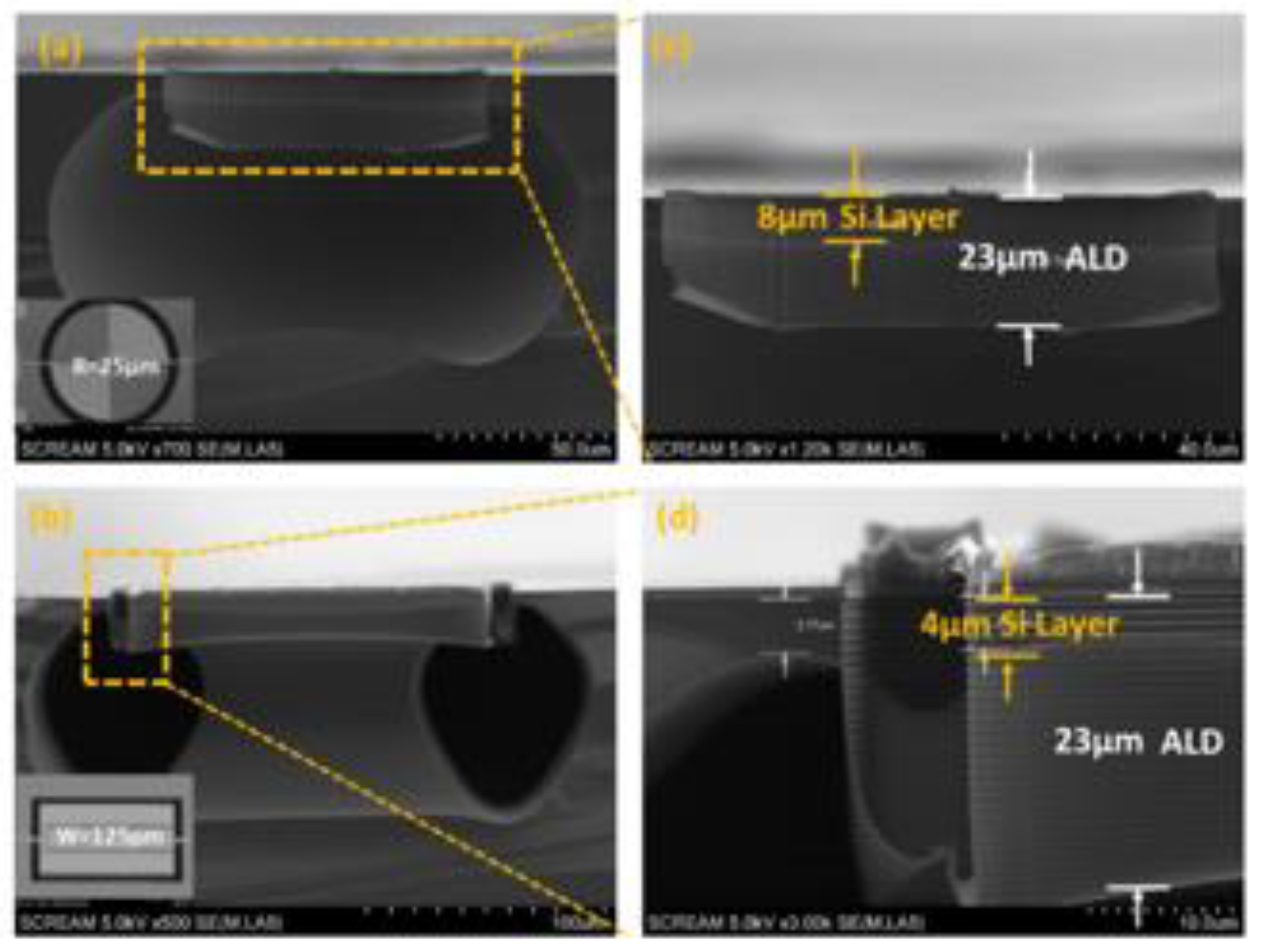

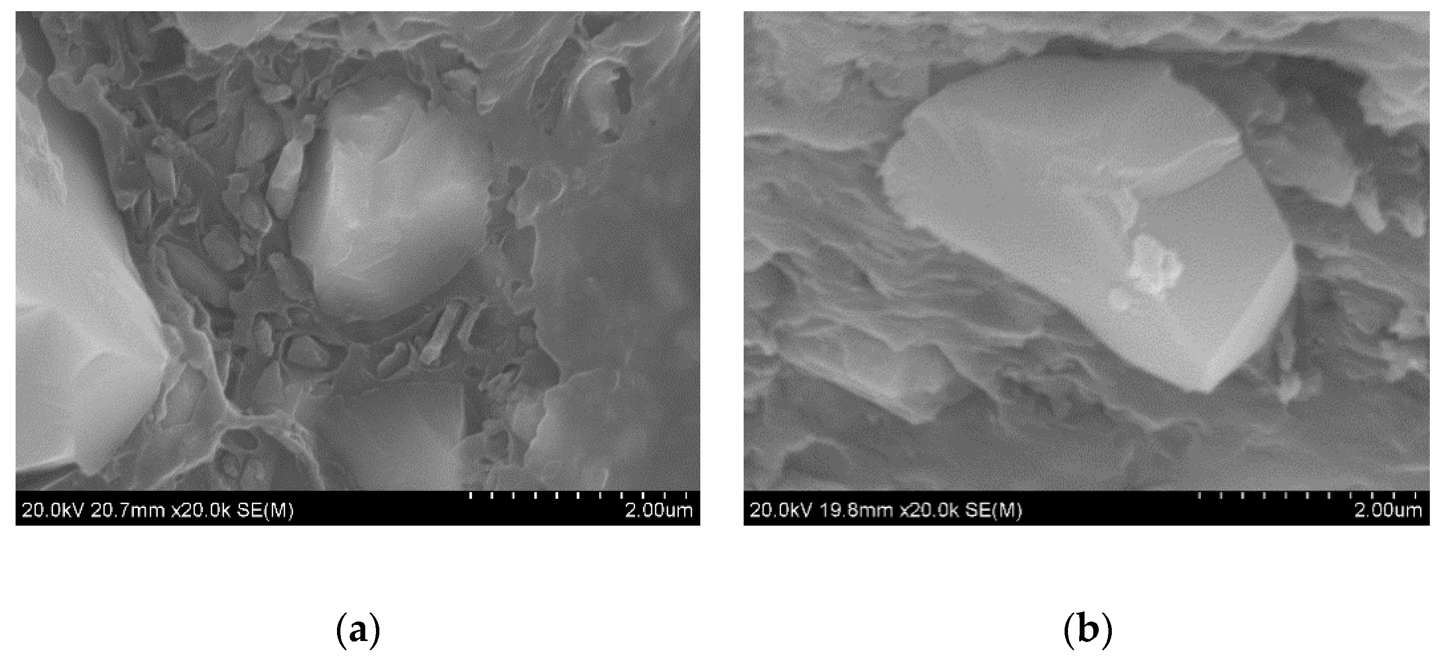
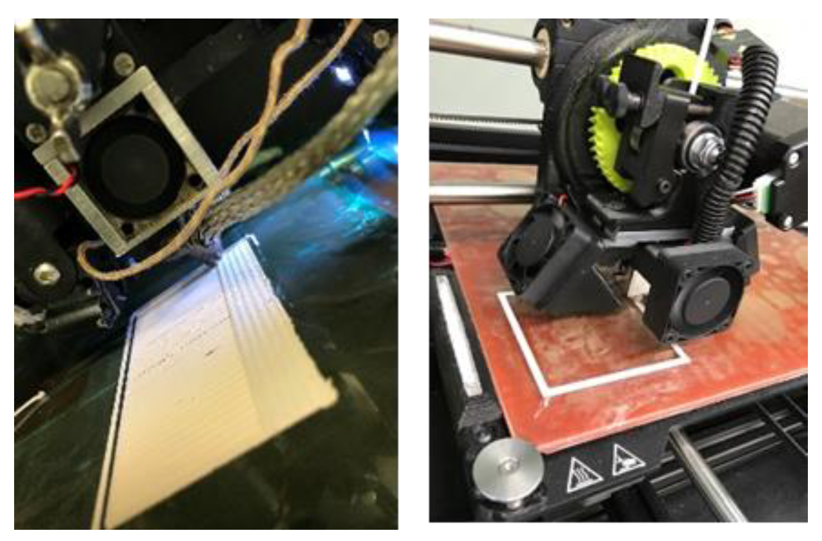
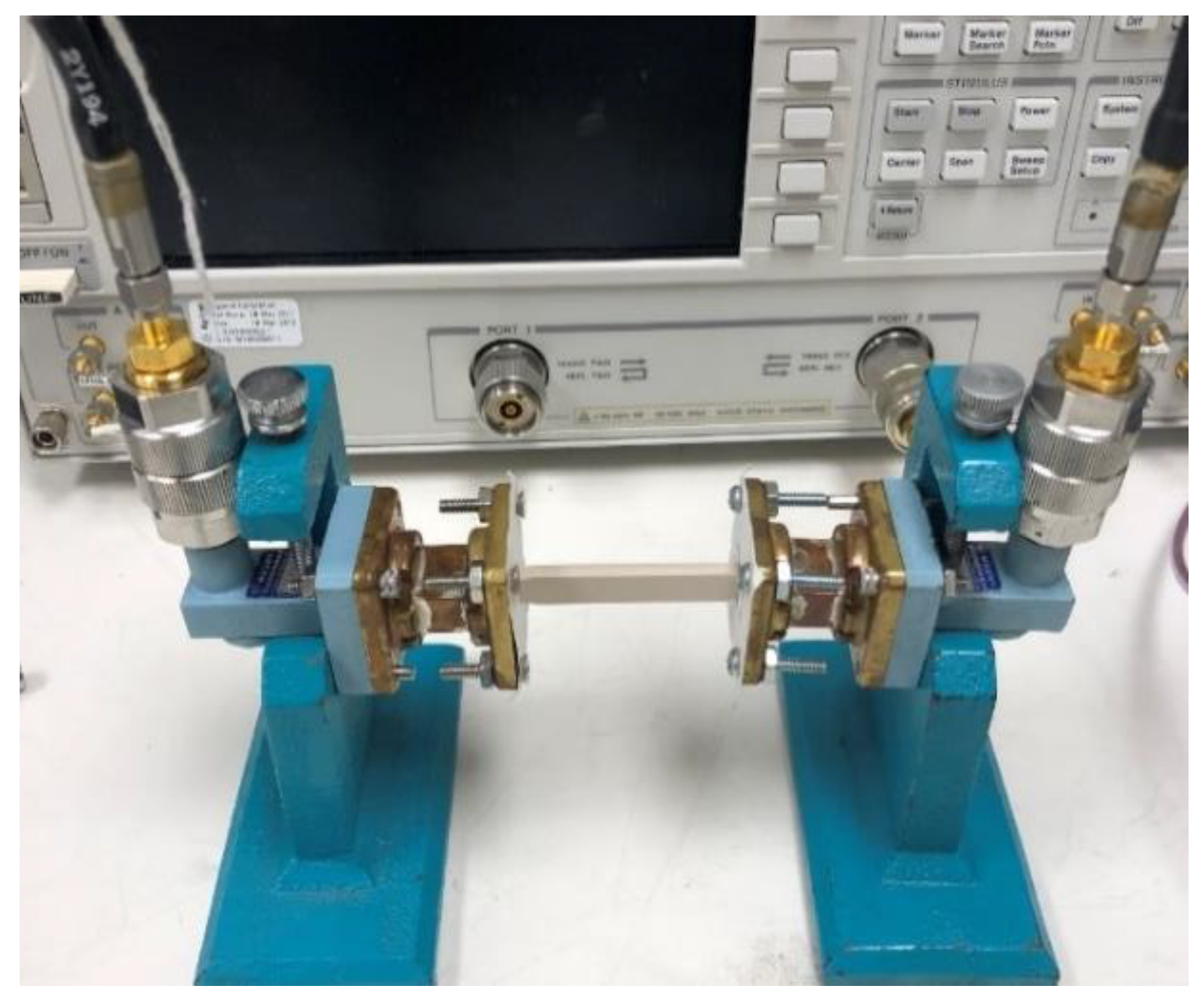
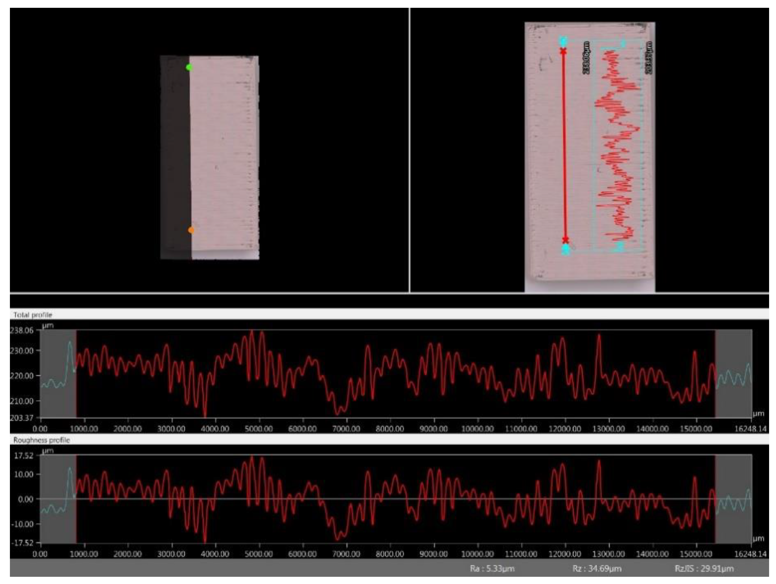


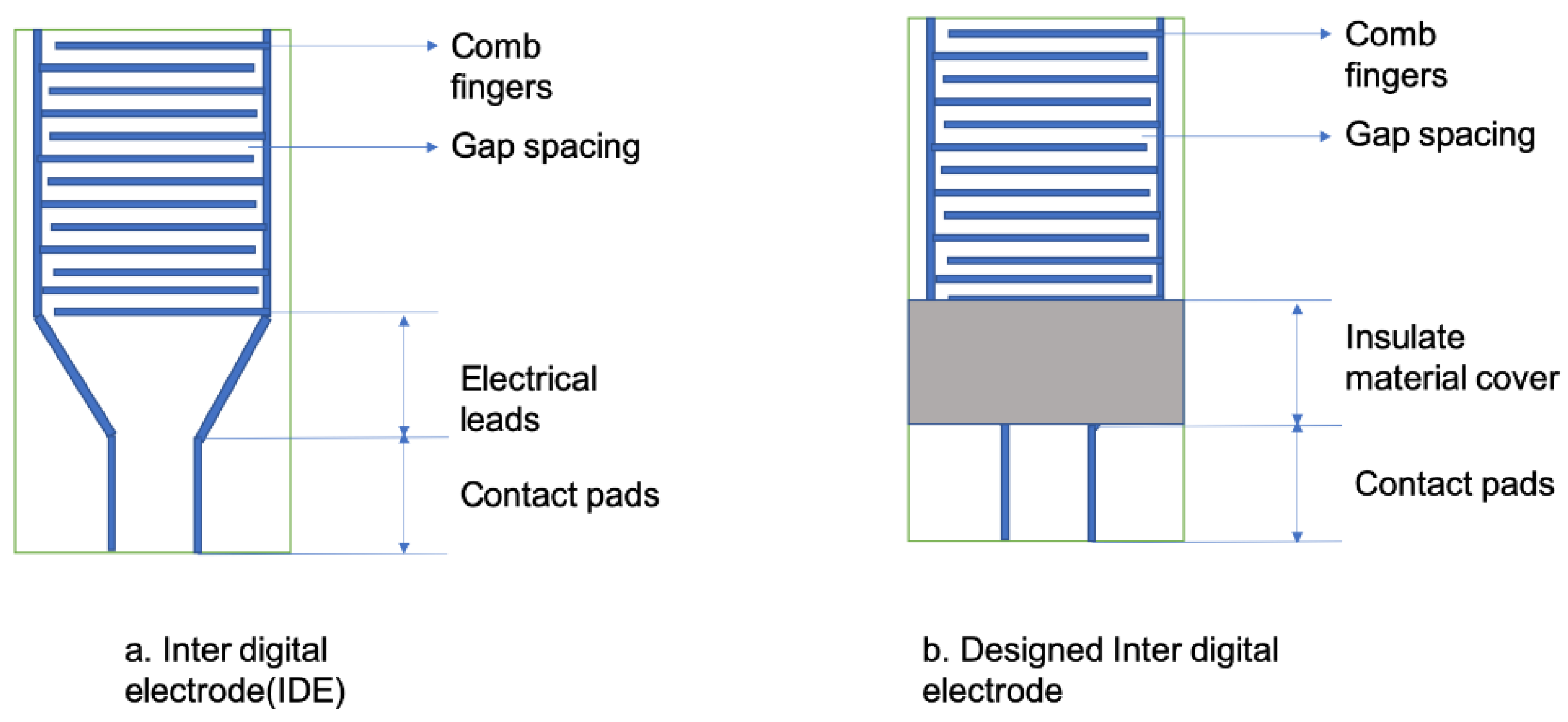
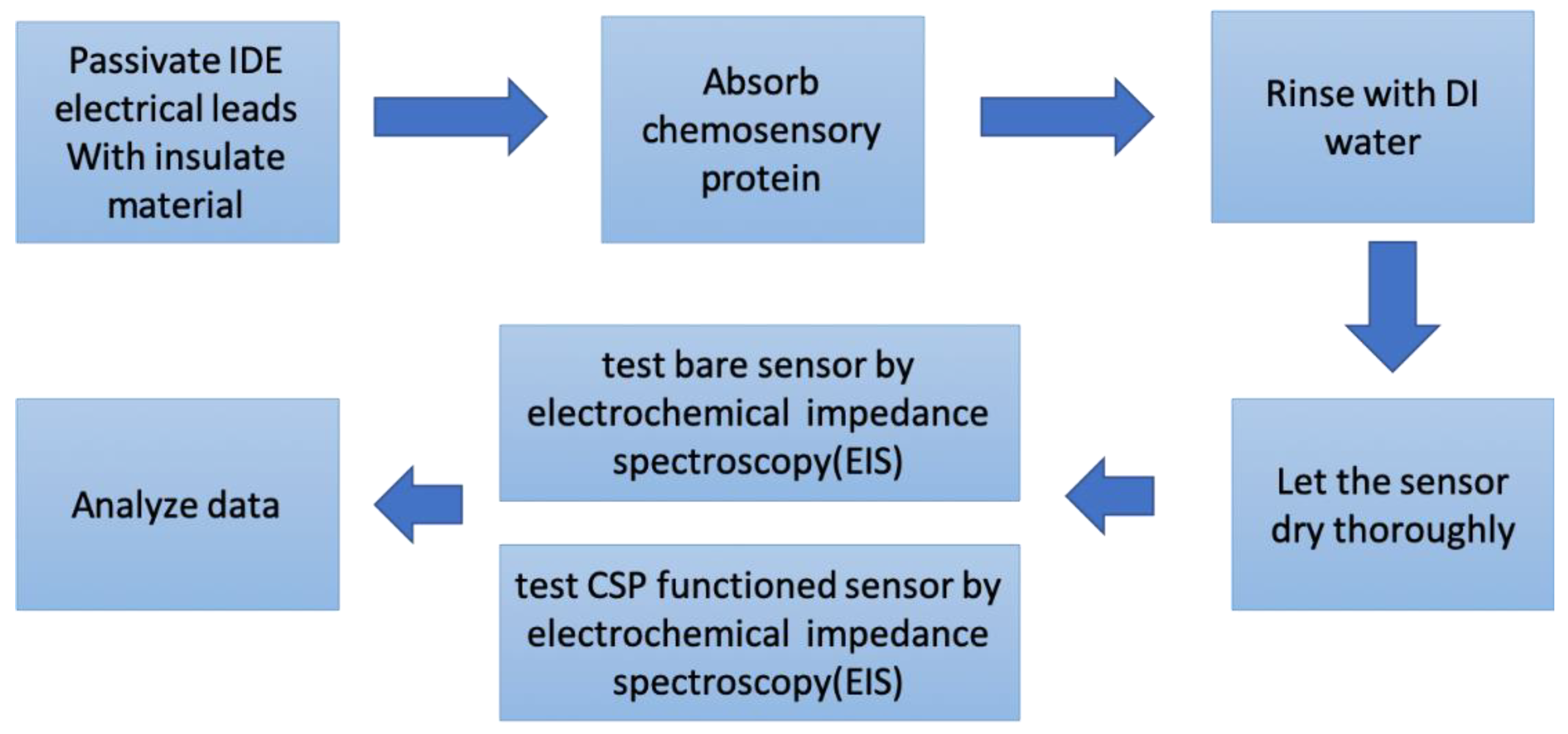
