Communication

Elastase/Collagenase Inhibition Compositions of Citrus unshiu and Its Association with Phenolic Content and Anti-Oxidant Activity

1
Subtropical Horticulture Research Institute, Jeju National University, Jeju 63243, Korea
2
Faculty of Biotechnology, College of Applied Life Science, SARI, Jeju National University, Jeju 63243, Korea
3
Faculty of Biotechnology, College of Applied Life Sciences and Research Institute for Subtropical Agriculture and Biotechnology, SARI, Jeju National University, Jeju 63243, Korea
*
Author to whom correspondence should be addressed.
Appl. Sci. 2020, 10(14), 4838; https://doi.org/10.3390/app10144838
Submission received: 23 June 2020 / Revised: 13 July 2020 / Accepted: 13 July 2020 / Published: 14 July 2020

Abstract

:
Citrus fruits are rich sources of different phytochemicals for human health due to their high anti-oxidant capacity. However, the anti-aging effect of citrus fruits has not been well understood. In this study, methanol extracts taken at various developmental stages from tissues of Citrus unshiu was used to investigate its anti-aging effect by an elastase/collagenase inhibition assay, and a gas chromatography-mass spectrometry (GC-MS) analysis was carried out to identify the potential anti-aging compositions. The elastase/collagenase inhibitory activity was greatest in the flesh of immature green fruit (i.e., early July flesh (EJF)), and four candidate compounds were selected by GC-MS and evaluated by a collagenase inhibition assay. Three of the four candidate compounds (heptadecanoic acid, D-allose, and 5-hydroxymethyl-2-furaldehyde (HMF)) showed anti-aging activity, and the activity was highest in heptadecanoic acid, followed by D-allose and HMF. The total phenolic content (TPC), total flavonoid content (TFC), and anti-oxidant activity (DPPH and ferric reducing anti-oxidant power (FRAP) assay) were also investigated. Interestingly, the patterns of the total phenolic/flavonoid content and the anti-oxidant activity were different from that of the elastase/collagenase inhibitory activity. Flowers had the most anti-oxidant activity followed by immature fruit, and the fruit peels had more anti-oxidant activity than its flesh at all stages of development. This study demonstrated that the flesh of immature fruit and flowers of C. unshiu could be sources of anti-aging and anti-oxidant agents for human health, respectively.

1. Introduction

Citrus is one of the most important horticultural crops on Jeju Island of South Korea, and the satsuma mandarin Citrus unshiu is the most important cultivar. Of all the land on the island used for citrus cultivation in 2017, more than 80% was dedicated to C. unshiu (18,020 ha out of 20,333 ha). Citrus fruits are not only flavorful and attractive to consumers, but they are also a valuable component of a healthy human diet. Citrus fruits contain a variety of phytochemicals, including vitamins, carotenoids, phenolic compounds, coumarins, limonoids, pectins, and minerals that serve diverse roles in human health [1]. Numerous studies have established the anti-oxidant activity of phenolics and carotenoids in extracts from various citrus species and plant parts, specifically the peel and flesh of the fruit and rootstocks [2,3,4,5,6]. Phenolic compounds from citrus fruits are not only associated with anti-oxidant activity, but they have anti-inflammatory and anti-microbial activity as well. They may also reduce the risk of cancer, alcoholic liver disease, and cardiovascular disease [7,8,9]. In addition to the numerous beneficial effects of the anti-oxidant activity mentioned above, they may also have anti-aging activity. Several studies have reported on the anti-aging of medicinal plants, such as Crocus sativus [10], Rosmarinus Officinalis [11], Manilkara zapota [12], Coccinia grandis [13], and Cucumis sativas [14]. However, to our knowledge there is little information on the anti-aging effect in citrus fruits during developmental stages and on their association with anti-oxidant activity.
We selected skin anti-aging as our metric for anti-aging activity. There are intrinsic factors (such as hormone levels, genotypes, endocrine metabolism) and extrinsic factors (such as ultraviolet radiation, nutritional levels, chemical pollution) in the aging process in human skin [15]. Intrinsic aging has a genetic basis and is characterized by cellular senescence. Skin exposure to solar radiation (photo-aging) accumulates reactive oxygen species (ROS) and activates dermal enzymes such as elastase and collagenase, and then these enzymes promote premature skin aging. As skin ages, the elastin and collagen levels decline, resulting in visible wrinkles and sagging. Elastin, the elastic protein in connective tissue, maintains skin elasticity [16] and is degraded in the extra-cellular matrix by elastase, a serine proteinase. Collagen is the main structural protein of connective tissues and maintains skin flexibility [17]. The enzyme collagenase is responsible for remodeling the extra-cellular matrix, which includes breaking the peptide bonds in collagen.
In the present study, elastase and collagenase inhibitory activities are investigated from the extracts of the leaves, flower, and fruits of C. unshiu at different stages of development, and we identify three candidate substances involved in the anti-aging by a gas chromatography-mass spectrometry (GC-MS) analysis and a collagenase inhibition assay. We also assess any correlation between anti-aging activity, total phenolic content (TPC), total flavonoid content (TFC), and anti-oxidant activity. We hope that the information in this study will be proven useful in the utilization of C. unshiu fruit in anti-oxidant products and in new applications in anti-aging products.

2. Materials and Methods

2.1. Chemicals

Methanol (MeOH) was purchased from Fisher Scientific (Seoul, Korea). Other chemicals are as follows: 5-hydroxymethyl-2-furaldehyde (HMF), heptadecanoic acid, D-allose, α-tocopherol acetate, 2,2-diphenyl–1 picrylhydrazyl (DPPH), a Folin–Ciocalteu reagent, sodium carbonate, NaNO2, AlCl3, NaOH, catechin, gallic acid, and dimethyl sulfoxide (DMSO); these were purchased from Sigma-Aldrich (St. Louis, MO, USA).

2.2. Plant Materials

The fruits, leaves, and flower of C. unshiu were sampled from the Research Institute for Subtropical Agriculture and Biotechnology in Jeju National University, South Korea. Fruits were harvested at the following stages of development: late June (green fruit stage: late June fruit (LJF)), early July (green fruits in the early stage of fruit expansion: early July peel (EJP) and flesh (EJF)), middle September (green fruits in the late stage of fruit expansion: middle September peel (MSP) and flesh (MSF)), early October (early stage of fruit color turning: early October peel (EOP) and flesh (EOF)), middle October (late stage of fruit color turning: middle October peel (MOP) and flesh (MOF)), and late November (maturity: late November peel (LNP) and flesh (LNF)). Except for the LJF, the peels and flesh of fruits were separated (Figure 1). All samples were lyophilized and powdered.

2.3. Sample Preparation

For the measurement of anti-elastase and anti-collagenase activity, 50 mg of lyophilized power of each sample was mixed with 1 mL of 80% (v/v) MeOH, shaken for 72 h in the dark, and centrifuged at 10,000 rpm for 5 min. The supernatant was transferred to a tube, and the residue was washed with 0.5 mL 80% (v/v) MeOH. Both supernatants were combined and filtered through a 0.45-μm syringe filter. The filtrate was removed by a centrifugal evaporator (CVE-3000, EYELA, Tokyo, Japan) and then dissolved in DMSO to a concentration of 100 mg/mL. These extracts were diluted to 10 mg/mL concentration with DMSO and stored at −20 °C until use for the assay.

2.4. In Vitro Elastase and Collagenase Inhibition Assay

For the elastase inhibition assay, we used the EnzChek Elastase Assay kit (Molecular Probes Inc., Eugene, OR, USA) and the previously described method by Chattuwatthana and Okello [13]. Porcine pancreatic elastase (100 μL; 0.5 units/mL) was mixed with 50 μL of plant extract (10 mg/mL) in a 96-well micro-plate and incubated for 30 min at 25 °C in the dark. DMSO (50 μL) and an elastase inhibitor (N-methoxysuccinyl-Ala-Ala-Pro-Val-chloromethyl ketone (MAAPV), 50 μL of 0.25 µM, and 1.25 µM) was used as the control (no inhibitor) and positive control, respectively. The blank contained distilled water (50 μL) and a 1× buffer working solution (100 μL). After the pre-incubation, 50 μL of the elastin working solution (100 µg/mL) was added to each well, and the solutions were incubated for 2 h at 25°C in the dark. The fluorescence intensity of the sample in each well was measured by a fluorescence micro-plate reader (Fluoroskan, Thermo Fisher, Vantaa, Finland) at an excitation wavelength of 485 nm and an emission wavelength of 520 nm. The collagenase inhibition assay was performed according to the manufacturer’s directions in the EnzChek Collagenase Assay kit (Molecular Probes Inc.). In this assay, the concentration of plant extracts and four candidate chemical compounds were 10 mg/mL and 0.05 mM, respectively. The fluorescence intensity of the sample in each well was measured by a fluorescence micro-plate reader (Fluoroskan, Thermo Fisher) at an excitation wavelength of 490 nm and an emission wavelength of 515 nm. The percentage of elastase/collagenase inhibitory activity was calculated as follows: inhibition (%) = (1 − S/C) × 100, where “S” and “C” indicate the fluorescence intensity of the sample and control, respectively.

2.5. GC-MS Analysis of Citrus Extracts

The citrus extracts were analyzed using gas chromatography-mass spectrometry (GC-MS, Shimadzu QP 2010 Plus, Tokyo, Japan). An Rtx-5MS column (30 m × 0.25 mm ID, film thickness 0.25 µm) was used with helium as a carrier gas. The column oven program was as follows: 70 °C (5 min), 70 °C to 310 °C (5 °C/min), 310 °C (5 min); injector temperature 230 °C; and detector temperature 250 °C. The chemical compounds were identified by their retention time and compared with the mass spectra from the National Institute of Standards and Technology (NIST) library and WILEY library.

2.6. Analysis of Total Phenolic Content (TPC)

The TPC of plant extracts was determined with a modified Folin–Ciocalteu method [18]. Twenty microliters of the plant samples (5 mg/mL), standard, and blank (DMSO) were added in a 96-well micro-plate and then mixed with a 10% (v/v) Folin–Ciocalteu reagent (100 µL). After incubation for 5 min at room temperature, a 7.5% (w/v) sodium carbonate solution (80 µL) was added to each assay well and was allowed to react for 60 min at 25 °C in the dark. Absorbance was measured by a micro-plate reader at the wavelength of 750 nm. Gallic acid was used as a standard at 12.5–400 µg/mL to generate a calibration curve (Figure S1A). All assays were performed in triplicate. The results were expressed as milligrams of gallic acid equivalent (GAE) per gram of the dried plant extract.

2.7. Analysis of Total Flavonoid Content (TFC)

The TFC of plant extracts was determined with a colorimetric aluminum chloride method as described by Herald et al. [19]. Briefly, 25 µL of each plant extract (5 mg/mL) and standard were added in a 96-well micro-plate and then mixed with 100 µL of distilled water and 10 µL of 50 g/L NaNO2. After incubation for 5 min at room temperature, 15 µL of 100 g/L AlCl3 was added to each assay well and then further incubated for 6 min at room temperature. After that, 50 µL of 1N NaOH and 50 µL of distilled water were added to each well, and absorbance was measured by a micro-plate reader at a wavelength of 510 nm. Catechin was used as a standard at 5–100 µg/mL to generate a calibration curve (Figure S1B). All assays were performed in triplicate. The results were expressed as milligrams of catechin equivalent per gram of the dried plant extract.

2.8. DPPH Radical Scavenging and Ferric Reducing Anti-Oxidant Power (FRAP) Activity Assay

Free radical scavenging activity of the extracts was determined by a stable 2,2-diphenyl–1 picrylhydrazyl (DPPH) assay according to the method of Herald et al. [19]. Catechin was used as a standard control at 0.05–0.4 mg/mL. The results were expressed as the DPPH radical scavenging activity (%) of the dried plant extract. The FRAP assay was performed according to the manufacturer’s directions in the FRAP assay kit (Abcam). Each assay was performed in triplicate. The results were expressed as µmol of the FRAP value per gram of the dried plant extract.

2.9. Cytotoxicity Assay

The cytotoxicity of the citrus extracts was determined by the MTT colorimetric assay as described by Mosmann [20], with some modification. Briefly, 293T and PANC-1 cells in suspension were seeded at a rate of 2.5 × 10⁴ cells per well in a 24-well micro-titer plate and placed in an incubator for 24 h at 37 °C in 5% CO2. Citrus extracts (250 μg/mL culture medium) were added to the wells, and the plates were incubated for another 72 h at 37 °C; the culture medium was then removed, and the MTT solution (0.25 mg in 500 μL of medium) was added to each well. DMSO, which was used to dissolve the citrus extracts, was used as a control. The micro-titer plate was incubated for another 30 min at 37 °C. The MTT solution was carefully removed with a paper towel, and DMSO (0.5 mL) was added. The DMSO (200 μL) in each well was transferred to a 96-well micro-plate, and the absorbance was measured at 540 nm in a micro-plate reader. The results were expressed as the cell viability (%) of the dried plant extract.

2.10. Statistical Analysis

The results reported in this study have been expressed as the mean ± SD of three replicates. All statistical analyses were carried out using IBM SPSS software (SPSS for Windows, version 20, SPSS Inc., Armonk, NY, USA). Significant differences among the samples were calculated using a one-way analysis of variance (ANOVA), followed by Duncan’s multiple-range test at the 5% level (p < 0.05).

3. Results

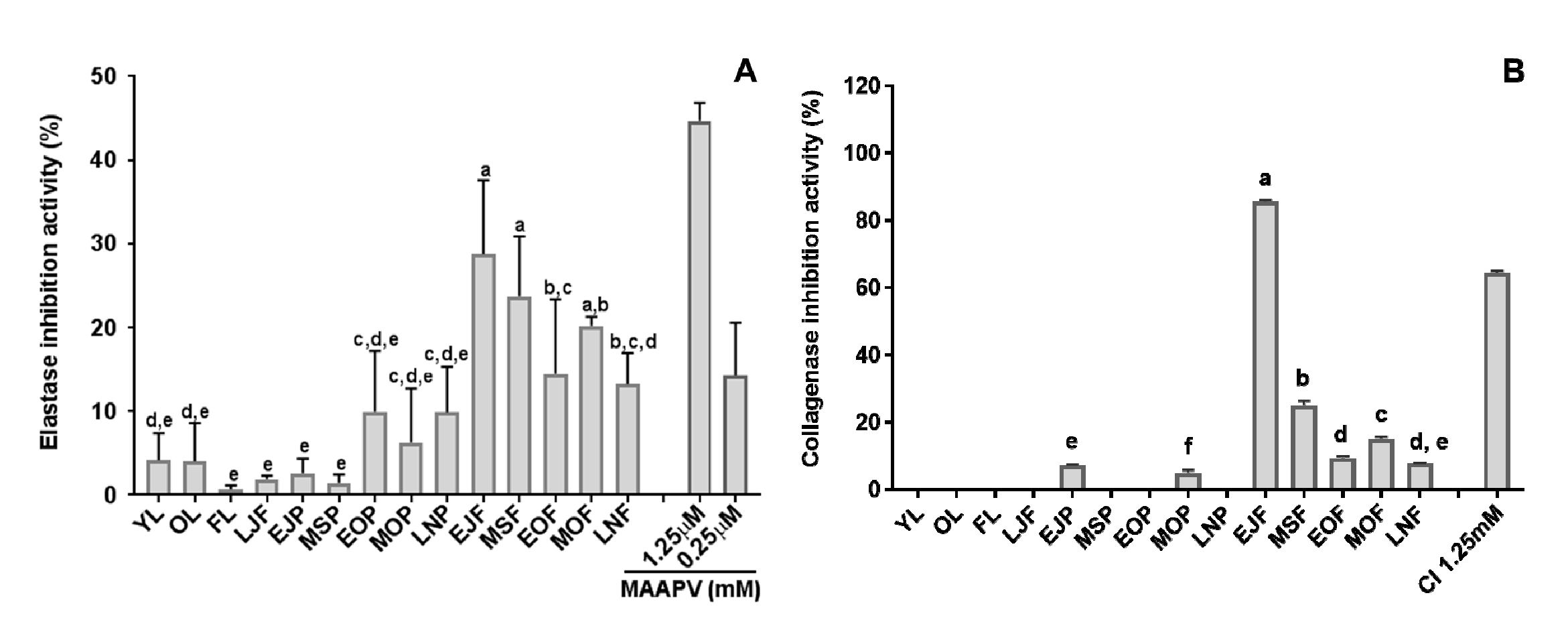
3.1. Elastase and Collagenase Inhibitory Activity

Skin aging is caused by the alteration of the dermal connective tissue and is due to intrinsic and extrinsic factors. Aging is an intrinsic factor, while chronic sun exposure is an example of an extrinsic factor. When skin ages, the amount of elastin and collagen decreases, resulting in the loss of strength and flexibility in the skin. Elastase and collagenase are responsible for the breakdown of elastin and collagen in the extra-cellular matrix, respectively. We assessed the anti-aging properties of the extracts by measuring their ability to inhibit elastase and collagenase activity. The results are shown in Figure 2. The elastase inhibitory activity was greater in the flesh of fruit (13–29%) than in the peels (1–10%) at all fruit-maturity stages (Figure 2A). Considering only the flesh of the fruit, the immature green stages (EJF and MSF) had more activity than the color-turning stages (EOF and MOF) and mature stages (LNF). The leaf and flower extracts had very little elastase inhibitory activity (0.6–4%). In the collagenase inhibition assay, the inhibitory activity was higher in the flesh (9.7–106.6%) than the peel (0–8.9%) at all fruit-maturity stages (Figure 2B). The most collagenase inhibition showed in the EJF stage (85%). The EJF extract was examined in more detail by a dilution concentration of the extract (Figure S2). The anti-collagenase activity was highly dependent on the concentration of the extract. Although the inhibitory activity of elastase and collagenase was somewhat different, the EJF stage had the greatest inhibitory activity in both enzyme assays.

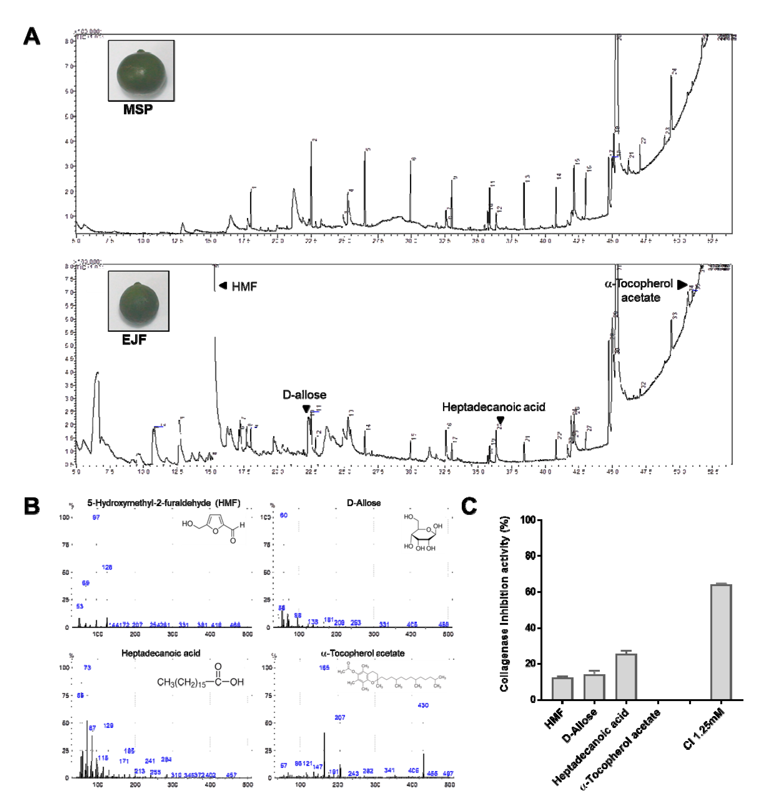
3.2. Chemical Composition by GC-MS Analysis and Anti-Aging Effect

We found that the EJF extract has the most elastase and collagenase inhibitory activity. A GC-MS analysis carried out to know which chemical compositions are involved in the elastase/collagenase inhibition from the EJF extract. In addition, the MSP extract, which showed no the inhibition activity, was used in the GC-MS analysis to be compared with the EJF extract. A total of 46 and 33 compounds were detected from the EJF and the MSP extracts, respectively (Figure 3A, Tables S1 and S2), and we subtracted the chemical compounds detected in both extracts. After this, from the remaining chemical compounds in the EJF extract, four candidate compounds related to anti-aging and anti-oxidant activity through previous literature search were selected as follows: 5-hydroxymethyl-2-furaldehyde (HMF, 26.67%), D-allose (2.77%), heptadecanoic acid (0.69), and α-tocopherol acetate (0.1%) (Figure 3B, Tables S1 and S2). Investigating which of these candidate substances has an anti-aging effect was performed through the collagenase inhibition assay (Figure 3C). The collagenase inhibitory activity was highest in heptadecanoic acid (25.6%), followed by D-allose (14.2%) and HMF (12.2%), but α-tocopherol acetate showed no inhibitory activity.

3.3. Total Phenolic and Flavonoid Contents of Citrus Fruits and Tissues

Phenolic and flavonoid compounds are secondary metabolites that are ubiquitously distributed in most plant tissues, and they have diverse benefits in the human diet. The total phenolic content (TPC) of citrus tissues and fruits at different fruit-maturity stages was determined by the Folin–Ciocalteu method and expressed as mg of GAE per g dry weight (DW) (Figure 4A). The TPCs of flowers (FL) averaged 17.3 mg GAE/g DW, which was significantly greater than the other plant parts; the second greatest TPC value was observed in whole fruits harvested in late June (late June fruits (LJF), 12.7 mg GAE/g DW). The TPC of the peel did not vary significantly at any of the stages of fruit maturity and ranged from 9.0 to 11.7 mg GAE/g DW. In contrast, the TPC of the flesh of fruits gradually decreased as the fruit matured and ripened, from 8.8 to 5.9 mg GAE/g DW. In addition, at all fruit-maturity stages the peels contained more phenolic compounds than the flesh. The pattern of total flavonoid content (TFC) of citrus tissues and fruits at different fruit-maturity stages was similar to that of TPC (Figure 4B). TFC was also greater in the peels than the flesh.

3.4. Anti-Oxidant Activity of Citrus Fruits and Tissues

The anti-oxidant activity of citrus tissues and fruits was determined by the 2,2-diphenyl–1 picrylhydrazyl (DPPH) and Ferric reducing anti-oxidant power (FRAP) assays. The DPPH free radical scavenging activity of the citrus extracts is shown in Figure 4C. Scavenging activity was greatest in the extracts from flowers (72%), and the second greatest levels were in LJF (66%). Moreover, the scavenging activity was greater in the peels (40–66%) than in the flesh of the fruit (19–37%) at all fruit-maturity stages. The patterns of anti-oxidant activity by the FRAP assay were generally consistent with the DPPH assay (Figure 4D).

3.5. Cytotoxicity

We wished to ensure that the extracts were not cytotoxic, since we anticipate that their anti-oxidant and anti-aging properties might be useful therapeutically. The cytotoxicity of the extracts was evaluated with the MTT assay using the 293T and PANC-1 cell line. We used extracts from flowers and immature fruits (LJF and EJF), which had high anti-oxidant and anti-aging activity. The cytotoxicity of the extracts was not significantly greater than the control (Figure 5). We conclude that citrus extracts can be used therapeutically for anti-oxidant and anti-aging purposes without cytotoxicity.

4. Discussion

In the present study, we first investigated the elastase/collagenase inhibitory activity of tissues and different fruit-maturity stages of C. unshiu. In both enzyme inhibition assays, the EJF extract showed the most enzyme inhibition activity of all tested samples. Therefore, we tried to determine potential chemical compounds from the EJF extract by a GC-MS analysis and selected four candidate chemical compounds that are related to anti-aging by a search of previous reported literatures. To confirm the four candidate compounds, we further examined these through a collagenase inhibition assay and identified three chemical compounds involved in anti-aging, namely heptadecanoic acid, D-allose, and HMF. Heptadecanoic acid, as a fatty acid, showed the highest collagenase inhibition activity of the three compounds in this study. Free fatty acids as essential structural components of biological membrane are known to play a regulatory effect on melanogenesis. Renneret and Melzig reported that the heptadecanoic acid of the free fatty acids tested showed the most inhibition activity of Clostridium histolyticum collagenase [21]. D-allose, a rare sugar, has various physiological functions with anti-cancer, anti-oxidant, anti-inflammatory properties and other health benefits [22,23,24]. Recently, Ju et al. (2020) reported that D-allose suppresses oxidative stress and protects skin flap from the injury of ischemia/reperfusion (I/R) [25]. This could explain the anti-aging potential of D-allose, together with our result. HMF has been extensively studied for its anti-proliferative, anti-oxidant, anti-cancer, and anti-inflammatory effects [26,27]. To our knowledge, there was no study on HMF to anti-aging, but a related study has recently been conducted by Niu et al. [28]. They found that mesosilica-supported 5-HMF has a protective function on UV-induced aging of human dermal fibroblasts. Together with our results and previous reports, it is suggested that heptadecanoic acid, D-allose, and HMF participate in anti-aging processes with other unknown chemical compositions. Although the three compounds for anti-aging were identified from C. unshiu, there are non-polar compounds from the GC-MS analysis. It is suggested that other compounds for preventing skin aging may also be present in polar compounds.
As for the correlation between the anti-aging and anti-oxidant properties of C. unshiu extracts, a positive correlation was not found in this study. Some recent reports support our anti-aging results. Pientaweeratch et al. (2016) also did not observe a correlation between the TPC and anti-aging properties of sapota fruit [12]. They observed low levels of phenolics and undetectable flavonoids, but they detected considerable elastase and collagenase inhibitory activity. Similarly, in extracts from the bark of Cochlospermum vitifolium, considerable elastase inhibition activity was present, despite little anti-oxidant activity [29]. In a Trolox-equivalent anti-oxidant capacity assay, the extract from lavender and witch hazel leaves had somewhat high anti-oxidant activity but had undetectable or low elastase inhibitory activity [16]. There are contrasting reports that link anti-aging and anti-oxidant activity. Certain phenolic compounds (for example, catechin, resveratrol, epicatechin) and the flavonoids (quercetin, kaempferol, and myricetin) have been shown to have anti-elastase activity that is dose-dependent [30,31]. The discrepancy in the results may be explained by whether the extracts contain different types of phenolic compounds or other bioactive components that have anti-aging properties.
The anti-oxidant activity of both DPPH and FRAP assays were positively correlated with the results of the TPC and TFC for the citrus extracts (Figure 4), that is, as phenolics increase, so does anti-oxidant activity. Two studies with highbush blueberry are consistent with this latter result. Kalt et al. [32] showed that the ability of the fruit to absorb oxygen radicals correlated with the TPC during ripening and storage. Castrejón et al. [33] observed a strong correlation between anti-oxidant activity and TPC during fruit maturation and ripening using the Trolox-equivalent anti-oxidant capacity assay. The TPC and TFC were higher in the peel than the flesh of C. unshiu. This result agrees with Park et al. [34], who found that orange peel contains more TPC than the flesh in extracts from various solvents. Elkhatim et al. [3] also measured the TPC and TFC of citrus and concluded that the peels contained much more total phenolics and flavonoids than the pulp and seeds, parts that are wasted during juice production. The correlation between the TPC and TFC that we observed are consistent with the studies cited above.
Based on these results, we suggest that flowers and fruits, particularly at the immature stage, which in our case are those of C. unshiu, may not only be a beneficial component of the human diet but may also have potential as important biological resources for therapeutic and cosmetic products, particularly when they are immature.

Supplementary Materials

The following are available online at https://www.mdpi.com/2076-3417/10/14/4838/s1, Figure S1: (A) Calibration curve of gallic acid for total phenolic content and ({B) Calibration curve of catechin for total flavonoid content, Figure S2: Collagenase inhibitory activity with different concentration of EJF extract, Table S1: Chemical compositions of citrus extract (MSP stage), Table S2: Chemical compositions of citrus extract (EJF stage).

Author Contributions

Conceptualization, C.-H.E. and I.-J.K.; performed the experiments, analyzed the data, and wrote the manuscript, C.-H.E. and M.-S.K.; edited the manuscript draft; I.-J.K. All authors have read and agreed to the published version of the manuscript.

Funding

This research was funded by Basic Science Research Program through the National Research Foundation of Korea (2019R1A6A1A11052070, 2017R1D1A1B06034883, 2016R1A6A1A03012862, and 2016R1A6A3A11933447) and 2019 scientific promotion program funded by Jeju National University.

Acknowledgments

We are grateful to the Subtropical/tropical Organism Gene Bank Center (SOGB) and the Research Institute for Subtropical Agriculture and Biotechnology and Sustainable Agriculture Research Institute (SARI) at Jeju National University for providing experimental facilities.

Conflicts of Interest

The authors declare no conflict of interest.

References

  1. Jayaprakasha, G.K.; Patil, B.S. In vitro evaluation of the antioxidant activities in fruit extracts from citron and blood orange. Food Chem. 2007, 101, 410–418. [Google Scholar] [CrossRef]
  2. Sun, Y.; Tao, W.; Huang, H.; Ye, Y.; Sun, P. Flavonoids, phenolic acids, carotenoids and antioxidant activity of fresh eating citrus fruits, using the coupled in vitro digestion and human intestinal HepG2 cells model. Food Chem. 2019, 279, 321–327. [Google Scholar] [CrossRef] [PubMed]
  3. Elkhatim, K.; Elagib, R.; Hassan, A. Content of phenolic compounds and vitamin C and antioxidant activity in wasted parts of Sudanese citrus fruits. Food Sci. Nutr. 2018, 6, 1214–1219. [Google Scholar] [CrossRef] [PubMed]
  4. Cardenosa, V.; Barros, L.; Barreira, J.C.M.; Arenas, F.; Moreno-Rojas, J.M.; Ferreira, I.C.F.R. Different Citrus rootstocks present high dissimilarities in their antioxidant activity and vitamins content according to the ripening stage. J. Plant Physiol. 2015, 174, 124–130. [Google Scholar] [CrossRef] [Green Version]
  5. Zhang, Y.; Sun, Y.; Xi, W.; Shen, Y.; Qiao, L.; Zhong, L.; Ye, X.; Zhou, Z. Phenolic compositions and antioxidant capacities of Chinese wild mandarin (Citrus reticulata Blanco) fruits. Food Chem. 2014, 145, 674–680. [Google Scholar] [CrossRef]
  6. Legua, P.; Forner, J.B.; Hernández, F.; Forner-Giner, M.A. Total phenolics, organic acids, sugars and antioxidant activity of mandarin (Citrus clementina Hort. ex Tan.): Variation from rootstock. Sci. Hortic. 2014, 174, 60–64. [Google Scholar] [CrossRef]
  7. AL-Jabri, N.N.; Hossain, M.A. Chemical composition and antimicrobial potency of locally grown lemon essential oil against selected bacterial strains. J. King Saud Univ.-Sci. 2018, 30, 14–20. [Google Scholar] [CrossRef] [Green Version]
  8. Rawson, N.; Ho, C.T.; Li, S. Efficacious anti-cancer property of flavonoids from citrus peels. Food Sci. Hum. Well. 2014, 3, 104–109. [Google Scholar] [CrossRef] [Green Version]
  9. Park, H.Y.; Choi, H.D.; Eom, H.; Choi, I. Enzymatic modification enhances the protective activity of citrus flavonoids against alcohol-induced liver disease. Food Chem. 2013, 139, 231–240. [Google Scholar] [CrossRef]
  10. Kumud, M.; Sanju, N. In-vitro evaluation of antioxidant, anti-elastase, anti-collagenase, anti-hyaluronidase activities of safranal and determination of its sun protection factor in skin photoaging. Bioorg. Chem. 2018, 77, 159–167. [Google Scholar]
  11. Duque, L.; Bravo, K.; Osorio, E. A holistic anti-aging approach applied in selected cultivated medicinal plants: A view of photoprotection of the skin by different mechanisms. Ind. Crops Products 2017, 97, 431–439. [Google Scholar] [CrossRef]
  12. Pientaweeratch, S.; Panapisal, V.; Tansirikongkol, A. Antioxidant, anti-collagenase and anti-elastase activities of Phyllanthus emblica, Manilkara zapota and silymarin: An in vitro comparative study for anti-aging applications. Pharm. Biol. 2016, 54, 1865–1872. [Google Scholar] [CrossRef] [PubMed] [Green Version]
  13. Chattuwatthana, T.; Okello, E. Anti-collagenase, anti-elastase and antioxidant activities of Pueraria candollei var. mirifica root extract and Coccinia grandis fruit juice extract: An In vitro study. Eur. J. Med. Plants 2015, 5, 318–327. [Google Scholar] [CrossRef] [Green Version]
  14. Nema, N.K.; Maity, N.; Sarkar, B.; Mukherjee, P.K. Cucumis sativus fruit-potential antioxidant, anti-hyaluronidase, and anti-elastase agent. Arch. Dermatol. Res. 2011, 303, 247–252. [Google Scholar] [CrossRef] [PubMed]
  15. Jenkins, G. Molecular mechanisms of skin aging. Mech. Aging Dev. 2002, 123, 801–810. [Google Scholar] [CrossRef]
  16. Thring, T.S.; Hili, P.; Naughton, D.P. Anti-collagenase, anti-elastase and anti-oxidant activities of extracts from 21 plants. BMC Comp. Alter. Med. 2009, 9, 27. [Google Scholar] [CrossRef] [Green Version]
  17. Mukherjee, P.K.; Maity, N.; Nema, N.K.; Sarkarm, B.K. Bioactive compounds from natural resources against skin aging. Phytomedicine 2011, 19, 64–73. [Google Scholar] [CrossRef]
  18. Zhang, Q.; Zhang, J.; Shen, J.; Silva, A.; Dennis, D.A.; Barrow, C.J. A simple 96-well microplate method for estimation of total polyphenol content in seaweeds. J. Appl. Phycol. 2006, 18, 445–450. [Google Scholar] [CrossRef] [Green Version]
  19. Herald, T.J.; Gadgil, P.; Tilley, M. High-throughput micro plate assays for screening flavonoid content and DPPH-scavenging activity in sorghum bran and flour. J. Sci. Food Agri. 2012, 92, 2326–2331. [Google Scholar] [CrossRef] [PubMed]
  20. Mosmann, T. Rapid colorimetric assay for cellular growth and survival: Application to proliferation and cytotoxicity assays. J. Immunol. Methods 1983, 65, 55–63. [Google Scholar] [CrossRef]
  21. Rennert, B.; Melzig, M.F. Free fatty acids inhibit the activity of Clostridium histolyticum collagenase and human neutrophil elastase. Planta Med. 2002, 68, 767–769. [Google Scholar] [CrossRef]
  22. Noguchi, C.; Kamitori, K.; Hossain, A.; Hoshikawa, H.; Katagi, A.; Dong, Y.; Sui, L.; Tokuda, M.; Yamaguchi, F. D-Allose inhibits cancer cell growth by reducing GLUT1 expression. Tohoku J. Exp. Med. 2016, 238, 131–141. [Google Scholar] [CrossRef] [PubMed] [Green Version]
  23. Nakamura, T.; Tanaka, S.; Hirooka, K.; Toyoshima, T.; Kawai, N.; Tamiya, T.; Shiraga, F.; Tokuda, M.; Keep, R.F.; Itano, T. Anti-oxidative effects of D-allose, a rare sugar, on ischemia–reperfusion damage following focal cerebral ischemia in rat. Neurosci. Lett. 2011, 48, 103–106. [Google Scholar] [CrossRef] [PubMed]
  24. Huang, T.; Gao, D.; Hei, Y.; Zhang, X.; Chen, X.; Fei, Z. D-Allose protects the blood brain barrier through PPAR gamma-mediated anti-inflammatory pathway in the mice model of ischemia reperfusion injury. Brain Res. 2016, 1642, 478–486. [Google Scholar] [CrossRef] [PubMed]
  25. Ju, J.; Hou, R.; Zhang, P. D-allose alleviates ischemia/reperfusion (I/R) injury in skin flap via MKP-1. Mol. Med. 2020, 26, 21. [Google Scholar] [CrossRef]
  26. Zhao, L.; Chen, J.; Su, J.; Ki, L.; Hu, S.; Li, B.; Zhang, X.; Xu, Z.; Chen, T. In vitro antioxidant and antiproliferative activities of 5-hydroxymethylfurfural. Agri. Food Chem. 2013, 61, 10604–10611. [Google Scholar] [CrossRef]
  27. Kim, H.K.; Choi, Y.W.; Lee, E.N.; Park, J.K.; Kim, S.G.; Park, D.J.; Kim, B.S.; Lim, Y.T.; Yoon, S. 5-Hydroxymethylfurfural from black garlic extract prevents TNFα-induced monocytic cell adhesion to HUVECs by suppression of vascular cell adhesion molecule-1 expression, reactive oxygen species generation and NF-κB activation. Phytother. Res. 2011, 25, 965–974. [Google Scholar] [CrossRef]
  28. Niu, X.; Wang, Z.; Zhang, L.; Quan, Y.; Wei, K. Experimental study of the protective effect of mesosilica-supported 5-hydroxymethylfurfural on UV-induced aging of human dermal fibroblasts. RSC Adv. 2018, 8, 25021–25030. [Google Scholar] [CrossRef] [Green Version]
  29. German-Báez, L.J.; Valdez-Flores, M.; Figureueroa-Pérez, M.G.; Garduño-Félix, K.G.; Valdez-Ortiz, R.; Meza-Ayala, K.A.; Valdez-Ortiz, A. Anti-aging and nutraceutical characterization of plant infusions used in traditional medicine. Pakistan J. Nutr. 2017, 16, 285–292. [Google Scholar]
  30. Kanashiro, A.; Souza, J.G.; Kabeya, L.M.; Azzolini, A.E.C.S.; Lucisano-Valim, Y.M. Elastase release by stimulated neutrophils inhibited by flavonoids: Importance of the catechol group. Verlag der Zeitschrift für Naturforschung 2007, 62, 357–361. [Google Scholar] [CrossRef]
  31. Wittenauer, J.; Mäckle, S.; Sußmann, D.; Schweiggert-Weisz, U.; Carle, R. Inhibitory effects of polyphenols from grape pomace extract on collagenase and elastase activity. Fitoterapia 2015, 101, 179–187. [Google Scholar] [CrossRef] [PubMed]
  32. Kalt, W.; Lawand, C.; Ryan, D.J.; McDonald, J.E.; Donner, H.; Forney, C.F. Oxygen radical absorbing capacity, anthocyanin and phenolic content of highbush blueberries (Vaccinium corymbosum L.) during ripening and storage. J. Amer. Soc. Horticul. Sci. 2003, 126, 917–923. [Google Scholar] [CrossRef]
  33. Castrejón, A.D.R.; Eichholz, I.; Rohn, S.; Kroh, L.W.; Huyskens-Keil, S. Phenolic profile and antioxidant activity of highbush blueberry (Vaccinium corymbosum L.) during fruit maturation and ripening. Food Chem. 2008, 109, 564–572. [Google Scholar]
  34. Park, J.H.; Lee, M.; Park, E. Antioxidant activity of orange flesh and peel extracted with various solvents. Prev. Nut. Food Sci. 2014, 19, 291–298. [Google Scholar] [CrossRef] [PubMed] [Green Version]
Figure 1. Photographs of leaves, flower, and fruits of C. unshiu at different stages of development. (YL, young leaf; OL, old leaf; FL, flower; LJF, late June fruit; EJP and EJF, early July peel and flesh; MSP and MSF, middle September peel and flesh; EOP and EOF, early October peel and flesh; MOP and MOF, middle October peel and flesh; LNP and LNF, late November peel and flesh).
Applsci 10 04838 g001
Figure 2. Anti-aging activity of tissues and fruit-maturity stages of C. unshiu. (A) Elastase inhibitory activity, (B) Collagenase inhibitory activity. Data are expressed as mean ± SD (n = 3), and different small letters indicate a significant difference by Duncan’s test; p < 0.05.
Applsci 10 04838 g002
Figure 3. (A) GC-MS chromatograph of C. unshiu extracts (MSP and EJF). (B) Candidate compounds of anti-aging activity by gas chromatography-mass spectrometry (GC-MS). (C) Collagenase inhibitory activity of four candidate compounds. Data are expressed as mean ± SD (n = 3), and different small letters indicate a significant difference by Duncan’s test; p < 0.05.
Applsci 10 04838 g003
Figure 4. Total phenolic content (TPC), total flavonoid content (TFC), and anti-oxidant activity of tissues and fruit-maturity stages of C. unshiu. (A) TPC, (B) TFC, (C) DPPH radical scavenging activity assay, with catechin (CAT) as a standard, (D) ferric reducing anti-oxidant power (FRAP) activity assay. Data are expressed as mean ± SD (n = 3), and different small letters indicate a significant difference by Duncan’s test; p < 0.05.
Applsci 10 04838 g004
Figure 5. Effect of C. unshiu extracts on 293T and PANC-1 cell viability. Cells were grown in the presence of C. unshiu extracts. The cell viability was determined by an MTT assay in triplicate and expressed in percentage of control (100%).
Applsci 10 04838 g005

