Article

Assessment of the Different Types of Failure on Anterior Cantilever Resin-Bonded Fixed Dental Prostheses Fabricated with Three Different Materials: An In Vitro Study

1
Department of Neurosciences, School of Dentistry, Section of Prosthodontics and Digital Dentistry, University of Padova, 35100 Padova, Italy
2
Departments of Civil, Environmental and Architectural Engineering, University of Padova, 35100 Padova, Italy
3
Departments of Management and Engineering, University of Padova, 36100 Vicenza, Italy
4
Department of Biomedical and Neuromotor Sciences (DIBINEM), Division of Prosthodontics and Maxillofacial Rehabilitation, Alma Mater Studiorum— University of Bologna, 40121 Bologna, Italy
*
Author to whom correspondence should be addressed.
Appl. Sci. 2020, 10(12), 4151; https://doi.org/10.3390/app10124151
Submission received: 19 May 2020 / Revised: 12 June 2020 / Accepted: 15 June 2020 / Published: 17 June 2020
(This article belongs to the Special Issue Mechanical Properties of Materials)

Abstract

:
background: resin-bonded fixed dental prosthesis (RBFDP) represents a highly aesthetic and conservative treatment option to replace a single tooth in a younger patient. The purpose of this in vitro study was to compare the fracture strength and the different types of failure on anterior cantilever RBFDPs fabricated using zirconia (ZR), lithium disilicate (LD), and PMMA-based material with ceramic fillers (PM) by the same standard tessellation language (STL) file. Methods: sixty extracted bovine mandibular incisives were embedded resin block; scanned to design one master model of RBFDP with a cantilevered single-retainer. Twenty cantilevered single-retainer RBFDPs were fabricated using ZR; LD; and PM. Static loading was performed using a universal testing machine. Results: the mean fracture strength for the RBFDPs was: 292.5 Newton (Standard Deviation (SD) 36.6) for ZR; 210 N (SD 37.6) for LD; and 133 N (SD 16.3) for PM. All the failures of RBFDPs in ZR were a fracture of the abutment tooth; instead; the 80% of failures of RBFDPs in LD and PM were a fracture of the connector. Conclusion: within the limitations of this in vitro study, we can conclude that the zirconia RBFDPs presented load resistance higher than the maximum anterior bite force reported in literature (270 N) and failure type analysis showed some trends among the groups

1. Introduction

Traumatic loss [1], or congenital absence of one anterior maxillary incisor [2] in adolescents, requires immediate treatment with temporary or definitive solutions for aesthetic and functional reasons. Resin-bonded fixed dental prosthesis (RBFDP) represents a highly aesthetic and conservative treatment option to replace a single tooth in a younger patient, before implant treatment becomes available [3] or after orthodontic treatment [4]. In literature, survival rates of RBFDPs were 87.7% in medium-term observation [5]. The main factors of failure include debonding [6], secondary caries on the abutment tooth [5], and fracture of the retainers [7]. These causes are determined by two prosthetic characteristics of RBFDPs: retainer designs [3,8] and properties of the materials [9]. The framework designed with two-retainer has been the most used by clinicians and dental technicians because it was considered with higher fracture resistance than the frameworks with one retainer (cantilever). However, several studies demonstrated that two-retainer RBFDPs have a higher fracture rate and lower survival rate than cantilever RBFDPs [10,11,12,13,14,15]. Traditionally, the framework of RBFDPs is made of metal alloy, but different metal-free materials with more aesthetics and bond strengths are available. Several authors showed excellent longevity of zirconia [16,17] and lithium ceramic [18] cantilever RBFDPs over 10–20 years with few mechanical complications. However, the mechanical complications were different, according to the material. Debonding rate of 8% and one loss of restoration was revealed by Kern et al. [17] for zirconia cantilever RBFDPs; no debonding was recorded by Sailer et al. [18] for lithium disilicate cantilever RBFDPs. However, the assessment of the mechanical performance of prosthetic materials through clinical trials is difficult because of several conditions and characteristics of the patients.
In the last few years, digital technologies have been introduced in dentistry to improve patient comfort, decrease operative time, and reduce clinical treatment [19,20,21,22]. The use of scanners, computer-aided design (CAD) software, and computer-aided manufacturing (CAM) machines have opened many possibilities to replicate and fabricate dental prosthesis in different materials.
Therefore, uniform fabrications of cantilever RBFDPs with different materials by the same standard tessellation language (STL) file allow knowing the real performance of these materials, and the possible adverse comportments on the abutment tooth in extreme conditions.
The purpose of this in vitro study was to compare, using the universal testing machine, the fracture strength and the different types of failure on anterior cantilever RBFDPs fabricated using zirconia, lithium disilicate, and PMMA-based material with ceramic fillers by the same STL file.

2. Materials and Methods

Sixty extracted bovine mandibular incisives were stored in physiological saline solution at a temperature between 5 °C and 10 °C [23]. They were embedded in autopolymerizable methacrylate resin block of dimensions 35 × 50 × 14 mm (ProBase Cold, Ivoclar Vivadent, Bologna, Italy), with the cement–enamel junction above 1 mm and orthogonal the resin base. After horizontal preparation of 1–1.5 mm using a diamond chamfer bur on lingual surface near the cementum–enamel junction of each tooth, air-polishing was performed (S2, EMS) with sodium bicarbonate-based powders (EMS) on 60 teeth for 30 s at distance of about 3 centimeters on total surface of each tooth (Figure 1).
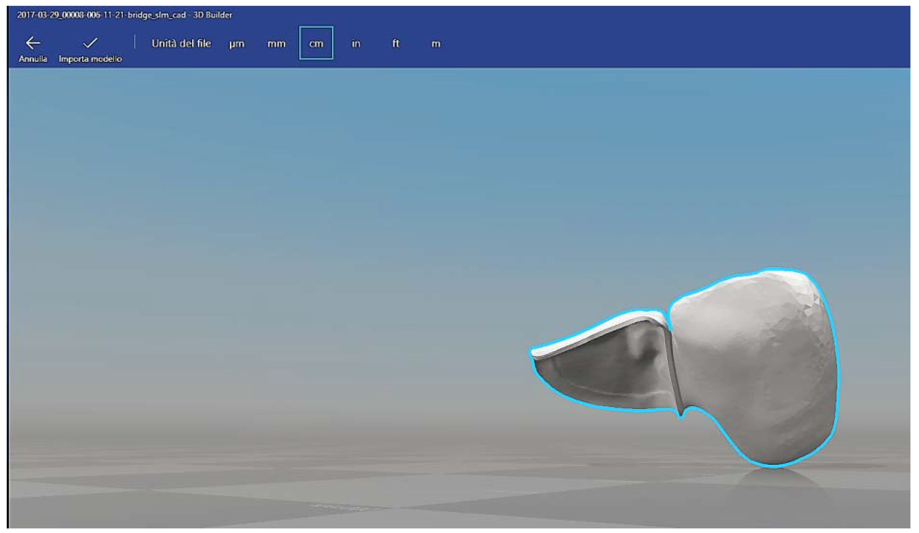
All teeth were imported in virtual environment by laboratory scanner (Smart Big Open Technology, Rezzato (BS), Italy). Using a Cad software (Exocad DentaCad, Darmstad, Germany), one master model of RBFDP with a cantilevered single-retainer was designed (Figure 2).
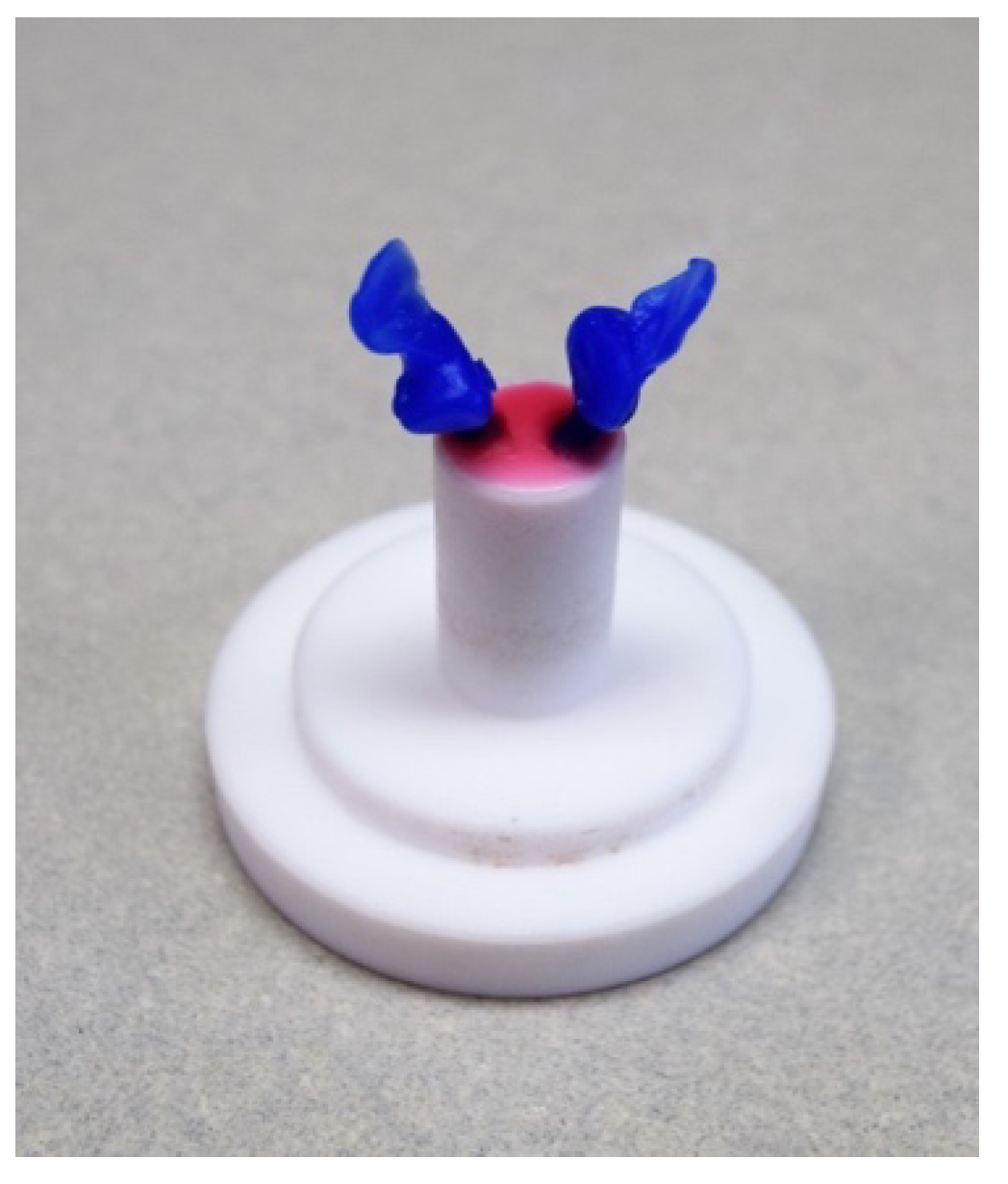
The RBFDP presented the same parameter setting, shape, and size of the cantilever tooth, independently from the anatomical morphology of the bovine mandibular incisives. The retainer wings were fabricated with a uniform thickness of 0.7 mm. The connectors were designed with a height of 3 mm and a width of 1.5 mm [24,25]. Twenty RBFDPs with a cantilevered single-retainer design for the groups were fabricated with 3 different materials: zirconia (Katana ML, Kuraray, Milano, Italy), lithium disilicate (IPS e.max Press LT A2, Ivoclar Vivadent, Bologna, Italy), and PMMA-based material with ceramic fillers (HIPC, Bredent GmbH, Senden Germany). Each RBFDP has been created by the same CAD design with different procedures based on the type of material. The twenty RBFDPs in HIPC were milled by a computer numerical control machine (Roland DWX-50, Roland DG Corporation, Osaka, Japan). The excessive material was removed with tungsten bur (Komet Dental; H250NEX). All of the samples were polished using a polisher machine (Sirio Dental, Meldola (FC), Italy). Twenty zirconia RBFDPs were milled by a CNC machine (Roland DWX-50, Roland DG Corporation, Osaka, Japan), polished using diamond burs, and sintered (Nabertherm LHT 01/17 D, Nabertherm, Lilienthal, Germany) for 12 h at 1450 °C following the manufacturing instructions. The 20 RBFDPs in lithium disilicate were fabricated with wax lost technique. A wax block (CeraWax, Co.N.Ce.P.T, Busseto (PR), Italy) was used to mill the RBFDPs. A sprue was waxed onto the cantilever tooth of all RBFDPs. They were then embedded in a universal investment material (IPS PressVest Premium investment, Ivoclar Vivadent, Bologna, Italy), with 2 RBFDPs (Figure 3).
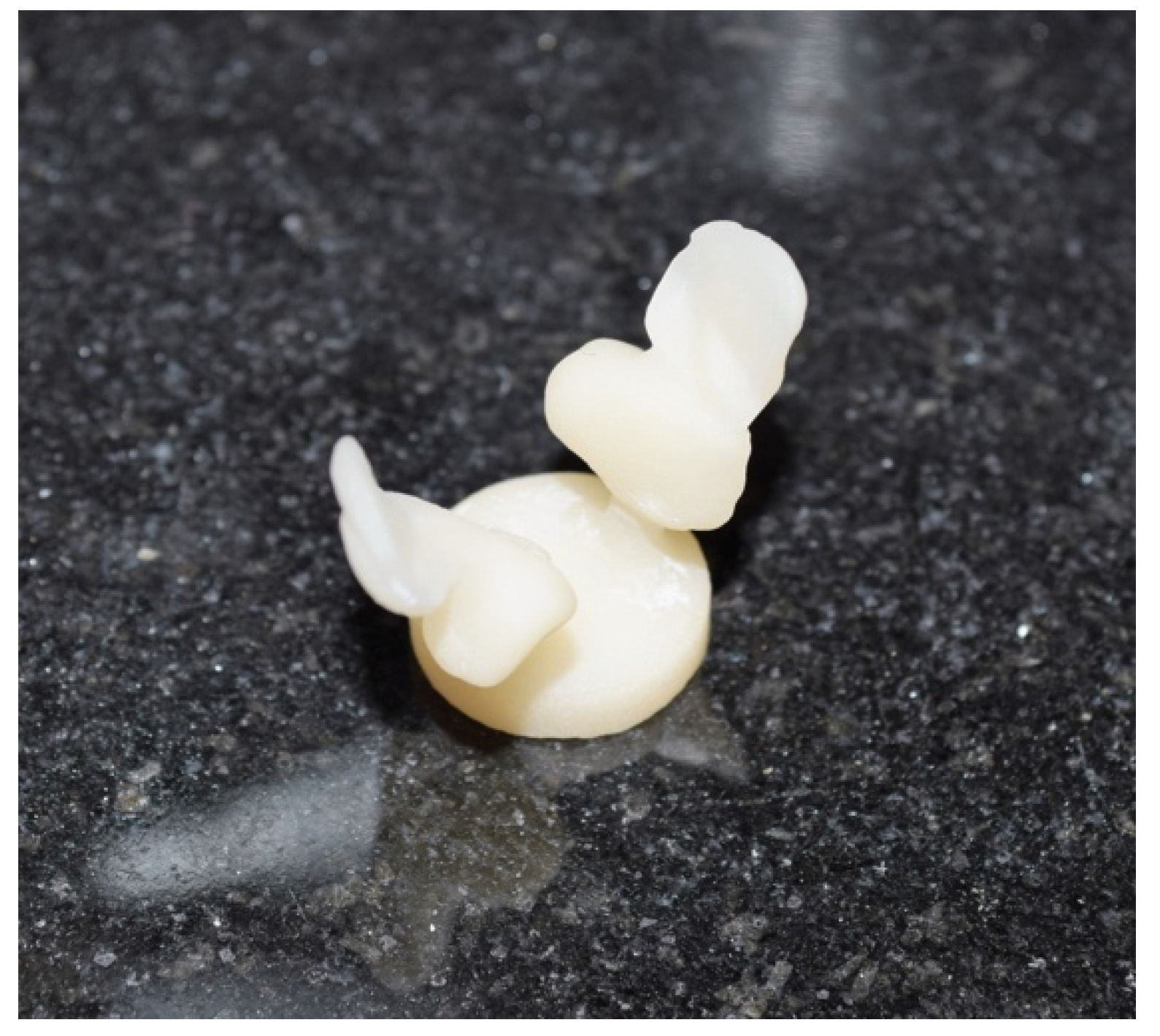
The wax was removed in a heated furnace (Sirio SR 750-In Fire, Sirio Dental S.r.l., Meldola (FC), Italy) and the RBFDPs were pressed with lithium disilicate glass-ceramic (IPS e-max Press LT A2, Ivoclar Vivadent, Bologna, Italy) in the machine-calibrated furnace (Luxor Press-SR 862, Sirio Dental S.r.l., Meldola (FC), Italy) following manufacturer recommendations (Figure 4).
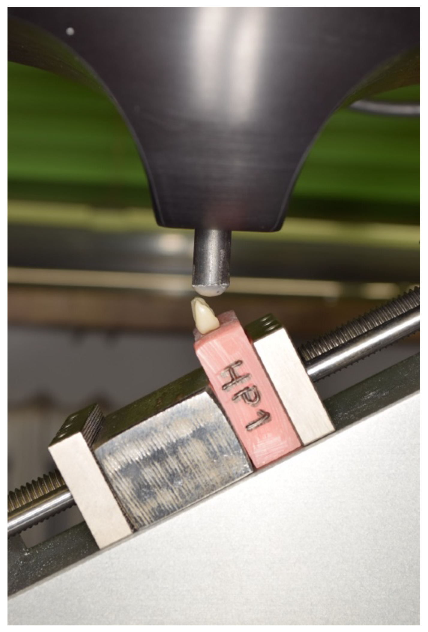
The sprue was removed using a metallic disk and all RBFDPs in lithium disilicate were polish with diamond burs. We obtained 60 comparable RBFDPs with a cantilever design without preparation of the abutment in three different materials. The thickness of the wings of all RBFDPs were measured using a caliper. All of the abutment teeth were treated with the same procedure: 15 s of etching (Scotchbond Universal Etching, 3M, Milano, Italy), 15 s of rinsing with water, 15 s of drying, and the bonding (Scotchbond Universal Adhesive, 3M, Milano, Italy) were applied for 20 s and polymerized for 60 s using a lamp (Valo, Ultradent, South Jordan, UT, USA). All of the RBFDPs were cemented with dual-cured resin cement (Relyx Ultimate, 3M, Milano, Italy) and polymerized for 60 s using led lamp (400–500 nm), but with different procedures. Air abrasion was performed with 50 μm Al2O3 particles at a 2.5 bar pressure for 15 s at a distance of 10 mm on the wings of RBFDPs in zirconia. After drying, the wings were cleaned with 96% isopropanol for 3 minutes. The bonding (Scotchbond Universal Adhesive, 3M, Milano italy) was applied on the wings for 20 s and polymerized for 60 s. Air abrasion was performed with 110 μm Al2O3 particles at a 2.5 bar pressure for 15 s, at a distance of 10 mm on the wings of RBFDPs in PMMA-based material with ceramic fillers. After drying, the primer (Visiolink, Bredent, Seden, Germany) was applied on the wings and polymerized for 60 s. Instead, the wings of RBFDPs in lithium disilicate were etching with hydrofluoric acid (Ceramic Etching gel, Ivoclar Vivadent, Bologna Italy) for 60 s, 60 s of rinsing, and 30 s of drying. The bonding (Scotchbond Universal Adhesive, 3M, Milano, Italy) was applied for 20 s and polymerized for 60 s. All procedures were performed according to the manufacturer’s instructions. The samples were numbered through the engraving on the resin of a code indicating the material. All samples were mounted on a 30° angled support of a universal testing machine (Acumen 3, MTS Systems Corporation, Eden Praire, MN, USA) and a static loading at a crosshead speed of 1.5 mm/min, until failure to the incisal edge of the pontic tooth was performed to simulate real situation (Figure 5). The load was transferred through a 6-mm diameter steatite ball in the middle of the incisal edge of the pontic tooth until failure [25].
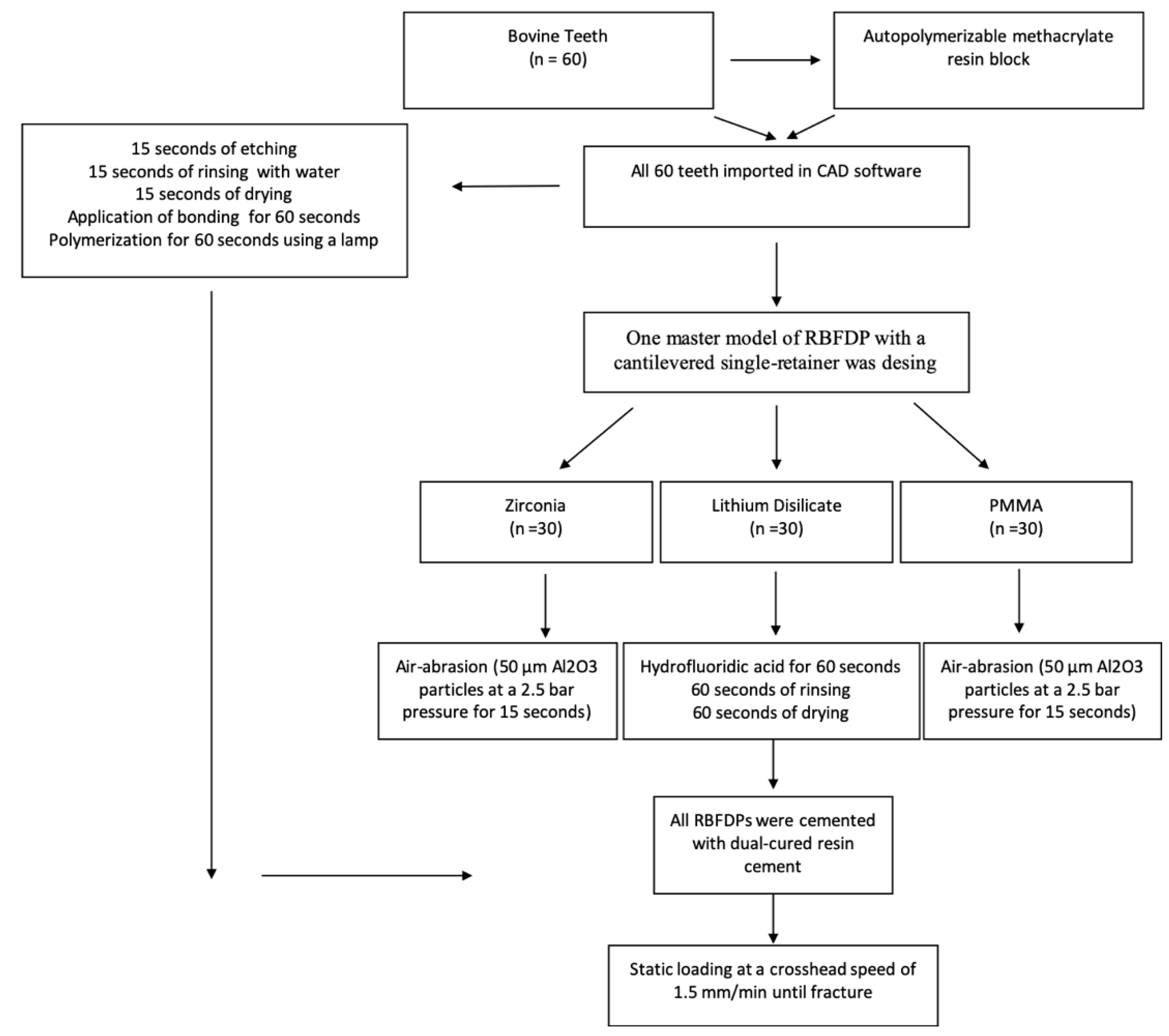
Four types of failure were recorded: debonding of RBFDPs, fracture of the connector, debonding of the RBFDPs with fracture of the abutment tooth, and fracture of the abutment tooth with the RBFDP still bonded. All of the methods were synthetized in the following flowchart (Figure 6).
The Kruskal–Wallis test with a post hoc analysis using Dunn’s test was used to compare the three groups. The level of statistical significance was set as α =0.05 and statistical power of 80%. A statistical software (SPSS v16.0; SPSS Inc., Chicago, IL, USA) was used for the analysis.

3. Results

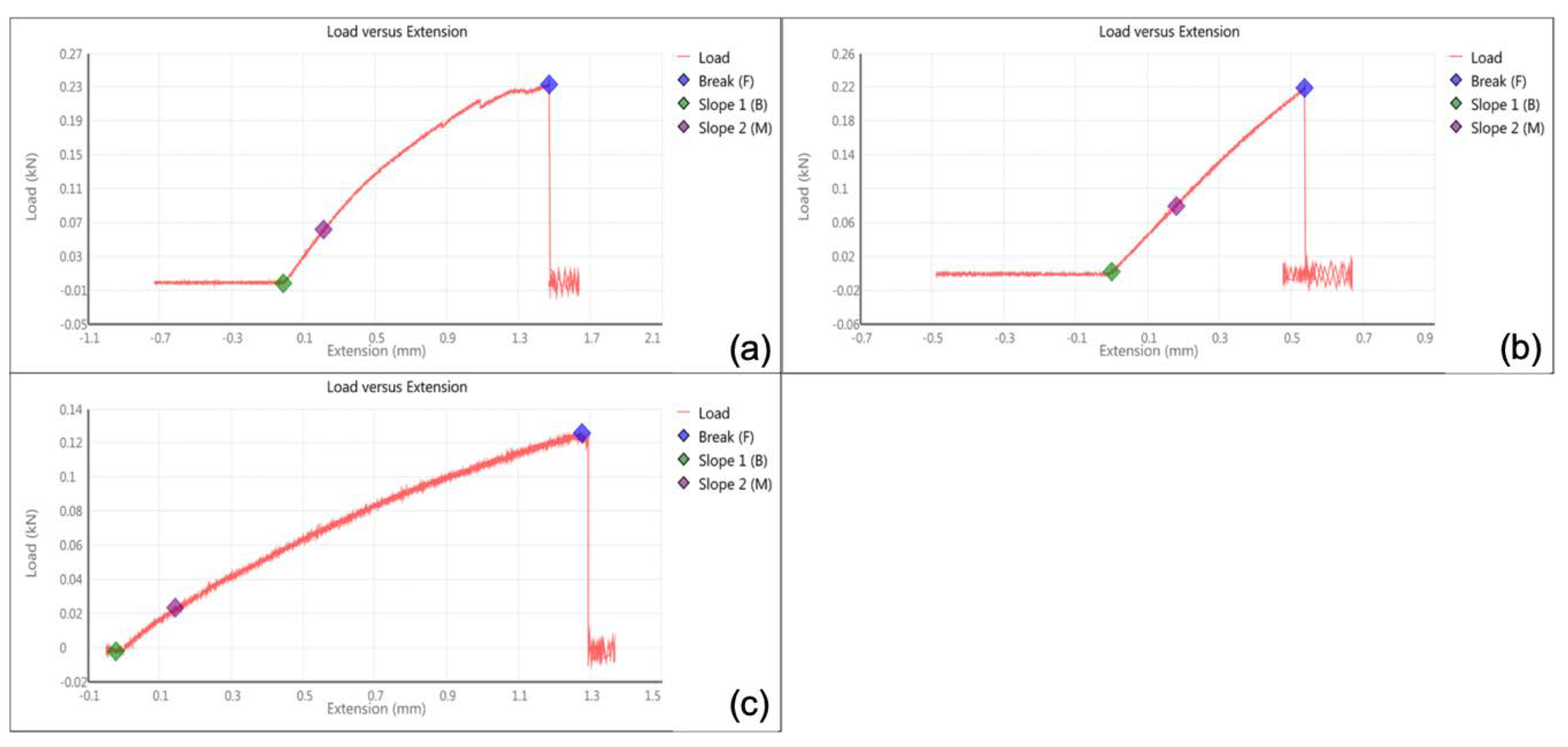
The mean fracture strength of the RBFDPs in zirconia was 292.5 N (SD 36.6) (Figure 7a), 210 N (SD 37.6) for lithium disilicate (Figure 7b), and 133 N (SD 16.3) for PMMA-based material with ceramic fillers (Figure 7c).
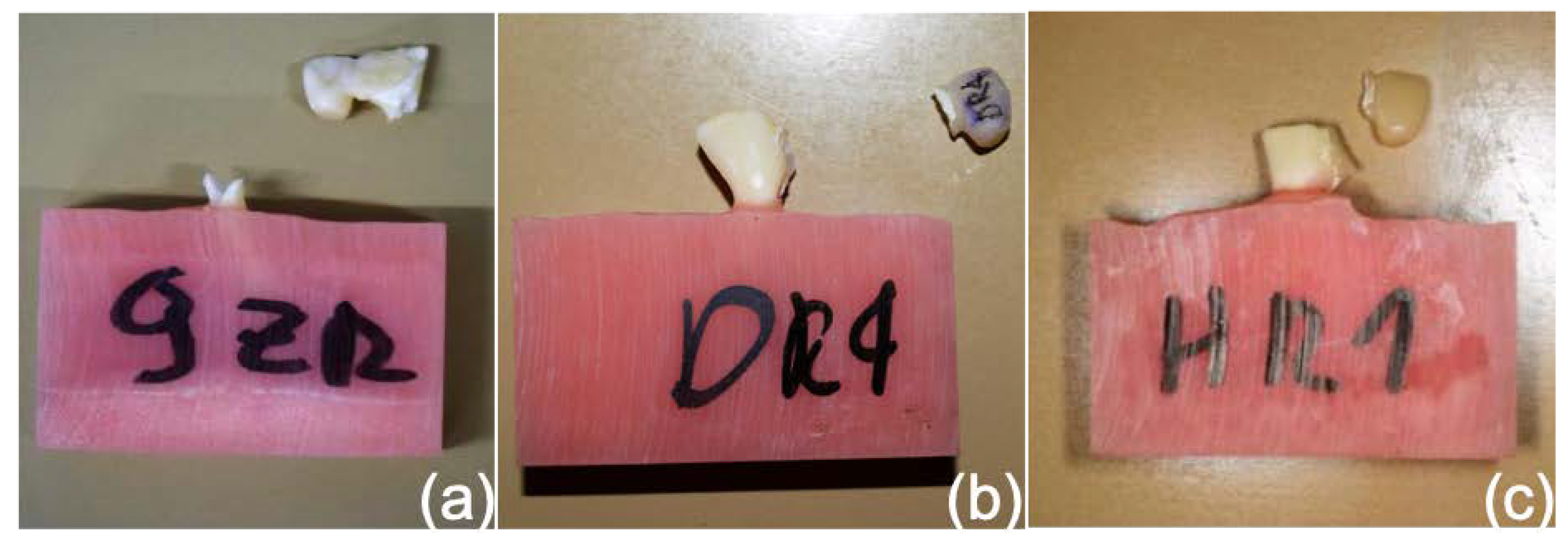
In the group of zirconia RBFDPs, all of the failures were fractures of the abutment tooth (Figure 8a); in the group lithium disilicate 80% of failures were fractures of the connector and 20% debonding of the RBFDPs, with fractures of the abutment tooth (Figure 8b). In the group of PMMA-based material with ceramic fillers, 80% of failures were fractures of the connector (Figure 8c) and 20% debonding. Kruskal–Wallis generated a P-value < 0.001, identifying statistically significant differences between groups. Dunn’s post hoc tests indicated, however, that the difference was statistically significant between the group RBFDPs in zirconia and PMMA-based material with ceramic fillers (P = 0.003), instead no differences were found between RBFDPs in zirconia and lithium disilicate (P = 0.43), and RBFDPs in lithium disilicate and PMMA-based material with ceramic fillers (P = 0.09).

4. Discussion

RBFDP is an aesthetic and conservative treatment option to replace a single tooth in a younger patient; therefore, it is a technique sensitive procedure because it requires proper planning of the clinical case and choice of materials. This in vitro study investigated the different types of failure after static fracture strength tests on RBFDPs fabricated using zirconia, lithium disilicate, and PMMA-based material with ceramic fillers. Zirconia, lithium disilicate, and PMMA-based material with ceramic fillers presented the mean values static fracture strength of 292.5 N (SD 36.6), 210 N (SD 37.6), and 133 N (SD 16.3), respectively. Moreover, different types of failure were collected according to the materials. The 80% of the RBFDPs made in lithium disilicate and PMMA-based material with ceramic fillers presented fracture of the connector; 20% of debonding and all of the RBFDPs in zirconia presented fracture of the abutment tooth.
The results can be explained by the different elastic modulus of materials. The lithium disilicate and PMMA-based material with ceramic fillers presented lower elastic modulus and, consequently, a greater deformation until the fracture of the materials. Another possible explanation of the fracture of the connector was recognizable to the superior bond strength, with respect to the materials. The authors highlight that the lithium disilicate is not indicated to fabricate RBFDPs by manufacturer; however, the results showed a mean fracture strength of 210 N. Therefore, clinical studies are necessary to evaluate the performance of this material used to fabricate RBFDPs. Instead, all the RBFDPs in zirconia presented a fracture of the abutment tooth. The reasons may be attributable to the high elastic modulus of the material or high bond strength of resin luting cement, with respect to the abutment tooth.
Opposite results were observed in clinical trials [16,17,18,19] compared to this in vitro study. The reasons could be several; however, the load strength of the RBFDP is different in each patient. Moreover, the pontic elements may not have static and dynamic contacts. In this in vitro study, the uniform fabrication of all the RBFDPs with different materials by the same STL file, and the strength applied through a universal machine until failure, allowed to know the mechanic performance of these prostheses with respect to an in vivo study.
In literature, the same authors in two in vitro studies evaluated the influence of framework design [14] and mode of loading on the fracture strength [26] on cantilever RBFDPs fabricated in pre-sintered aluminum-oxide blocks (In-Ceram alumina blanks). The microscopic examination revealed that 58% of the specimens fractured at the connector only, exactly at the framework-to-veneer interface, 17% fractured at the connector, including veneered parts of the pontic, and 25% fractured at the retainer only. The possible difference of the performance should be explained that, in this research, the authors used bovine teeth. Physiologic occlusal forces for adults in the anterior region were determined to be in a range of 10 to 35 N [27]. Maximal incisive biting forces may vary up to 270 N, primarily depending on facial morphology and age [28]. Compared to the maximal incisive biting forces, only zirconia RBFPDs reached values higher than the physiologic values. Even though the mean fracture strength of lithium disilicate RBFDPs was lower compared to the maximal incisive biting force, it is possible to use the material to fabricate RBFDPs. However, the clinicians should evaluate, with attention, the static and dynamic contacts of the clinical case before use of lithium disilicate RBFDP. Whereas, PMMA-based material with ceramic filler RBFDPs represents a cheap and aesthetic solution for a temporary prosthesis for patients waiting for implant treatments. Therefore, the choice of a correct material, according to therapeutic needs, is crucial for the survival of RBFDPs.
Long-term clinical evidence is needed to evaluate the performance of these materials, especially for use of polymer with ceramic fillers. The drawbacks of this in vitro study were the lack of clinical conditions, as artificial aging, dynamic loading, and of physiologic tooth mobility; however, the uniform fabrications of all the RBFDPs with different materials by the same STL file and the same laboratory conditions allowed to know the real performance of this material, with respect to an in vivo study where the RBFDPs have different shapes and dimensions.

5. Conclusions

Within the limitations of this in vitro study, we can conclude that the zirconia RBFDPs presented load resistance higher than the maximum anterior bite force; furthermore, the lithium disilicate RBFDPs showed a mean fracture strength similar the anterior bite force.

Author Contributions

Conceptualization, A.D.F. and E.S.; methodology, R.M.; validation, S.G. and G.S.; formal analysis, S.G. and S.R.; investigation, G.S. and L.G.; data curation, L.G.; writing—original draft preparation, A.D.F. and E.S.; writing—review and editing, C.M. All authors have read and agreed to the published version of the manuscript.

Funding

This research received no external funding.

Acknowledgments

The authors thank Giorgio Calgaro for materials used for experiments and technical support.

Conflicts of Interest

The authors declare no conflict of interest.

References

  1. Borum, M.K.; Andreasen, J.O. Therapeutic and economic implications of traumatic dental injuries in Denmark: An estimate based on 7549 patients treated at a major trauma centre. Int. J. Paediatr. Dent. 2001, 11, 249–258. [Google Scholar] [CrossRef]
  2. Andrade, D.C.; Loureiro, C.A.; Araujo, V.E.; Riera, R.; Atallah, A.N. Treatment for agenesis of maxillary lateral incisors: A systematic review. Orthod. Craniofac. Res. 2013, 16, 129–136. [Google Scholar] [CrossRef] [PubMed]
  3. Wei, Y.R.; Wang, X.D.; Zhang, Q.; Li, X.X.; Blatz, M.B.; Jian, Y.T.; Zhao, K. Clinical performance of anterior resin-bonded fixed dental prostheses with different framework designs: A systematic review and meta-analysis. J. Dent. 2016, 47, 1–7. [Google Scholar] [CrossRef]
  4. Robertsson, S.; Mohlin, B. The congenitally missing upper lateral incisor: A retrospective study of orthodontic space closure versus restorative treatment. Eur. J. Orthod. 2000, 22, 697–709. [Google Scholar] [CrossRef] [Green Version]
  5. Thoma, D.S.; Sailer, I.; Ioannidis, A.; Zwahlen, M.; Makarov, N.; Pjetursson, B.E. A systematic review of the survival and complication rates of resin-bonded fixed dental prostheses after a mean observation period of at least 5 years. Clin. Oral. Implant. Res. 2017, 28, 1421–1432. [Google Scholar] [CrossRef] [PubMed]
  6. Botelho, M.G.; Ma, X.; Cheung, G.J.; Law, R.K.; Tai, M.T.; Lam, W.Y. Long-term clinical evaluation of 211 two-unit cantilevered resin-bonded fixed partial dentures. J. Dent. 2014, 42, 778–784. [Google Scholar] [CrossRef] [PubMed]
  7. Sasse, M.; Eschbach, S.; Kern, M. Randomized clinical trial on single retainer all-ceramic resin-bonded fixed partial dentures: Influence of the bonding system after up to 55 months. J. Dent. 2012, 40, 783–786. [Google Scholar] [CrossRef]
  8. El-Mowafy, O.; Rubo, M.H. Resin-bonded fixed partial dentures-a literature review with presentation of a novel approach. Int. J. Prosthodont. 2000, 13, 460–467. [Google Scholar]
  9. Keulemans, F.; Shinya, A.; Lassila, L.V.; Vallittu, P.K.; Kleverlaan, C.J.; Feilzer, A.J.; De Moor, R.J. Three-dimensional finite element analysis of anterior two-unit cantilever resin-bonded fixed dental prostheses. Sci. World J. 2015, 2015, 864389. [Google Scholar] [CrossRef] [Green Version]
  10. Kern, M.; Sasse, M. Ten-year survival of anterior all-ceramic resin-bonded fixed dental prostheses. J. Adhes. Dent. 2011, 13, 407–410. [Google Scholar]
  11. Kern, M. Clinical long-term survival of two-retainer and single-retainer all-ceramic resin-bonded fixed partial dentures. Quintessence Int. 2005, 36, 141–147. [Google Scholar] [PubMed]
  12. Botelho, M.G.; Leung, K.C.; Ng, H.; Chan, K. A retrospective clinical evaluation of two-unit cantilevered resin-bonded fixed partial dentures. J. Am. Dent. Assoc. 2006, 137, 783–788. [Google Scholar] [CrossRef] [PubMed]
  13. Botelho, M.G.; Chan, A.W.; Leung, N.C.; Lam, W.Y. Long-term evaluation of cantilevered versus fixed-fixed resin-bonded fixed partial dentures for missing maxillary incisors. J. Dent. 2016, 45, 59–66. [Google Scholar] [CrossRef] [PubMed] [Green Version]
  14. Mourshed, B.; Samran, A.; Alfagih, A.; Samran, A.; Abdulrab, S.; Kern, M. Anterior cantilever resin-bonded fixed dental prostheses: A review of the literature. J. Prosthodont. 2018, 27, 266–275. [Google Scholar] [CrossRef]
  15. Kern, M. Fifteen-year survival of anterior all-ceramic cantilever resin-bonded fixed dental prostheses. J. Dent. 2017, 56, 133–135. [Google Scholar] [CrossRef]
  16. Sasse, M.; Kern, M. Survival of anterior cantilevered all-ceramic resin-bonded fixed dental prostheses made from zirconia ceramic. J. Dent. 2014, 42, 660–663. [Google Scholar] [CrossRef]
  17. Kern, M.; Passia, N.; Sasse, M.; Yazigi, C. Ten-year outcome of zirconia ceramic cantilever resin-bonded fixed dental prostheses and the influence of the reasons for missing incisors. J. Dent. 2017, 65, 51–55. [Google Scholar] [CrossRef]
  18. Sailer, I.; Bonani, T.; Brodbeck, U.; Hämmerle, C.H. Retrospective clinical study of single-retainer cantilever anterior and posterior glass-ceramic resin-bonded fixed dental prostheses at a mean follow-up of 6 years. Int. J. Prosthodont. 2013, 26, 443–450. [Google Scholar] [CrossRef] [Green Version]
  19. Di Fiore, A.; Meneghello, R.; Graiff, L.; Savio, G.; Vigolo, P.; Monaco, C.; Stellini, E. Full arch digital scanning systems performances for implant-supported fixed dental prostheses: A comparative study of 8 intraoral scanners. J. Prosthodont. Res. 2019, 63, 396–403. [Google Scholar] [CrossRef]
  20. Di Fiore, A.; Vigolo, P.; Graiff, L.; Stellini, E. Digital vs Conventional Workflow for Screw-Retained Single-Implant Crowns: A Comparison of Key Considerations. Int. J. Prosthodont. 2018, 31, 577–579. [Google Scholar] [CrossRef]
  21. Granata, S.; Giberti, L.; Vigolo, P.; Stellini, E.; Di Fiore, A. Incorporating a facial scanner into the digital workflow: A dental technique. J. Prosthet. Dent. 2019, 19, 30356–30357. [Google Scholar] [CrossRef] [PubMed]
  22. Malaguti, G.; Rossi, R.; Marziali, B.; Esposito, A.; Bruno, G.; Dariol, C.; Di Fiore, A. In vitro evaluation of prosthodontic impression on natural dentition: A comparison between traditional and digital techniques. Oral Implantol. 2017, 9, 21–27. [Google Scholar] [CrossRef] [PubMed]
  23. Sirisha, K.; Rambabu, T.; Ravishankar, Y.; Ravikumar, P. Validity of bond strength tests: A critical review-Part II. J. Conserv. Dent. 2014, 17, 420–426. [Google Scholar] [CrossRef] [Green Version]
  24. Koutayas, S.O.; Kern, M.; Ferraresso, F.; Strub, J.R. Influence of framework design on fracture strength of mandibular anterior all-ceramic resin-bonded fixed partial dentures. Int. J. Prosthodont. 2002, 15, 223–229. [Google Scholar]
  25. Yokoyama, D.; Shinya, A.; Lassila, L.V.; Gomi, H.; Nakasone, Y.; Vallittu, P.K.; Shinya, A. Framework design of an anterior fiber-reinforced hybrid composite fixed partial denture: A 3D finite element study. Int. J. Prosthodont. 2009, 22, 405–412. [Google Scholar] [PubMed]
  26. Koutayas, S.O.; Kern, M.; Ferraresso, F.; Strub, J.R. Influence of design and mode of loading on the fracture strength of all-ceramic resin-bonded fixed partial dentures: An in vitro study in a dual-axis chewing simulator. J. Prosthet. Dent. 2000, 84, 112. [Google Scholar] [CrossRef]
  27. De Boever, J.A.; McCall, W.D., Jr.; Holden, S.; Ash, M.M., Jr. Functional occlusal forces: An investigation by telemetry. J. Prosthet. Dent. 1978, 40, 326–333. [Google Scholar] [CrossRef]
  28. Kiliaridis, S.; Kjellberg, H.; Wenneberg, B.; Engstrom, C. The relationship between maximal force, bite force endurance, and facial morphology during growth. A cross-sectional study. Acta. Odontol. Scand. 1993, 51, 323–331. [Google Scholar] [CrossRef]
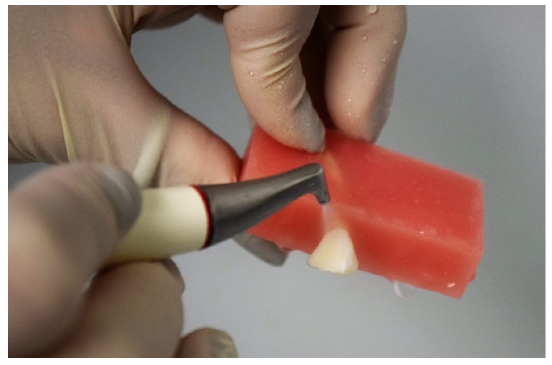
Figure 1. Air-polishing used to clean incisive embedded in resin block.
Applsci 10 04151 g001
Figure 2. Design of master model of cantilever BFDP.
Applsci 10 04151 g002
Figure 3. Cantilever RBFDPs milled in wax.
Applsci 10 04151 g003
Figure 4. RBFDPs in lithium disilicate fabricated with wax lost technique.
Applsci 10 04151 g004
Figure 5. A sample mounted on a 30° angled support of the universal testing machine.
Applsci 10 04151 g005
Figure 6. Flowchart of the methods.
Applsci 10 04151 g006
Figure 7. Graphic load-extension of the sample: (a) number 3 in zirconia, (b) number 5 in lithium disilicate, and (c) number 2 in PMMA-based material with ceramic fillers.
Applsci 10 04151 g007
Figure 8. Different types of failure: (a) fracture of the abutment tooth in the group RBFDP in zirconia, (b) fracture of the connector in the group RBFDP in lithium disilicate, and (c) fracture of the connector in the group RBFDP in PMMA-based material with ceramic fillers.
Applsci 10 04151 g008