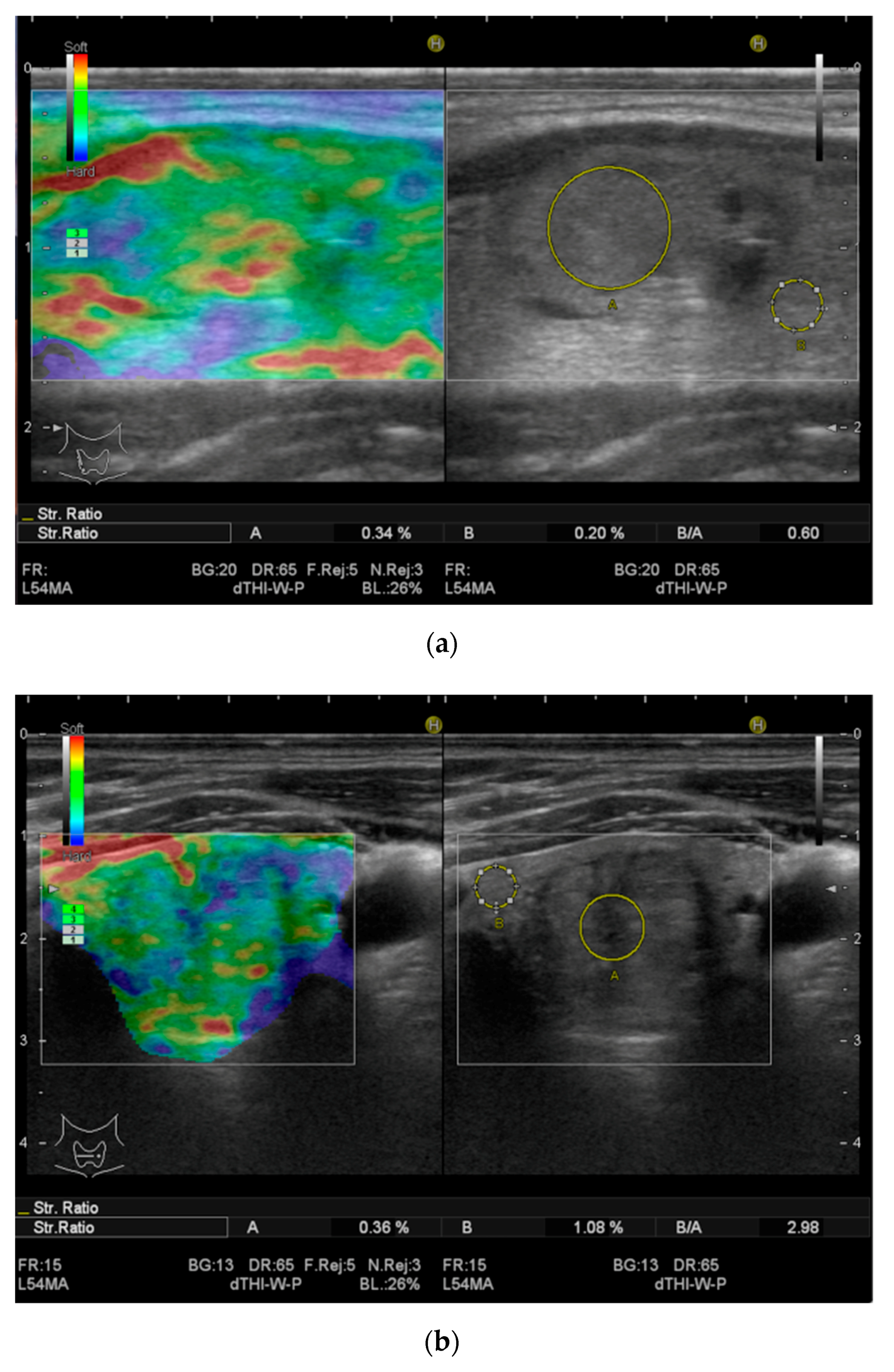
Thyroid Multimodal Ultrasound Evaluation—Impact on Presurgical Diagnosis of Intermediate Cytology Cases
Abstract
:1. Introduction
2. Material and Methods
2.1. Subjects
2.2. Ultrasound Techniques
2.3. FNA
2.4. Surgical Intervention and Pathological Examination
2.5. Statistical Analysis
3. Results
4. Discussion
5. Conclusions
Author Contributions
Funding
Conflicts of Interest
References
- Vanderpump, M.P.J. The Epidemiology of Thyroid Disease. Br. Med. Bul. 2011, 99, 39–51. [Google Scholar] [CrossRef] [Green Version]
- Kent, W.D.; Hall, S.F.; Isotalo, P.A.; Houlden, R.L.; George, R.L.; Groome, P.A. Incresed incidence of differentiated thyroid carcinoma and detection of subclinical disease. CMAJ 2007, 177, 1357–1361. [Google Scholar] [CrossRef] [PubMed] [Green Version]
- Pellegriti, G.; Frasca, F.; Regalbuto, C.; Squatrito, S.; Vigneri, R. Worldwide Increasing Incidence of Thyroid Cancer: Update on Epidemiology and Risk Factors. J. Cancer Epidemiol. 2013, 2013, 965212. [Google Scholar] [CrossRef] [PubMed] [Green Version]
- Gharib, H.; Papini, E.; Garber, J.R.; Duick, D.S.; Harrell, R.M.; Hegedüs, L.; Paschke, R.; Valcavi, R.; Vitti, P. AACE/ACE/AME Task Force on Thyroid Nodules. American Association of Clinical Endocrinologists, American College of Endocrinology, and Associazione Medici Endocrinologi Medical Guidelines for Clinical Practice for The Diagnosis and Management of Thyroid Nodules. Endocr. Pract. 2016, 22, 1–60. [Google Scholar]
- Cooper, D.S.; Doherty, G.M.; Haugen, B.R.; Kloos, R.T.; Lee, S.L.; Mandel, S.J.; Mazzaferri, E.L.; McIver, B.; Pacini, F.; Schlumberger, M.; et al. American Thyroid Association (ATA) Guidelines Taskforce on Thyroid Nodules and Differentiated Thyroid Cancer1; Revised ATA management Guidelines for Patients with thyroid nodules and differentiated thyroid cancer. Thyroid 2009, 19, 1167–1214. [Google Scholar] [CrossRef] [PubMed] [Green Version]
- Haugen, B.R.; Alexander, E.K.; Bible, K.C.; Doherty, G.M.; Mandel, S.J.; Nikiforov, Y.E.; Pacini, F.; Randolph, G.W.; Sawka, A.M.; Schlumberger, M.; et al. 2015 ATA management guidelines for adults with thyroid nodules and differentiated thyroid cancer. Thyroid 2016, 1, 1–133. [Google Scholar] [CrossRef] [PubMed] [Green Version]
- Cibas, E.S.; Ali, S.Z. The 2017 Bethesda system for reporting thyroid cytopathology. Thyroid 2017, 27, 1341–1346. [Google Scholar] [CrossRef]
- Jo, V.Y.; Stelow, E.B.; Dustin, S.M.; Hanley, K.Z. Malignancy risk for fine-needle aspiration of thyroid lesions according to the Bethesda System for Reporting Thyroid Cytopathol- ogy. Am. J. Clin. Pathol. 2010, 134, 450–456. [Google Scholar] [CrossRef] [Green Version]
- Wang, C.-C.; Friedman, L.; Kennedy, G.C.; Wang, H.; Kebebew, E.; Steward, D.L.; Zeiger, M.A.; Westra, W.H.; Wang, Y.; Khanafshar, E.; et al. A large multi- center correlation study of thyroid nodule cytopathology and histopathology. Thyroid 2011, 21, 243–251. [Google Scholar] [CrossRef]
- Mistry, S.G.; Mani, N.; Murthy, P. Investigating the value of fine needle aspiration cytology in thyroid cancer. J. Cytol. 2011, 28, 185–190. [Google Scholar]
- Bongiovanni, M.; SPitale, A.; Gaquin, W.C.; Mazzucchelli, L.; BAloch, Z.W. The Bethesda System for Reporting Thyroid Cytopathology: A meta-analysis. Acta Cytol. 2012, 56, 333–339. [Google Scholar] [CrossRef] [PubMed]
- Ohori, N.P.; Nikiforova, M.N.; Schoedel, K.E.; LeBeau, S.O.; Hodak, S.P.; Seethala, R.R.; Carty, S.E.; Ogilvie, J.B.; Yip, L.; Nikiforov, Y.E. Contrbution of molecular testing to thyroid fine needle aspiration cytology of “follicular lesion of undeterminated significance/atypia of undeterminated significance”. Cancer Cytopathol. 2010, 118, 17–23. [Google Scholar] [CrossRef] [PubMed]
- Bongiovanni, M.; Crippa, S.; Baloch, Z.; Piana, S.; Spitale, A.; Pagni, F.; Mazzucchelli, L.; Di Bella, C.; Faquin, W. Comparison of 5-tiered and 6-tiered diagnostic systems for the reporting of thyroid cytopathology: A multi-institutional study. Cancer Cytopathol. 2012, 120, 117–125. [Google Scholar] [CrossRef] [PubMed]
- Stoian, D.; Borcan, F.; Petre, I.; Mozos, I.; Varcus, F.; Ivan, V.; Cioca, A.; Apostol, A.; Dehelean, C.A. Strain elastography as a valuable diagnostic tool in intermediate citology (Bethesda III) thyroid nodules. Diagnostics 2019, 9, 119. [Google Scholar] [CrossRef] [Green Version]
- Moifo, B.; Takoeta, E.O.; Tambe, J.; Blanc, F.; Fotsin, J.G. Reliability of Thyroid Imaging Reporting and Data System (TIRADS) Classification in Differentiating Benign from Malignant Thyroid Nodules. Open J. Radiol. 2013, 3, 103–107. [Google Scholar] [CrossRef] [Green Version]
- Wei, X.; Li, Y.; Zhang, S.; Gao, M. Thyroid imaging reporting and data system (Ti-RADS) in the diagnostic value of thyroid nodules: A systematic review. Tumor Biol. 2014, 35, 6769–6776. [Google Scholar] [CrossRef]
- Friedrich-Rust, M.; Meyer, G.; Dauth, N.; Berner, C.; Bogdanou, D.; Herrmann, E.; Zeuzem, S.; Bojunga, J. Interobserver agreement of Thyroid Reporting and Data System (TIRADS) and strain elastography for assessment of thyroid nodules. PLoS ONE 2013, 8, e77927. [Google Scholar] [CrossRef] [Green Version]
- Russ, G.; Royer, B.; Bigorgne, C.; Rouxel, A.; Bienvenu-Perrard, M.; Leenhardt1, L. Prospective evaluation of thyroid imaging system reporting and data system on 4550 nodules with and without elastography. Eur. J. Endocrinol. 2013, 168, 649–655. [Google Scholar] [CrossRef]
- Stoian, D.; Timar, B.; Derban, M.; Pantea, S.; Varcus, F.; Craina, M.; Craciunescu, M. TI-RADS: The impact of qualitative strain elastography for better stratification of the cancer risk. Med. Utrason. 2015, 17, 327–333. [Google Scholar] [CrossRef]
- Stoian, D.; Cornianu, M.; Dobrescu, A.; Lazar, F. Nodular thyroid cancer. Diagnostic value of real time elastography. Chirurgia 2012, 107, 39–46. [Google Scholar]
- Grazhdani, H.; Drakonaki, E.; D’Andrea, V.; di Segni, M.; Kaleshi, E.; Calliada, F.; Catalano, C.; Redler, A.; Brunese, L.; Drudi, F.M.; et al. Strain US Elastography for the Characterization of Thyroid Nodules: Advantages and Limitation. Int. J. Endocrinol. 2015, 2015, 908575. [Google Scholar] [CrossRef]
- Migda, B.; Migda, M.; Migda, A.M.; Bierca, J.; Slowniska-Srzednicka, J.; Jakubowski, W.; Slapa, R.Z. Evaluation of Four Variantsof the Thyroid Imaging Reporting and Data System (TIRADS) Classification inPatients with Multinodular Goitre - initial study. Endokrynol Pol. 2018, 69, 156–162. [Google Scholar] [CrossRef] [PubMed] [Green Version]
- Stoian, D.; Ivan, V.; Sporea, I.; Varcus, F.; Mozos, I.; Navolan, D.; Nemescu, D. Advanced ultrasound application- impact on presurgical risk stratification of the thyroid nodules. Ther. Clin. Risk Manag. 2020, 16, 21–30. [Google Scholar] [CrossRef] [PubMed] [Green Version]
- Slapa, R.Z.; Jakubowski, W.S.; Slowinska-Srzednicka, J.; Szopinski, K.T. Advantages and disadvantages of 3D ultrasound of thyroid nodules including thin slice volume rendering. Thyroid Res. 2011, 4, 1–12. [Google Scholar] [CrossRef] [Green Version]
- Cantisani, V.; David, E.; Grazhdani, H.; Rubini, A.; Radzina, M.; Dietrich, C.F.; Durante, C.; Lamartina, L.; Grani, G.; Valeria, A.; et al. Prospective evaluation of semiquantitative strain ratio and quantitative 2D Ultrasound shear wave elastography (SWE) in association with TIRADS classification for thyroid module characterization. Ultraschall Med. 2019, 40, 495–503. [Google Scholar] [CrossRef]
- Cappelli, C.; Pirola, I.; Gandossi, E.; Marini, F.; Cristiano, A.; Casella, C.; Lombardi, D.; Agosti, B.; Ferlin, A.; Castellano, M. Ultrasound microvascular blood flow evaluation: A new tool for the management of thyroid nodules? Int. J. Endocrinol. 2019, 2019, 7874890. [Google Scholar] [CrossRef] [Green Version]
- Grani, G.; Lamartina, L.; Ascoli, V.; Bosco, D.; Biffoni, M.; Giacomelli, L.; Maranghi, M.; Falcone, R.; Ramundo, V.; Cantisani, V.; et al. Reducing the number of unnecessaru thyroid biopsies while improving diagnostic accuracy: Toward the “right” TIRADS. J. Clin. Endocrinol. Metab. 2019, 104, 95–102. [Google Scholar]
- Valderrabano, P.; McIver, B. Evaluation and management of indeterminate thyroid nodules: The revolution of risk stratification beyond cytological diagnosis. Cancer Control. 2017, 24. [Google Scholar] [CrossRef]
- He, Y.P.; Xu, H.X.; Zhao, C.K.; Sun, L.P.; Li, X.L.; Yue, W.W.; Guo, L.; Wang, D.; Ren, W.W.; Wang, Q.; et al. Cytologically indeterminate thyroid nodules: Increased diagnostic performance with combination of US TI-RADS and a new scoring system. Sci. Rep. 2017, 7, 6906. [Google Scholar] [CrossRef] [Green Version]
- Asteria, C.; Giovanardi, A.; Pizzocaro, A.; Cozzaglio, L.; Morabito, A.; Somalvico, F.; Zoppo, A. US-elastography in the differential diagnosis of benign and malignant thyroid nodules. Thyroid 2008, 18, 523–531. [Google Scholar] [CrossRef]
- Kakudo, K. How to handle borderline/precursor thyroid tumors in management of patients with thyroid nodules. Gnad Surg. 2018, 7 (Suppl. 1), 8–18. [Google Scholar] [CrossRef]
- Bychkov, A.; Hirokawa, M.; Jung, C.K.; Liu, Z.; Zhu, Y.; Hong, S.W.; Satoh, S.; Lai, C.; Huynh, L.; Kakudo, K. Low rate of noninvasive follicular thyroid neoplasm with papillary like nuclear features in Asian practice. Thyroid 2017, 27, 983–984. [Google Scholar] [CrossRef] [PubMed]
- Liu, Z.; Song, Y.; Han, B.; Zhang, X.; Su, P.; Cui, X. Non-invasive Follicular Thyroid Neoplasm with Papillary-like Nuclear Features and the Practice in Qilu Hospital of Shandong University, China. J. Basic Clin. Med. 2017, 6, 22–25. [Google Scholar]
- Jung, C.K.; Kim, C. Effect of lowering the diagnostic threshold for encapsulated follicular variant of papillary thyroid carcinoma on prevalence of noninvasive follicular neoplasm with papillary like nuclear features: A single Institution experience in Korea. J. Basic Clin. Med. 2017, 6, 26–28. [Google Scholar]
- Trimboli, P.; Treglia, G.; Guidobaldi, L.; Saggiorato, E.; Nigri, G.; Crescenzi, A.; Romanelli, F.; Orlandi, F.; Valabrega, S.; Sadeghi, R.; et al. Clinical characteristics as predictors of malignancy in patients with indeterminate thyroid cytology: A meta-analysis. Endocrine 2014, 46, 52–59. [Google Scholar] [CrossRef]
- Varshney, R.; Forest, V.I.; Zawawi, F.; Rochon, L.; Hier, M.P.; Mlynarek, A.; Tamilia, M.; Payne, R.J. Ultrasound-guided fine-needle aspiration of thyroid nodules: Does size matter? Am. J. Otolaryngol. 2014, 35, 373–376. [Google Scholar] [CrossRef]
- Banks, N.D.; Kowalski, J.; Tsai, H.; Somervell, H.; Tufano, R.; Dackiw, A.P.B.; Marohn, M.R.; Clark, D.P.; Umbricht, C.B.; Zeiger, M.A. A Diagnostic Predictor Model for Indeterminate or Suspicious Thyroid FNA Samples. Thyroid 2008, 18, 933–941. [Google Scholar] [CrossRef]
- Jeong, S.H.; Hong, H.S.; Lee, E.H.; Cha, J.G.; Park, J.S.; Kwak, J.J. Outcome of thyroid nodules characterized as atypia of undetermined significance or follicular lesions of undetermined significance and correlation with Ultrasound features and BRAF mutation analysis. Am. J. Roentgenol. 2013, 201, 854–860. [Google Scholar] [CrossRef]
- Kihara, M.; Ito, Y.; Hirokawa, M.; Masuoka, H.; Yabuta, T.; Tomoda, C.; Higashiyama, T.; Inoue, H.; Fukushima, M.; Takamura, Y.; et al. Role of ultrasonography in patients with cytological follicular thyroid tumor. Auris Nasus Larynx 2011, 38, 508–511. [Google Scholar] [CrossRef]
- Chng, C.I.; Kurazawinski, T.R.; Bale, T. Value of sonographic features in predicting malignancy in thyroid nodules diagnosed as follicular neoplasm on cytology. Clin. Endocrinol. (Oxf.) 2015, 83, 711–716. [Google Scholar] [CrossRef]
- Rago, T.; Di Coscio, G.; Basolo, F.; Scutari, M.; Elisei, R.; Berti, P.; Miccoli, P.; Romani, R.; Faviana, P.; Pinchera, A.; et al. Combined clinical, thyroid ultrasound and cytological features help to predict thyroid malignancy in follicular and Hurtle cell thyroid lesions: Results from a series of 505 consecutive patients. Clin. Endocrinol. 2007, 66, 13–20. [Google Scholar]
- Rago, T.; Scutari, M.; Santini, F.; Loiacono, V.; Piaggi, P.; Di Coscio, G.; Basolo, F.; Berti, P.; Pinchera, A.; Vitti, P. Real-time elastosonography: Useful tool for refining the presurgical diagnosis in thyroid nodules with indeterminate or nondiagnostic cytology. J. Clin. Endocrinol. Metab. 2010, 95, 5274–5280. [Google Scholar] [CrossRef] [PubMed] [Green Version]
- Sorrenti, S.; Trimboli, P.; Catania, A.; Ulisse, S.; de Antoni, E.; D’Armiento, M. Comparison of malignancy rate in thyroid nodules with cytology of indeterminate follicular or indeterminate Hürthle cell neoplasm. Thyroid 2009, 19, 355–360. [Google Scholar] [CrossRef] [PubMed]
- Garino, F.; Deandrea, M.; Motta, M.; Mormile, A.; Ragazzoni, F. Diagnostic performance of elastography in cytological indeterminate thyroid nodules. Endocrine 2015, 49, 175–183. [Google Scholar] [CrossRef]
- Mufti, S.T.; Molah, R. The Bethesda System for Reporting Thyroid Cytopathology: A Five-Year Retrospective Review of One Center Experience. Int. J. Health. Sci. (Qassim) 2012, 6, 159–173. [Google Scholar] [CrossRef]
- Park, J.H.; Yoon, S.O.; Son, E.J.; Kim, H.M.; Nahm, J.H.; Hong, S. Incidence and malignancy rates of diagnoses in the Bethesda system for reporting thyroid aspiration cytology: An institutional experience. Korean J. Pathol. 2014, 48, 133–139. [Google Scholar] [CrossRef]
- Park, S.Y.; Hahn, S.Y.; Shin, J.H.; Ko, E.Y.; Oh, Y.L. The Diagnostic Performance of Thyroid US in Each Category of the Bethesda System for Reporting Thyroid Cytopathology. PLoS ONE 2016, 11, e0155898. [Google Scholar] [CrossRef] [Green Version]
- Lin, K.; Xiang, Y.; Qiao, L.; Liu, R.; Dong, S.; Zhang, X.A. predictive model for selecting malignant thyroid nodules in patients with nondiagnostic or intermediate fine needle aspiration cytological findings. J. Ultrasound Med. 2015, 34, 1245–1251. [Google Scholar] [CrossRef]
- Habib, L.; Abdrabou, A.; Geneidi, E.; Sultan, Y. Role of elastography in assessment of intermediate thyroid nodules. Egypt. J. Radiol. Nucl. Med. 2016, 47, 141–147. [Google Scholar] [CrossRef] [Green Version]
- Rago, T.; Santini, F.; Scutari, M.; Pinchera, A.; Vitti, P. Elastography: New developments in ultrasound for predicting malignancy in thyroid nodules. J. Clin. Endocrinol. Metab. 2007, 92, 2917–2922. [Google Scholar] [CrossRef] [Green Version]


| Characteristic | CA | BN | ||
|---|---|---|---|---|
| 21 cases | 43 cases | |||
| No. | % | No. | % | |
| Intense hypoechoic | 5 | 23.8 | 15 | 34.8 |
| Iso-/hyper-/mild hypoechoic | 16 | 76.1 | 28 | 65.1 |
| Ill-defined margins | 11 | 52.3 | 11 | 25.6 |
| Well-defined margins | 10 | 47.6 | 32 | 74.4 |
| Taller than wide | 11 | 52.4 | 4 | 9.3 |
| Wider than tall | 9 | 42.8 | 39 | 90.6 |
| Inhomogeneity | 16 | 76.2 | 6 | 13.9 |
| Homogeneity | 5 | 23.8 | 37 | 86.1 |
| Intranodular microcalcifications | 17 | 80.9 | 43 | 100 |
| Absent/perinodular calcifications | 4 | 19.1 | 0 | 0 |
| No lymph nodes | 0 | 0 | 43 | 100 |
| Lymph nodes | 7 | 33.4 | 0 | 0 |
| ES 1,2 | 5 | 23.8 | 37 | 86.1 |
| ES 3,4 | 16 | 76.2 | 6 | 13.9 |
| Absent/low vascularization | 6 | 28.5 | 39 | 90.7 |
| Moderate/intense vascularization | 15 | 71.4 | 4 | 9.3 |
| Stiffness Score | Prevalence of Cancer (%) | |
|---|---|---|
| Low-risk SE | Score 1 Score 2 | 2/22 (9.1%) 3/20 (15.0%) |
| High-risk SE | Score 3 Score 4 | 8/12 (66.6%) 8/10 (80.0%) |
| 3D Vascularization | Prevalence of Cancer (%) | |
|---|---|---|
| Low-risk 3D | Absent Low | 2/28 (7.1%) 4/17 (23.5%) |
| High-risk 3D | Moderate Severe | 6/8 (75%) 9/11 (81.8%) |
| Se | Sp | PPV | NPV | Acc | |
|---|---|---|---|---|---|
| 2B | 57.14 | 67.44 | 46.15 | 82.88 | 64.06 |
| 2B + SE | 80.95 | 88.37 | 77.27 | 92.68 | 85.93 |
| 2B + 3D | 71.42 | 67.44 | 51.72 | 82.85 | 68.75 |
| 2B + SE + 3D | 85.71 | 88.37 | 78.26 | 92.68 | 90.32 |
© 2020 by the authors. Licensee MDPI, Basel, Switzerland. This article is an open access article distributed under the terms and conditions of the Creative Commons Attribution (CC BY) license (http://creativecommons.org/licenses/by/4.0/).
Share and Cite
Borlea, A.; Stoian, D.; Cotoi, L.; Sporea, I.; Lazar, F.; Mozos, I. Thyroid Multimodal Ultrasound Evaluation—Impact on Presurgical Diagnosis of Intermediate Cytology Cases. Appl. Sci. 2020, 10, 3439. https://doi.org/10.3390/app10103439
Borlea A, Stoian D, Cotoi L, Sporea I, Lazar F, Mozos I. Thyroid Multimodal Ultrasound Evaluation—Impact on Presurgical Diagnosis of Intermediate Cytology Cases. Applied Sciences. 2020; 10(10):3439. https://doi.org/10.3390/app10103439
Chicago/Turabian StyleBorlea, Andreea, Dana Stoian, Laura Cotoi, Ioan Sporea, Fulger Lazar, and Ioana Mozos. 2020. "Thyroid Multimodal Ultrasound Evaluation—Impact on Presurgical Diagnosis of Intermediate Cytology Cases" Applied Sciences 10, no. 10: 3439. https://doi.org/10.3390/app10103439
APA StyleBorlea, A., Stoian, D., Cotoi, L., Sporea, I., Lazar, F., & Mozos, I. (2020). Thyroid Multimodal Ultrasound Evaluation—Impact on Presurgical Diagnosis of Intermediate Cytology Cases. Applied Sciences, 10(10), 3439. https://doi.org/10.3390/app10103439


