Review

Therapeutic Potential of Polymer-Coated Mesoporous Silica Nanoparticles

by
Kuldeep K. Bansal
1,2,*,†,
Deepak K. Mishra
3,†,
Ari Rosling
4 and
Jessica M. Rosenholm
1,*
1
Pharmaceutical Sciences Laboratory, Faculty of Science and Engineering, Åbo Akademi University, 20520 Turku, Finland
2
Department of Pharmacy, Faculty of Mathematics and Natural Sciences, University of Oslo, 0316 Oslo, Norway
3
Laboratory of Nanotechnology and Chemical Biology, Regional Centre for Biotechnology, Faridabad 121001, India
4
Laboratory of Organic Chemistry, Åbo Akademi University, Biskopsgatan 8, 20500 Turku, Finland
*
Authors to whom correspondence should be addressed.
These authors contributes equally.
Appl. Sci. 2020, 10(1), 289; https://doi.org/10.3390/app10010289
Submission received: 26 November 2019 / Revised: 23 December 2019 / Accepted: 24 December 2019 / Published: 31 December 2019
(This article belongs to the Special Issue Nanoscale Materials for Drug Delivery and Tissue Engineering)

Abstract

:
Mesoporous silica nanoparticles (MSNs) find tremendous applications in drug delivery due to several advantages such as their easy fabrication process, high drug loading, biodegradability, biocompatibility, and so forth. Nevertheless, despite several advantages, the use of this striking drug delivery carrier is restricted due to premature drug release owing to the porous structure. Coating of the pores using polymers has emerged as a great solution to this problem. Polymer coatings, which act as gatekeepers, avoid the premature release of loaded content from MSNs and offers the opportunity for controlled and targeted drug delivery. Therefore, in this review, we have compiled the polymer-based coating approaches used in recent years for improving the drug delivery capability of MSNs. This manuscript provides an insight into the research about the potential of polymer-coated MSNs, allowing the selection of right polymer for coating purposes according to the desired application.

1. Introduction

With the advent of nanotechnology in biomedical sciences, the application of different materials has increased exponentially to offer solutions for drug delivery and diagnostic challenges [1,2]. Mesoporous silica nanoparticles (MSNs) are an example of such a material that has been widely used for the development of modern nanoparticulate drug delivery systems targeting various diseases, ranging from cancer to neurodegenerative disorders as well as metabolic disorders such as diabetes.
Although discovered in the early 1990s [3], mesoporous silica found their way into the biomedical sciences in 2001 when an mobil composition of matter-41 (MCM-41) material was used for drug loading and the release of ibuprofen [4]. A few years later, mesoporous silica saw a significant boost in this area when the preparation of this material in a nanoparticulate form (i.e., MSNs) began to be reported on. One of the foremost advantages of MSNs are their large surface area and pore volume, which lead to exceptionally high drug loading capacity, but, in turn, may also result in the premature release of the drugs [5,6]. Premature drug release before reaching the target site hampers the utility of MSNs in targeted therapy. However, the tunable surface chemistry of MSNs allows surface polymer coating, thus providing sustainable and controlled release functions to be integrated by polymers acting as gatekeepers [6,7]. The optimal biodistribution, biodegradation, and biocompatibility of polymer coated MSNs favour them as a potential type of drug delivery system. Just like any nanocarrier, MSNs absorption and distribution mainly depend on the administration route. For instance, MSNs with a 110-nm size were delivered via intravenous (i.v.), hypodermic, intramuscular, and oral routes in mice to compare absorption and distribution. After oral administration, MSNs were absorbed after 24 h through the intestine followed by transpose to the liver. Delivery via hypodermic and intramuscular route demonstrated poor absorption whereas after i.v. injection, MSNs were mainly deposited in liver and spleen. However, particle size also played an important role in determining the fate of MSNs in vivo [8]. In a recent study, a decrease in particle size from 142 to 32 nm of MSNs, increased bioavailability regardless of the administration route; intravenous (i.v.), or intraperitoneal with accumulation in the liver and spleen. The surface chemistry of MSNs played a significant role in excretion, where a positive charge with shielded surface amines demonstrated a higher excretion rate and thus displayed lower liver accumulation compared to cationic and neutral MSNs [9]. The excretion of MSNs was monitored through urine and faeces with no change in kidney structure, suggesting the low toxicity of these nanocarriers [8]. The adsorption, distribution, metabolism, excretion (ADME), and safety of MSNs have been reviewed multiple times in the literature [10,11,12,13]. Since the physical and chemical properties of MSNs can be easily modulated by organic functionalization, the majority of future work is focused on polymer coated MSNs to optimize the properties of these multifunctional hybrid nanocarriers. Though there is a long way to go for MSNs to appear in clinics, some of the parent silica particles have appeared in clinical trials and are presented in Table 1.
Numerous published reports discuss the applications of MSNs in the biomedical field [14,15], but literature presenting the significance of polymer coating on MSNs is still somewhat lacking and, therefore, this review will shed light on various polymers utilized for the synthesis of polymer coated MSNs for effective management of their properties under biological conditions (Table 2). Coating of the polymer onto MSN and drug loading are usually performed via two different approaches. In the first approach, pristine MSNs are coated with polymer followed by drug loading, whereas in the second approach, drug loaded MSNs are prepared first, followed by the polymer coating (Figure 1). The selection of the right approach for drug loading depends largely on the physicochemical properties of the drug molecule, as well as the polymer characteristics and coating procedure.
Natural polymers such as chitosan and poly(dopamine), as well as chemically synthesised artificial polymers such as poly(ethyleneimine) (PEI) and poly(ethyleneglycol) (PEG), have been widely utilized for coating purposes (Figure 2). The selection of a polymer for coating is mostly based on the predefined properties required for the desired outcome, such as sustained release, biocompatibility, targeting, or stimuli responsive release of cargo from MSNs [16]. The stimuli-responsive drug release characteristic in MSNs could be of great benefit, which allows the release of the loaded drug only at the target site and thus enhances the therapeutic response and minimizes the side effects. The stimuli-responsive trigger can be either exogenous (external stimuli such as magnet, light, temperature) or endogenous (internal stimuli such as pH, redox, enzyme). However, a combination of more than one stimuli-responsive character to generate multiresponsive systems are reported to be superior to single systems. Specifically, the endogenous stimuli responses are highly variable in different patients due to changes in the pathophysiology of disease in each individual. Thus, a dual or triple responsive carrier system with a combination of endogenous and/or exogenous characteristics is capable of releasing the drug more precisely at target sites [17,18].
The pathophysiological condition of the targeted disease forms the basis of properties required to design a polymer coated MSN, and effective treatments of numerous diseases through polymer coated MSNs have already been explored (Figure 3). Therefore, in this review, the polymer coated MSNs have been categorized based on the disease conditions, and the associated advantages are discussed in detail.

2. Polymer Coated MSNs for Cancer

Nanocarriers find enormous application potential in the diagnosis and treatment of cancer due to their capability of providing controlled release and targeted therapy. MSNs, due to their high surface area, tunable pore size, and flexible surface modification possibilities, along with good biocompatibility, have been prominently employed in cancer treatment research. In addition, the surface coating of MSNs offers the opportunity of achieving stimuli-responsive drug release at target sites, thus decreasing the systemic toxic effect of antineoplastic drugs [19]. Internal as well as external stimuli are widely explored for the selective release of drugs at tumor sites. Several polymers have been utilized to coat the MSNs in order to increase their potential in this respect and are discussed below.
Li et al. prepared polydopamine (PDA) coated MSNs for dual stimuli-responsive drug delivery of doxorubicin (DOX). A simple stirring of bare MSNs and dopamine in Tris buffer allowed the coating of PDA over the MSN surface. The developed MSNs were responsive to ultrasound and pH. The accelerated release of DOX was observed at low pH owing to the high solubility of DOX and degradation of PDA under acidic conditions. In addition, a unique “On-Off” release of DOX was observed in the presence of HIFU (high-intensity focused ultrasound) irradiation due to the mechanical changes in the structure (Figure 4) [20]. Similarly, Rahoui et al. developed gold modified PDA-coated DOX-loaded MSNs for synergistic chemo-photo therapy. High DOX release was observed at low pH from MSNs with an average diameter of 60 ± 5 nm due to the presence of PDA coating and a reduction in electrostatic interaction between MSN and DOX. On the other hand, less than 10% release was observed at physiological pH, advocating the advantage of PDA coating to avoid premature drug release. In addition, the irradiation of NIR light (808 nm) upsurged DOX release at low pH (pH—5.0) with the possibility of an on-off mechanism. It was proposed that NIR light increases the solution temperature due to the presence of gold particles and thus promote DOX diffusion from MSN [21].
In another example, PDA was used to coat MSN to facilitate pH sensitive drug release. Polyethylene glycol conjugated with epithelial cell adhesion molecule (EpCAM) aptamer was later attached to the MSNs to prepare a targeted drug delivery system for the treatment of colorectal cancer. DM-1, a cytotoxic agent, loaded MSNs demonstrate an increase in cytotoxicity to SW 480 cell lines by up to 50% after 48 h incubation compared to the free drug [22]. Recently, PDA coated MSNs were employed by us to further evaluate the effect of MSN shape on intracellular uptake and drug release rate. We demonstrated that rod-shaped MSNs exhibited delayed intracellular drug release with higher uptake compared to spherical MSNs [23] thus suggesting that polymer surface coating in combination with other design aspects can be utilized to fine-tune the drug delivery properties of the system [24]. This was a follow-up study to another one where we showed that on one hand, high drug loading (>30 wt% for water soluble compounds DOX and calcein) can be achieved owing to the π–π stacking interactions between the abundant aromatic rings of PDA and the aromatic backbones of drug compounds, and on the other hand catechol–metal–drug coordination systems can easily be constructed based on the coordination bonding between catechols in PDA and transition metal ions (Fe3+, Zn2+) as well as that between metal ions and anthracycline drugs such as DOX, resulting in an acid-triggered drug release (Figure 5) [25].
PDA coated metal dichalcogenides (MoSe2) sheets were also employed to cap MSNs to develop a modified nanocarrier for chemo-photothermal therapy in cancer treatment (Figure 6). These ~300 nm particles loaded with DOX (427 mg/g) demonstrated accelerated drug release in response to low pH and exposure of NIR laser (808 nm). A superior antitumor efficacy with high safety was observed in vivo for these nanoparticles in combination with laser exposure compared to free DOX and other tested monotherapies [26].
Alpha-tocopheryl polyethylene glycol 1000 succinate (TPGS) functionalized polydopamine-coated MSNs were also investigated for the treatment of multidrug-resistant lung cancer. An additional coating of TPGS is supposed to suppress P-glycoprotein (P-gp) meditated resistance in cancer cells. The TPGS conjugated MSNs enhanced cellular uptake in drug resistant A549 cells, thus suggesting the possibility of drug resistance reversal [27].
A polyelectrolyte complex approach was utilized by Szegedi et al. to prepare polymer coated MSNs for the controlled delivery of curcumin. Curcumin loaded amine terminated MSNs were coated using the “layer-by-layer” technique by depositing oppositely charged polyelectrolytes. Further, κ-carrageenan and chitosan were used as polyelectrolyte polymers due to the presence of opposite charges. Four alternating layers of coating were deposited around the particles, starting with carrageenan followed by chitosan. The loss of anti-neoplastic activity of the drug was not observed after polymer coating, but the coating aided the sustained release of curcumin over a 24-h period [28]. Chitosan coated MSNs modified with dual targeting ligands, i.e., folic acid and hyaluronic acid, for the active targeting of DOX to tumor cells was developed by Hu et al. for effective cancer therapy [29]. Chitosan coated MSNs were also employed to sustain the release of gallic acid [30].
A pH-sensitive MSN coated with poly(L-histidine) (PLH) and poly(ethylene glycol) (PEG) were synthesized by Mu et al. for tumor specific release of sarafenib [31]. PLH exhibit solubility alteration based on the surrounding pH and thus, the coating provided an “on-off” mechanism of drug release (Figure 7). The PLH was grafted through a Michael addition reaction from thiol functionalized MSNs followed by the grafting of PEG as a hydrophilic coating. Coated MSNs with a size of 160 nm and with 20-nm thick coating were capable to trigger the release of drugs in lysosomes after the uptake of nanoparticles, due to the low lysosomal pH as evidenced by confocal microscopy in HepG2 cells. Further, nanoparticles showed negligible haemolysis activity and no visible tissue toxicity along with good anti-proliferative and tumour growth regression in vivo [31].
In another example, PEGylated lipid bilayer coated MSNs were utilized for the co-delivery of paclitaxel and curcumin. A film hydration method was used for MSN coating with 1:1.1 MSN/lipid ratio, which provided a 13–15 nm uniform coating of the surface. An in vitro drug release assay demonstrated the sustained release of drugs with improved aqueous solubility, which persistently promoted the cytotoxic effect against breast cancer cells (7364 cells) [32]. A “dendritic” structured mesoporous silica nanoparticle with large pores, coated with a PEI–PEG copolymer was also explored for the controlled delivery of highly toxic tumour necrosis factor-alpha (TNF-α). The PEI–PEG coated MSN led to the pH dependent sustained release of TNF-α, reduced its systemic toxicity, and demonstrated the efficient regression of 3D melanoma spheroids in a time and dose-dependent manner [33].
PEG coated MSNs were also utilized for chemo-photodynamic therapy using chlorin-e6 as a photosensitizer. As such, (3-aminopropyl) triethyoxysilane (APTES) conjugated with chlorine-e6 was employed to prepare MSNs and DOX was loaded into the pores. PEG was later conjugated onto MSNs surface via oxygen sensitive bis(alkylthio)alkene linker to control the drug release by light irradiation. On irradiation of red light (660 nm), chlorin-e6 generates killer ROS, which along with initiating cell apoptosis, also cleaved the oxygen sensitive linker leading to PEG de-shielding and allowing the accelerated release of DOX for chemotherapy. The superior anticancer effect of MSNs was further demonstrated in vivo, in which a significant anticancer effect was observed in mice treated with modified MSNs when irradiated with a red light. In addition, the photodynamically triggered endosomal escape of the carrier by light irradiation owing to the presence of chlorin-e6 was successfully confirmed in vitro [34].
A triple responsive drug delivery system was fabricated by applying a thermo-responsive polymer poly(N-isopropylacrylamide) (PNIPAAm) coating to MSN via disulfide linkage. In addition to the temperature and redox responsiveness, iron oxide nanoparticles were incorporated inside MSNs to make them triple responsive (i.e., magnetic field). DOX was encapsulated as an anticancer agent and its release was controlled via a magnetic field, temperature, and the presence of a reductive environment (Figure 8) [35].
Dual responsive MSNs were also utilized to co-deliver evodiamine and berberine (herbal anticancer drugs) by Feng et al. Poly(NIPAM-co-MA) was used as a dual sensitive polymer to seal the pores, which imparted pH and thermo-responsive drug release characteristics. MSNs were further coated with DSPE-PEG2000-modified lipid bilayer to enhance the biocompatibility of the nanoparticles. An accelerated release of drugs was observed at pH 5.0 and at high temperature (41 °C). The dual drug loaded nanoparticles having a 1:6 ratio of evodiamine:berberine led to an increase in apoptosis rate and was found to be almost similar to taxol to inhibit tumor growth in vivo, but with fewer side effects [36].
Synergistic cancer starvation and chemotherapy was attempted by Du et al. using a poly (L-lysine) and hyaluronic acid (HA) coated MSN for the co-delivery of glucose oxidase and paclitaxel (PTX) (Figure 9). Poly (L-lysine) was attached to the MSN surface via a pH-sensitive benzoic-imine bond to prevent drug leakage and to facilitate endosomal escape. An additional coating of HA was performed to avoid the toxic effect of poly (L-lysine) due to the presence of amine groups, consequently increasing biocompatibility and simultaneously HA is acting as a targeting ligand. It was observed that the surface coating of HA enhanced the cellular uptake of nanoparticles via CD44 receptor-dependent mechanism in HepG2 cells. A significant reduction in tumor growth was observed in vivo with modified MSNs compared to PTX alone. It was suggested that the glucose oxidase elevated the toxic H2O2 level by oxidizing intratumoral glucose (which block the energy supply to tumor cells) and thus demonstrated remarkable anticancer activity along with PTX [37].
Sodium alginate coated MSNs was developed for dual responsive controlled release of DOX. Sodium alginate was attached to MSNs via disulphide linkage to add reduction sensitive property, whereas alginate itself exhibit pH responsive characteristics. A higher DOX release (more than 60%) was observed at pH 5.0 in the presence of glutathione (10 mM) whereas less than 20% drug was released in the absence of stimuli. The biocompatibility of blank nanocarrier was ascertained by MTT assay using HeLa cells (above 90% survivability) while DOX loaded MSNs killed 54% of the cells [38].
Iron oxide core@shell MSNs coated with pH-responsive poly(ethyleneimine) (PEI) and folic acid was fabricated for the effective delivery of erlotinib. The iron oxide core aided in tumor-targeted magnetic resonance imaging, whereas PEI was integrated to support the controlled release of the drug. Folic acid was attached to PEI by a DCC coupling reaction before coating the surface of MSNs. The resulting nanoparticles demonstrated higher toxicity in HeLa cell lines at 62.5 μg mL−1 concentration of drug compared to free erlotinib [39]. A 30% boost in drug release was observed at pH 5.5 when compared to physiological pH for the PEI-coated MSN, compared to the non-coated MSNs for which the release behaviour under different pH conditions was not pronounced. The authors attributed this to the lower attraction between the silica surface and PEI at a lower pH, through which the silanols become less deprotonated (i.e., loses some negative charge) and any loaded drug molecules can escape through the polymer coating. We note that, in this study, the drug release behaviour was correctly compared between coated and non-coated MSNs. Thereby, especially with regard to DOX release studies, often only the release for the final MSN design is shown, this being a very frequently encountered misconception upon which “pH-responsive release” is claimed, even though the observed release differences are solely due to the pH-dependent solubility of DOX. We also note that, given most drug molecules are weak acids or bases, when using pH-sensitive polymer coatings such as polyamines (in essence, any polyacid or polybase) one needs to take into account the pH-induced changes a coating may impart on any loaded agents [40], thus altering their physicochemical properties.
β-cyclodextrin coating was employed by Liu et al. to control the release of indocyanine green from mesoporous silica coated gold nanorods. Adamantane decorated dual-functional RLA peptide was then attached to modified MSNs via host–guest interaction for membrane penetration and mitochondrial targeting. Later, to impart biocompatibility and stability, a charge-switchable and anti-fouling polymer (2,3-dimethylmaleic anhydride modified chitosan oligosaccharide-b-poly(ethylene glycol)) was coated through electrostatic interaction. It was proposed that at an acidic pH of the tumor microenvironment, an electrostatic interaction between the anti-fouling polymer and RLA peptide weaken, leading the exposer of RLA for enhanced cellular internalization and mitochondrial accumulation. A combined photodynamic and photothermal therapy was demonstrated upon NIR exposure, due to the presence of indocyanine green and gold rods, leading to the excessive generation of killer ROS and subsequently resulting in tumor cell apoptosis in vitro and in vivo [41].
Poly(acrylic acid) (PAA) coated MSNs loaded with arsenic trioxide were prepared by Tao et al. for targeted and pH triggered release in glioma. The MSNs were further encapsulated within liposomes coated with angioprep-2 as a targeting ligand to prepare a hybrid nanocarrier for targeted therapy (Figure 10). The amine functionalized MSNs were reacted with poly(acrylic acid) to achieve surface coating and pH sensitive properties. These 140-nm particles with negative zeta potential (−13 mV) showed improved cellular uptake in HBMEC cells and blood brain barrier (BBB) permeation in an in vitro model, ascribed to the presence of the targeting peptide. A remarkable antitumor activity was observed in C6 glioblastoma orthotopic rats when treated with ligand targeted hybrid carrier (tumor volume = 27.43 ± 11.92 mm3) compared to free arsenic trioxide (80.91 ± 13.45 mm3) or with polymer coated MSNs (52.82 ± 10.82 mm3) [42].
Likewise, gold core@shell MSNs have also been investigated in cancer chemo-photothermal therapy. These MSNs (nanorods) were coated with poly-2-ethyl-2-oxazoline in order to increase the biocompatibility and blood circulation time. Polymer coated MSNs were able to load more DOX compared to bare MSNs and provide a slow and controlled release of drug. It was observed that polymer coating leads to the improvement of the performance of gold core MSNs for multifunctional combinatorial cervical cancer therapy [43].
Yu et al. used a temperature and ROS dual responsive polymer, 4-(4,4,5,5-tetramethyl-1,3,2-dioxaborolan-2-yl)benzyl acrylate, to coat MSNs for the stimuli sensitive delivery of DOX. The drug was loaded at low temperature (4° C) and the pores were then blocked by raising the temperature (37° C). A zero burst release was observed under physiological conditions, whereas a rapid release of cargo was observed in the presence of H2O2 due to the phase transition of coated polymer from hydrophobic to hydrophilic [44].
Liu et al. developed MSNs coated with lipid galactosylceramide for the sustained delivery of hydroxycamptothecine. The MSNs were prepared by a modified Stöber method and the lipid membrane was coated on the MSN surface using a lipid film hydration method. These nanoparticles showed that up to 30% of drug loading and approx. 48% of the drug was released in a simulated tumor environment in a sustained manner. The cell viability studies using these nanoparticles suggested that drug loaded nanoparticles had up to three times more stronger inhibitory effects on hepatocellular carcinoma cells compared to free hydroxycamptothecine [45].
Ma et al. prepared DOX loaded MSNs coated with pH sensitive calcium phosphate for chemo-photodynamic therapy. To impart photosensitivity, these MSNs were encapsulated in PEGylated liposomes containing zinc phthalocyanine as a photosensitizer. The liposomes were passively targeted and showed outstanding tumour accumulation, which was investigated by confocal laser scanning microscopy [46].

3. Polymer Coated MSNs for Neurodegenerative Diseases

The treatment of neurodegenerative diseases, such as Parkinson’s, Alzheimer’s, and so forth, represent a great challenge due to the existence of the BBB. However, MSNs demonstrate their potential to cross the BBB and it has been reported that polymer coated MSNs could perform better than their uncoated counterparts in terms of BBB penetration [66,67].
Negatively charged MSNs loaded with pramipexole were coated with chitosan and sodium alginate via electrostatic interaction for the treatment of oxidative stress associated with Parkinson’s disease. The polymer coated MSNs showed sustained drug release and minimized initial burst release at acidic and neutral pH. Cytotoxicity assay results suggested that the toxicity of the free drug was greatly reduced upon encapsulation in coated MSNs, and the designed drug delivery system showed promising potential in preventing H2O2-induced oxidative damage in human neuroblastoma SH-SY5Y cells [47]. The same group later demonstrated the ability of chitosan and sodium alginate coated hollow MSNs for the delivery of dopamine-receptor agonist pramipexole [68].
Polylactic acid (PLA)-coated MSNs, attached with low-density lipoprotein receptor (LDLR) as a ligand were evaluated for brain targeting. These targeted particles were capable to cross the BBB to deliver resveratrol in CNS. In this study, PLA was explored as a stimuli-responsive gatekeeper. It was demonstrated that PLA significantly hinders the release of drug in PBS, but in the presence of superoxide (ROS), drug release increased owing to the accelerated degradation of PLA [48].
In an interesting study, a plasmid RhoG-DsRed was used to coat the MSNs to control the release rate of loaded curcumin and simultaneously for promoting neurite outgrowth in treating oxidative stress. Negatively charged plasmid was adsorbed on the surface of positively charged MSN via electrostatic interaction. Further, to promote cellular uptake, RhoG was mixed with cell penetrating peptide TAT, which subsequently showed higher cellular uptake in N2a cells compared to TAT unmodified MSNs. Curcumin loaded engineered MSNs significantly reduced paraquat (superoxide anion radical generator) induced signals in N2a cells [49].
PEG coated MSNs were explored to deliver flavonoids (quercetin, myricetin and myricitrin) with strong antioxidant properties in order to improve their aqueous solubility, release characteristics, and stability. High drug loading was observed for the flavonoids (up to 27 weight %) and the loaded flavonoids were capable of suppressing H2O2-induced changes in model lipid membrane’s elasticity and morphological properties. The results suggested that flavonoid loaded MSNs showed neuroprotective activity, and that atomic force microscopy could be a valuable technique for monitoring the drug-induced effects at the membrane level, which may be extended to the cellular level [50].

4. Polymer Coated MSNs for Inflammation

Ibuprofen was the first drug molecule to be loaded and released from mesoporous silica in 2001 and has been vastly employed as a model drug in the context of mesoporous silica ever since. Also, stimuli-responsive systems have been designed based on ibuprofen release. For instance, MSNs grafted with thermo-responsive polymer poly(N-isopropylacrylamide-co-3-(methacryloxypropyl) trimethoxysilane) (PNIPAM-co-MPS, LCST—40 °C) were developed by Peralta et al. (Figure 11). The MSNs were magnetised by encapsulating iron oxide nanoparticles using the solvothermal method. The author’s observed low loading efficiency of the drug in polymer coated MSNs (6.2 wt%) compared to uncoated MSNs (24 wt%), and this was attributed to the poor diffusion of drug through the polymer chains during the loading process. A temperature dependent release behaviour was observed from hybrid MSNs, where only 20% release was observed at 20 °C in 16 h but complete drug release at 40° C was observed within 24 h due to the coil-to-globule transition of the LCST polymer [51].
PEI-b-PEG coated MSNs loaded with dexamethasone was reported by Gulin-Sarfraz et al. for the treatment of airway inflammation. PEG–PEI coating improved the dispersibility of drug-loaded particles and suppressed unfavorable interactions within the airways. The coating was performed by electrostatically adsorbing PEI-b-PEG onto silica particles loaded with the drug. The aerosol formulation prepared using MSNs were administered in vivo via a nebulizer in two different mice groups with pulmonary inflammation induced by either melphalan or lipopolysaccharide. The obtained results suggested that drug loaded MSNs were as effective as free drug for treating the inflammation induced by melphalan. However, surprisingly, it was found that the blank particles were also capable of reducing inflammation in a lipopolysaccharide induced animal model [52].
Eudragit coated santa barbara amorphous-15 (SBA-15) and MCM-41 silica nanoparticles were developed with the aim to enhance the efficiency of sulfasalazine through the oral route. Amine terminated MSNs were prepared using 3-aminopropyltriethoxysilane (APTES) and loaded with drug followed by coating with pH sensitive Eudragit S and Eudragit RL to control the drug release. The polymer coating restrained the drug release at pH 1.2 but accelerated the release at target pH, i.e., 6.2, as expected [53].

5. Polymer Coated MSNs for Infectious Diseases

Growing antibiotic resistance and persistent biofilms are the major reasons for the failure of potent antibiotics to combat infections. Nanomedicine, particularly MSNs, have recently demonstrated their capability in the treatment of bacterial infections owing to enhanced penetration through biological barriers [69].
SBA-15-type MSNs coated with PDA containing silver as an antibacterial agent were prepared by Song et al. to prolong the inhibition of bacterial growth via in situ formed silver nanoparticles. PDA coated MSNs control the release of silver particles and a prolonged inhibitory effect was observed with E. coli (60 h) and S. aureus (36 h) bacterial strains [57]. Similarly, whisker-like SBA-15 particles coated with 10% tannic acid were utilized as a carrier system for metronidazole for the treatment of trichomoniasis. The coating of tannic acid over the surface of nanoparticles was assisted by glutaraldehyde. The presence of tannic acid allowed pH sensitive drug release and improved the drug loading capacity of metronidazole by 7%. The coated MSNs demonstrate 100% protozoal growth inhibition for 180 min in vitro, which was superior to metronidazole solution [58].
In one of our own studies, the antibacterial properties of the naturally derived polymer chitosan were used as a coating on silver-doped MSNs to introduce multimodal antibacterial properties [70]. Also, in this case, a clear shape-dependent effect was observed especially on E. coli. when administered concomitantly with the antibiotic drug kanamycin, an enhanced antibacterial effect against Vibrio cholerae (V. cholerae) was achieved. Since MSNs are especially suitable for modular design, great promise can be envisaged in producing multifunctional MSNs including several antibacterial constructs in one system, prospectively making them more efficient in combating antibacterial resistance than drugs alone.
Tamanna et al. reported the preparation of gentamicin loaded MSNs, coated with Nafion polymer as a thin film on a slide (Figure 12). Nafion polymer provides stability to the film along with pH responsive release behaviour. Prepared films of MSNs were found stable for up to 90 days at pH 7.4 and release the loaded drug sustainably over up to 38 h in a mildly acidic solution, whereas up to 56 days at physiological pH. The utilization of this novel thin film system to protect against bacterial infections was proposed for implantable medical devices [59].

6. Polymer Coated MSNs for Bone Related Disorders

MSNs also find applications in treating bone related diseases. Recently, lithium doped silica nanospheres coated with PDA was prepared by Zhang et al. to improve the bioactivity and osseointegration ability of polyetheretherketone (PK) orthopaedic implant. Nanospheres were coated on PK surface by direct immersion and coated PK demonstrate excellent cell proliferation suggesting bone tissue formation and enhanced osseointegration in vivo. The improvement in PK performance after nanosphere coating was attributed to the change in the surface characteristics and release of Si and Li ions, which supported the osteogenic activity of osteoblasts [60].
In another study, Sun et al. developed phospholipid coated MSNs to provide additional lubricating effects during treatment of osteoarthritis via nanoparticles injected directly to joint capsules. Tribiological test results suggested that coated MSNs provide effective lubrication via hydration lubrication mechanism owing to the amphiphilic nature of phospholipids. The release kinetics of nanoparticles were studied using rhodamine B as a model compound and a sustained release was observed from lipid coated MSNs compared to uncoated particles. Cytotoxicity assay on MC3T3-E1 cells suggests the low toxic behaviour of coated MSNs, advocating the use of developed particles in biomedical applications [61].

7. Polymer Coated MSNs for Gene Delivery

Cationic particles are mostly investigated for gene delivery due to nucleic acids being highly negatively charged, thus allowing complexation and, ultimately, better transfection efficiency. PEI is therefore, the most employed and efficient in vitro gene transfection vector, but due to its cytotoxicity, it is not suitable for therapeutic applications. Nevertheless, when derivatized, or used as a construct in drug delivery system design, the cytotoxicity of plain PEI can be significantly suppressed [40]. Therefore, MSNs ~100 nm in size coated with PEI and PEG polymers were utilized by Ngamcherdtrakul et al. to deliver siRNA to HER2+ breast cancer cells. PEI coating was performed followed by cross-linking (by bio-reducible cross linker), to promote the endosomal escape of particles via the alleged proton sponge effect, whereas PEG coating protected the siRNA from degradation and avoid aggregation and adverse immune response from the cationic nanoparticles. Trastuzumab was used as a ligand for the selective uptake of nanoparticles by cancerous cells. These nano-constructs were selectively taken up by HER2+ cells via the antibody–receptor interaction and successfully silenced HER2 expression, as evident from in vitro and in vivo experiments [62]. Furthermore, similar nanoparticles were used to deliver Polo-like kinase 1 siRNA (PLK1) for the treatment of triple negative breast cancer. The treatment was able to bring down the onset of death in mice and improved the overall survival in a TNBC mice model. The authors also claimed that these modified MSNs are capable to scavenge intracellular ROS and modulate NOX4 activity, which results in the inhibition of cellular invasion and migration in breast cancer cells in vitro [71].
PEI coated phosphonated MSNs were also evaluated for the delivery of plasmid DNA by Zarei et al. The coating was performed by electrostatic complexation between cationic PEI on anionic phosphonate modified MSNs, which also lead to a reduction in PEI toxicity. The transfection of particles was tested on Neuro-2A cells and found that transfection efficiency of MSN-PEI modified with 10 and 25 KDa respectively at C/P of 0.5 was significantly higher than PEI 10 and 25 kDa polymer. The authors also demonstrated that the loading of chloroquine, a lysosomotropic compound, does not always enhance the transfection efficiency [63].
PEI is not only useful for enhancing the transfection efficiency, but also for complexing/loading the negatively charged nucleic acids. Therefore, in one of our own studies, we functionalized MSNs with surface-hyperbranched PEI for the efficient loading of siRNA, which was covalently attached to the MSNs tethered via redox-cleavable linkers. The hyperbranched structure of surface-grown PEI provided a high surface concentration of attachment sites for the siRNA, yielding loading degrees of 120 mg·g−1. To ensure efficient cellular uptake, a second layer of short (low MW) PEI was attached to the surface of MSNs, yielding an overall positive surface charge. Triggered by the intracellular redox conditions, the siRNA was sustainably released inside the cells over a period of several days. These hybrid nanocarriers prospectively allow for more efficient and long-term siRNA delivery, suitable also for therapeutic gene silencing in RNA interference (RNAi) therapy [72] while yielding an overall positive net surface charge for efficient intracellular uptake.
In another study, MSNs loaded with lanthanide-doped upconversion nanoparticles (UCNPs) covered with PEG or PEG-folic acid was employed for effective delivery of siRNA. PEG was coated via a photocleavable linker to introduce a photoresponsive characteristic, which controls the release of loaded siRNA. The irradiation of 980 nm light cleaves the linker and eradicates PEG from the MSN surface to release siRNA at the target site. However, the presence of PEG hindered the endosomal escape of siRNA, and thus a photosensitizer, hypocrellin A, was also loaded into the MSNs which, upon light irradiation, generate ROS to disrupt the endosomal membrane and facilitate siRNA release into the cytoplasm as well as promote cell apoptosis (Figure 13). Photo-controlled gene therapy was efficiently demonstrated in vitro and in vivo for the treatment of cancer [64].

8. Polymer Coated MSNs for Diabetes

The oral delivery of insulin is always attractive for the treatment of diabetes, but, at the same time, challenging due to the degradation of insulin in the physiological environment. To overcome this issue, Esmaeli et al. prepared poly(amidoamine) (PAMAM) dendrimer coated MSNs and investigated their capability to deliver insulin via oral route. The MSNs were loaded with insulin and coated with PAMAM dendrimer through urea linkage to provide better control over drug release. Cinnamaldehyde was loaded within the PAMAM structure to provide synergistic effects, and these nanoparticles were suspended in a chitosan-gelatine scaffold to further control the release rate of drugs. In vivo studies suggested that the prepared insulin loaded MSNs were able to decrease the blood glucose level for up to 10 h in streptozotocin-induced diabetic rat [65].

9. Polymer Coated MSNs for Imaging

The synthesis of mesoporous silica not only allows for the precise control of pore size and structure, as well as particle size and shape, but is also suitable for constructing core@shell composites by coating other nanoparticles with porous silica shells. For imaging purposes, inorganic materials are frequently used as core materials due to their inherent detectability by several (bio)medical imaging techniques, e.g., iron oxide cores for MRI as exemplified above. Amorphous silica itself is optically transparent, so in lieu of a detectable core material, MSNs can be loaded or conjugated to molecular imaging probes for introducing traceability in the physiological environment [73]. Simultaneously, a silica layer does not interfere with the imaging signal of optically active core material and provides better dispersibility to the otherwise most often hydrophobic inorganic cores [74]. Consequently, these core@shell constructs are fully composed of inorganic materials and usually require polymer coatings as well to render them more functional or responsive to the physiological environment, as outlined above for plain MSNs. In one example, fluorescent nanodiamonds (ND) were used as an optically active core material, which were coated with porous silica shells to introduce drug delivery ability to the system. Coating with silica yielded more uniform particles as compared to the irregularly shaped and sized ND cores, and the porous shells could be amply loaded with cargo molecules instead of adsorption to the ND surface, which is otherwise a customary strategy for ND drug delivery [75].
In an example, gold nanorods coated with mesoporous silica (Au@MSNs) were fabricated by Liu et al. as a theranostic agent for NIR imaging and photothermal cancer therapy. PEG was used to cover the silica pores and to introduce stealth property. PEG was also used as a linker for attaching targeting ligand polypeptide (tLyp-1). This indocyanine green (tracker dye) loaded nano-system offered a promising theranostic system for near-infrared (NIR) light-induced imaging-guided photothermal cancer therapy [54].
In another study, cisplatin loaded MSNs were prepared using starting material (3-aminopropyl)-trimethoxysilane (APTMS) labelled with fluorescein isothiocyanate (FITC) for theranostic application in cancer therapy. MSNs were further coated with PDA and graphene oxide (GO) to control the release of drug and for better dispensability of the particles. The thin layer of PDA over the MSN surface was created by oxidative self-polymerization, and later, coated MSNs were double wrapped with GO sheets via electrostatic interaction, followed by the conjugation of epidermal growth factor receptor (EGFR) peptide as a targeting ligand (Figure 14). In addition to pH responsive drug release, NIR light responsive accelerated drug release was also observed due to the higher NIR absorbance of the GO/PDA coating, which in turn produced an excellent photothermal effect. Confocal microscopy demonstrated the localisation of FITC-MSNs in the cytoplasm and the results also suggest that GO promotes the internalization of MSNs in H-SY5H cells through clathrin-mediated endocytosis. As expected, EGFR functionalized MSNs demonstrate more pronounced cytotoxicity due to specific targeting via receptor mediated endocytosis [55].
In an interesting study, curcumin polymer was prepared and used as a multifunctional material, which provides MSNs with a coating, acts as fluorescent probe and offers stimuli-sensitive drug release. Curcumin polymer was prepared from bis-acrylated curcumin derivative and later coupled to MSNs via thiol-ene coupling reaction in the presence of 3-mercaptopropyltrimethoxysilane (MPTMS) and dithiothreitol (DTT) to impart a reduction sensitive property. The accelerated release of loaded DOX was observed in the presence of glutathione owing to the degradation of curcumin polymer, and the polymer also demonstrated fluorescence imaging capability as evidenced by confocal microscopy [56].

10. Toxicity Aspects of Polymers

One of the major parameters in selecting the polymer for coating purposes of MSNs is its inherent toxicity. Although in most cases polymers are known to reduce the toxicity of the wrapped material, e.g., PEG coatings, in other cases, the polymer might induce the overall toxicity. As discussed in this paper, many studies have utilized poly(dopamine) as a polymer for MSN coating. In one study, polydopamine coating was used to attenuate the in vivo toxicity of two well-known low toxic materials, i.e., quantum dots and poly(lactic acid) while in contact with blood or tissues, suggesting the highly biocompatible behavior of poly(dopamine) [76]. Another natural polymer used considerably for coating is chitosan, which is also known to be a biocompatible material and its toxicity is mainly based on the surface charge [77]. Hyaluronic acid and poly(L-histidine) are usually considered as safe materials [78,79] whereas high molecular weight poly (L-lysine) (>30 kDa) with LD50 of 15 mg/kg is capable to produce undesired immunogenic responses [80].
Being a cationic molecule, PEI poses cytotoxicity issues where high molecular weight branched PEI has been found to be more toxic than the linear or low molecular weight counterparts [81,82]. However, the toxicity of PEI is concentration dependent and can be reduced by masking the polymer’s cationic charge via derivatization, e.g., additional polymer coating. Although monomer NIPAM is toxic, poly(NIPAM) has been found to be less toxic in vitro and in vivo within working concentration ranges. The biodegradability of poly(NIPAM) can be tuned by making its copolymers, which also move the toxicity window of the copolymer to a higher concentration [83,84]. All the other polymers reported in this article usually demonstrate concentration and time dependent toxicity, whereas the reported concentrations in the literature were well below the toxic level.

11. Conclusions

Polymer coatings provide tremendous advantages in introducing functionality and responsiveness to MSNs in the biological/physiological environment, as well as facilitating the multifunctionality of MSNs in theranostic applications. With the aid of a polymer coating, MSNs biocompatibility, blood circulation time, and colloidal stability can be improved. Further, drug release in a spatially and temporally controlled manner, e.g., using targeting ligands and stimuli-responsive release, is possible by applying polymer coatings, thus providing innovative concepts in drug delivery research using MSNs. However, despite being comprehensively explored for the treatment of numerous diseases, the translation of this carrier system in clinics is still hampered. The major problems could be connected to reproducibility, scale-up, and in vivo fate in the human body. The more complex the design, the more complex these aspects are to realize and explore. MSNs as a drug carrier and reservoir with a polymer coating and a loaded drug, could already go a long way. Therefore, future research should be more focused on the translational aspects of this remarkable carrier system, so that patients and subsequently the wider society will benefit from the research.

Author Contributions

Conceptualization, K.K.B., D.K.M., A.R. and J.M.R.; writing—original draft preparation, K.K.B. and D.K.M.; writing—review and editing, J.M.R. and A.R.; funding acquisition, K.K.B. and J.M.R. All authors have read and agreed to the published version of the manuscript.

Funding

This work was supported by The Ella and Georg Ehrnrooth Foundation, Maire Lisko Foundation and by Academy of Finland (project #309374) grants to Kuldeep K. Bansal and Jessica M. Rosenholm

Conflicts of Interest

The authors declare no conflict of interest.

References

  1. Wang, Y.; Zhao, Q.; Hu, Y.; Sun, L.; Bai, L.; Jiang, T.; Wang, S. Ordered nanoporous silica as carriers for improved delivery of water insoluble drugs: A comparative study between three dimensional and two dimensional macroporous silica. Int. J. Nanomed. 2013, 8, 4015. [Google Scholar] [CrossRef] [Green Version]
  2. Kilpeläinen, M.; Riikonen, J.; Vlasova, M.A.; Huotari, A.; Lehto, V.P.; Salonen, J.; Herzig, K.H.; Järvinen, K. In vivo delivery of a peptide, ghrelin antagonist, with mesoporous silicon microparticles. J. Control. Release 2009, 137, 166–170. [Google Scholar] [CrossRef]
  3. Yanagisawa, T.; Shimizu, T.; Kuroda, K.; Kato, C. The preparation of alkyltrimethylammonium-kanemite complexes and their conversion to microporous materials. Bull. Chem. Soc. Jpn. 1990, 63, 988–992. [Google Scholar] [CrossRef] [Green Version]
  4. Yang, P.; Gai, S.; Lin, J. Functionalized mesoporous silica materials for controlled drug delivery. Chem. Soc. Rev. 2012, 41, 3679–3698. [Google Scholar] [CrossRef]
  5. Vallet-Regí, M.; Colilla, M.; Izquierdo-Barba, I.; Manzano, M. Mesoporous Silica Nanoparticles for Drug Delivery: Current Insights. Molecules 2017, 23, 47. [Google Scholar] [CrossRef] [PubMed] [Green Version]
  6. Wen, J.; Yang, K.; Liu, F.; Li, H.; Xu, Y.; Sun, S. Diverse gatekeepers for mesoporous silica nanoparticle based drug delivery systems. Chem. Soc. Rev. 2017, 46, 6024–6045. [Google Scholar] [CrossRef] [PubMed]
  7. Nadrah, P.; Porta, F.; Planinšek, O.; Kros, A.; Gaberšček, M. Poly(propylene imine) dendrimer caps on mesoporous silica nanoparticles for redox-responsive release: Smaller is better. Phys. Chem. Chem. Phys. 2013, 15, 10740–10748. [Google Scholar] [CrossRef] [PubMed]
  8. Fu, C.; Liu, T.; Li, L.; Liu, H.; Chen, D.; Tang, F. The absorption, distribution, excretion and toxicity of mesoporous silica nanoparticles in mice following different exposure routes. Biomaterials 2013, 34, 2565–2575. [Google Scholar] [CrossRef]
  9. Dogra, P.; Adolphi, N.L.; Wang, Z.; Lin, Y.S.; Butler, K.S.; Durfee, P.N.; Croissant, J.G.; Noureddine, A.; Coker, E.N.; Bearer, E.L.; et al. Establishing the effects of mesoporous silica nanoparticle properties on in vivo disposition using imaging-based pharmacokinetics. Nat. Commun. 2018, 9, 4551. [Google Scholar] [CrossRef] [Green Version]
  10. Lindén, M. Biodistribution and Excretion of Intravenously Injected Mesoporous Silica Nanoparticles: Implications for Drug Delivery Efficiency and Safety. In Enzymes; Academic Press: Cambridge, MA, USA, 2018; Volume 43, pp. 155–180. ISBN 9780128151129. [Google Scholar]
  11. Croissant, J.G.; Fatieiev, Y.; Almalik, A.; Khashab, N.M. Mesoporous Silica and Organosilica Nanoparticles: Physical Chemistry, Biosafety, Delivery Strategies, and Biomedical Applications. Adv. Healthc. Mater. 2018, 7, 1700831. [Google Scholar] [CrossRef] [Green Version]
  12. Murugadoss, S.; Lison, D.; Godderis, L.; Van Den Brule, S.; Mast, J.; Brassinne, F.; Sebaihi, N.; Hoet, P.H. Toxicology of silica nanoparticles: An update. Arch. Toxicol. 2017, 91, 2967–3010. [Google Scholar] [CrossRef] [PubMed]
  13. Li, Z.; Zhang, Y.; Feng, N. Mesoporous silica nanoparticles: Synthesis, classification, drug loading, pharmacokinetics, biocompatibility, and application in drug delivery. Expert Opin. Drug Deliv. 2019, 16, 219–237. [Google Scholar] [CrossRef] [PubMed]
  14. Iturrioz-Rodríguez, N.; Correa-Duarte, M.A.; Fanarraga, M.L. Controlled drug delivery systems for cancer based on mesoporous silica nanoparticles. Int. J. Nanomed. 2019, 14, 3389–3401. [Google Scholar] [CrossRef] [PubMed] [Green Version]
  15. Chen, L.; Zhou, X.; He, C. Mesoporous silica nanoparticles for tissue-engineering applications. Wiley Interdiscip. Rev. Nanomed. Nanobiotechnol. 2019, 11, e1573. [Google Scholar] [CrossRef]
  16. Castillo, R.R.; Lozano, D.; González, B.; Manzano, M.; Izquierdo-Barba, I.; Vallet-Regí, M. Advances in mesoporous silica nanoparticles for targeted stimuli-responsive drug delivery: An update. Expert Opin. Drug Deliv. 2019, 16, 415–439. [Google Scholar] [CrossRef]
  17. Fu, X.; Hosta-Rigau, L.; Chandrawati, R.; Cui, J. Multi-Stimuli-Responsive Polymer Particles, Films, and Hydrogels for Drug Delivery. Chem 2018, 4, 2084–2107. [Google Scholar] [CrossRef] [Green Version]
  18. Song, Y.; Li, Y.; Xu, Q.; Liu, Z. Mesoporous silica nanoparticles for stimuli-responsive controlled drug delivery: Advances, challenges, and outlook. Int. J. Nanomed. 2017, 12, 87–110. [Google Scholar] [CrossRef] [Green Version]
  19. Rosenholm, J.M.; Mamaeva, V.; Sahlgren, C.; Lindén, M. Nanoparticles in targeted cancer therapy: Mesoporous silica nanoparticles entering preclinical development stage. Nanomedicine 2012, 7, 111–120. [Google Scholar] [CrossRef]
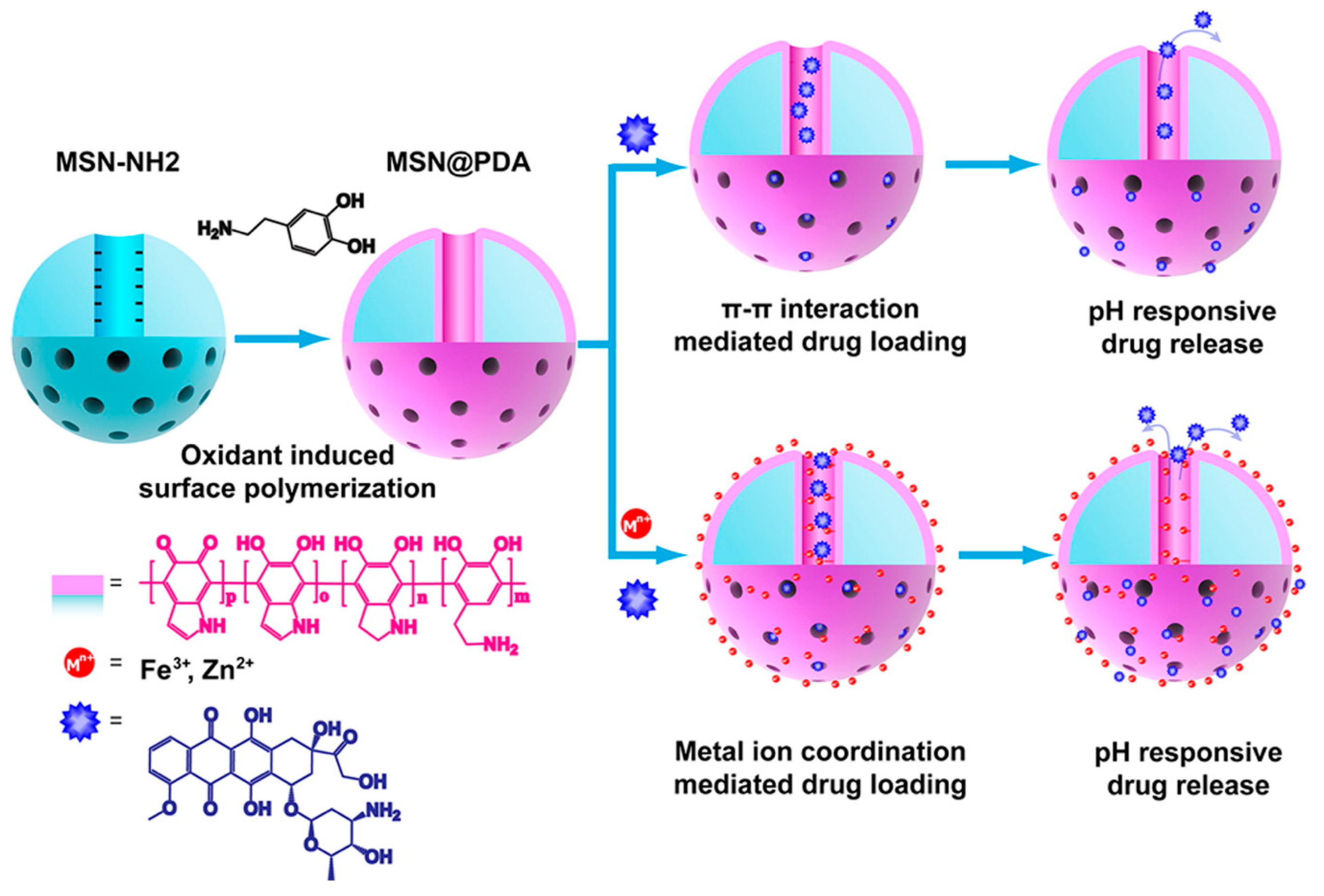
  20. Li, X.; Xie, C.; Xia, H.; Wang, Z. pH and Ultrasound Dual-Responsive Polydopamine-Coated Mesoporous Silica Nanoparticles for Controlled Drug Delivery. Langmuir 2018, 34, 9974–9981. [Google Scholar] [CrossRef]
  21. Rahoui, N.; Jiang, B.; Hegazy, M.; Taloub, N.; Wang, Y.; Yu, M.; Huang, Y.D. Gold modified polydopamine coated mesoporous silica nano-structures for synergetic chemo-photothermal effect. Colloids Surf. B Biointerfaces 2018, 171, 176–185. [Google Scholar] [CrossRef]
  22. Li, Y.; Duo, Y.; Bao, S.; He, L.; Ling, K.; Luo, J.; Zhang, Y.; Huang, H.; Zhang, H.; Yu, X. EpCAM aptamer-functionalized polydopamine-coated mesoporous silica nanoparticles loaded with DM1 for targeted therapy in colorectal cancer. Int. J. Nanomed. 2017, 12, 6239–6257. [Google Scholar] [CrossRef] [PubMed] [Green Version]
  23. Pada, A.-K.; Desai, D.; Sun, K.; Prakirth Govardhanam, N.; Törnquist, K.; Zhang, J.; Rosenholm, J.M. Comparison of Polydopamine-Coated Mesoporous Silica Nanorods and Spheres for the Delivery of Hydrophilic and Hydrophobic Anticancer Drugs. Int. J. Mol. Sci. 2019, 20, 3408. [Google Scholar] [CrossRef] [PubMed] [Green Version]
  24. Karaman, D.; Desai, D.; Senthilkumar, R.; Johansson, E.M.; Råtts, N.; Odén, M.; Eriksson, J.E.; Sahlgren, C.; Toivola, D.M.; Rosenholm, J.M. Shape engineering vs organic modification of inorganic nanoparticles as a tool for enhancing cellular internalization. Nanoscale Res. Lett. 2012, 7, 358. [Google Scholar] [CrossRef] [PubMed] [Green Version]
  25. Zheng, X.; Zhang, J.; Wang, J.; Qi, X.; Rosenholm, J.M.; Cai, K. Polydopamine Coatings in Confined Nanopore Space: Toward Improved Retention and Release of Hydrophilic Cargo. J. Phys. Chem. C 2015, 119, 24512–24521. [Google Scholar] [CrossRef]
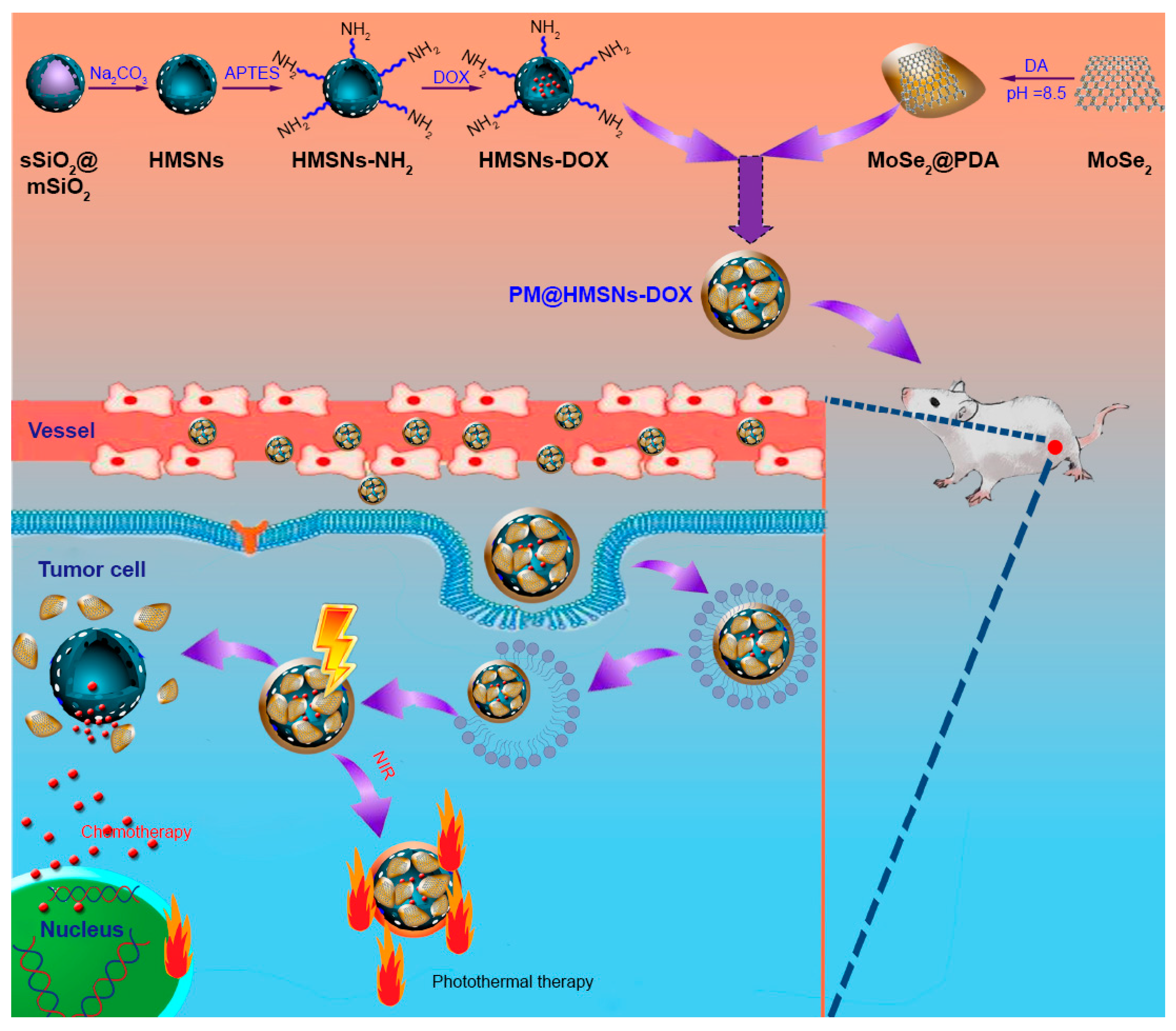
  26. Chai, S.; Kan, S.; Sun, R.; Zhou, R.; Sun, Y.; Chen, W.; Yu, B. Fabricating polydopamine-coated MoSe2-wrapped hollow mesoporous silica nanoplatform for controlled drug release and chemo-photothermal therapy. Int. J. Nanomed. 2018, 13, 7607–7621. [Google Scholar] [CrossRef] [Green Version]
  27. Cheng, W.; Liang, C.; Xu, L.; Liu, G.; Gao, N.; Tao, W.; Luo, L.; Zuo, Y.; Wang, X.; Zhang, X.; et al. TPGS-Functionalized Polydopamine-Modified Mesoporous Silica as Drug Nanocarriers for Enhanced Lung Cancer Chemotherapy against Multidrug Resistance. Small 2017, 13, 1700623. [Google Scholar] [CrossRef]
  28. Szegedi, Á.; Shestakova, P.; Trendafilova, I.; Mihayi, J.; Tsacheva, I.; Mitova, V.; Kyulavska, M.; Koseva, N.; Momekova, D.; Konstantinov, S.; et al. Modified mesoporous silica nanoparticles coated by polymer complex as novel curcumin delivery carriers. J. Drug Deliv. Sci. Technol. 2019, 49, 700–712. [Google Scholar] [CrossRef]
  29. Hu, Y.; Ke, L.; Chen, H.; Zhuo, M.; Yang, X.; Zhao, D.; Zeng, S.; Xiao, X. Natural material-decorated mesoporous silica nanoparticle container for multifunctional membrane-controlled targeted drug delivery. Int. J. Nanomed. 2017, 12, 8411–8426. [Google Scholar] [CrossRef] [Green Version]
  30. Iraji, S.; Ganji, F.; Rashidi, L. Surface modified mesoporous silica nanoparticles as sustained-release gallic acid nano-carriers. J. Drug Deliv. Sci. Technol. 2018, 47, 468–476. [Google Scholar] [CrossRef]
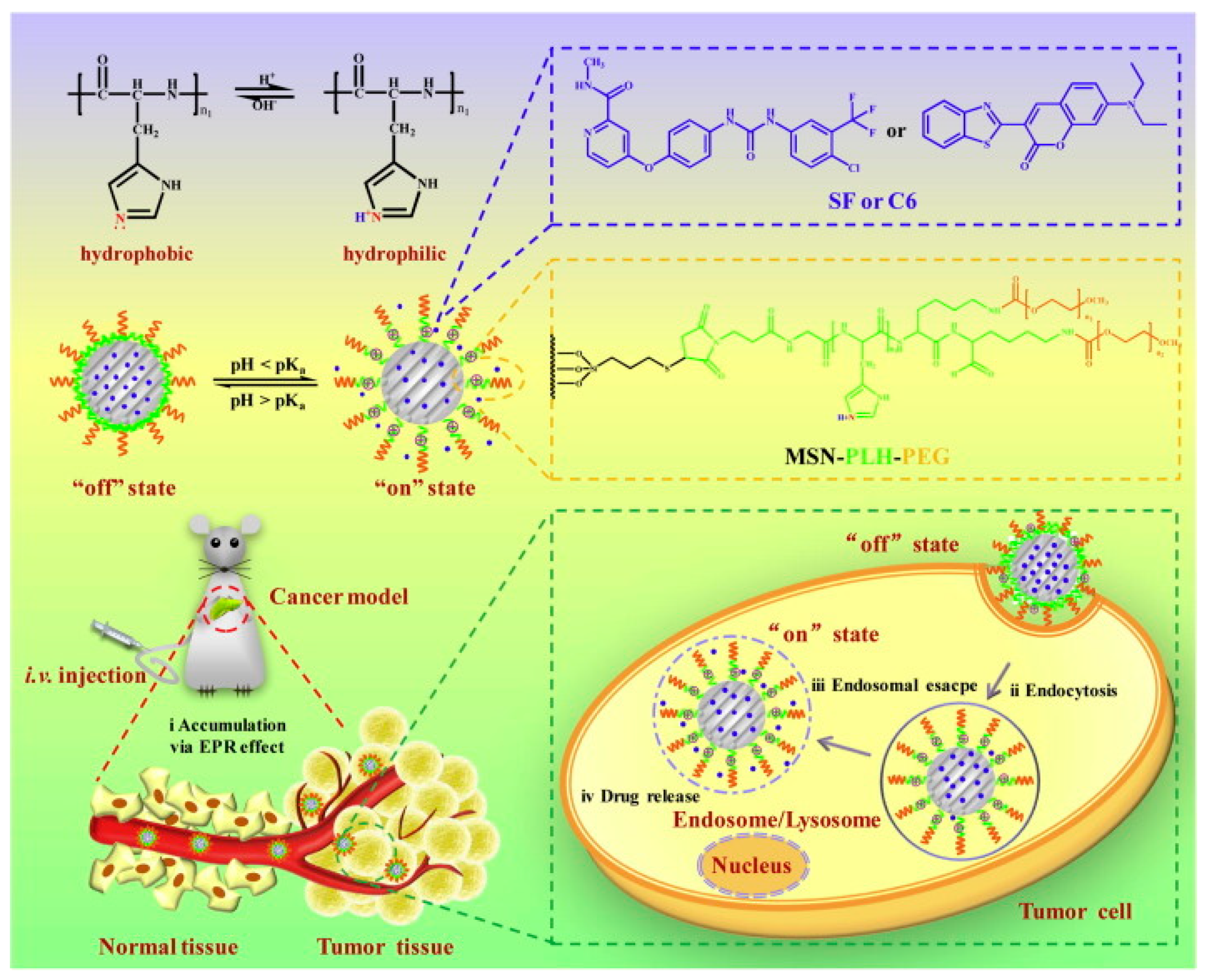
  31. Mu, S.; Liu, Y.; Wang, T.; Zhang, J.; Jiang, D.; Yu, X.; Zhang, N. Unsaturated nitrogen-rich polymer poly(l-histidine) gated reversibly switchable mesoporous silica nanoparticles using “graft to” strategy for drug controlled release. Acta Biomater. 2017, 63, 150–162. [Google Scholar] [CrossRef]
  32. Lin, J.; Cai, Q.; Tang, Y.; Xu, Y.; Wang, Q.; Li, T.; Xu, H.; Wang, S.; Fan, K.; Liu, Z.; et al. PEGylated Lipid bilayer coated mesoporous silica nanoparticles for co-delivery of paclitaxel and curcumin: Design, characterization and its cytotoxic effect. Int. J. Pharm. 2018, 536, 272–282. [Google Scholar] [CrossRef]
  33. Kienzle, A.; Kurch, S.; Schlöder, J.; Berges, C.; Ose, R.; Schupp, J.; Tuettenberg, A.; Weiss, H.; Schultze, J.; Winzen, S.; et al. Dendritic Mesoporous Silica Nanoparticles for pH-Stimuli-Responsive Drug Delivery of TNF-Alpha. Adv. Healthc. Mater. 2017, 6, 1700012. [Google Scholar] [CrossRef] [PubMed]
  34. Lee, J.; Lee, Y.M.; Kim, J.; Kim, W.J. Doxorubicin/Ce6-Loaded Nanoparticle Coated with Polymer via Singlet Oxygen-Sensitive Linker for Photodynamically Assisted Chemotherapy. Nanotheranostics 2017, 1, 196–207. [Google Scholar] [CrossRef] [PubMed] [Green Version]
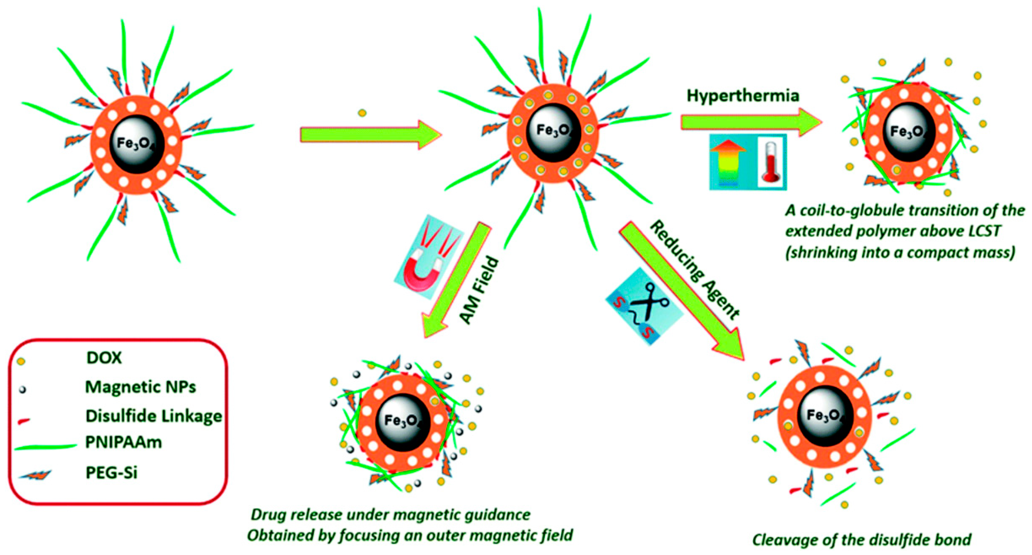
  35. Hegazy, M.; Zhou, P.; Wu, G.; Wang, L.; Rahoui, N.; Taloub, N.; Huang, X.; Huang, Y. Construction of polymer coated core–shell magnetic mesoporous silica nanoparticles with triple responsive drug delivery. Polym. Chem. 2017, 8, 5852–5864. [Google Scholar] [CrossRef]
  36. Feng, Y.; Li, N.; Yin, H.; Chen, T.; Yang, Q.; Wu, M. Thermo- and pH-responsive, Lipid-coated, Mesoporous Silica Nanoparticle-based Dual Drug Delivery System To Improve the Antitumor Effect of Hydrophobic Drugs. Mol. Pharm. 2019, 16, 422–436. [Google Scholar] [CrossRef] [PubMed]
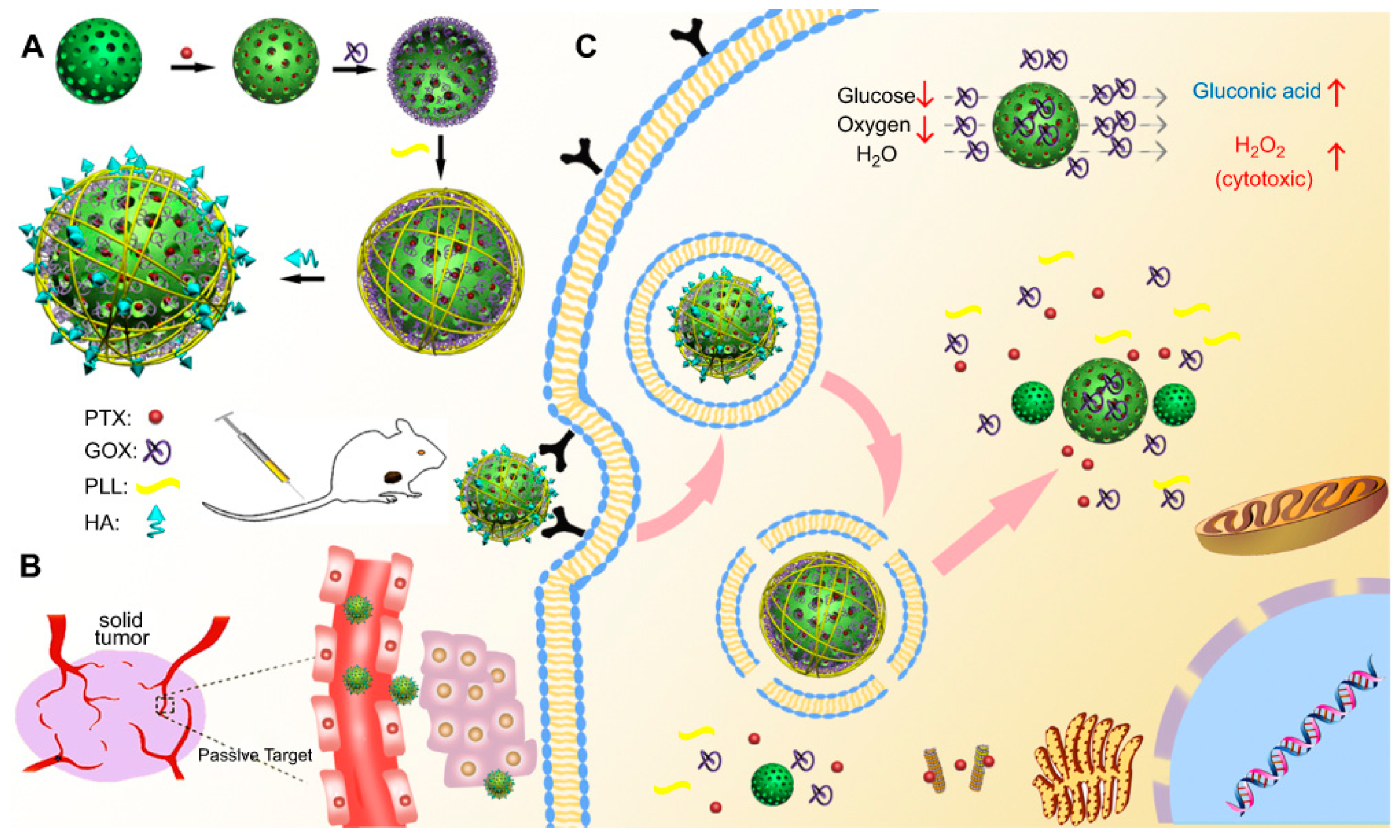
  37. Du, X.; Zhang, T.; Ma, G.; Gu, X.; Wang, G.; Li, J. Glucose-responsive mesoporous silica nanoparticles to generation of hydrogen peroxide for synergistic cancer starvation and chemistry therapy. Int. J. Nanomed. 2019, 14, 2233–2251. [Google Scholar] [CrossRef] [Green Version]
  38. Yuan, N.; Li, S.; Li, G. Sodium alginate coated mesoporous silica for dual bio-responsive controlled drug delivery. J. Drug Deliv. Sci. Technol. 2018, 46, 348–353. [Google Scholar] [CrossRef]
  39. Avedian, N.; Zaaeri, F.; Daryasari, M.P.; Akbari Javar, H.; Khoobi, M. pH-sensitive biocompatible mesoporous magnetic nanoparticles labeled with folic acid as an efficient carrier for controlled anticancer drug delivery. J. Drug Deliv. Sci. Technol. 2018, 44, 323–332. [Google Scholar] [CrossRef]
  40. Desai, D.; Karaman, D.S.; Prabhakar, N.; Tadayon, S.; Duchanoy, A.; Toivola, D.M.; Rajput, S.; Näreoja, T.; Rosenholm, J.M. Design considerations for mesoporous silica nanoparticulate systems in facilitating biomedical applications. Open Mater. Sci. 2014, 1, 16–43. [Google Scholar] [CrossRef] [Green Version]
  41. Liu, J.; Liang, H.; Li, M.; Luo, Z.; Zhang, J.; Guo, X.; Cai, K. Tumor acidity activating multifunctional nanoplatform for NIR-mediated multiple enhanced photodynamic and photothermal tumor therapy. Biomaterials 2018, 157, 107–124. [Google Scholar] [CrossRef]
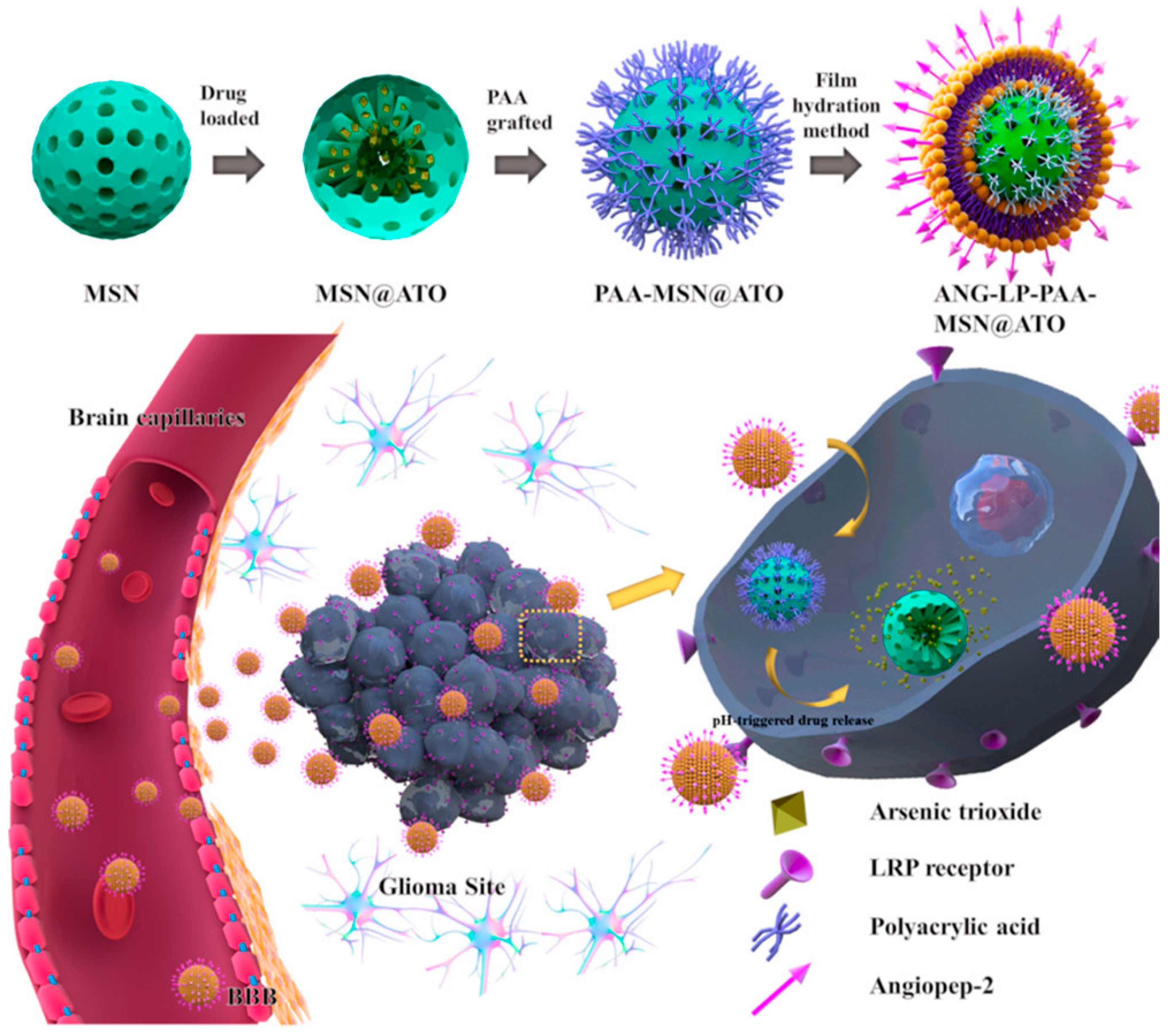
  42. Tao, J.; Fei, W.; Tang, H.; Li, C.; Mu, C.; Zheng, H.; Li, F.; Zhu, Z. Angiopep-2-Conjugated “Core–Shell” Hybrid Nanovehicles for Targeted and pH-Triggered Delivery of Arsenic Trioxide into Glioma. Mol. Pharm. 2019, 16, 786–797. [Google Scholar] [CrossRef] [PubMed]
  43. Moreira, A.F.; Rodrigues, C.F.; Reis, C.A.; Costa, E.C.; Ferreira, P.; Correia, I.J. Development of poly-2-ethyl-2-oxazoline coated gold-core silica shell nanorods for cancer chemo-photothermal therapy. Nanomedicine 2018, 13, 2611–2627. [Google Scholar] [CrossRef] [PubMed]
  44. Yu, F.; Wu, H.; Tang, Y.; Xu, Y.; Qian, X.; Zhu, W. Temperature-sensitive copolymer-coated fluorescent mesoporous silica nanoparticles as a reactive oxygen species activated drug delivery system. Int. J. Pharm. 2018, 536, 11–20. [Google Scholar] [CrossRef] [PubMed]
  45. Liu, X.; Li, M.; Yuan, W.; Liu, Y.; Wang, Y. Lipid-coated mesoporous silica nanoparticles of hydroxycamptothecin for sustained release and cancer therapy. Pharmazie 2018, 73, 447–453. [Google Scholar]
  46. Ma, J.; Wu, H.; Li, Y.; Liu, Z.; Liu, G.; Guo, Y.; Hou, Z.; Zhao, Q.; Chen, D.; Zhu, X. Novel Core-Interlayer-Shell DOX/ZnPc Co-loaded MSNs@ pH-Sensitive CaP@PEGylated Liposome for Enhanced Synergetic Chemo-Photodynamic Therapy. Pharm. Res. 2018, 35, 57. [Google Scholar] [CrossRef] [PubMed]
  47. Tzankov, B.; Tzankova, V.; Aluani, D.; Yordanov, Y.; Spassova, I.; Kovacheva, D.; Avramova, K.; Valoti, M.; Yoncheva, K. Development of MCM-41 mesoporous silica nanoparticles as a platform for pramipexole delivery. J. Drug Deliv. Sci. Technol. 2019, 51, 26–35. [Google Scholar] [CrossRef]
  48. Shen, Y.; Cao, B.; Snyder, N.R.; Woeppel, K.M.; Eles, J.R.; Cui, X.T. ROS responsive resveratrol delivery from LDLR peptide conjugated PLA-coated mesoporous silica nanoparticles across the blood–brain barrier. J. Nanobiotechnol. 2018, 16, 13. [Google Scholar] [CrossRef] [Green Version]
  49. Cheng, C.S.; Liu, T.P.; Chien, F.C.; Mou, C.Y.; Wu, S.H.; Chen, Y.P. Codelivery of Plasmid and Curcumin with Mesoporous Silica Nanoparticles for Promoting Neurite Outgrowth. ACS Appl. Mater. Interfaces 2019, 11, 15322–15331. [Google Scholar] [CrossRef]
  50. Mandić, L.; Sadžak, A.; Strasser, V.; Baranović, G.; Domazet Jurašin, D.; Sikirić, M.D.; Šegota, S. Enhanced Protection of Biological Membranes during Lipid Peroxidation: Study of the Interactions between Flavonoid Loaded Mesoporous Silica Nanoparticles and Model Cell Membranes. Int. J. Mol. Sci. 2019, 20, 2709. [Google Scholar] [CrossRef] [Green Version]
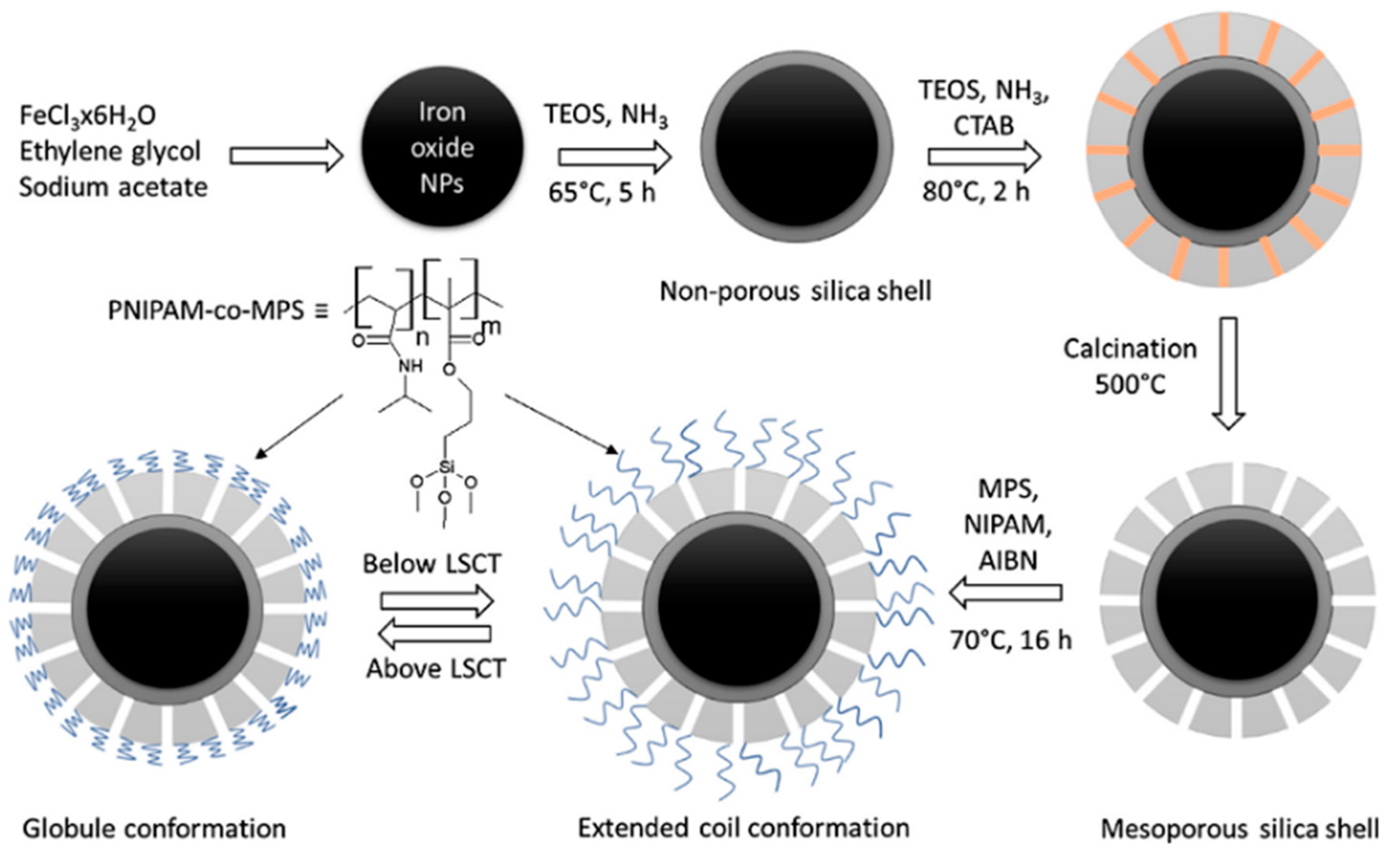
  51. Peralta, M.E.; Jadhav, S.A.; Magnacca, G.; Scalarone, D.; Mártire, D.O.; Parolo, M.E.; Carlos, L. Synthesis and in vitro testing of thermoresponsive polymer-grafted core-shell magnetic mesoporous silica nanoparticles for efficient controlled and targeted drug delivery. J. Colloid Interface Sci. 2019, 544, 198–205. [Google Scholar] [CrossRef]
  52. Gulin-Sarfraz, T.; Jonasson, S.; Wigenstam, E.; von Haartman, E.; Bucht, A.; Rosenholm, J. Feasibility Study of Mesoporous Silica Particles for Pulmonary Drug Delivery: Therapeutic Treatment with Dexamethasone in a Mouse Model of Airway Inflammation. Pharmaceutics 2019, 11, 149. [Google Scholar] [CrossRef] [PubMed] [Green Version]
  53. Popova, M.; Trendafilova, I.; Zgureva, D.; Kalvachev, Y.; Boycheva, S.; Novak Tušar, N.; Szegedi, A. Polymer-coated mesoporous silica nanoparticles for controlled release of the prodrug sulfasalazine. J. Drug Deliv. Sci. Technol. 2018, 44, 415–420. [Google Scholar] [CrossRef]
  54. Liu, Y.; Xu, M.; Chen, Q.; Guan, G.; Hu, W.; Zhao, X.; Qiao, M.; Hu, H.; Liang, Y.; Zhu, H.; et al. Gold nanorods/mesoporous silica-based nanocomposite as theranostic agents for targeting near-infrared imaging and photothermal therapy induced with laser. Int. J. Nanomed. 2015, 10, 4747. [Google Scholar] [CrossRef] [PubMed] [Green Version]
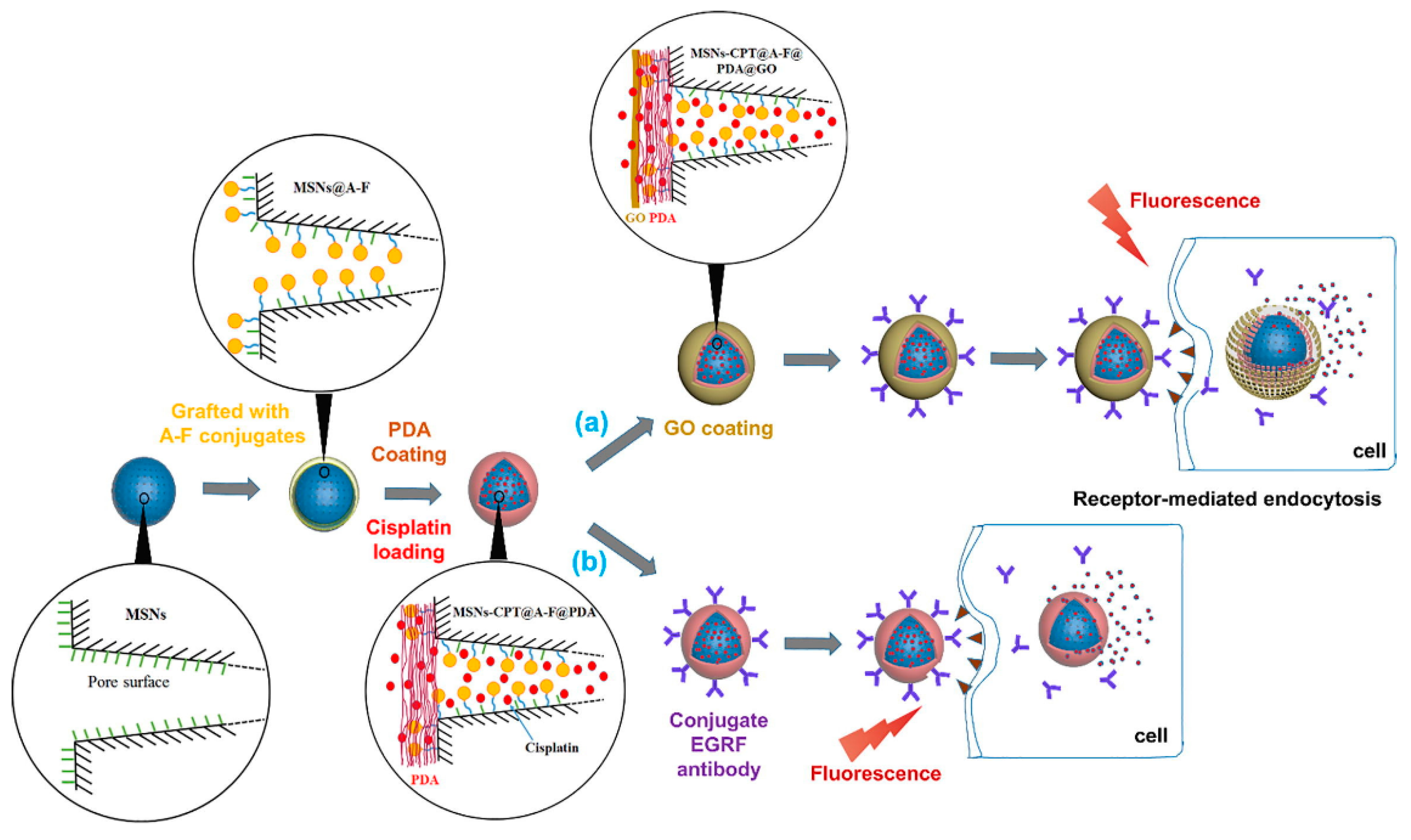
  55. Tran, A.-V.; Shim, K.; Vo Thi, T.-T.; Kook, J.-K.; An, S.S.A.; Lee, S.-W. Targeted and controlled drug delivery by multifunctional mesoporous silica nanoparticles with internal fluorescent conjugates and external polydopamine and graphene oxide layers. Acta Biomater. 2018, 74, 397–413. [Google Scholar] [CrossRef] [PubMed]
  56. Xu, X.; Lü, S.; Wu, C.; Wang, Z.; Feng, C.; Wen, N.; Liu, M.; Zhang, X.; Liu, Z.; Liu, Y.; et al. Curcumin polymer coated, self-fluorescent and stimuli-responsive multifunctional mesoporous silica nanoparticles for drug delivery. Microporous Mesoporous Mater. 2018, 271, 234–242. [Google Scholar] [CrossRef]
  57. Song, Y.; Jiang, H.; Wang, B.; Kong, Y.; Chen, J. Silver-Incorporated Mussel-Inspired Polydopamine Coatings on Mesoporous Silica as an Efficient Nanocatalyst and Antimicrobial Agent. ACS Appl. Mater. Interfaces 2018, 10, 1792–1801. [Google Scholar] [CrossRef]
  58. Yunessnia lehi, A.; Shagholani, H.; Nikpay, A.; Ghorbani, M.; Soleimani lashkenari, M.; Soltani, M. Synthesis and modification of crystalline SBA-15 nanowhiskers as a pH-sensitive metronidazole nanocarrier system. Int. J. Pharm. 2019, 555, 28–35. [Google Scholar] [CrossRef]
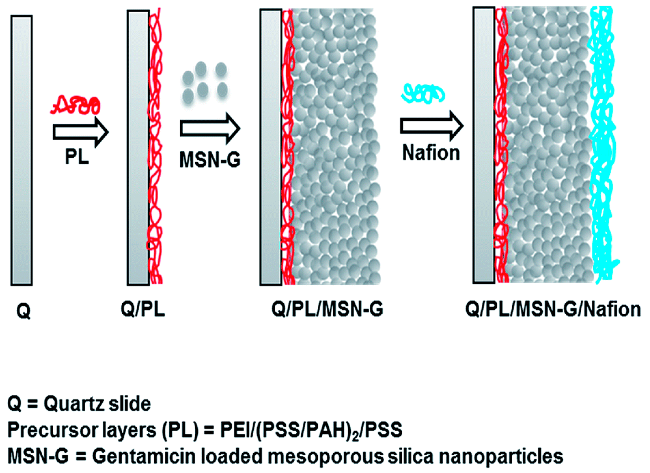
  59. Tamanna, T.; Bulitta, J.B.; Landersdorfer, C.B.; Cashin, V.; Yu, A. Stability and controlled antibiotic release from thin films embedded with antibiotic loaded mesoporous silica nanoparticles. RSC Adv. 2015, 5, 107839–107846. [Google Scholar] [CrossRef]
  60. Zhang, J.; Cai, L.; Wang, T.; Tang, S.; Li, Q.; Tang, T.; Wei, S.; Qian, J.; Wei, J.; Su, J. Lithium doped silica nanospheres/poly(dopamine) composite coating on polyetheretherketone to stimulate cell responses, improve bone formation and osseointegration. Nanomed. Nanotechnol. Biol. Med. 2018, 14, 965–976. [Google Scholar] [CrossRef]
  61. Sun, T.; Sun, Y.; Zhang, H. Phospholipid-Coated Mesoporous Silica Nanoparticles Acting as Lubricating Drug Nanocarriers. Polymers 2018, 10, 513. [Google Scholar] [CrossRef] [Green Version]
  62. Ngamcherdtrakul, W.; Morry, J.; Gu, S.; Castro, D.J.; Goodyear, S.M.; Sangvanich, T.; Reda, M.M.; Lee, R.; Mihelic, S.A.; Beckman, B.L.; et al. Cationic Polymer Modified Mesoporous Silica Nanoparticles for Targeted siRNA Delivery to HER2 + Breast Cancer. Adv. Funct. Mater. 2015, 25, 2646–2659. [Google Scholar] [CrossRef] [PubMed] [Green Version]
  63. Zarei, H.; Kazemi Oskuee, R.; Hanafi-Bojd, M.Y.; Gholami, L.; Ansari, L.; Malaekeh-Nikouei, B. Enhanced gene delivery by polyethyleneimine coated mesoporous silica nanoparticles. Pharm. Dev. Technol. 2019, 24, 127–132. [Google Scholar] [CrossRef] [PubMed]
  64. Zhang, Y.; Ren, K.; Zhang, X.; Chao, Z.; Yang, Y.; Ye, D.; Dai, Z.; Liu, Y.; Ju, H. Photo-tearable tape close-wrapped upconversion nanocapsules for near-infrared modulated efficient siRNA delivery and therapy. Biomaterials 2018, 163, 55–66. [Google Scholar] [CrossRef] [PubMed]
  65. Esmaeili, A.; Mousavi, S.N. Synthesis of a novel structure for the oral delivery of insulin and the study of its effect on diabetic rats. Life Sci. 2017, 186, 43–49. [Google Scholar] [CrossRef] [PubMed]
  66. Mendiratta, S.; Hussein, M.; Nasser, H.A.; Ali, A.A.A. Multidisciplinary Role of Mesoporous Silica Nanoparticles in Brain Regeneration and Cancers: From Crossing the Blood-Brain Barrier to Treatment. Part. Part. Syst. Charact. 2019, 36, 1900195. [Google Scholar] [CrossRef] [Green Version]
  67. Le Joncour, V.; Karaman, S.; Laakkonen, P.M. Predicting in vivo payloads delivery using a blood-brain tumor-barrier in a dish. J. Vis. Exp. 2019, 146, e59384. [Google Scholar] [CrossRef] [Green Version]
  68. Tzankov, B.; Voycheva, C.; Yordanov, Y.; Aluani, D.; Spassova, I.; Kovacheva, D.; Lambov, N.; Tzankova, V. Development and in vitro safety evaluation of pramipexole-loaded hollow mesoporous silica (HMS) particles. Biotechnol. Biotechnol. Equip. 2019, 33, 1204–1215. [Google Scholar] [CrossRef] [Green Version]
  69. Şen Karaman, D.; Manner, S.; Rosenholm, J.M. Mesoporous silica nanoparticles as diagnostic and therapeutic tools: How can they combat bacterial infection? Ther. Deliv. 2018, 9, 241–244. [Google Scholar] [CrossRef] [Green Version]
  70. Şen Karaman, D.; Sarwar, S.; Desai, D.; Björk, E.M.; Odén, M.; Chakrabarti, P.; Rosenholm, J.M.; Chakraborti, S. Shape engineering boosts antibacterial activity of chitosan coated mesoporous silica nanoparticle doped with silver: A mechanistic investigation. J. Mater. Chem. B 2016, 4, 3292–3304. [Google Scholar] [CrossRef] [Green Version]
  71. Morry, J.; Ngamcherdtrakul, W.; Gu, S.; Reda, M.; Castro, D.J.; Sangvanich, T.; Gray, J.W.; Yantasee, W. Targeted Treatment of Metastatic Breast Cancer by PLK1 siRNA Delivered by an Antioxidant Nanoparticle Platform. Mol. Cancer Ther. 2017, 16, 763–772. [Google Scholar] [CrossRef] [Green Version]
  72. Prabhakar, N.; Zhang, J.; Desai, D.; Casals, E.; Gulin-Sarfraz, T.; Näreoja, T.; Westermarck, J.; Rosenholm, J.M. Stimuli-responsive hybrid nanocarriers developed by controllable integration of hyperbranched PEI with mesoporous silica nanoparticles for sustained intracellular siRNA delivery. Int. J. Nanomed. 2016, 11, 6591–6608. [Google Scholar] [CrossRef] [PubMed] [Green Version]
  73. Rosenholm, J.M.; Gulin-Sarfraz, T.; Mamaeva, V.; Niemi, R.; Özliseli, E.; Desai, D.; Antfolk, D.; von Haartman, E.; Lindberg, D.; Prabhakar, N.; et al. Prolonged Dye Release from Mesoporous Silica-Based Imaging Probes Facilitates Long-Term Optical Tracking of Cell Populations In vivo. Small 2016, 12, 1578–1592. [Google Scholar] [CrossRef] [PubMed]
  74. Prabhakar, N.; Rosenholm, J.M. Nanodiamonds for advanced optical bioimaging and beyond. Curr. Opin. Colloid Interface Sci. 2019, 39, 220–231. [Google Scholar] [CrossRef]
  75. Von Haartman, E.; Jiang, H.; Khomich, A.A.; Zhang, J.; Burikov, S.A.; Dolenko, T.A.; Ruokolainen, J.; Gu, H.; Shenderova, O.A.; Vlasov, I.I.; et al. Core-shell designs of photoluminescent nanodiamonds with porous silica coatings for bioimaging and drug delivery I: Fabrication. J. Mater. Chem. B 2013, 1, 2358–2366. [Google Scholar] [CrossRef]
  76. Hong, S.; Kim, K.Y.; Wook, H.J.; Park, S.Y.; Lee, K.D.; Lee, D.Y.; Lee, H. Attenuation of the in vivo toxicity of biomaterials by polydopamine surface modification. Nanomedicine 2011, 6, 793–801. [Google Scholar] [CrossRef] [PubMed]
  77. Svirshchevskaya, E.V.; Zubareva, A.A.; Boyko, A.A.; Shustova, O.A.; Grechikhina, M.V.; Shagdarova, B.T.; Varlamov, V.P. Analysis of toxicity and biocompatibility of chitosan derivatives with different physico-chemical properties. Appl. Biochem. Microbiol. 2016, 52, 483–490. [Google Scholar] [CrossRef]
  78. Becker, L.C.; Bergfeld, W.F.; Belsito, D.V.; Klaassen, C.D.; Marks, J.G.; Shank, R.C.; Slaga, T.J.; Snyder, P.W.; Cosmetic Ingredient Review Expert Panel; Andersen, F.A. Final report of the safety assessment of hyaluronic acid, potassium hyaluronate, and sodium hyaluronate. Int. J. Toxicol. 2009, 28, 5–67. [Google Scholar] [CrossRef]
  79. Johnson, R.P.; John, J.V.; Kim, I. Poly(l-histidine)-containing polymer bioconjugate hybrid materials as stimuli-responsive theranostic systems. J. Appl. Polym. Sci. 2014, 131, 40796. [Google Scholar] [CrossRef]
  80. Isaksson, K.; Åkerberg, D.; Posaric-Bauden, M.; Andersson, R.; Tingstedt, B. In vivo toxicity and biodistribution of intraperitoneal and intravenous poly-l-lysine and poly-l-lysine/poly-l-glutamate in rats. J. Mater. Sci. Mater. Med. 2014, 25, 1293–1299. [Google Scholar] [CrossRef]
  81. Moghimi, S.M.; Symonds, P.; Murray, J.C.; Hunter, A.C.; Debska, G.; Szewczyk, A. A two-stage poly(ethylenimine)-mediated cytotoxicity: Implications for gene transfer/therapy. Mol. Ther. 2005, 11, 990–995. [Google Scholar] [CrossRef]
  82. Kazemi Oskuee, R.; Dabbaghi, M.; Gholami, L.; Taheri-Bojd, S.; Balali-Mood, M.; Mousavi, S.H.; Malaekeh-Nikouei, B. Investigating the influence of polyplex size on toxicity properties of polyethylenimine mediated gene delivery. Life Sci. 2018, 197, 101–108. [Google Scholar] [CrossRef]
  83. Lanzalaco, S.; Armelin, E. Poly(N-isopropylacrylamide) and Copolymers: A Review on Recent Progresses in Biomedical Applications. Gels 2017, 3, 36. [Google Scholar] [CrossRef]
  84. Cooperstein, M.A.; Canavan, H.E. Assessment of cytotoxicity of (N-isopropyl acrylamide) and poly(N-isopropyl acrylamide)-coated surfaces. Biointerphases 2013, 8, 1–12. [Google Scholar] [CrossRef] [Green Version]
Figure 1. Graphical representation of the approaches used to prepare drug loaded MSNs coated with polymer.
Applsci 10 00289 g001
Figure 2. Polymers of natural and synthetic origin that have been used as coating agent for MSN for providing control over drug release.
Applsci 10 00289 g002
Figure 3. Biological disorders that have been targeted using polymer coated mesoporous silica nanoparticles.
Applsci 10 00289 g003
Figure 4. Pictorial presentation of pH/HIFU dual-responsive release characteristic of DOX loaded MSN–PDA. Reproduced with permission from reference [20].
Applsci 10 00289 g004
Figure 5. Schematic illustration of the progress in constructing polydopamine-coated MSN particles for drug loading and release. Adapted with permission from [25], copyright (2015) American Chemical Society.
Applsci 10 00289 g005
Figure 6. Graphical representation of the preparation of dual sensitive MSNs for pH/NIR-responsive chemo-photothermal therapy [26].
Applsci 10 00289 g006
Figure 7. Schematic presentation of pH responsive drug release with the aid of poly(L-histidine) (PLH) coated MSNs. Reproduced with permission from reference [31].
Applsci 10 00289 g007
Figure 8. Schematic representation of a triple responsive release mechanism of drug from PNIPAAm coated magnetic MSNs. Reproduced with permission from reference [35].
Applsci 10 00289 g008
Figure 9. Schematic presentation of coated MSNs for synergistic tumor-targeted therapy [37].
Applsci 10 00289 g009
Figure 10. Graphical representation of the synthesis of PAA coated MSNs and their encapsulation in liposomes for glioma therapy [42].
Applsci 10 00289 g010
Figure 11. Preparation of polymer coated magnetic MSNs for delivery of Ibuprofen. Reproduced with permission from reference [51].
Applsci 10 00289 g011
Figure 12. Pictorial presentation of thin film preparation of MSNs coated with Nafion. Reproduced with permission from reference [59].
Applsci 10 00289 g012
Figure 13. Graphical representation of (a) synthesis of nanoparticles, and (b) receptor-mediated endocytosis and NIR controlled intracellular delivery of siRNA. Reproduced with permission from reference [64].
Applsci 10 00289 g013
Figure 14. Illustration of preparation of theranostic MSNs (a) graphene oxide coated and (b) EGRF antibody conjugated without graphene oxide coating, for targeted delivery and imaging in cancer cells. Reproduced with permission from reference [55].
Applsci 10 00289 g014
Table 1. Clinical trials on silica nanoparticles.
S.
No
TitleSubmission DateIdentifierRecruitment Status
1Plasmonic Photothermal Therapy of Flow-Limiting Atherosclerotic Lesions with Silica-Gold Nanoparticles: a First-in-Man Study30 December 2010NCT01270139Completed
2Targeted Silica Nanoparticles for Real-Time Image-Guided Intraoperative Mapping of Nodal Metastases3 April 2014NCT02106598Recruiting
3Molecular Phenotyping and Image-Guided Surgical Treatment of Prostate Cancer Using Ultra small Silica Nanoparticles18 November 2019NCT04167969Recruiting
Table 2. Tabular presentation of different coating materials used for MSNs along with their investigated applications.
Sr NoInvestigatorsSilica CorePolymer Used Drug Application
1Li et al. [20]MSNsPolydopamineDOXCancer
2Rahoui et al. [21]Gold modified MSNsPolydopamineDOXCancer
3Yang et al. [22]MSNsPolydopamine, PEG and EpCAM aptamerDM-1Colorectal cancer
4Chai et al. [26]MoSe2 wrapped MSNsPolydopamineDOXCancer
5Cheng et al. [27]MSNsAlpha-tocopheryl polyethylene glycol 1000 succinate functionalized polydopamineDOXCancer
6Szegedi et al. [28]KIL-2 and KIT-6k-Carrageenan and ChitosanCurcuminCancer
7Hu et al. [29]MSNsFolic acid modified Chitosan and Hyaluronic acidDOXCancer
8Iraji et al. [30]MSNsChitosanGallic acidCancer
9Mu et al. [31]MSNsPoly-(L-Histidine) and PEG SorafenibCancer
10Hegazy et al. [35]Core-shell magnetic MSNsPoly(N-isopropylacrylamide) (PNIPAAm)DOXCancer
11Feng et al. [36]MSNsPoly(N-Isopropylacrylamide-co-methacrylic acid) and DSPE-PEG2000Evodiamine and BerberineCancer
12Du et al. [37]MSNsPoly(L-lysine) and Hyaluronic acidGlucose Oxidase and PaclitaxelCancer
13Yuan et al. [38]MSNsSodium AlginateDOXCancer
14Avedian et al. [39]Fe3O4 core MSNsPolyethyleneimine and Folic acidErlotinibCancer
15Moreira et al. [43]Gold core MSNsPoly-2-ethyl-2-oxazolineDOXCancer
16Yu et al. [44]MSNs4-(4,4,5,5-tetramethyl-1,3,2-dioxaborolan-2-yl)benzyl acrylate polymerDOXCancer
17Liu et al. [45]MSNsLipid Galactosyl-CeramideHydroxy-campothecineCancer
18Ma et al. [46]MSNs encapsulated in liposomesCalcium PhosphateDOX and Zinc PhthalocyanineCancer
19Liu et al. [41]Gold Nanorods coated with Mesoporous silicaβ-cyclodextrin and (2,3-dimethylmaleic anhydride modified chitosan oligosaccharide-b-poly(ethylene glycol))Indocyanine Green dyeCancer
20Lin et al. [32]Highly ordered MSNsPEGylated Lipid bilayerPaclitaxel and CurcuminCancer
21Kienzle et al. [33]Dendritic MSNsPolyethylene imine and Poly ethylene glycolTNF-αCancer
22Lee et al. [34]Chlorin-e6 loaded MSNsPEGDOXCancer
23Tao et al. [42]MSNs encapsulated in liposomes Poly(acrylic acid) Arsenic trioxideGlioma
24Tzankov et al. [47]MCM-41Chitosan and Sodium alginatePramipexoleNeuroblastoma
25Shen et al. [48]MSNsPoly(lactic acid) ResveratrolParkinson’s disease
26Cheng et al. [49]MSNsplasmid RhoG-DsRedCurcuminOxidative stress
27Mandic et al. [50]MSNsPEGQuercetin, myricetin and myricitrinOxidative stress
28Peralta et al. [51]MSNsPoly(N-isopropylacrylamide-co-3-(methacryloxypropyl) trimethoxysilane)IbuprofenInflammation
29Gulin-Sarfaz et al. [52]MSNsPEI and PEGDexamethasonePulmonary Inflammation
30Popova et al. [53]SBA-15 and MCM- 41Eudragit S and Eudragit RLSulfasalazine inflammatory bowel disease
31Liu et al. [54]Gold nanorods coated mesoporous silica PEGIndocyanine greenLight-induced imaging-guided cancer therapy
32Tran et al. [55]FITC-MSNsPolydopamine and Graphene oxideCisplatinTheranostic Cancer therapy
33Xu et al. [56]MSNsPoly CurcuminDOXTheranostic Cancer therapy
34Song et al. [57]SBA-15PolydopamineSilverAntibacterial therapy
35Lehi et al. [58]SBA-15 nanowishkers Tannic acid MetronidazoleTrichomonasis
36Tamanna et al. [59]MSNsNafion PolymerGentamicinImmunoassay
37Zhang et al. [60]Lithium Doped Silica Nanospheres Polydopamine Polyetherether ketone implantBone regeneration
38Sun et al. [61]MSNsDistearoyl phosphatidylcholineRhodamine- BOsteoarthritis
39Ngamcherdtrakul et al. [62]MSNsPEI and PEG siRNAGene Delivery
40Zarei et al. [63]Phosphonate modified MSNsPEIPlasmid DNA and chlooquineGene delivery
41Zhang et al. [64]Lanthanide doped upconversion Silica nanoparticlesPEGsiRNA and hypocrellin AGene delivery
42Esmaeli et al. [65]MCM-41PAMAM dendrimer and chitosan-gelatine scaffoldInsulin and cinnamaldeydeDiabetes

