Article

Comparative Toxicological Evaluation of Solubilizers and Hydrotropic Agents Using Daphnia magna as a Model Organism

by
Iulia Ioana Olaru
,
Dragos Paul Mihai
*,
Octavian Tudorel Olaru
*,
Cerasela Elena Gird
,
Anca Zanfirescu
,
Gheorghe Stancov
,
Corina Andrei
,
Emanuela-Alice Luta
and
George Mihai Nitulescu
Faculty of Pharmacy, “Carol Davila” University of Medicine and Pharmacy, Traian Vuia 6, 020956 Bucharest, Romania
*
Authors to whom correspondence should be addressed.
Environments 2025, 12(5), 172; https://doi.org/10.3390/environments12050172
Submission received: 16 April 2025 / Revised: 15 May 2025 / Accepted: 19 May 2025 / Published: 21 May 2025

Abstract

:
Improving the aqueous solubility of poorly soluble pharmaceuticals is essential for accurate pharmacotoxicological testing, but the biological safety of solubilizers and hydrotropic agents used for this purpose requires careful evaluation. This study assessed the acute toxicity, physiological parameters (heart rate, claw and appendage movement), behavioral responses (swimming speed), and embryotoxicity of 15 commonly used solubilizers and hydrotropes using Daphnia magna as a biological model. Compounds included surfactants (polysorbate 20 (Tween 20), polysorbate 80 (Tween 80), sodium lauryl sulfate (SLS)), sulfonated hydrotropes (sodium xylene sulfonate (SXS), sodium benzenesulfonate (SBS), sodium p-toluenesulfonate (PTS), sodium 1,3-benzenedisulfonate (SBDS)), and solubilizing solvents (dimethyl sulfoxide (DMSO), glycerol (GLY), propylene glycol (PDO), dimethylformamide (DMF), N,N’-Dimethylbenzamide (DMBA), N,N-Diethylnicotinamide (DENA), N,N-Dimethylurea (DMU), urea). Acute lethality was evaluated across concentration ranges appropriate to each compound group (e.g., 0.0005–0.125% for surfactants; up to 5% for less toxic solvents). Surfactants exhibited extreme toxicity, with Tween 20 and SLS causing 100% lethality even at 0.0005%, while Tween 80 induced 40–50% lethality at that concentration. In contrast, DMSO, GLY, and PDO showed low acute toxicity, maintaining normal heart rate (202–395 bpm), claw and appendage movement, and swimming speed at ≤1%, though embryotoxicity became evident at higher concentrations (≥1–2%). SXS, SBS, PTS, and SBDS displayed clear dose-dependent toxicity but were generally tolerated up to 0.05%. DMBA, DENA, and DMU caused physiological suppression, including reduced heart rate (e.g., DMBA: 246 bpm vs. control 315 bpm) and impaired mobility. Behavioral assays revealed biphasic effects for DMSO and DMBA, with early stimulation (24 h) followed by inhibition (48 h). Embryotoxicity assays demonstrated significant morphological abnormalities and developmental delays at elevated concentrations, especially for DMSO, GLY, and PDO. Overall, DMSO, GLY, PDO, SXS, and DMF can be safely used at tightly controlled concentrations in Daphnia magna toxicity assays to ensure accurate screening without solvent-induced artifacts.

1. Introduction

Daphnia magna (D. magna) is a freshwater zooplankton species widely used in ecotoxicology due to its short life cycle, high fecundity, and parthenogenetic reproduction, which enable the isolation of environmental effects [1,2]. It undergoes 4–6 pre-adult instars and can live up to 960 h [3]. Its distinctive size and life-history traits influence its sensitivity to pollutants compared to other zooplankters [1]. Due to its ecological relevance, D. magna is commonly used in life-cycle and toxicity tests [4,5], and standardized protocols further reinforce its reliability for assessing a wide variety of contaminants.
Ecologically, D. magna functions as a key primary consumer in freshwater ecosystems, linking microbial and algal producers to higher trophic levels such as other invertebrates or fish [1,6]. Its natural occurrence in ponds, lakes, and other freshwater sources and tolerance to abiotic stressors such as temperature, pH, and chemical exposure make it suitable for pollutant screening. It exhibits compound-specific uptake and elimination mechanisms, with physiological responses like the modulation of antioxidant enzymes (SOD, CAT) under heavy metal or endocrine disruptor exposure [7,8]. These stress responses and generation-specific sensitivity increase the value of the crustacean in different studies. Moreover, Daphnia has emerged as a sentinel species for chemical pollution monitoring, particularly through the application of omics-based tools that reveal molecular effects of complex chemical mixtures [9].
In recent years, the scope of endpoints evaluated in D. magna has expanded. While Organisation for Economic Co-operation and Development (OECD) tests primarily focus on immobilization and reproduction, sublethal endpoints—such as swimming speed, heart rate, feeding rate, and oxygen consumption—offer valuable insights, especially at environmentally relevant concentrations [10]. Its transparent embryos also allow the direct observation of developmental abnormalities, making it ideal for teratogenicity assessments [11]. Studies have shown that maternal exposure to compounds like 4-nonylphenol induces embryotoxic effects [12]. As behavioral assays gain importance in fish embryo models, Daphnia offers a complementary platform for assessing toxicity and developmental potential [13,14]. An advantage of using D. magna is that such bioassays can be conducted under a wide range of testing conditions, as the parameters—including apparatus, endpoints, and exposure setups—can vary greatly depending on the specific goals of the study [10,15,16,17,18].
The discovery and development of new bioactive compounds is a complex, time-consuming, and costly process, often spanning 12–15 years and requiring investments of up to USD 1 billion [19]. Despite extensive testing, only a small fraction of candidate compounds reaches the market [20]. One critical determinant of success in drug development is solubility, especially in aqueous systems. Solubility affects multiple stages of testing, with the use of solubilizers being required to ensure the compatibility of compounds with biological assays [21]. Many pharmacologically active structures have low water solubility and thus require co-solvents such as dimethyl sulfoxide (DMSO), glycerol (GLY), propylene glycol (PDO), or dimethylformamide (DMF) [22]. However, studies have demonstrated that even standard concentrations of DMSO can exert toxic effects, prompting interest in safer alternatives [23,24].
Among these alternatives are surfactants and hydrotropic agents, which enhance solubilization via diverse mechanisms. Hydrotropic compounds, commonly found in detergents and industrial products, have historically been considered low risk due to their rapid biodegradability and low bioaccumulation potential [25]. However, recent findings suggest that their environmental impacts may be underestimated, especially when combined with other stressors or under chronic exposure. For instance, copper—a common co-contaminant—disrupts predator–prey interactions in Daphnia by impairing the detection of Chaoborus kairomones, thereby increasing vulnerability to predation [26]. Similarly, modern hydrotropic compounds like glycerol ethers improve solubility but lack thorough environmental toxicity profiles [27,28]. The use of cosolvents is mandatory in the evaluation of biological effects of pharmaceuticals such as ketoprofen, which impacts Daphnia behavior and physiology even at low concentrations, further highlighting the need for detailed toxicity screening [29]. Also, the environmental impact of these substances has been increasingly evaluated in the recent literature, revealing potential ecological risks that warrant further investigation [30,31,32,33].
Current toxicity assessments primarily rely on a standard approach, starting with in silico models, followed by in vitro tests on cell lines, and culminating in in vivo assays using organisms of increasing complexity [34]. Within this framework, D. magna provides an essential early-stage indicator of biological activity and ecological safety, especially for compounds expected to reach aquatic environments during manufacturing, use, or disposal [35].
The present study aims to assess the acute and developmental toxicity of selected cosolvents, including hydrotropic compounds on D. magna. Behavioral and embryotoxic endpoints were focused to provide insights into both their potential use in pharmaceutical research and their ecological safety. Specifically, the study assessed the effects of these compounds and solubilizers on D. magna over 48 h, investigating their influence on physiological activity, swimming behavior, and further on embryonic development. Therefore, the findings aim to inform guidelines for solubilizer use in toxicity assays of pharmaceutical ingredients. To enhance environmental relevance, future research should incorporate environmentally realistic concentrations and chronic exposure scenarios to better understand the long-term ecological risks associated with these solubilizers.

2. Materials and Methods

2.1. Chemicals

Sodium xylenesulfonate (SXS), sodium benzenesulfonate (SBS), sodium p-toluenesulfonate (PTS), and sodium 1,3-benzenedisulfonate (SBDS)—all classified as hydrotropic agents—along with N,N-Dimethylbenzamide (DMBA), N,N-Diethylnicotinamide (DENA), N,N’-Dimethylurea (DMU), sodium lauryl sulfate (SLS, a surfactant), and propylene glycol (PDO, a solubilizer/solvent)—were purchased from Roth (Steinheim, Germany). Urea (a urea derivative), polysorbate 20 (Tween 20), and polysorbate 80 (Tween 80, both non-ionic surfactants), dimethylformamide (DMF, a solvent/solubilizer), and dimethyl sulfoxide (DMSO, a solvent/solubilizer) were obtained from Scharlau Carl Roth (Steinheim, Germany). Glycerol (GLY, a solubilizer) was purchased from Chimopar Trading SRL (Bucharest, Romania). All substances used were of analytical reagent grade.

2.2. Toxicity Assay

Daphnia magna Straus neonates (<24 h old) were obtained from a continuously maintained culture incubated parthenogenetically at 25 °C under a 16 h light/8 h dark photoperiod in a climatic chamber (Sanyo MLR-351H, Sanyo, San Diego, CA, USA) [36]. The neonates were obtained from D. magna females, isolated 24 h prior from a mother culture that has been maintained in our laboratory since 2012 (University of Medicine and Pharmacy Carol Davila, Faculty of Pharmacy, Department of Pharmaceutical Botany and Cell Biology) in a Elendt M7-modified medium. The toxicity of each compound was evaluated at six concentrations (5%, 2.5%, 1%, 0.5%, 0.1%, and 0.05% w/v), using two replicates of 10 individuals per concentration. The concentrations were selected based on their use as solubilizers for low-soluble substances. Tween 20, SLS, and Tween 80 were also tested at concentrations ranging from 0.0005 to 0.125%, due to their known toxicity on aquatic organisms and based on other bioassay studies [31,33,37,38,39]. The test was performed in 12-well tissue culture plates (Greiner Bio-One, Kremsmünster, Austria), with each well containing 3 mL of the test solution, since this volume is typically used in this type of assays [16,17,18]. Distilled water (5%) in culture medium was used as negative control. Daphnids were exposed to each concentration for up to 48 h, and lethality was assessed at 24 h and 48 h time points. Immobile organisms were recorded as dead.
The 50% lethal concentration (LC50) values and their corresponding 95% confidence intervals (CI95%) were calculated using the least squares fit method with GraphPad Prism 5.1 (GraphPad Software Inc., La Jolla, CA, USA).

2.3. Physiological Activity Assessment

The physiological activity of selected compounds was assessed using adult Daphnia magna individuals. Daphnids were selected based on size (from 2 mm to 3 mm) and developmental stage to ensure uniformity. For each tested compound, three replicate groups were prepared, each consisting of 10 daphnids. The tested concentrations were chosen based on prior toxicity assay results to avoid lethality and allow physiological monitoring. Thus, the experimental groups were as follows: group 1: SXS, 0.05%; group 2: DMBA, 0.05%; group 3: DMF; group 4: GLY, 0.5%; group 5: PDO, 0.05%; group 6: DMSO, 1.0%; and group 7: untreated control—distilled water (5%) in culture medium (group 7).
All exposures were conducted under controlled culture conditions (25 °C, 16 h light/8 h dark photoperiod) for a period of 48 h. The experiment was performed in Petri dishes 15 mm diameter in three replicates/group. After exposure, individuals were subjected to physiological monitoring.

2.3.1. Recording of Heart Rate

Following the 48 h exposure period, daphnids were individually transferred onto microscope slides and observed under a stereomicroscope equipped with a digital camera at 25 fps [40]. The daphnids were immobilized using a microscope slide, on which several smaller cover slips were glued together to form a narrow channel. Video recordings were conducted for each individual for a minimum of 30 s. For each recording, two points were selected to measure the heart rate, and the average of the two was considered the representative value. The heart rate was determined by counting the number of contractions (peaks) per unit time using Tracker® software (v6.1.2) [41]. Data were plotted in Microsoft Excel (2021), and heartbeats were manually counted from the waveform plot.

2.3.2. Recording of Thoracic Appendages and Post-Abdominal Claw Movement

The same video recordings used for heart rate analysis were also used to assess thoracic appendages/limb movement and post-abdominal claw activity. Two specific observation points per individual were analyzed for each parameter.
Thoracic appendages movement was assessed by counting the number of appendages movements per time unit, applying the same averaging and validation method as described for the heart rate.
Post-abdominal claw movements were quantified by measuring high-amplitude peaks from the movement waveform. A baseline calibration was applied to isolate significant peaks, which were then counted. Measurements were validated against manual counts under slow-motion playback. All analysis was performed using Tracker® software (v6.1.2) [41] and Microsoft Excel.

2.3.3. Motion Recording and Swimming Speed Evaluation

Groups 1–7 of daphnids were tested to evaluate their swimming speed through video recording. Prior to each recording, the Petri dishes were kept outside the climatic chamber for 15 min to allow acclimatization. Vertical movement of the crustaceans was limited due to the shallow depth of the Petri dishes. One-minute video recordings were performed for each sample (3 replicates per group) using a digital camera mounted on a stable stand. The videos were then converted to a resolution of 640×480 pixels and a frame rate of 15.00 frames per second for further analysis.
Distance expressed in mm was assessed using the ImageJ—TrackMate 2 plugin [42], and the average velocity (speed), expressed in millimeters per second, was calculated using Microsoft Excel.
The effect relative to the initial time point was calculated at 24 and 48 h and expressed as a percentage. Negative values indicate inhibition of swimming speed, while positive values represent stimulation.

2.4. Embryotoxicity

Based on previous toxicity results, for each compound, the low-toxicity to non-lethal concentrations were selected to evaluate the embryotoxic potential using the Daphnia magna embryonic development assay. The assay was conducted in 12-well plates, with two replicates of 5 embryos per sample, and results were compared against an untreated control. The protocol was adapted from the protocol of Wang et al. [43], with slight modifications [44]. Embryos (parthenogenic) were collected from adult females of D. magna from our culture and exposed to the test substances under controlled laboratory conditions: constant temperature (25 °C) and relative humidity (75%), in the absence of light to prevent photodegradation or stress-related interference (Sanyo MLR-351H, Sanyo, San Diego, CA, USA). The substances were tested at the 0.05% concentrations for SXS, SBS, PTS, SBDS, DMBA, DENA, DMU, urea, and DMF. DMSO was tested at three concentrations—1%, 2%, and 2.5%, as well as GLY and PDO—0.5%, 1%, and 2%. Tween 20, Tween 80, and SLS were tested at the concentration of 0.0005%.
After exposure, embryonic development was evaluated at 24 h and 48 h using light microscopy (dark field) (bScope® microscope, Euromex Microscope BV, Arnhem, The Netherlands; and Optika B-383FL, Italy). Key parameters measured in this assessment included the progression of developmental stages, embryo morphology, and the presence of abnormalities or delayed development. The developmental process was categorized into four main phases, and the maximum phase was indicated: Phase 1 was characterized by the formation of the head and naupliar segments; Phase 2 involved the development of post-naupliar segments; Phase 3 included the formation of antennae, the appearance of a hook-shaped abdomen, and the development of a carapace that partially covered the appendages; and Phase 4 represented the young neonates that were mobile. In addition to morphological observations, the average embryo mobility and the rate of complete development were recorded at 48 h and used for comparative analysis across all test conditions.

2.5. Statistical Analysis

All data are presented as mean ± standard deviation. Data distribution was assessed using the D’Agostino–Pearson normality test. As the data for each replicate followed a normal distribution, one-way ANOVA followed by Tukey’s post-hoc test was applied to evaluate differences between treated groups and the untreated control. Statistical significance was considered at p < 0.05. In swimming evaluation, the data were analyzed versus the initial time point of the experiment (0 h of exposure). All statistical analyses and calculations were performed using GraphPad Prism version 5.0 (GraphPad Software, La Jolla, CA, USA).

3. Results

3.1. Toxicity Evaluation

At 24 h of exposure, Tween 20 and SLS induced 100% lethality, being the most toxic compounds. DMBA and DENA showed high toxicity at concentrations between 0.5 and 5% (90–100%) but low or lack of toxicity below these concentrations. SXS, SBS, and PTS showed a similar trend, being toxic at concentrations 1–5%, but showed a lack of toxicity in the range from 0.05 to 0.5%. SBDS, DMBA, DENA, DMU, urea, and DMF were toxic at the first two concentrations (75 to 100%) but lacked toxicity in the interval of 0.05–1%. DMSO, GLY, and PDO showed the lowest toxicities, being low or nontoxic in the range from 0.05 to 2.5%.
At 48 h, Tween 20, SLS, Tween 80, DENA, DMBA, DMU, and SBDS, all of which caused high lethality (≥85%) across most concentrations, including the lowest tested (0.05%), indicating severe cytotoxicity with no evident safe threshold. Furthermore, Tween 20, SLS, and Tween 80 were tested at concentrations ranging from 0.0005 to 0.125%, when Tween 20 and SLS induced as well lethality of 100% at all concentrations, except 0.0005%, whereas Tween 80 induced at concentrations of 0.0025 and 0.0005% and lethality outcomes of 50 and 40%, respectively; however, the values were too high in order to calculate the LC50. SBS, PTS, urea, DMF, and SXS, which exhibited concentration-dependent toxicity, with high lethality at ≥1%, with moderate to significant effects (10–40%) at 0.05–0.1%. DMSO, GLY, and PDO showed low or negligible lethality (≤15%) at 0.1% and 0.05%, suggesting better biocompatibility and safer profiles at low concentrations.
Calculated LC50 values expressed in % (g/100 mL) are presented in Table 1. For Tween 20 and SLS, the LC50 could not be calculated, as the lethality was 100% even at the smallest concentration. The LC50 of Tween 80, DENA, DMU, DMBA, and SBDS at 48 h was lower than 0.1%, therefore being noted as Highly Toxic Compounds. LC50 values for SBS, PTS, SXS, urea, and DMF were between 0.2% and 1%, at the same time showing clear dose-dependent toxicity. DMSO, GLY, and PDO showed the lowest toxicity, with LC50 values over 1%.

3.2. Physiological Activity Assessment
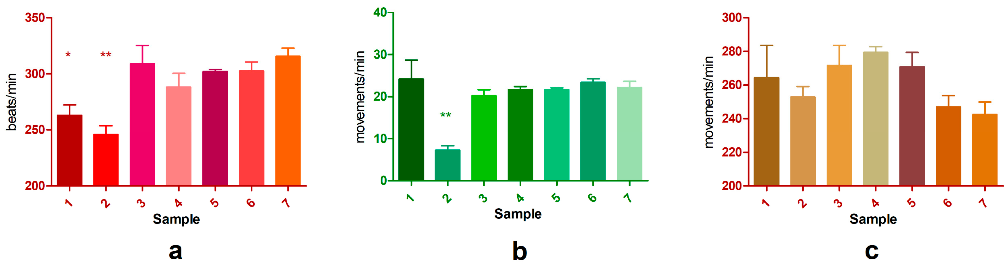
Following the results obtained from the acute toxicity assay, six compounds were selected for further physiological evaluation: DMSO, GLY, and PDO—which demonstrated low toxicity—and SXS, DMBA, and DMF, which showed moderate but time-dependent toxicity, with a marked increase in lethality between 24 h and 48 h. These were chosen to assess their potential impact on physiological parameters beyond lethality. The data are graphically represented in Figure 1a–c.

3.2.1. Heart Rate

The heart rate of test organisms ranged from 155 to 426 beats per minute (bpm) across all groups, compared to a control range from 240 to 395 bpm (Figure 1a). One-way ANOVA revealed significant differences within groups (p = 0.0017), with a statistically significant decrease in heart rate (Tukey post-hoc) being observed for SXS (263.10 ± 16.21 bpm) and DMBA (246.04 ± 13.47 bpm), both showing lower average heart rates than the control group (315.64 ± 12.94 bpm, p < 0.05). GLY induced a slight, non-significant reduction (288.08 ± 21.65 bpm), while DMSO (302.50 ± 13.81), DMF (309.0 ± 28.25), and PDO (302.10 ± 3.33) maintained values similar to those obtained in the control group, suggesting minimal impact on cardiac rhythm.

3.2.2. Claw Movement

The frequency of claw movement varied between 3 and 52 movements per minute, with the control group recording from 3 to 42 movements per minute (Figure 1b). Among all tested substances, only DMBA caused a statistically significant reduction in claw activity (7.30 ± 1.77 movements/min, p < 0.05), indicating possible neuromuscular suppression. The other compounds—including SXS, GLY, DMF, PDO, and DMSO—did not produce significant alterations in this parameter, showing comparable activity levels to control.

3.2.3. Appendages Movement

The evaluation of general appendage movement rates revealed no statistically significant differences among test groups when analyzed via Tukey’s post-hoc test. Average movement rates were slightly higher in all test samples compared to control (242.66 ± 12.80 movements/min), with values ranging from 247.07 ± 11.59 (DMSO) to 279.33 ± 6.18 (GLY) (Figure 1c). This mild increase was not significant, suggesting that general motor coordination and peripheral nerve function were preserved in most test conditions.

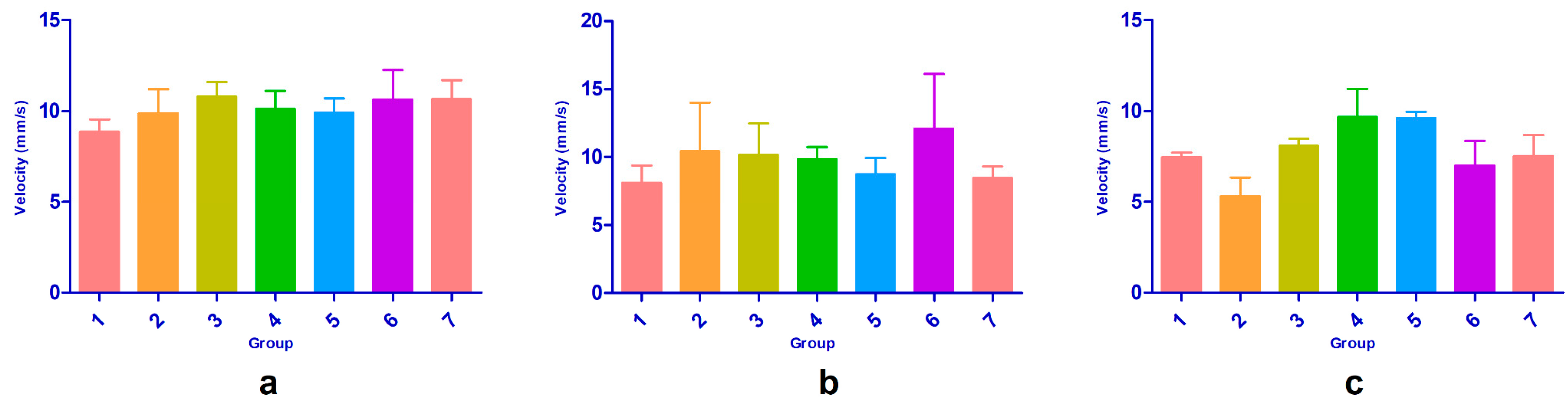
3.2.4. Behavioral Evaluation

A swimming speed-based bioassay was conducted to assess the sublethal effects of selected samples on Daphnia over 24 and 48 h (Figure 2, Figure 3 and Figure 4). Each compound was tested in triplicate, and average percentage changes in swimming speed were calculated relative to the initial time point (0 h).
At the 24 h point, DMSO was the only compound that produced the highest stimulatory effect on swimming speed (+12.6% average), despite high variability across replicates (CV% > 200%), however without statistical significance compared to control. Most other compounds induced mild inhibition or inconsistent effects, with changes ranging from −10.9% (PDO) to +3.9% (DMBA). The control group showed a significant decrease of swimming speed (−19.7%), indicating that natural physiological decline or environmental stress may have influenced the baseline behavior of Daphnia over time. At this stage, no statistically significant differences were observed based on one-way ANOVA (p > 0.05).
At 48 h, all groups—including the control—showed a reduction in swimming speed, suggesting cumulative stress. The most pronounced inhibitory effects were observed with DMBA (−44.7%) and DMSO (−32.3%), indicating a time-dependent shift in their action profiles. DMF also demonstrated consistent and reproducible inhibition (−25.1%) with low variability, suggesting a reliable negative impact on motility. At the 48 h time point, statistical analysis using ANOVA revealed significant differences among the groups (p = 0.013). Although not quantitatively measured, the swimming trajectories of Daphnia magna revealed noticeable behavioral differences (Figure 4). A general reduction in swimming distance was observed across all tested samples.

3.2.5. D. magna Embryotoxicity

D. magna embryo toxicity test was conducted to evaluate the developmental and sublethal effects of various substances at specific concentrations. Embryos were monitored at 24 and 48 h for morphological development and mobility, with a focus on identifying any delays, malformations, or signs of toxicity. A mobility score was defined as the percentage of individuals displaying active movement relative to the total number of organisms tested.
At a concentration of 0.05%, the group of sulfonated aromatic compounds—including SXS, SBS, PTS, and SBDS—displayed low toxicity. Even though slight developmental delays were observed at 24 h (reaching Phase 3), all embryos progressed to Phase 4 by 48 h. SXS resulted in a lower mobility score (40%) despite eventual complete development, suggesting a moderate sublethal effect, whereas the others maintained high mobility and development levels, comparable with the control group (Phase 4 at 48 h).
Among the amide and urea derivatives tested at 0.05%, results varied significantly. DMBA caused pronounced embryotoxicity, with disorganized embryos observed as early as 24 h and disorganized cellular structures persisting at 48 h (Figure 5 and Figure 6e). No mobility was recorded, and only 30% of the embryos developed (Phase 3), indicating a high level of embryotoxicity at this concentration. In contrast, DENA, DMU, and urea were much better tolerated (Phase 3 at 24 h). These compounds produced only minor early effects such as partial development or slight signs of embryotoxicity, but embryos generally completed development (Phase 4) by 48 h with moderate to high mobility. DMF stood out as particularly safe, with embryos reaching Phase 3 at 24 h and Phase 4 at 48 h, and exhibiting full mobility at 0.05%, indicating no adverse effects at the tested dose (Figure 5 and Figure 6i).
DMSO demonstrated a clear concentration-dependent toxicity profile. At 1%, it was well tolerated and comparable to the control, with embryos showing high mobility and complete development (to Phase 3 at 24 h and Phase 4 at 48 h). However, at 2% and 2.5%, DMSO became markedly embryotoxic. At 2.5%, mobility dropped to just 20%, and only 30% of embryos completed development. Disorganized development and abnormal embryonic morphology were observed. At 2%, despite some embryos appearing to begin development, the majority failed to form properly by 48 h (Phase 3), and morphological abnormalities were prominent. These findings confirm that DMSO becomes harmful to Daphnia embryos at concentrations above 1% (Figure 5 and Figure 6j).
The three tested surfactants—Tween 20, Tween 80, and SLS—were applied at very low concentrations (0.0005%). Tween 20 caused a reduction in both mobility (20%) and development (only Phase 1 at 24 h; Phase 4 reached by few embryos at 48 h) (40%), suggesting some adverse developmental influence despite the low concentration. Meanwhile, Tween 80 and SLS were better tolerated, each showing 80% mobility and development, indicating minimal embryotoxic potential at the tested level (Phase 4).
GLY and PDO both displayed concentration-dependent effects. At 0.5%, both substances produced results comparable to the control, with high mobility and complete development. However, at 1% and 2%, both showed severe developmental delays and morphological abnormalities. Embryos failed to complete development in most cases. In GLY-treated embryos, malformed structures and incomplete morphogenesis were common, including partial eye formation (Phase 3). For PDO, morphological damage of Phase 3 embryos included underdeveloped rostrum, missing or incomplete appendages, and affected compound eyes. Development was severely impaired, with mobility dropping to zero in some cases.
The control group exhibited moderate mobility and development scores (70% each), establishing a baseline for comparison, with the embryos being in Phase 3 at 24 h and in Phase 4 at 48 h. Substances performing similarly to or better than the control were considered non-toxic under the test conditions.

4. Discussions

D. magna is a crustacean widely used in toxicity evaluation, mostly in ecotoxicological studies, but also in pharmaceutical development in early-stage screenings [10]. The use of solubilizes is mandatory when testing poorly water-soluble compounds; thus, this study aimed to evaluate the most common co-solubilizers alongside with hydrotropic substances that enhance solubility in aqueous media [22].
Among all tested compounds, only six were selected based on their lowest toxicity—SXS, DMBA, DMF, GLY, PDO, and DMSO. Of these, only GLY, PDO, and DMSO could be used safely at concentrations between 0.5% and 1%.
Heart rate in Daphnia magna is highly variable and influenced by stress, handling, and experimental conditions. In our study, the control group showed an average heart rate of 316 bpm, which, although lower than some reported values, remained within the range found in the literature—such as 354 bpm [45], around 400 bpm [46,47], and 484 bpm [48]. These differences often reflect variations in recording methods and frame rates. Our videos were recorded at 25 fps, which could explain the relatively lower values observed for the control group. Nonetheless, acclimation procedures helped stabilize physiological activity, and consistent trends across all groups support the reliability of our findings. DMSO, widely used in biological assays due to its low toxicity across cells, invertebrates, and vertebrates [49,50], was tolerated at 1% in lethality terms. However, assessment of physiological activity revealed that the heart rate, appendage movement, and claw movement were minimally affected, while the swimming speed showed a biphasic response. Therefore, the swimming speed was significantly increased (at 24 h) or decreased (at 48 h), showing an incompatibility with the evaluation of this endpoint on Daphnia. This biphasic effect likely reflects initial neural stimulation and enhanced membrane permeability, followed by metabolic exhaustion or neuromotor suppression, as supported by earlier studies [46,51]. Importantly, inhibitory effects on heart rate and movement at even 0.1–1% DMSO have been previously reported [46]. Moreover, toxic effects were reported even at 1%, possibly due to variability across replicates and between populations, arising from individual physiological differences, environmental microconditions, or additive toxicity [52,53]. The embryotoxicity at ≥1% DMSO was marked by significant morphological alterations, consistent with previous findings [51]. The results suggest that some compounds exert either delayed or biphasic effects on Daphnia behavior. DMSO at 24 h increased the swimming speed, and at 48 h, swimming speed was reduced compared with the initial moment, and it was comparable with the control at 48 h. In contrast, DMBA induced the highest inhibition of swimming speed at 48 h (−44.7%). The lowest influence on swimming speed were registered for GLY (−4.7%) and PDO (−2.6%), indicating minimal effects under the tested conditions.
GLY, though often considered benign, exhibited harmful effects at high concentrations (>1%), both as a direct toxicant and as a pollutant or metabolite in environmental contexts [54,55]. However, GLY should thus be limited to ≤0.5% in embryotoxicity assays, as it induced significant developmental abnormalities at 1%. Similarly, PDO, which is a less commonly used solubilizer, can be used only at concentrations ≤1% in embryotoxicity assays. In toxicity evaluations, PDO was slightly more toxic than GLY.
DMBA strongly inhibited physiological functions, especially heart rate, by ~20–25% compared to controls. This reduction has critical biological implications, as lowered heart rate impairs oxygen delivery, reduces metabolic capacity, and can result in neuromotor impairment and decreased organismal fitness. Therefore, DMBA is unsuitable for Daphnia assays. In contrast, SXS and DMF could be applied safely at ≤0.5%, as they did not significantly alter heart rate or movement. The class of sulfonated aromatic compounds, including SXS, SBS, PTS, and SBDS, exhibited clear concentration-dependent toxicity trends. These hydrotropes were relatively well tolerated at 0.05%, suggesting that sulfonates could be applied in short-term solubilization protocols if concentrations are strictly controlled, being, at the same time, considered dangerous for the environment [56]. Embryo assays further reinforced these findings. SBDS showed greater toxicity than the other sulfonates, consistent with its higher molecular weight and dual sulfonate groups, which may contribute to increased osmotic or membrane-disruptive stress. These compounds are widely recognized for their hydrotropic properties and compatibility in formulation, but their use in in vivo bioassays, especially those extending beyond 24 h, may require tight concentration limits to avoid delayed-onset toxicity or sublethal physiological effects.
The three surfactants tested—Tween 20, Tween 80, and SLS—were among the most toxic compounds in the study. Their toxicity is well explained by their strong membrane-disrupting and emulsifying activities, which compromise membrane integrity, alter ion gradients, and disrupt cellular homeostasis. Similar mechanisms have been reported in zebrafish embryos, where surfactant exposure rapidly leads to developmental arrest and death [51]. Although all three surfactants showed high acute toxicity, embryotoxicity assays revealed nuanced differences: at 0.0005%, SLS and Tween 80 preserved ~80% embryonic mobility and development, whereas Tween 20 reduced these endpoints to 20–40%. While there may be a narrow safety margin at submicromolar levels, the margin is unpredictable, and we recommend avoiding surfactants in Daphnia assays unless diluted well below 0.001%, with rigorous controls and endpoint-specific validation [57].
Urea and its derivatives (DMU, DENA) represent structurally diverse yet pharmacologically relevant derivatives. Acute toxicity was moderate at >1%, with LC50 values between 0.2% and 1% at 48 h but negligible below 0.1%. In embryonic development assays, urea and DMU supported near-complete development with moderate mobility at 0.05%, with only minor delays or irregularities. DENA showed a slightly narrower safety window. While these compounds appear suitable for short-term Daphnia assays at ≤0.05–0.1%, it is worth noting that urea derivatives can affect metabolism and behavior in aquatic organisms [58], warranting careful concentration selection.
From an environmental perspective, the persistence of surfactants and hydrotropes in wastewater systems, their resistance to biodegradation, and their potential to accumulate and interact with co-occurring pollutants raise serious ecological concerns. Surfactants, for instance, can alter the bioavailability and toxicity of other contaminants, potentially amplifying their environmental impact [59].
Study limitations include the use of high test concentrations, short-term (48 h) exposure protocols, and a single-species model. While these constraints are typical of exploratory assays, they limit the extrapolation of findings to chronic exposures, lower environmentally relevant concentrations, and multi-species systems. Future research should address chronic and sublethal exposure scenarios, conduct multi-species assessments, and explore environmentally realistic co-exposure effects to better inform both pharmaceutical safety and ecological risk.
This study identified DMSO, GLY, PDO, SXS, and DMF as relatively safe solubilizers for D. magna assays at controlled concentrations, while surfactants and some urea derivatives pose high toxicity risks. These findings emphasize the need for informed solubilizer selection in aquatic bioassays and highlight important directions for future pharmaceutical and ecotoxicological research.
Although this study does not fully adhere to OECD guideline protocols, it employs scientifically validated methods adapted for early-stage ecotoxicological and pharmacotoxicological screening. The use of 12-well plates, small-volume assays, and sublethal endpoints is consistent with miniaturized and alternative bioassay strategies commonly reported in the literature. These adaptations allow greater flexibility in testing a broad panel of solubilizers under tightly controlled conditions and are particularly suited to exploratory studies focused on relative toxicity rankings rather than regulatory hazard classification. While OECD compliance is crucial for standardized inter-laboratory comparisons and regulatory acceptance, such alternative methods provide valuable insight during the early assessment phase, enabling the identification of compounds with acceptable toxicological profiles for further investigation.

5. Conclusions

This study evaluated the acute toxicity, physiological effects, behavior, and embryotoxicity of several commonly used solubilizers and hydrotropic agents using D. magna as a biological model. Surfactants such as Tween 20, Tween 80, and SLS exhibited the highest toxicity and should be excluded from Daphnia-based bioassays unless used at ultra-low concentrations (<0.001%), where minimal embryotoxicity was observed. Among the sulfonates, SXS, SBS, PTS, and SBDS demonstrated clear dose-dependent toxicity but were generally well tolerated at 0.05%, making them suitable only for short-term assays under strict concentration control. Urea and its derivatives (DMU, DENA) showed moderate toxicity and were generally safe below 0.1%, though they may influence Daphnia metabolism and behavior. Compounds such as DMSO, GLY, and PDO showed the lowest toxicity profiles and are recommended for use at concentrations ≤1% to minimize both acute and embryotoxic effects, aligning with established pharmacotoxicological guidelines. These compounds are preferable for pharmaceutical formulation in Daphnia assays where solvent safety is critical. Therefore, DMSO, GLY, PDO, SXS, and DMF can be considered relatively safe solubilizers in Daphnia assays if used at carefully selected concentrations.
Daphnia magna embryos are sensitive indicators of developmental toxicity, capable of detecting both overt and subtle effects depending on the concentration and nature of the test substance. While many compounds—including sulfonates, urea derivatives, and DMF—were well tolerated at 0.05%, substances like DMSO, GLY, and PDO caused concentration-dependent embryotoxicity, with observable developmental delays and morphological defects at concentrations of 1% and above. These results emphasize the importance of dose selection in pharmacotoxicological and ecotoxicological testing and support the use of Daphnia embryo assays in the early hazard screening of chemical substances.
Lastly, the findings highlight the need for chronic exposure studies to assess long-term environmental risks, especially considering the persistence of surfactants and hydrotropes in wastewater systems and their potential for cumulative or synergistic effects with co-pollutants. Future research should expand to multi-species models and chronic endpoints, helping translate these insights into environmentally relevant risk assessments and refining solubilizer guidelines for ecotoxicological applications.

Author Contributions

Conceptualization, I.I.O., D.P.M., O.T.O., C.E.G., C.A., E.-A.L., and G.M.N.; data curation, I.I.O., O.T.O., and G.S.; formal analysis, I.I.O., D.P.M., C.E.G., A.Z., G.S., and E.-A.L.; investigation, I.I.O., D.P.M., O.T.O., A.Z., and G.S.; methodology, I.I.O., D.P.M., O.T.O., A.Z., and G.M.N.; project administration, G.M.N.; resources, O.T.O.; software, D.P.M., O.T.O., C.A., and E.-A.L.; supervision, G.M.N.; validation, I.I.O., O.T.O., C.E.G., and C.A.; visualization, I.I.O., C.E.G., A.Z., and G.M.N.; writing—original draft, I.I.O., D.P.M., O.T.O., C.E.G., A.Z., and G.S.; writing—review and editing, C.A., E.-A.L., and G.M.N. All authors have read and agreed to the published version of the manuscript.

Funding

This research received no external funding. The APC was funded by the authors.

Data Availability Statement

The original contributions presented in this study are included in the article. Further inquiries can be directed to the corresponding authors.

Acknowledgments

This study was supported by “Carol Davila” University of Medicine and Pharmacy, Bucharest, Romania.

Conflicts of Interest

The authors declare no conflicts of interest.

References

  1. Koivisto, S. Is Daphnia magna an Ecologically Representative Zooplankton Species in Toxicity Tests? Environ. Pollut. 1995, 90, 263–267. [Google Scholar] [CrossRef] [PubMed]
  2. Ebert, D. Daphnia as a Versatile Model System in Ecology and Evolution. EvoDevo 2022, 13, 16. [Google Scholar] [CrossRef] [PubMed]
  3. Anderson, B.G.; Jenkins, J.C. A Time Study of Events in the Life Span of Daphnia magna. Biol. Bull. 1942, 83, 260–272. [Google Scholar] [CrossRef]
  4. Sánchez, M.; Ferrando, M.D.; Sancho, E.; Andreu-Moliner, E.; Sánchez, M.; Ferrando, M.D.; Sancho, E.; Andreu-Moliner, E. Evaluation of a Daphnia magna Renewal Life-Cycle Test Method with Diazinon. J. Environ. Sci. Health B 1998, 33, 785–797. [Google Scholar] [CrossRef]
  5. Nebeker, A.V. Evaluation of a Daphnia magna Renewal Life-Cycle Test Method with Silver and Endosulfan. Water Res. 1982, 16, 739–744. [Google Scholar] [CrossRef]
  6. Tomasik, P.; Warren, D.M. The Use of Daphnia in Studies of Metal Pollution of Aquatic Systems. Environ. Rev. 2011, 4, 25–64. [Google Scholar] [CrossRef]
  7. Kim, H.; Yim, B.; Bae, C.; Lee, Y.M. Acute Toxicity and Antioxidant Responses in the Water Flea Daphnia magna to Xenobiotics (Cadmium, Lead, Mercury, Bisphenol A, and 4-Nonylphenol). Toxicol. Environ. Health Sci. 2017, 9, 41–49. [Google Scholar] [CrossRef]
  8. Tsui, M.T.K.; Wang, W.X. Biokinetics and Tolerance Development of Toxic Metals in Daphnia magna. Environ. Toxicol. Chem. 2007, 26, 1023–1032. [Google Scholar] [CrossRef] [PubMed]
  9. Abdullahi, M.; Li, X.; Abdallah, M.A.E.; Stubbings, W.; Yan, N.; Barnard, M.; Guo, L.H.; Colbourne, J.K.; Orsini, L. Daphnia as a Sentinel Species for Environmental Health Protection: A Perspective on Biomonitoring and Bioremediation of Chemical Pollution. Environ. Sci. Technol. 2022, 56, 14237–14248. [Google Scholar] [CrossRef]
  10. Tkaczyk, A.; Bownik, A.; Dudka, J.; Kowal, K.; Ślaska, B. Daphnia magna Model in the Toxicity Assessment of Pharmaceuticals: A Review. Sci. Total Environ. 2021, 763, 143038. [Google Scholar] [CrossRef]
  11. Mishra, M. Daphnia magna as a Model Organism to Predict the Teratogenic Effect of Different Compounds. Methods Mol. Biol. 2024, 2753, 261–281. [Google Scholar] [CrossRef]
  12. LeBlanc, G.A.; Mu, X.; Rider, C.V. Embryotoxicity of the Alkylphenol Degradation Product 4-Nonylphenol to the Crustacean Daphnia magna. Environ. Health Perspect. 2000, 108, 1133. [Google Scholar] [CrossRef] [PubMed]
  13. Weichert, F.G.; Floeter, C.; Meza Artmann, A.S.; Kammann, U. Assessing the Ecotoxicity of Potentially Neurotoxic Substances—Evaluation of a Behavioural Parameter in the Embryogenesis of Danio rerio. Chemosphere 2017, 186, 43–50. [Google Scholar] [CrossRef] [PubMed]
  14. Wittlerová, M.; Jírová, G.; Vlková, A.; Kejlová, K.; Maly, M.; Heinonen, T.; Wittlingerová, Z.; Zimová, M. Sensitivity of Zebrafish (Danio rerio) Embryos to Hospital Effluent Compared to Daphnia magna and Aliivibrio fischeri. Physiol. Res. 2020, 69, S681–S691. [Google Scholar] [CrossRef] [PubMed]
  15. Reilly, K.; Ellis, L.J.A.; Davoudi, H.H.; Supian, S.; Maia, M.T.; Silva, G.H.; Guo, Z.; Martinez, D.S.T.; Lynch, I. Daphnia as a Model Organism to Probe Biological Responses to Nanomaterials—From Individual to Population Effects via Adverse Outcome Pathways. Front. Toxicol. 2023, 5, 1178482. [Google Scholar] [CrossRef]
  16. Sakka, Y.; Koeser, J.; Filser, J. How Test Vessel Properties Affect the Fate of Silver Nitrate and Sterically Stabilized Silver Nanoparticles in Two Different Test Designs Used for Acute Tests with Daphnia magna. Environ. Sci. Pollut. Res. 2017, 24, 2495–2506. [Google Scholar] [CrossRef]
  17. Powell, R.L.; Moser, E.M.; Kimerle, R.A.; McKenzie, D.E.; McKee, M. Use of a Miniaturized Test System for Determining Acute Toxicity of Toxicity Identification Evaluation Fractions. Ecotoxicol. Environ. Saf. 1996, 35, 1–6. [Google Scholar] [CrossRef]
  18. Küster, E.; Addo, G.G.; Aulhorn, S.; Kühnel, D. Miniaturisation of the Daphnia magna Immobilisation Assay for the Reliable Testing of Low Volume Samples. UCL Open Environ. 2025, 7, e3037. [Google Scholar] [CrossRef]
  19. Hughes, J.P.; Rees, S.S.; Kalindjian, S.B.; Philpott, K.L. Principles of Early Drug Discovery. Br. J. Pharmacol. 2011, 162, 1239–1249. [Google Scholar] [CrossRef]
  20. Newman, D.J.; Cragg, G.M. Natural Products As Sources of New Drugs over the 30 Years from 1981 to 2010. J. Nat. Prod. 2012, 75, 311–335. [Google Scholar] [CrossRef]
  21. Kumari, L.; Choudhari, Y.; Patel, P.; Gupta, G.D.; Singh, D.; Rosenholm, J.M.; Bansal, K.K.; Kurmi, B. Das Advancement in Solubilization Approaches: A Step towards Bioavailability Enhancement of Poorly Soluble Drugs. Life 2023, 13, 1099. [Google Scholar] [CrossRef] [PubMed]
  22. Kerns, E.; Di, L.; Carter, G. In Vitro Solubility Assays in Drug Discovery. Curr. Drug Metab. 2008, 9, 879–885. [Google Scholar] [CrossRef]
  23. Galvao, J.; Davis, B.; Tilley, M.; Normando, E.; Duchen, M.R.; Cordeiro, M.F. Unexpected Low-Dose Toxicity of the Universal Solvent DMSO. FASEB J. 2014, 28, 1317–1330. [Google Scholar] [CrossRef] [PubMed]
  24. Kais, B.; Schneider, K.E.; Keiter, S.; Henn, K.; Ackermann, C.; Braunbeck, T. DMSO Modifies the Permeability of the Zebrafish (Danio rerio) Chorion-Implications for the Fish Embryo Test (FET). Aquat. Toxicol. 2013, 140–141, 229–238. [Google Scholar] [CrossRef]
  25. Stanton, K.; Tibazarwa, C.; Certa, H.; Greggs, W.; Hillebold, D.; Jovanovich, L.; Woltering, D.; Sedlak, R. Environmental Risk Assessment of Hydrotropes in the United States, Europe, and Australia. Integr. Environ. Assess. Manag. 2010, 6, 155–163. [Google Scholar] [CrossRef]
  26. DeMille, C.M.; Arnott, S.E.; Pyle, G.G. Variation in Copper Effects on Kairomone-Mediated Responses in Daphnia Pulicaria. Ecotoxicol. Environ. Saf. 2016, 126, 264–272. [Google Scholar] [CrossRef] [PubMed]
  27. Soares, B.P.; Abranches, D.O.; Sintra, T.E.; Leal-Duaso, A.; García, J.I.; Pires, E.; Shimizu, S.; Pinho, S.P.; Coutinho, J.A.P. Glycerol Ethers as Hydrotropes and Their Use to Enhance the Solubility of Phenolic Acids in Water. ACS Sustain. Chem. Eng. 2020, 8, 5742–5749. [Google Scholar] [CrossRef]
  28. Mazaud, A.; Lebeuf, R.; Laguerre, M.; Nardello-Rataj, V. Improved Hydrotropic Extraction of Carnosic Acid from Rosemary and Sage with Short-Chain Monoalkyl Glycerol Ethers. ACS Sustain. Chem. Eng. 2022, 10, 3673–3681. [Google Scholar] [CrossRef]
  29. Bownik, A.; Jasieczek, M.; Kosztowny, E. Ketoprofen Affects Swimming Behavior and Impairs Physiological Endpoints of Daphnia magna. Sci. Total Environ. 2020, 725, 138312. [Google Scholar] [CrossRef]
  30. Semenova, M.N.; Kuptsova, T.S.; Semenov, V.V. Toxicity of Organic Solvents and Surfactants to the Sea Urchin Embryos. Chemosphere 2024, 353, 141589. [Google Scholar] [CrossRef]
  31. Coelho, M.; Palma, M.; Tavares, L.C.; Panserat, S.; Viegas, I.; Magnoni, L.J. Glycerol Supplementation in Farmed Fish Species: A Review from Zootechnical Performance to Metabolic Utilisation. Rev. Aquac. 2024, 16, 1901–1917. [Google Scholar] [CrossRef]
  32. García, J.I.; Pires, E.; Aldea, L.; Lomba, L.; Perales, E.; Giner, B. Ecotoxicity Studies of Glycerol Ethers in Vibrio Fischeri: Checking the Environmental Impact of Glycerol-Derived Solvents. Green. Chem. 2015, 17, 4326–4333. [Google Scholar] [CrossRef]
  33. Lee, P.A.; De Mora, S.J.; Levasseur, M. A Review of Dimethylsulfoxide in Aquatic Environments. Atmos. Ocean 1999, 37, 439–456. [Google Scholar] [CrossRef]
  34. Roncaglioni, A.; Toropov, A.A.; Toropova, A.P.; Benfenati, E. In Silico Methods to Predict Drug Toxicity. Curr. Opin. Pharmacol. 2013, 13, 802–806. [Google Scholar] [CrossRef] [PubMed]
  35. Freires, I.A.; Morelo, D.F.C.; Soares, L.F.F.; Costa, I.S.; de Araújo, L.P.; Breseghello, I.; Abdalla, H.B.; Lazarini, J.G.; Rosalen, P.L.; Pigossi, S.C.; et al. Progress and Promise of Alternative Animal and Non-Animal Methods in Biomedical Research. Arch. Toxicol. 2023, 97, 2329–2342. [Google Scholar] [CrossRef]
  36. Olaru, O.T.; Nitulescu, G.M.; Codreanu, A.M.; Calmuc, V.A.; Venables, L.; van de Venter, M.; Gird, C.E.; Duta-Bratu, C.G.; Nitulescu, G. Inhibitory Effects on Staphylococcus aureus Sortase A by Aesculus Sp. Extracts and Their Toxicity Evaluation. Plants 2024, 13, 1405. [Google Scholar] [CrossRef] [PubMed]
  37. Hajir, S.; Jobst, K.J.; Kleywegt, S.; Simpson, A.J.; Simpson, M.J. Do Co-Solvents Used in Exposure Studies Equally Perturb the Metabolic Profile of Daphnia magna? Environ. Toxicol. Chem. 2025, vgaf068. [Google Scholar] [CrossRef] [PubMed]
  38. Kawakami, K.; Oda, N.; Miyoshi, K.; Funaki, T.; Ida, Y. Solubilization Behavior of a Poorly Soluble Drug under Combined Use of Surfactants and Cosolvents. Eur. J. Pharm. Sci. 2006, 28, 7–14. [Google Scholar] [CrossRef]
  39. Cheleski, J.; Wiggers, H.J.; Citadini, A.P.; da Costa Filho, A.J.; Nonato, M.C.; Montanari, C.A. Kinetic Mechanism and Catalysis of Trypanosoma cruzi Dihydroorotate Dehydrogenase Enzyme Evaluated by Isothermal Titration Calorimetry. Anal. Biochem. 2010, 399, 13–22. [Google Scholar] [CrossRef]
  40. Greene, M.; Pitts, W.; Dewprashad, B. Using Videography to Study the Effects of Stimulants on Daphnia magna. Am. Biol. Teach. 2017, 79, 35–40. [Google Scholar] [CrossRef]
  41. Tracker® Software. Available online: https://opensourcephysics.github.io/tracker-website/ (accessed on 3 February 2025).
  42. Schindelin, J.; Arganda-Carreras, I.; Frise, E.; Kaynig, V.; Longair, M.; Pietzsch, T.; Preibisch, S.; Rueden, C.; Saalfeld, S.; Schmid, B.; et al. Fiji: An Open-Source Platform for Biological-Image Analysis. Nat. Methods 2012, 9, 676–682. [Google Scholar] [CrossRef] [PubMed]
  43. Wang, K.S.; Lu, C.Y.; Chang, S.H. Evaluation of Acute Toxicity and Teratogenic Effects of Plant Growth Regulators by Daphnia magna Embryo Assay. J. Hazard. Mater. 2011, 190, 520–528. [Google Scholar] [CrossRef]
  44. Ivan, I.M.; Popovici, V.; Chițescu, C.L.; Popescu, L.; Luță, E.A.; Ilie, E.I.; Brașoveanu, L.I.; Hotnog, C.M.; Olaru, O.T.; Nițulescu, G.M.; et al. Phytochemical Profile, Antioxidant and Cytotoxic Potential of Capsicum annuum (L.) Dry Hydro-Ethanolic Extract. Pharmaceutics 2024, 16, 245. [Google Scholar] [CrossRef] [PubMed]
  45. Corotto, F.; Ceballos, D.; Lee, A.; Vinson, L. Making the Most of the Daphnia Heart Rate Lab: Optimizing the Use of Ethanol, Nicotine & Caffeine. Am. Biol. Teach. 2010, 72, 176–179. [Google Scholar] [CrossRef]
  46. Bownik, A.; Stępniewska, Z.; Skowroński, T. Effects of Ectoine on Behavioural, Physiological and Biochemical Parameters of Daphnia magna. Comp. Biochem. Physiol. Part C Toxicol. Pharmacol. 2015, 168, 2–10. [Google Scholar] [CrossRef]
  47. Saputra, F.; Farhan, A.; Suryanto, M.E.; Kurnia, K.A.; Chen, K.H.C.; Vasquez, R.D.; Roldan, M.J.M.; Huang, J.C.; Lin, Y.K.; Hsiao, C. Der Automated Cardiac Chamber Size and Cardiac Physiology Measurement in Water Fleas by U-Net and Mask RCNN Convolutional Networks. Animals 2022, 12, 1670. [Google Scholar] [CrossRef]
  48. Lari, E.; Steinkey, D.; Pyle, G.G. A Novel Apparatus for Evaluating Contaminant Effects on Feeding Activity and Heart Rate in Daphnia spp. Ecotoxicol. Environ. Saf. 2017, 135, 381–386. [Google Scholar] [CrossRef] [PubMed]
  49. Jacob, S.W.; Wood, D.C. Dimethyl Sulfoxide (DMSO) Toxicology, Pharmacology, and Clinical Experience. Am. J. Surg. 1967, 114, 414–426. [Google Scholar] [CrossRef]
  50. Colucci, M.; Maione, F.; Bonito, M.C.; Piscopo, A.; Di Giannuario, A.; Pieretti, S. New Insights of Dimethyl Sulphoxide Effects (DMSO) on Experimental in Vivo Models of Nociception and Inflammation. Pharmacol. Res. 2008, 57, 419–425. [Google Scholar] [CrossRef]
  51. Hoyberghs, J.; Bars, C.; Ayuso, M.; Van Ginneken, C.; Foubert, K.; Van Cruchten, S. DMSO Concentrations up to 1% Are Safe to Be Used in the Zebrafish Embryo Developmental Toxicity Assay. Front. Toxicol. 2021, 3, 804033. [Google Scholar] [CrossRef]
  52. Andrade-Vieira, L.F.; Bojic, C.; Santana Alvarenga, I.F.; de Carvalho, T.S.; Masfaraud, J.F.; Cotelle, S. Ecotoxic Effects of the Vehicle Solvent Dimethyl Sulfoxide on Raphidocelis subcapitata, Daphnia magna and Brachionus calyciflorus. Chem. Ecol. 2022, 38, 471–483. [Google Scholar] [CrossRef]
  53. Haap, T.; Triebskorn, R.; Köhler, H.R. Acute Effects of Diclofenac and DMSO to Daphnia magna: Immobilisation and Hsp70-Induction. Chemosphere 2008, 73, 353–359. [Google Scholar] [CrossRef]
  54. Pelizaro, B.I.; Braga, F.C.; Crispim, B.d.A.; de Barros, L.G.M.L.; Pessatto, L.R.; Oliveira, E.J.T.; Vani, J.M.; de Souza, A.P.; Grisolia, A.B.; Antoniolli-Silva, A.C.M.B.; et al. Assessment of Acute Toxicity and Cytotoxicity of Fluorescent Markers Produced by Cardanol and Glycerol, Which Are Industrial Waste, to Different Biological Models. Environ. Sci. Pollut. Res. 2019, 26, 9193–9202. [Google Scholar] [CrossRef] [PubMed]
  55. Cui, R.; Kim, S.W.; An, Y.J. Polystyrene Nanoplastics Inhibit Reproduction and Induce Abnormal Embryonic Development in the Freshwater Crustacean Daphnia Galeata. Sci. Rep. 2017, 7, 12095. [Google Scholar] [CrossRef] [PubMed]
  56. Verge, C.; Moreno, A. Effects of Anionic Surfactants on Daphnia magna. Tenside Surfactants Deterg. 2000, 37, 172–175. [Google Scholar] [CrossRef]
  57. Yuan, D.; Long, Y.; Liu, D.; Zhou, F.; Liu, C.; Chen, L.; Pan, Y. Ecological Impact of Surfactant Tween-80 on Plankton: High-Scale Analyses Reveal Deeper Hazards. Sci. Total Environ. 2024, 912, 169176. [Google Scholar] [CrossRef]
  58. Ma, Y.; Liu, Y.; Sun, J.; Min, P.; Liu, W.; Li, L.; Yi, P.; Guo, R.; Chen, J. Ecological Risks of High-Ammonia Environment with Inhibited Growth of Daphnia magna: Disturbed Energy Metabolism and Oxidative Stress. Sci. Total Environ. 2024, 948, 174959. [Google Scholar] [CrossRef]
  59. Lechuga, M.; Fernández-Serrano, M.; Jurado, E.; Núñez-Olea, J.; Ríos, F. Acute Toxicity of Anionic and Non-Ionic Surfactants to Aquatic Organisms. Ecotoxicol. Environ. Saf. 2016, 125, 1–8. [Google Scholar] [CrossRef]
Figure 1. The physiological activity evaluation results. (a) Heart rate; (b) claw movement; (c) appendages movement. The columns represent average values, and the error bars represent the standard deviation; Statistical significance: p values between 0.01 and 0.05 are marked with *, and p values between 0.001 and 0.01 with **, based on Tukey’s post-hoc test. All comparisons were made against the control group. Groups 1–7: 1—SXS; 2—DMBA; 3—DMF; 4—GLY; 5—PDO; 6—DMSO; 7—control.
Environments 12 00172 g001
Figure 2. Average swimming speed of Daphnia magna measured at the initial time point (a), after 24 h of exposure (b), and after 48 h of exposure (c). Bars represent mean values; error bars indicate standard deviation (n = 3). Groups 1–7: 1—SXS; 2—DMBA; 3—DMF; 4—GLY; 5—PDO; 6—DMSO; 7—control.
Environments 12 00172 g002
Figure 3. Effect on Daphnia magna swimming speed relative to the initial time point after 24 h (a) and 48 h (b). Bars represent mean values; error bars indicate standard deviation (n = 3). Groups 1–7: 1-SXS; 2-DMBA; 3-DMF; 4-GLY; 5-PDO; 6-DMSO; 7-control.
Environments 12 00172 g003
Figure 4. Sample paths of D. magna—one replicate from group 2 (a), group 6 (b), and group 7 (c) over the 48 h of exposure. The pink circles mark the end of the path. Each path was colored different.
Environments 12 00172 g004
Figure 5. Daphnia magna embryonic development assay at 24 h of exposure (objective 10×). (a) SXS; (b) SBS; (c) PTS; (d) SBDS; (e) DMBA; (f) DENA; (g) DMU; (h) urea; (i) DMF; (j) DMSO; (k) Tween 20; (l) Tween 80; (m) SLS; (n) GLY; (o) PDO; scale bar = 500 µm.
Environments 12 00172 g005
Figure 6. Daphnia magna embryonic development assay at 48 h of exposure (objective 10×). (a) SXS; (b) SBS; (c) PTS; (d) SBDS; (e) DMBA; (f) DENA; (g) DMU; (h) urea; (i) DMF; (j) DMSO; (k) Tween 20; (l) Tween 80; (m) SLS; (n) GLY; (o) PDO; scale bar = 500 µm.
Environments 12 00172 g006
Table 1. Results of the acute toxicity assay.
LC50
(%)
95%CI of LC50
(%)
r2
Substance24 h48 h24 h48 h24 h48 h
1SXS1.5690.08631.023–2.4050.0536–0.13900.86050.8162
2SBS2.0780.09281.447–2.9830.0750–0.11480.85360.9555
3PTS1.1530.06060.6764–1.9650.0553–0.06640.78380.962
4SBDS2.5160.48231.824–3.4730.2127–1.0940.83710.722
5DMBA0.1358ND *ND ***ND0.998ND
6DENA1.0830.14910.8912–1.3160.0780–0.28490.95250.8074
7DMU2.6640.39091.477–4.8070.2097–0.72850.94890.8307
8Urea3.270.92862.369–4.5160.5688–1.5160.66240.7353
9DMF2.4640.305 0.1717–0.54180.92130.8575
10DMSOND **ND **NDNDNDND
11Tween 20ND *ND *NDNDNDND
12Tween 80ND *ND *NDNDNDND
13SLSND *ND *NDNDNDND
14GLYND **ND **NDNDNDND
15PDOND **2.725ND2.086–3.559ND0.7268
LC50—50% lethal concentrations; 95%CI—95% confidence intervals; ND—not determined due to the obtained results; *—the lethality values were too high to calculate LC50; **—the lethality values were too low to calculate LC50; ***—95%CI is very wide.
Disclaimer/Publisher’s Note: The statements, opinions and data contained in all publications are solely those of the individual author(s) and contributor(s) and not of MDPI and/or the editor(s). MDPI and/or the editor(s) disclaim responsibility for any injury to people or property resulting from any ideas, methods, instructions or products referred to in the content.

