Article

Current to Biomass: Media Optimization and Strain Selection from Cathode-Associated Microbial Communities in a Two-Chamber Electro-Cultivation Reactor

Institute of Chemistry and Bioengineering, Department of Physical Chemistry and Microreaction Technology, Technische Universität Ilmenau, 98693 Ilmenau, Germany
*
Author to whom correspondence should be addressed.
Environments 2023, 10(6), 97; https://doi.org/10.3390/environments10060097
Submission received: 12 April 2023 / Revised: 1 June 2023 / Accepted: 3 June 2023 / Published: 7 June 2023

Abstract

:
Cathode-associated microbial communities (caMCs) are the functional key elements in the conversion of excess electrical energy into biomass. In this study, we investigated the development of electrochemical caMCs based on two-chamber microbial electrolytic cells (MECs) after optimization of media composition. Microbial communities obtained from a historical soil sample were inoculated into the cathode chamber of MECs. The inorganic medium with (A) carbon dioxide in air or (B) 100 mM sodium bicarbonate as carbon source was used in the absence of any organic carbon source. After 12 days of operation, the experimental results showed that (1) the bacterial community in group B exhibited lush growth and (2) a single strain TX168 Epilithonimonas bovis isolated from group A indicated electrochemical activity and synthesized large volumes of biomass using sodium bicarbonate. We also analyzed the caMCs of the MECs and reference samples without electro-cultivation using 16S rRNA gene sequencing. The results showed that the caMCs of MECs in groups A and B were dominated by the genera Acinetobacter and Pseudomonas. The caMCs were further inoculated and cultured on different agars to isolate specific electroactive bacterial strains. Overall, our study highlights the possibility of converting excess energy into biomass by electro-cultivation and the importance of selecting appropriate media to enrich specific microbial communities and single strains in MECs.

1. Introduction

Electroactive microorganisms (EAMs) are very interesting for coupling microbial physiological activities with technical processes converting energy. On the one hand, EAMs can oxidize organic matter in microbial fuel cells (MFCs), generating electric potential [1]. On the other hand, EAMs are used in microbial electrolysis cells (MECs) to produce methane or hydrogen [2]. The key step in these processes is the microorganisms involved in electron transfer [3]. This transfer can occur either through direct contact between biological cells and the electrode surface or through an electrochemical active mediator. Some bacteria are, in principle, capable of transferring electrons from the cathode to inorganic species, reducing inorganic ions or generating biomass and producing valuable biomolecules [4].
One focus of research is the application of MFCs or MECs to search for and evaluate suitable electroactive bacterial strains and consortia [5]. Among others, Geobacter sulfurreducens can be used in MECs as a biocatalyst for hydrogen formation at the cathode [6]. The production of acetate by microbial electrosynthesis using sulfate-reducing bacteria has been reported [7]. Pseudomonas putida has been proved to be suitable for the bioelectrochemical production of rhamnolipids [8].
Efficient EAMs are not only used to produce specific chemicals in electrochemical synthesis. They are also used to convert electrical energy into biomass, which is a promising strategy for storing energy from green energy production [9]. In both cases, microbial communities can play a crucial role in addition to single strains [10]. The communication and physiological interaction between electrochemically active and redox-active bacteria are key issues for stimulating biomass production [11].
Until now, only a tiny fraction of potentially electroactive bacteria have been cultivated. Many species can only be identified by the presence of their genes. However, finding and cultivating electroactive strains are essential for biotechnological processes. In principle, electroactive bacteria can be found in different soil environments [12]. However, finding the most suitable strains in the vast soil microbial community is like finding a needle in a haystack. It is assumed that special soils and sites may be more suitable than others for such a search. This is because the input of special substances and the high density and diversity of special groups can promote the formation of different bacterial communities in soil, such as nitrate- and sulfate-reducing or ammonia-oxidizing bacterial communities, methanogenic bacteria, and bacteria with high tolerance to redox-active heavy metals [13,14,15]. Among recently contaminated soils, soil bacterial communities from sites with ancient human impacts appear promising for tailored searches [16,17,18]. Soil samples from archaeological excavation sites with a complex spectrum of former exposure factors could be particularly attractive for this search. Ancient tannery sites represent such a type of formerly massively polluted ancient environment, presenting animal residues, tools, and chemicals, including the release of chromium and other toxic metals. It is known that tannery waste is characterized by particular types and communities of bacteria [19,20]. Therefore, the possibility of finding cathodically active bacteria has been investigated using a soil sample from a historical tannery area [21].
The main objective of this study was to investigate and demonstrate the suitability of the presented MEC for searching for cathodically activated and CO2-binding bacteria, on the one hand, and to show the potential of soil samples from a place with ancient human impact for such a search for and isolation of promising bacterial strains, on the other hand. In the following, the experimental setup, the electro-cultivation procedure, and the electroactive strains found are reported. Our study used two-chamber MECs with air-fixed carbon dioxide or sodium bicarbonate as the carbon source for cathodic electrochemical screening of bacterial communities from a historical tannery site. After electrochemical or non-electrochemical cultivation, the cathode-associated microbial communities were analyzed using 16S rRNA gene sequencing, and single strains of cathode-associated electroactive bacteria were isolated and identified. The electrochemical properties of the identified single strain Epilithonimonas bovis were characterized.

2. Materials and Methods

2.1. Soil Sample Preparation

The soil sample HB35 (pH: 8.1, conductivity: 512.3 µS/cm) was collected during an archaeological excavation from wet, buried soil in a medieval suburb of the city of Jena. After collection, the soil samples were air-dried under sterile conditions. HB35 has a pH value of 8.1 and a conductivity of 512.3 µS/cm. For the experiment, 2 g of soil was mixed with 10 mL of inorganic medium and vortexed thoroughly. Following centrifugation at 200× g for 20 min, the supernatant was passed through filter paper of size 20 µm (VWR, Germany) to remove excess soil particles. A final concentration of 75 mg/L of cyclohexane (BioChemica, Sauerlach, Germany) was added to prevent the growth of soil-borne fungi during incubation.

2.2. Chemicals and Medium

For the electrochemical and non-electrochemical cultivation of media-dependent bacterial communities, an inorganic medium was utilized with 0.1 g/L NH4NO3, 0.5 g/L K2HPO4, 0.2 g/L MgSO4·7H2O, and 0.01 g/L FeSO4·7H2O (all chemicals from Merck, Darmstadt, Germany). To precisely control the carbon source during the mixture culture, the inorganic medium was optimized by using carbon dioxide fixed from air and 100 mM sodium bicarbonate as the carbon source. For the enrichment of mixed cultures after electrochemical and non-electrochemical cultivation, minimal medium (CMM1) and modified actinomycete minimal medium (AM), which was modified to AM1 and AM2, were utilized, as well as the rich media Luria-Bertani (LB) and Trypticase soya agar (TSA) (Table 1). Mixed colonies grown on these solid media were isolated and purified by using a TSA agar medium.

2.3. Electrochemical Experiment Concept and Arrangement

In this study, we constructed a two-chamber H-type MEC system, which consisted of an anode chamber and a cathode chamber separated by a 1.5 cm diameter proton exchange membrane (Nafion® N117, Ion power, München-Flughafen, Germany) (Figure 1). The effective volume of the system was 40 mL (20 mL each for the anode and cathode sides). The electrodes of the cathode were stainless-steel electrodes with a length of 5 cm and a diameter of 0.8 mm. The anode was made of a platinum electrode and had a length of 15 cm and a diameter of 0.25 mm. Both electrodes were connected by a digital power source meter (Hameg, Frankfurt, Germany).
Unlike conventional two-chamber MECs, we completed the experimental setup with an additional mini-peristaltic pump (DC12V, G528 32 × 23 mm, Chongqing, China). Its functions were (1) to circulate the bacterial culture solution and electrolyte at the cathode and anode, respectively; (2) to speed up the transfer of ions, especially protons, from the anode to the cathode, which sped up the electron transfer rate; (3) to promote the fresh exchange of culture solution in the cathode chamber; and (4) to export the gas produced at the electrode surfaces. First, 100 mM of sodium bicarbonate was circulated in the anode chamber for ionic equilibration with a bacterial culture solution containing 100 mM of sodium bicarbonate in the cathode chamber. The pH of the anode and cathode chambers during the reaction was measured using pH indicator papers (GE Healthcare, Amersham, UK). The change in pH during the reaction was observed to, on the one hand, facilitate reaction with any protons that were transported from the anode to the cathode and, on the other hand, prevent the production of excess hydrogen gas at the cathode due to the decrease in pH affecting the bacteria involved in electron transport at the electrode surface.
For electrochemical cultivation, bacteria were inoculated in the cathode chamber and subjected to electrochemical reactions at a constant voltage of 1.6 V. This voltage was found to be the most suitable voltage after our experiments, beyond which a large amount of hydrogen gas was produced on the electrode surface. This affected the electron transport of the bacteria at the electrode surface in the cathode chamber. In the anode chamber, a lot of oxygen was produced due to the oxidation reaction taking place. Therefore, the culture flask connected to the valve was not sealed. The whole reaction took place at room temperature over 12 days. The electrochemical processes can be described as follows:
Anodic reaction:
2·H2O → 4H+ + O2 + 4e
Possible cathodic reactions:
HCO3 + n·e (bacteria) → CxHyOz (biomass)
Mediator(oxidation) + n·e → mediator(reduction)
HCO3 + mediator(reduction) (bacteria) → CxHyOz (biomass) + mediator(oxidation)
In the cathodic chamber, on the one hand, bicarbonate could be converted into organic substances with the assistance of bacteria. On the other hand, the oxidation mediator (Mox) in the medium could accept electrons at the cathode surface and be converted into a reduction mediator (Mrd), which bacteria could use, and then converted into Mox again to form a cycle. Therefore, the bacteria in the cathode chamber could not only metabolize bicarbonate to grow but also participated in the redox cycle related to the mediator.

2.4. Next-Generation Gene Sequencing and Data Processing

All samples were investigated using NGS (Illumina) according to the following procedures. The microbial DNA of the soil sample HB35 was extracted and purified from a 250 mg soil sample using the Power Soil Isolation Kit (Qiagen, Hilden, Germany). The cultured microbiome of each 0.5 µL sample of DNA extracted was used for the Illumina PCR.
The amplification of the V4 region of the 16S rRNA was carried out in an Edvocycler (94 °C for 5 min, 30 cycles of denaturation at 94 °C for 30 s; annealing at 50 °C for 30 s; extension at 72 °C for 30 s; and a final extension at 72 °C for 5 min). The sequence of the adaptor primer A519F-Ad was 5’-TCGTCGGCAGCGTCAGATGTGTATAAGAGACAGCAGCMGCCGCGGTAA-3’ and that of the reverse adaptor primer Bact_805R-Ad was 5’–GTCTCGTGGGCTCGGAGATGTGTATAAGAGACAGGACTACHVGGGTATCTAATC; both were obtained from Eurofins Genomics (Ebersberg, Germany) in concentrations of 1 µM. The presence of the desired 353 bp DNA fragment was confirmed by gel electrophoresis. Per the manufacturer’s instructions, PCR products were purified using proNex beads (Promega, Madison, WI, USA).
For the index PCR, the total volume of the master mixture was 25 µL and it contained 2.5 µL of the amplified PCR described above, 25 mM MgCl2, 10 mM PCR nucleotide mixture, 5 units of GoTaq®Mdx Hot Start DNA polymerase, nuclease-free water (all reagents from Promega, Madison, WI, USA), and 125 nM of each primer. The index PCR program was set up as follows: 95 °C for 3 min, 8 cycles (denaturation at 95 °C for 30 s; annealing at 55 °C for 30 s; extension at 72 °C for 30 s), and a final extension at 72 °C for 5 min. Index PCR products were first purified with proNex beads, then pooled and purified again using proNex beads. Finally, the prepared samples were sent to Eurofins for next-generation sequencing with an Illumina HiSeq system.
For data processing, the obtained fastq files for the forward- and reverse-aligned 16S rRNA data were first converted to fasta-format contig files and quality files (mothur (version 1.39.5)) in the open-source Galaxy platform (https://usegalaxy.org; accessed on 18 May 2022). The contig files were then aligned to the NCBI cloud-based rRNA databases using the SILVAngs data analysis service (https://ngs.arb-silva.de/silvangs; accessed on 18 May 2022). Furthermore, for all analyses, the preset parameter configurations from the SILVAngs database, version 138.1, were used [22]. Sequencing data allowed assignment to genus-level taxonomic groups in most cases. However, there were cases where only higher taxonomic levels could be identified, such as family, order, class, or phylum. The lowest level of each distinguished bacterial type was called an operational taxonomic unit (OTU).

2.5. Single Strain Isolation and Identification

Mixed colonies grown on different solid media were characterized by Sanger sequencing after successful isolation on TSA agar. The protocol for the sample preparation for Sanger sequencing and sequence data analysis was as follows. First, for DNA extraction, one or two single colonies on agar plates were transferred to PCR tubes containing 50 µL of nuclease-free water (Promega, Madison, WI, USA) and vortexed. The cells were then heat-lysed in an Edvocycler (Edvotek, Washington, DC, USA) at 95 °C for 5 min, which was followed by centrifugation of the samples at 10,000× g for 2 min. Then, 2 µL of DNA template solution was taken and mixed with 2 mM MgCl2, 250 µM PCR nucleotide mixture, 1.25 units of GoTaq G2 Flexi DNA polymerase, nuclease-free water (all reagents from Promega, Madison, WI, USA), and 0.5 µmol/L of the two primers. The primers addressing the 16S rRNA from the V1–V9 region used—namely, primers Bakt_27f (5′-AGAGTTTGATCMTGGCTCAG-3′) and Bakt_1492r (5′-TACGGYTACCTTGTTACGACTT-3′)—were obtained from Eurofins Genomics (Ebersberg, Germany) at concentrations of 100 pmol/µL. After mixing, PCR amplification of 16S rDNA fragments (95 °C for 2 min, 30 cycles of denaturation at 94 °C for 30 s; annealing at 52 °C for 30 s; extension at 72 °C for 90 s; and a final extension at 72 °C for 7 min) was performed with the Edvocycler. The extracted DNA was then purified with the proNex beads. Finally, the presence of the desired DNA fragment of approximately 1500 bp was confirmed using gel electrophoresis. The DNA samples were sent to Eurofins (Ebersberg, Germany), where a second PCR reaction was performed with primers Bakt_27f and Bakt_1492r, respectively, and sequenced with an ABI 3730xl DNA analyzer system.
The received ab1-files of the 16S rRNA sequences were processed with SeqTrace software, version 0.9.0, which processed the related fwd and rev DNA sequences into a contig file in fasta format [23]. For the item settings in the program, we chose 32 as the minimum confidence score; i.e., the Bayesian consensus algorithm used to generate the contig sequences. The trimming parameters of the sequence were defined until 9 out of 10 bases were called correctly, and the ends of the sequences were set to be automatically trimmed. The obtained fasta files of the sequences were uploaded to the BLAST identification service, where the sequences were matched with the target genes in a quality-controlled 16S rRNA sequence database. According to the literature, a minimum of 99% similarity is required to achieve species-level identification, and for genus-level identification, a minimum of 97% similarity is required [24].

2.6. Confirmation of the Dry Weight of the Microbial Biomass

Microbial biomass is an essential variable in microbial research [25]. Biomass expressed as dry weight is a basic parameter for determining growth kinetics [26] and the metabolic quotient [27]. To obtain the biomass of individual strains after electrochemical or non-electrochemical cultivation, after 10 days of cultivation, a cell suspension of 50 mL was collected in a sterile 50 mL tube (Labsolute, Renningen, Germany) and then centrifuged at 200× g for 15 min. The supernatant was removed, and the remaining biomass was exposed in the biosafety cabinet (Thermo, Dreieich, Germany) and dried at room temperature. The biomass was weighed after two days of drying.

3. Results

3.1. Growth Kinetics and pH Development during Electrochemical Incubation with Soil Sample HB35

The bacterial community in the soil sample HB35 cultured in the cathodic chamber showed growth kinetics that depended on the electrode activity and culture medium. In group B, after 12 days of electrochemical incubation in an inorganic medium with 100 mM sodium bicarbonate as the only carbon source, a continuous increase in optical density up to 0.5 could be observed, related to the increase in bacterial growth and cell density. Analysis of the carbon balance and determination of carbohydrates, fatty acids, fats, and other lipophilic bioproducts should be the task of further metabolism-related studies. Here, the main focus was using an MEC to search for cathodically active bacteria.
Electrochemically stimulated physiological activity is not only detected from increasing absorbance inside the cultivation solution but is also reflected by the pH change. During the fast-growing time, the pH value changed from 9.5 to 8 (Figure 2, circle). Regarding the use of carbon dioxide in the air as the only carbon source (group A), HB35 grew slightly and showed lower optical density between 0 and0.05, and its pH gradually increased from 6.5 to 7.5 within 12 days (Figure 2, square). In parallel, the reference group (ref. A without NaHCO3 and ref. B with NaHCO3) was also treated in the same medium in culture flasks without additional voltage. However, in both reference experiments, HB35 bacterial communities appeared to develop co-aggregation and deposition at the bottom of the culture flasks, forming a small number of fine co-aggregation pellets. Thus, the change in optical density was not measurable in the culture medium. The pH values of refs. A and B also did not change after 12 days of culture (Figure 2a, triangles).
In general, when the community in HB35 was cultured without an electrochemical power supply, negligible biomass was seen to be synthesized and co-aggregation occurred with and without the addition of 100 mM sodium bicarbonate. When the bacterial community in HB35 was cultured electrochemically in the cathode chamber, a 10 times greater amount of biomass was synthesized in the presence of 100 mM sodium bicarbonate compared to the sample without addition of NaHCO3. The reasons could have been that (1) when sodium bicarbonate was added to the culture medium, it could react with water to produce CO2, which could have been utilized by the electroactive microorganisms in the electrochemical system; (2) sodium bicarbonate could have increased the concentration of inorganic carbon in the medium [28], which was metabolized by the microorganisms that used it; or (3) sodium bicarbonate could have helped to buffer the pH of the culture medium and, by maintaining the pH within the appropriate range, NaHCO3 could have promoted the growth of certain microorganisms.

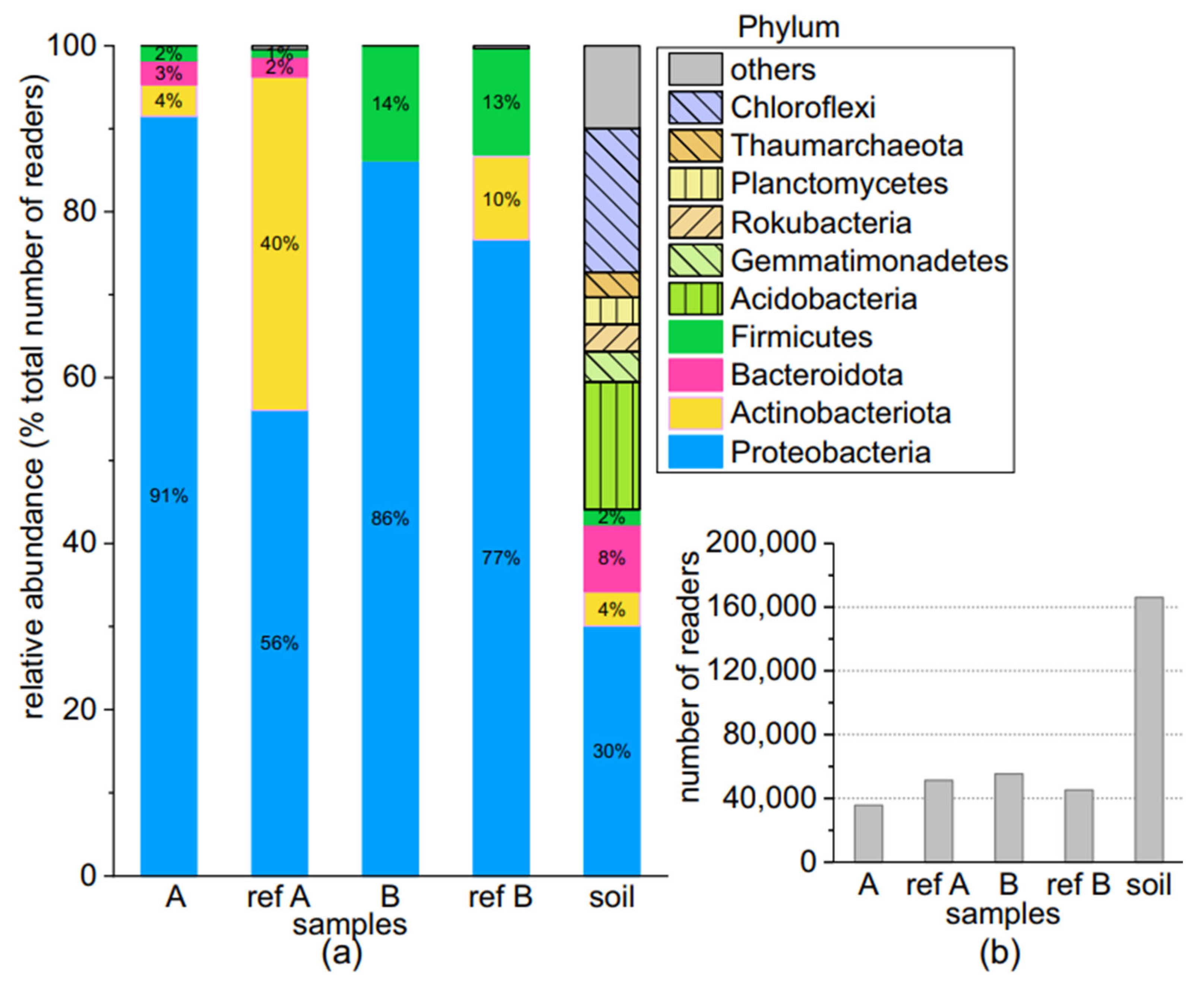
3.2. Comparison of the Microbial Community Abundance of HB35

3.2.1. With and without Electrochemical Incubation

The 16S rRNA sequences of the HB35 bacterial communities obtained under four groups of culture conditions (A, B, ref. A, and ref. B) were analyzed with NGS (Figure 3). The NGS analyses showed the following: (1) The total numbers of reads in the four groups (40,000 reads in average) after incubation were about 75% lower than in the soil sample (about 160,000 reads). (2) Compared with the HB35 soil sample, the bacterial community structures of the four groups after incubation changed significantly and were reduced to mainly four phyla: Firmicutes, Bacteroidota, Actinobacteriota, and Proteobacteria. Chloroflexi and Acidobacteria, which were highly abundant in the soil, were almost invisible here. (3) They also showed that the relative abundances of Proteobacteria were 91% and 86% higher in electrochemical culture groups A and B than the values of 56% and 77% in the non-electrochemical culture groups ref. A and ref. B, and they dominated all groups. (4) The relative abundances of Actinobacteriota in groups A and B under electrochemical cultivation were only 4% and less than 1%, whereas in ref. A and ref. B, significantly higher relative abundances were found (40% and 10%). (5) Bacteroidota had 3% and 2% relative abundances in groups A and ref. A, while its relative abundances in groups B and ref. B were less than 1%. (6) The relative abundances of Firmicutes were 14% and 13% in groups B and ref. B, significantly higher than in groups A and ref. A (2% and 1%).
The phylum Proteobacteria was dominant in all four groups of culture conditions, while Actinobacteriota, Bacteroidota, and Firmicutes were selectively enriched under different culture conditions. Proteobacteria—as a dominant phylum for bioelectrochemical studies, especially α- and γ-proteobacteria—are involved in microbial extracellular electron transfer [29]. Firmicutes can be enriched on biocathodes and are involved in bio-electrochemical denitrification [30]. Bacteroidota and Actinobacteriota were present in the biofilm growing on the electrode, but the exact electroactive function is unknown [31]. Moreover, bacterial community structure is mainly influenced by the environment [32], suggesting that these four phyla may have been screened from the bacterial community by the culture medium and electrochemical incubation method applied here.
In general, the bacterial communities differed in structure and abundance at the phylum level when using CO2 in air and sodium bicarbonate as the carbon sources, respectively. In contrast, microbial communities cultured in the same inorganic medium with either electrochemical or non-electrochemical methods were structurally identical and differed only in abundance.

3.2.2. Comparison at the Genus Level

The distribution of the HB35 bacterial communities at the genus level was compared for the aforementioned four groups of culture conditions (Figure 4). (1) Compared to the non-electrochemical culture group (ref. A and ref. B), the electrochemical culture group showed significant specificity; i.e., 70% Acinetobacter in group A (CO2 in air as carbon source) and 80% Pseudomonas in group B (NaHCO3 as carbon source). (2) Comparison of group B with ref. B showed that Pseudomonas was dominant in both cases, indicating that it is likely to be able to use sodium bicarbonate for metabolism. (3) Ref. A showed almost no Acinetobacter compared to group A, suggesting that it can be enriched with CO2 from the air as a carbon source during electrochemical incubation. (4) Chryseobacterium, which belonged to group A, was not present in any of the other three groups. Chryseobacterium can form biofilms of metabolized chlorobenzenes in MECs [33].
The high abundances of Acinetobacter and Pseudomonas suggest that they can be enriched in the bacterial community after electrochemical cultivation. Pseudomonas sp. has been found to exhibit high rates of furfural degradation in wastewater samples pretreated electrochemically at 2.0 V [34]. Pseudomonas aeruginosa has been found to be associated with the production of current [35]. Acinetobacter calcoaceticus can perform a reduction reaction at the cathode of an electrochemical reactor [36].
Overall, the distribution at the genus level (1) clarified the specific distribution of the community under various conditions, as well as (2) the differentiates of the culture conditions, especially when using carbon dioxide from air and when incubating with sodium bicarbonate, and (3) provided the possibility of subsequently isolating multiple single strains from the community.

3.2.3. Comparison of the Unique OTUs of HB35

The unique OTUs of the bacterial community in HB35 found in four groups of culture conditions are listed in Table 2, and these OTUs were found in only one culture condition and not in the other three. Compared to the non-electrochemical incubation group (ref. A and ref. B), less unique OTUs were found in groups A and B with electrochemical incubation. This is because electrochemical incubation conditions in microbial electrochemical systems are optimized for the growth and activity of electrochemically active microorganisms, which may be a subset of the total microbial community. Delftia, Diaphorobacter, and Methylophilaceae were the unique OTUs belonging to group A. Species of the genus Delftia are known for their special ability to metabolize different pollutants; for example, herbicides [37], chloraniline [38], and polycyclic aromatic hydrocarbons [39]. In addition, some Delftia strains are known to tolerate and bind toxic heavy metals, such as cadmium [40]. The genus Diaphorobacter has been described with a strain from active sludge capable of degrading environmentally polluting polymers, such as polyhydroxybutyrate and hydroxybutyratehydroxyvalerate copolymers [41]. Methylophilaceae are a family of methylotrophic Betaproteobacteria. For example, they metabolize C1-compounds, such as formic acid, methanol, formaldehyde, and methylamine [42]. The electron transport capacities of Delftia, Diaphorobacter, and Methylophilaceae in MECs have been of interest in the field of bioelectrochemistry because these bacteria have been shown to exhibit high electroactivity and can efficiently transfer electrons to the anode surface [43,44,45]. The unique OTU Paenisporosarcina in group B had a significantly high number of reads (5370), and strains under this genus have been reported by Harris et al. to form biofilms [46], which are essential for electron transport in electrochemical systems. Some species of the Sporosarcina group can decompose urea and are observed in urine-contaminated soils with high abundances [47]. In group B, Achromobacter had a high number of reads (1340). Achromobacter is a genus of bacteria that includes both electroactive and non-electroactive species. In this genus are found species able to use molecular hydrogen as an electron source [47]. This is important for cathodically stimulated bacterial growth because hydrogen typically evolves under cathodic polarization and could act as an electrochemical mediator metabolized by bacteria in the near-electrode solution. Some strains of Achromobacter have been shown to have electrochemical activity and can transfer electrons to solid surfaces, including electrodes. For example, a recent study found that Achromobacter xylosoxidans isolated from wastewater could produce a significant amount of current in a microbial electrochemical cell [48]. Kohzuma et al. have reported that Achromobacter stains are involved in reduction reactions connected with electrochemical processes [49].
Some unique OTUs in group B belonged to genera described up to now as uncultured. They belonged to different families (i.e., Planococcaceae, Bacillaceae, Nitrosomonadaceae, and Colwelliaceae), which means they were related to genera and species that were not specified in the applied database.
Among the strains found under electrochemical processing (A and B in Table 2) were types that reflected the historical environmental conditions of the sampling site. The ability to degrade different hydrocarbons, the ability to degrade urea, and tolerances against toxic metals are typical properties that could be expected from bacteria communities from soil contaminated by tannery activities. Thus, on the one side, the appearance of taxonomic groups related to such organisms can be interpreted as an ecological echo of the former use of the sampling place, with tannery activities dating back for centuries. On the other hand, this finding confirmed the strategy of searching for new bacterial strains with interesting metabolic features in such places with specific ancient human impacts.

3.3. Comparison of the Community Abundances in HB35 Related to Different Culture Agar Media

3.3.1. Comparison at the Phylum Level

The structure of the HB35 bacterial community under the four groups of conditions was clarified with 16S rRNA analysis to isolate and purify as many single strains with electroactivity or medium specificity from them as possible. Mixed cultures from the four groups of samples were inoculated onto three synthetic solid media (AM1, AM2, and CMM1) and two complex solid media (LB and TSA) for incubation. After 7 days of incubation at room temperature, the 16S rRNA sequences of the grown mixed colonies were analyzed and their selective specificities on the five media were compared.
Figure 5 shows the phylum-level classification of the microbial communities in relation to the five different solid media. The bacterial community compositions on the AM1, AM2, and CMM1 solid media were identical (i.e., Proteobacteria, Actinobacteria, and Bacteroidota), differing only in abundance. While the abundance of Bacteroidota was significantly higher (AM1: 20% AM2: 35%, and CMM1: 58%), the abundance of Actinobacteriota was slightly higher (11%, 7%, and 10%). The abundance of Proteobacteria was lower (70%, 57%, and 32%) compared to the initial mixed culture (group A, 91%). The abundance of Firmicutes with the complex solid media was more than five times higher than that in the initial mixed culture A on LB and TSA media.
These NGS data suggest that inoculation onto different solid media favors the enrichment of low-abundance bacteria from mixed cultures. It can be concluded that here the low abundances of the electrochemically cultured Bacteroidota and Actinobacteriota could be enriched on AM1, AM2, and CMM1 agar, while the electrochemically cultured Firmicutes could be enriched by using complex agar (LB and TSA).

3.3.2. Comparison at the Genus Level

To characterize the structure and abundance of mixed colonies grown on the five solid media, they were analyzed with a smaller taxonomic unit at the genus level (Figure 6). Compared to the initial sample (group A), the abundances of Pseudomonas, Lysobacter, and Bacillus increased on the complex solid media LB and TSA, and the abundances of Chryseobacterium, Pseudarthrobacter, and Sphingopyxis increased on the synthetic solid media AM1, AM2, and CMM1. The increased abundances suggest that these genera can be enriched on the corresponding solid media. In contrast, 10% of Ramlibacter and 5% of Aminobacter were found only on AM1 agar, while both genera had very low abundances in the initial sample A. These data suggest that AM1 has a specific selectivity for them.
The genus structure of the mixed cultures of group A cultured on five solid media confirmed that (1) the mixed cultures of group A with low growth density could not only be cultured on different solid media but could also (2) expand species abundance and (3) retain diversity. In addition, (4) the low-abundance communities were more detailed in the genus-level analysis after expanding their abundance on solid media, which facilitated comparison with the real isolates (Table 3).

3.4. Electrochemical Incubation of Isolated Strains

After isolation and identification, 16 strains were identified. These strains were characterized specifically in terms of the initial culture conditions, the solid medium that was enriched, and the identification results after comparison with the genetic database (@BLAST) (Table 3). Ten of them were strains from electrochemical cultures and six were isolated from the non-electrochemical reference group.
Chryseobacterium bovis was observed in group A cultures after cultivation on CMM1. It was first described as Epilithonimonas bovis, which was transferred to the genus Epilithonimonas in 2020 [50]; in 2008, it was isolated from raw cow’s milk and identified as Chryseobacterium bovis [51]. The abundance of Chryseobacterium was significantly high, with up to 58% on CMM1 (Figure 7). On TSA agar, Acinetobacter johnsonii and Arthrobacter humicola were isolated, and the abundance of Acinetobacter on TSA was 46%, while the abundance of Arthrobacter was less than 1%, and it is not visible in Figure 6. Sphingopyxis chilensis was isolated on AM2, and the abundance of Sphingopyxis on AM2 was 2% (Figure 6).
A literature search revealed that, of the 16 strains isolated here, Pseudomonas alcaliphila can also show denitrification activity in a two-chamber bioelectrochemical system with electrodes as the sole electron donor [52]. Pseudomonas stutzeri can use cytochromes for electron transfer [53]. Acinetobacter johnsonii has been isolated from electroactive cathodic biofilms [54]. Very few studies on the electroactivity of other isolated strains have been reported.
In short, on the one hand, single strains could be isolated and characterized after enrichment on different solid media. On the other hand, the electroactive characteristics of several strains isolated here have already been reported, and the electroactive characteristics of other strains isolated by electrochemical culture deserve specific study.
Table 3. Information on the isolated strains.
NameCulture ConditionAgar MediumPhylumIdentification Result
TX166ACMM1BacteroidotaEpilithonimonas bovis DSM 19482
TX167ACMM1BacteroidotaEpilithonimonas bovis DSM 19482
TX168ACMM1BacteroidotaEpilithonimonas bovis DSM 19482
TX169ATSAActinobacteriotaArthrobacter humicola
TX183BAM1ProteobacteriaPseudomonas alcaliphila
TX184Ref. BAM1ProteobacteriaPseudomonas stutzeri
TX185BCMM1ProteobacteriaPseudomonas azotoformans
TX186Ref. ALBActinobacteriotaArthrobacter humicola
TX187Ref. ATSAProteobacteriaLysobacter caeni
TX188Ref. BTSAActinobacteriotaPseudarthrobacter oxydans
TX190AAM2ProteobacteriaSphingopyxis chilensis
TX191ATSAProteobacteriaAcinetobacter johnsonii
TX296Ref. ACMM1ActinobacteriotaArthrobacter humicola
TX297Ref. ALBActinobacteriotaArthrobacter humicola
TX298BTSAProteobacteriaPseudomonas alcaliphila
TX299BTSAProteobacteriaPseudomonas sp. J380
To verify the electrochemical characteristics of an example of the isolated individual strains, strain TX168 was again inoculated into an electrochemical cathodic culture with 100 mM sodium bicarbonate as a carbon source in an inorganic medium. The reference group was cultured in the same medium in culture flasks without additional voltage. During the 10 days of cultivation, it was observed that the biofilm formed during electrochemical cultivation increased significantly with the incubation time, while the TX168 strain in the reference group automatically aggregated at the bottom of the culture flask.
Figure 7 shows the weight of the biomass after 10 days of cultivation and the weight after 2 days of drying at room temperature. After two days of drying, the biomass of TX168 was 2.45 ± 0.15 mg compared to 0.94 ± 0.035 mg for the reference group. This difference in biomass suggests that (1) weighing biomass via direct drying is meaningful, (2) TX168 can not only be cultured in an electrochemical cathode chamber but also metabolize sodium bicarbonate as an inorganic carbon source, and (3) electrochemical incubation can support biomass production by TX168. In addition, Chryseobacterium is known to play a role in removing tetrachloroethylene enrichment and nitrate with cathodic enrichment biofilms [55]. Besides the electrochemical processes, biofilms play an important role in electron transport [56].

4. Conclusions

The study suggests that the growth media used in MECs can significantly impact the microbial communities associated with the cathode. A microbial community from the soil sample HB35 collected during an archaeological excavation grew better when cultured under cathodic polarization in MECs using sodium bicarbonate as a carbon source than when using carbon dioxide from air only. In addition, the non-electrochemical batch culture of the bacterial communities from HB35 (reference experiment) showed only weak growth. It seems that adding sodium bicarbonate promotes the growth of soil bacterial communities during electrochemical cultivation. This is a strong argument for cathodically supported biological fixation of carbon from carbonate.
The analysis based on the 16S rRNA gene identified OTUs from the HB35 bacterial community after two-chamber MEC culturing using NaHCO3 as a carbon source. Proteobacteria (86%) and Firmicutes (14%) were the dominating phyla and, among them, 80% belonged to the genus Pseudomonas and 10% to Paenisporosarcina in terms of abundance. When carbon dioxide from air was used as the carbon source, an abundance of 74% for Acinetobacter at the genus level was identified. It is well-known that both Acinetobacter and Pseudomonas are highly abundant bacteria in anode biofilms and play important roles in energy production [57]. The analysis of the unique OTUs showed that many different uncultured species could be found under electrochemical cultivation conditions with NaHCO3 as a carbon source. In addition, this study showed that a soil sample from a historical tannery site was able to supply new, interesting strains. Our results support the idea of using soil microbial communities from places with special ancient human impacts as promising sources in the search for bacteria applicable to new biotechnological processes.
To obtain the pure isolate, the microbial communities were inoculated onto five solid media. The 16S rRNA gene analysis showed that bacteria with low abundance in the mixed cultures (air as carbon source) could be enriched on different solid media; among them, Chryseobacterium and Bacillus were significantly enriched on synthetic agars (AM1, AM2, and CMM1) and complex agars (LB and TSA), respectively. Finally, a total of 16 strains were isolated. Further characterization with TX168 Epilithonimonas bovis showed high biomass production using the MEC setup compared to non-electrochemical cultivation.
Overall, our findings have important implications for optimizing MEC performance, as different microbial communities may have different electrochemical properties and efficiencies. Additionally, the study highlights the potential of MECs for the study of microbial ecology and bioremediation applications. First, future research will focus on the investigation of specially composed soil bacterial communities from other places with ancient human impacts due to their potential to provide new cathodically active strains. This will include soil bacterial communities from medieval or pre-industrial copper mining areas, as well as from prehistoric settlement places. Furthermore, the MEC setup will be improved to enable efficient searching for new cathodically stimulable bacteria with CO2-fixation potential. To this end, the MEC cell will be combined with the microfluidic screening process and microsegmentation operations to (1) automatically modify the composition of the medium, (2) optimize the screening process for new strains, and (3) optimize biomass production using excess current.

Author Contributions

Laboratory measurements and data analysis, T.X. and J.C.; conceptualization and writing—original draft preparation, J.C. and T.X.; bioinformatics software, P.M.G.; NGS sample preparation and data analysis, L.E.; writing—review and editing, J.C., J.M.K. and L.E. All authors have read and agreed to the published version of the manuscript.

Funding

T.X. and L.E. were financed by Thuringian State Graduate Funding. J.C. gratefully acknowledges the financial support provided by a habilitation scholarship from the Technische Universität Ilmenau.

Data Availability Statement

The data used to support the findings of this study are available from the corresponding author upon request.

Acknowledgments

We thank Steffen Schneider for lab assistance.

Conflicts of Interest

The authors declare no conflict of interest.

References

  1. Ahmad, A.; Ibrahim, M.N.M.; Yaqoob, A.A.; Setapar, S.H.M. Microbial Fuel Cells for Environmental Remediation; Springer Nature: Singapore, 2022; Volume 1, p. 450. [Google Scholar]
  2. He, K.; Li, W.; Tang, L.; Lv, S.; Xing, D. Suppressing Methane Production to Boost High-Purity Hydrogen Production in Microbial Electrolysis Cells. Environ. Sci. Technol. 2021, 56, 11931–11951. [Google Scholar] [CrossRef] [PubMed]
  3. Zhao, J.; Li, F.; Cao, Y.; Zhang, X.; Chen, T.; Song, H.; Wang, Z. Microbial extracellular electron transfer and strategies for engineering electroactive microorganisms. Biotechnol. Adv. 2020, 53, 107682. [Google Scholar] [CrossRef] [PubMed]
  4. Thapa, B.S.; Kim, T.; Pandit, S.; Song, Y.E.; Afsharian, Y.P.; Rahimnejad, M.; Kim, J.R.; Oh, S.-E. Overview of electroactive microorganisms and electron transfer mechanisms in microbial electrochemistry. Bioresour. Technol. 2022, 347, 126579. [Google Scholar] [CrossRef] [PubMed]
  5. González-Paz, J.R.; Becerril-Varela, K.; Guerrero-Barajas, C. Iron reducing sludge as a source of electroactive bacteria: Assessing iron reduction in biofilm bacteria, planktonic cells and isolates from a microbial fuel cell. Arch. Microbiol. 2022, 204, 632. [Google Scholar] [CrossRef] [PubMed]
  6. Geelhoed, J.S.; Stams, A.J.M. Electricity-Assisted Biological Hydrogen Production from Acetate by Geobacter sulfurreducens. Environ. Sci. Technol. 2011, 45, 815–820. [Google Scholar] [CrossRef] [PubMed]
  7. Xiang, Y.; Liu, G.; Zhang, R.; Lu, Y.; Luo, H. Acetate production and electron utilization facilitated by sulfate-reducing bacteria in a microbial electrosynthesis system. Bioresour. Technol. 2017, 241, 821–829. [Google Scholar] [CrossRef] [PubMed]
  8. Askitosari, T.D.; Berger, C.; Tiso, T.; Harnisch, F.; Blank, L.M.; Rosenbaum, M.A. Coupling an Electroactive Pseudomonas putida KT2440 with Bioelectrochemical Rhamnolipid Production. Microorganisms 2020, 8, 1959. [Google Scholar] [CrossRef]
  9. Choi, O.; Sang, B.-I. Extracellular electron transfer from cathode to microbes: Application for biofuel production. Biotechnol. Biofuels 2016, 9, 11. [Google Scholar] [CrossRef] [Green Version]
  10. Schmitz, S.; Rosenbaum, M.A. Boosting mediated electron transfer in bioelectrochemical systems with tailored defined microbial cocultures. Biotechnol. Bioeng. 2018, 115, 2183–2193. [Google Scholar] [CrossRef]
  11. Paquete, C.M.; Rosenbaum, M.A.; Bañeras, L.; Rotaru, A.E.; Puig, S. Let’s chat: Communication between electroactive microorganisms. Bioresour. Technol. 2022, 347, 126705. [Google Scholar] [CrossRef]
  12. Chabert, N.; Ali, O.A.; Achouak, W. All ecosystems potentially host electrogenic bacteria. Bioelectrochemistry 2015, 106, 88–96. [Google Scholar] [CrossRef]
  13. Zhang, G.; Bai, J.; Zhao, Q.; Jia, J.; Wang, W.; Wang, X. Bacterial Succession in Salt Marsh Soils Along a Short-term Invasion Chronosequence of Spartina alterniflora in the Yellow River Estuary, China. Microb. Ecol. 2020, 79, 644–661. [Google Scholar] [CrossRef] [PubMed]
  14. Liao, S.; Wang, Y.; Liu, H.; Fan, G.; Sahu, S.K.; Jin, T.; Chen, J.; Zhang, P.; Gram, L.; Strube, M.L.; et al. Deciphering the Microbial Taxonomy and Functionality of Two Diverse Mangrove Ecosystems and Their Potential Abilities to Produce Bioactive Compounds. mSystems 2020, 5, e00851-19. [Google Scholar] [CrossRef] [PubMed]
  15. Wang, X.; Teng, Y.; Ren, W.; Li, Y.; Yang, T.; Chen, Y.; Zhao, L.; Zhang, H.; Kuramae, E.E. Variations of Bacterial and Diazotrophic Community Assemblies throughout the Soil Profile in Distinct Paddy Soil Types and Their Contributions to Soil Functionality. Msystems 2022, 7, e01047-21. [Google Scholar] [CrossRef] [PubMed]
  16. Canarini, A.; Schmidt, H.; Fuchslueger, L. Ecological memory of recurrent drought modifies soil processes via changes in soil microbial community. Nat. Commun. 2021, 12, 5308. [Google Scholar] [CrossRef]
  17. Köhler, J.M.; Kalensee, F.; Cao, J.; Günther, P.M. Hadesarchaea and other extremophile bacteria from ancient mining areas of the East Harz region (Germany) suggest an ecological long-term memory of soil. SN Appl. Sci. 2019, 1, 839. [Google Scholar] [CrossRef] [Green Version]
  18. Ehrhardt, L.; Günther, P.M.; Böhme, M.; Köhler, J.M.; Cao, J. Three Soil Bacterial Communities from an Archaeological Excavation Site of an Ancient Coal Mine near Bennstedt (Germany) Characterized by 16S r-RNA Sequencing. Environments 2022, 9, 115. [Google Scholar] [CrossRef]
  19. Araújo, A.S.F.; Lima, L.M.; Santos, V.M.; Schmidt, R. Repeated application of composted tannery sludge affects differently soil microbial biomass, enzymes activity, and ammonia-oxidizing organisms. Environ. Sci. Pollut. Res. 2016, 23, 19193–19200. [Google Scholar] [CrossRef]
  20. Miranda, A.R.L.; Mendes, L.W.; Lemos, L.N.; Antunes, J.E.L.; Amorim, M.R.; Melo, V.M.M.; de Melo, W.J.; Brink, P.J.V.D.; Araujo, A.S.F. Dynamics of archaeal community in soil with application of composted tannery sludge. Sci. Rep. 2019, 9, 7347. [Google Scholar] [CrossRef] [Green Version]
  21. Leigh, J.A.; Albers, S.-V.; Atomi, H.; Allers, T. Model organisms for genetics in the domain Archaea: Methanogens, halophiles, Thermococcales and Sulfolobales. FEMS Microbiol. Rev. 2011, 35, 577–608. [Google Scholar] [CrossRef] [Green Version]
  22. Yilmaz, P.; Parfrey, L.W.; Yarza, P.; Gerken, J.; Pruesse, E.; Quast, C.; Schweer, T.; Peplies, J.; Ludwig, W.; Glöckner, F.O. The SILVA and “all-species living tree project (LTP)” taxonomic frameworks. Nucleic Acids Res. 2014, 42, D643–D648. [Google Scholar] [CrossRef] [PubMed] [Green Version]
  23. Stucky, B.J. SeqTrace: A Graphical Tool for Rapidly Processing DNA Sequencing Chromatograms. J. Biomol. Tech. JBT 2012, 23, 90. [Google Scholar] [CrossRef] [PubMed] [Green Version]
  24. Drancourt, M.; Berger, P.; Raoult, D. Systematic 16S rRNA Gene Sequencing of Atypical Clinical Isolates Identified 27 New Bacterial Species Associated with Humans. J. Clin. Microbiol. 2004, 42, 2197–2202. [Google Scholar] [CrossRef] [Green Version]
  25. Li, E.; de Orduña, R.M. A rapid method for the determination of microbial biomass by dry weight using a moisture analyser with an infrared heating source and an analytical balance. Lett. Appl. Microbiol. 2010, 50, 283–288. [Google Scholar] [CrossRef]
  26. Stanbury, P.; Whittaker, A.; Hall, S. Principles of Fermentation Technology (Chap. 6); Butterworth-Heinemann: Oxford, UK, 1995. [Google Scholar]
  27. Peppler, H.J.; Perlman, D. Microbial Technology: Microbial Processes; Academic Press: Cambridge, MA, USA, 1979; Volume 1. [Google Scholar]
  28. Kim, J.; Lee, J.Y.; Ahting, C.; Johnstone, R.; Lu, T. Growth of Chlorella vulgaris using sodium bicarbonate under no mixing condition. Asia-Pac. J. Chem. Eng. 2014, 9, 604–609. [Google Scholar]
  29. Yuan, Y.; Cai, X.; Wang, Y.; Zhou, S. Electron transfer at microbe-humic substances interfaces: Electrochemical, microscopic and bacterial community characterizations. Chem. Geol. 2017, 456, 1–9. [Google Scholar] [CrossRef]
  30. Lee, S.-H.; Kondaveeti, S.; Min, B.; Park, H.-D. Enrichment of Clostridia during the operation of an external-powered bio-electrochemical denitrification system. Process. Biochem. 2013, 48, 306–311. [Google Scholar] [CrossRef]
  31. Khater, D.Z.; El-Khatib, K.; Hassan, H.M. Microbial diversity structure in acetate single chamber microbial fuel cell for electricity generation. J. Genet. Eng. Biotechnol. 2017, 15, 127–137. [Google Scholar] [CrossRef]
  32. Carson, J.K.; Campbell, L.; Rooney, D.; Clipson, N.; Gleeson, D.B. Minerals in soil select distinct bacterial communities in their microhabitats. FEMS Microbiol. Ecol. 2009, 67, 381–388. [Google Scholar] [CrossRef]
  33. Ying, Z.; Yu, Y.; Chen, H.; Zhao, J.; You, J.; Ye, J.; Hu, J.; Zhang, S. Structural Characteristics and Functional Genes of Biofilms in Microbial Electrolysis Cells for Chlorobenzene Abatement. ACS ES T Water 2023, 3, 500–509. [Google Scholar] [CrossRef]
  34. Silva, J.R.; Santos, D.S.; Santos, U.R.; Eguiluz, K.I.; Salazar-Banda, G.R.; Schneider, J.K.; Krause, L.C.; López, J.A.; Hernández-Macedo, M.L. Electrochemical and/or microbiological treatment of pyrolysis wastewater. Chemosphere 2017, 185, 145–151. [Google Scholar] [CrossRef]
  35. Venkataraman, A.; Rosenbaum, M.; Arends, J.B.; Halitschke, R.; Angenent, L.T. Quorum sensing regulates electric current generation of Pseudomonas aeruginosa PA14 in bioelectrochemical systems. Electrochem. Commun. 2010, 12, 459–462. [Google Scholar] [CrossRef]
  36. Freguia, S.; Tsujimura, S.; Kano, K. Electron transfer pathways in microbial oxygen biocathodes. Electrochimica Acta 2010, 55, 813–818. [Google Scholar] [CrossRef]
  37. Leibeling, S.; Schmidt, F.; Jehmlich, N.; von Bergen, M.; Müller, R.H.; Harms, H. Declining capacity of starving Delftia acidovorans MC1 to degrade phenoxypropionate herbicides correlates with oxidative modification of the initial enzyme. Environ. Sci. Technol. 2010, 44, 3793–3799. [Google Scholar] [CrossRef]
  38. Zhang, L.-L.; He, D.; Chen, J.-M.; Liu, Y. Biodegradation of 2-chloroaniline, 3-chloroaniline, and 4-chloroaniline by a novel strain Delftia tsuruhatensis H1. J. Hazard. Mater. 2010, 179, 875–882. [Google Scholar] [CrossRef]
  39. Wu, W.; Huang, H.; Ling, Z.; Yu, Z.; Jiang, Y.; Liu, P.; Li, X. Genome sequencing reveals mechanisms for heavy metal resistance and polycyclic aromatic hydrocarbon degradation in Delftia lacustris strain LZ-C. Ecotoxicology 2016, 25, 234–247. [Google Scholar] [CrossRef] [PubMed]
  40. Liu, Y.; Tie, B.; Li, Y.; Lei, M.; Wei, X.; Liu, X.; Du, H. Inoculation of soil with cadmium-resistant bacterium Delftia sp. B9 reduces cadmium accumulation in rice (Oryza sativa L.) grains. Ecotoxicol. Environ. Saf. 2018, 163, 223–229. [Google Scholar] [CrossRef] [PubMed]
  41. Khan, S.T.; Hiraishi, A. Diaphorobacter nitroreducens gen. nov., sp. nov., a poly (3-hydroxybutyrate)-degrading denitrifying bacterium isolated from activated sludge. J. Gen. Appl. Microbiol. 2002, 48, 299–308. [Google Scholar] [CrossRef] [PubMed] [Green Version]
  42. Kalyuzhnaya, M.; Bowerman, S.; Lara, J.C.; Lidstrom, M.E.; Chistoserdova, L. Methylotenera mobilis gen. nov., sp. nov., an obligately methylamine-utilizing bacterium within the family Methylophilaceae. Int. J. Syst. Evol. Microbiol. 2006, 56, 2819–2823. [Google Scholar] [CrossRef] [Green Version]
  43. Jangir, Y.; French, S.; Momper, L.M.; Moser, D.P.; Amend, J.P.; El-Naggar, M.Y. Isolation and Characterization of Electrochemically Active Subsurface Delftia and Azonexus Species. Front. Microbiol. 2016, 7, 756. [Google Scholar] [CrossRef] [Green Version]
  44. Jiang, L.; Huang, L.; Sun, Y. Recovery of flakey cobalt from aqueous Co(II) with simultaneous hydrogen production in microbial electrolysis cells. Int. J. Hydrogen Energy 2014, 39, 654–663. [Google Scholar] [CrossRef]
  45. Yang, Y.; Wang, H.; Zheng, Y.; Zhu, B.; Wu, X.; Zhao, F. Extracellular electron transfer of Methylophilus methylotrophs. Process. Biochem. 2020, 94, 313–318. [Google Scholar] [CrossRef]
  46. Harris, D.; Ummadi, J.G.; Koley, D. Scanning Electrochemical Microscopy: A Tool to Study the Biomediated Calcification Process by Sporosarcina Pasteurii Biofilm. In Electrochemical Society Meeting Abstracts 229; The Electrochemical Society, Inc.: San Diego, CA, USA, 2016; p. 1667. [Google Scholar]
  47. Madigan, M.T.; Martinko, J.M. Brock Mikrobiologie, 11th ed.; Pearson Studium: München, Germany, 2006. [Google Scholar]
  48. Diba, F.; Khan, Z.H.; Uddin, S.Z.; Istiaq, A.; Shuvo, S.R.; Alam, A.S.M.R.U.; Hossain, M.A.; Sultana, M. Bioaccumulation and detoxification of trivalent arsenic by Achromobacter xylosoxidans BHW-15 and electrochemical detection of its transformation efficiency. Sci. Rep. 2021, 11, 21312. [Google Scholar] [CrossRef] [PubMed]
  49. Kohzuma, T.; Shidara, S.; Suzuki, S. Direct Electrochemistry of Nitrite Reductase from Achromobacter cycloclastes IAM 1013. Bull. Chem. Soc. Jpn. 1994, 67, 138–143. [Google Scholar] [CrossRef]
  50. Nicholson, A.C.; Gulvik, C.A.; Whitney, A.M.; Humrighouse, B.W.; Bell, M.E.; Holmes, B.; Steigerwalt, A.G.; Villarma, A.; Sheth, M.; Batra, D.; et al. Division of the genus Chryseobacterium: Observation of discontinuities in amino acid identity values, a possible consequence of major extinction events, guides transfer of nine species to the genus Epilithonimonas, eleven species to the genus Kaistella, and three species to the genus Halpernia gen. nov., with description of Kaistella daneshvariae sp. nov. and Epilithonimonas vandammei sp. nov. derived from clinical specimens. Int. J. Syst. Evol. Microbiol. 2020, 70, 4432. [Google Scholar] [CrossRef]
  51. Hantsis-Zacharov, E.; Senderovich, Y.; Halpern, M. Chryseobacterium bovis sp. nov., isolated from raw cow’s milk. Int. J. Syst. Evol. Microbiol. 2008, 58, 1024–1028. [Google Scholar] [CrossRef]
  52. Su, W.; Zhang, L.; Li, D.; Zhan, G.; Qian, J.; Tao, Y. Dissimilatory nitrate reduction by Pseudomonas alcaliphila with an electrode as the sole electron donor. Biotechnol. Bioeng. 2012, 109, 2904–2910. [Google Scholar] [CrossRef]
  53. Raffalt, A.C.; Schmidt, L.; Christensen, H.E.; Chi, Q.; Ulstrup, J. Electron transfer patterns of the di-heme protein cytochrome c4 from Pseudomonas stutzeri. J. Inorg. Biochem. 2009, 103, 717–722. [Google Scholar] [CrossRef]
  54. Logan, B.E.; Rossi, R.; Ragab, A.; Saikaly, P.E. Electroactive microorganisms in bioelectrochemical systems. Nat. Rev. Genet. 2019, 17, 307–319. [Google Scholar] [CrossRef]
  55. Chen, F.; Ye, Y.; Fan, B.; Lv, M.; Liang, B.; Liu, W.; Cheng, H.-Y.; Chen, Y.; Liu, Y.; Wang, Y.; et al. Simultaneous removal of tetrachloroethylene and nitrate with a novel sulfur-packed biocathode system: The synergy between bioelectrocatalytic dechlorination and sulfur autotrophic denitrification. Chem. Eng. J. 2022, 439, 135793. [Google Scholar] [CrossRef]
  56. Bond, D.R.; Strycharz-Glaven, S.M.; Tender, L.M.; Torres, C.I. On Electron Transport through Geobacter Biofilms. Chemsuschem 2012, 5, 1099–1105. [Google Scholar] [CrossRef] [PubMed]
  57. Chignell, J.F.; De Long, S.K.; Reardon, K.F. Meta-proteomic analysis of protein expression distinctive to electricity-generating biofilm communities in air-cathode microbial fuel cells. Biotechnol. Biofuels 2018, 11, 121. [Google Scholar] [CrossRef] [PubMed]
Figure 1. Setup of the two-chamber MECs. (a) The scheme shows, from left to right, that the bacterial population or individual strains in the culture flasks could be circulated via pumping into the cathode chamber. A constant voltage was applied to the MECs. Electrolytes from an additional bottle were pumped into the anode chamber for recycling. (b) Laboratory setup.
Environments 10 00097 g001
Figure 2. Growth kinetics of HB35 with and without sodium bicarbonate after 12 days of culturing in cathode chamber with MECs at a constant voltage of 1.6 V (A and B) and in culture flasks (refs. A and B). Development of (a) pH value and (b) optical density of HB35 during 12 days of culture.
Environments 10 00097 g002
Figure 3. Phylum-level classification. (a) Comparison of microbial communities at the phylum level: A—sample cultured with electrochemical incubation and CO2 in air as carbon source, ref. A—reference sample without electrochemical incubation and with CO2 in air as carbon source, B—sample with electrochemical incubation and NaHCO3 as carbon source, ref. B—reference sample without electrochemical incubation and with NaHCO3 as carbon source, and soil sample. The relative abundance shows the percentage of reads for each phylum compared to the total number of reads. (b) Total number of reads in each group.
Environments 10 00097 g003
Figure 4. Genus-level classification. Comparison of microbial communities at the genus level: A—sample cultured with electrochemistry and CO2 in air as carbon source, ref. A—reference sample without electrochemistry and with CO2 in air as carbon source, B—sample cultured with electrochemistry and NaHCO3 as carbon source, and ref. B—reference sample without electrochemistry and with NaHCO3 as carbon source. The relative abundance refers to the percentage of reads for each genus compared to the total number of reads.
Environments 10 00097 g004
Figure 5. Phylum-level classification. Comparison of microbial communities at the phylum level by inoculating electrochemically cultured bacteria from group A with CO2 in air as a carbon source onto AM1, AM2, CMM1, LB, and TSA solid media. The relative abundance indicates the percentage of reads for each phylum compared to the total number of reads.
Environments 10 00097 g005
Figure 6. Genus-level classification. Comparison of microbial communities at the genus level by inoculating electrochemically cultured bacteria from group A with CO2 in air as a carbon source onto AM1, AM2, CMM1, LB, and TSA solid media. The relative abundance indicates the percentage of reads for each genus compared to the total number of reads.
Environments 10 00097 g006
Figure 7. Biomass of TX168 Epilithonimonas bovis cultured under electrochemical and non-electrochemical conditions for 10 days using 100 mM sodium bicarbonate as a carbon source. Both culture sets started with the same inoculum cell density of 3 × 10⁶ cells/mL. The biomass was weighed after 48 h drying at room temperature.
Environments 10 00097 g007
Table 1. Ingredients and composition of solid media.
NameIngredient and Composition
AM10.1 g/L NH4NO3, 0.5 g/L K2HPO4, 0.2 g/L MgSO4·7H2O, 0.01 g/L FeSO4·7H2O, and 20 g/L agar
AM20.1 g/L NH4NO3, 0.5 g/L K2HPO4, 0.2 g/L MgSO4·7H2O, 0.01 g/L FeSO4·7H2O, 2g/L glucose, and 20 g/L agar
CMM10.1 g/L K2HPO4, 0.1 g/L (NH4)2SO4, 1 g/L MgSO4·7H2O, 0.02 g/L CaCl2·7H2O 1% methanol, 1 mL/L trace element solution (5 g/L NaEDTA, 2 g/L FeSO4·7H2O, 0.1 g/L ZnSO4·7H2O, 0.03 g/L MnCl2·4H2O, 0.2 g/L CoCl2·6H2O, 0.1 g/L CuCl2·5H2O, 0.02 g/L NiCl2·4H2O, 0.03 g/L Na2MoO4), and 16 g/L Gelrite
LB10 g/L tryptone, 5 g/L yeast extract, 10 g/L NaCl, and 15 g/L agar
TSA15 g/L tryptone, 5 g/L soya peptone, 5 g/L NaCl, and 15 g/L agar
Table 2. Unique OTUs found in each group. Comparison of unique OTUs found in A: sample with electrochemical incubation and CO2 in air as carbon source, ref. A: reference sample without electrochemical incubation and with CO2 in air as carbon source, B: sample with electrochemical incubation and NaHCO3 as carbon source, and ref. B: reference sample without electrochemical incubation and with NaHCO3 as carbon source. Numbers show the exact numbers of sequences for each OUT; only OTUs with more than two reads are listed.
Aref. ABref. B
Delftia137Terrimonas586Paenisporosarcina5370Salipaludibacillus280
Diaphorobacter10Alphaproteobacteria uncultured163Achromobacter1340Fontimonas95
Methylophilaceae OM43 clade6Chitinophagaceae Edaphobaculum162Planococcaceae Uncultured356Solimonadaceae uncultured15
Delftia137Chitinophagaceae uncultured107Sporosarcina162Nitrosarchaeum13
Panacagrimonas72Bacillaceae uncultured125Marmoricola10
Pedobacter45Nitrosomonadaceae uncultured46Xanthomonadaceae9
Desulfovibrio25Morganellaceae Candidatus Hamiltonella11Heimdallarchaeia8
Chitinophagaceae Taibaiella21Colwelliaceae uncultured3Thermoanaerobaculaceae Subgroup 106
Gemmatimonadota BD2-11 terrestrial group16 Paracoccus5
Bdellovibrio14 Gammaproteobacteria uncultured5
Oxalobacteraceae Undibacterium12 Planctomycetales uncultured5
Gammaproteobacteria JTB2311 Candidatus Nitrosotenuis4
Rickettsiales Candidatus Jidaibacter9 Clostridium sensu stricto 133
Aquicella8 Ruminiclostridium3
Rickettsiaceae uncultured8 Xanthobacteraceae uncultured3
Candidatus Paracaedibacter8 Holosporaceae uncultured3
Defluviicoccales uncultured7 Anaerolineae RBG-13-54-93
Candidatus Accumulibacter6 Anaerolineae SBR10313
Bradyrhizobium6
Cytophaga6
Iamiaceae6
Methyloligellaceae uncultured6
Sphingobium5
Candidatus Obscuribacter5
Falsarthrobacter5
Fimbriimonas5
Procabacter5
Rurimicrobium5
Rhizobiales Incertae Sedis uncultured5
Actinomarinales uncultured5
Acidobacteriae Subgroup 25
Disclaimer/Publisher’s Note: The statements, opinions and data contained in all publications are solely those of the individual author(s) and contributor(s) and not of MDPI and/or the editor(s). MDPI and/or the editor(s) disclaim responsibility for any injury to people or property resulting from any ideas, methods, instructions or products referred to in the content.

