
Electrochemical Biosensor for Evaluation of Environmental Pollutants Toxicity


Abstract
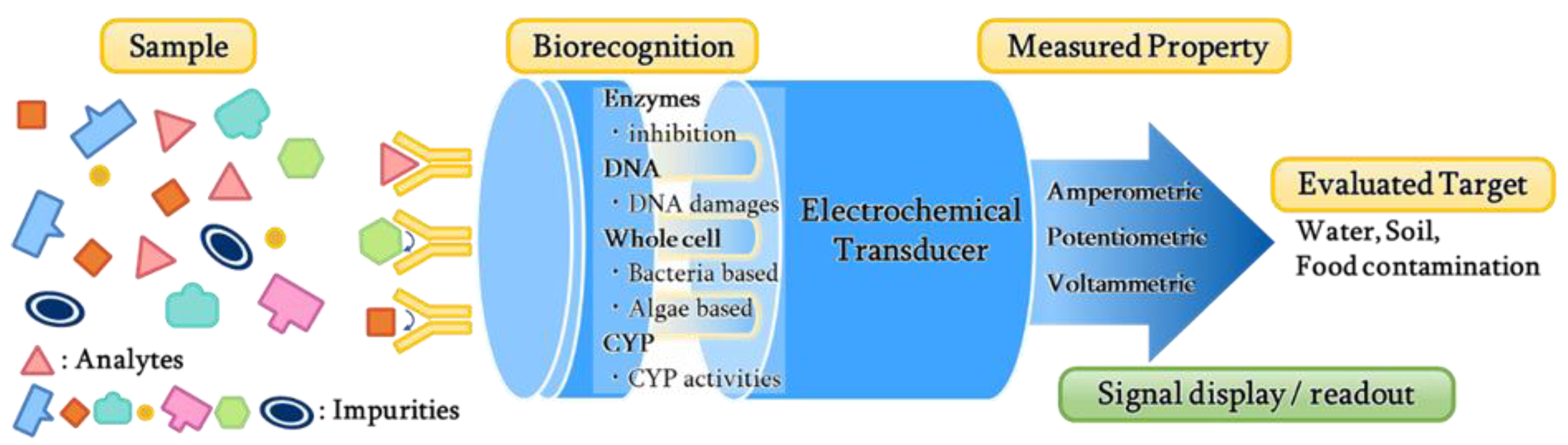
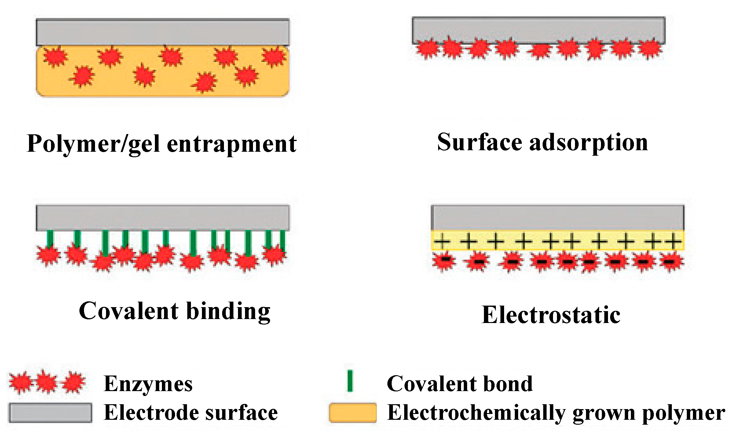
1. Introduction
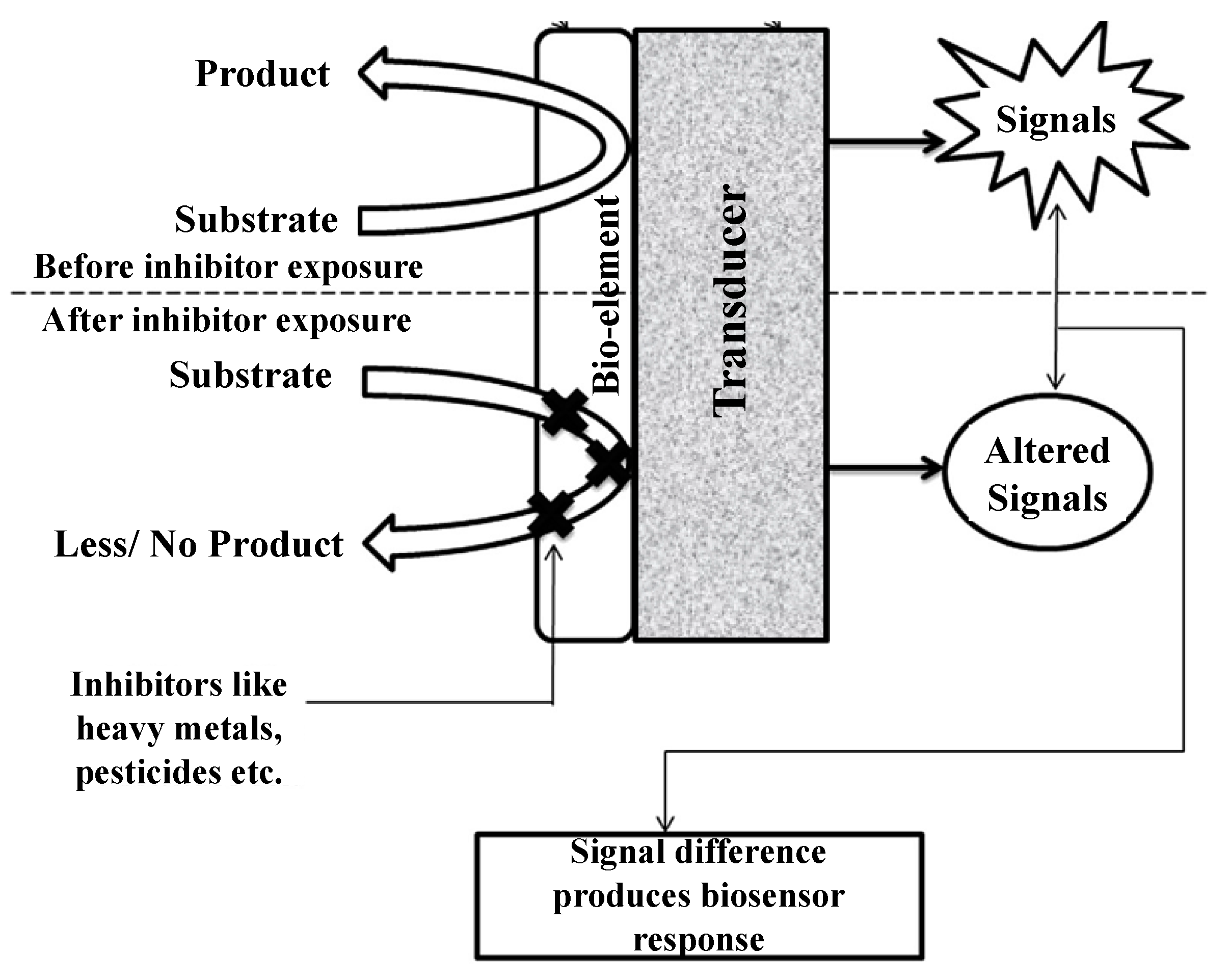
2. Biosensors Based on Enzyme Inhibition Activity
2.1. Glucose Oxidase Inhibition
2.2. Urease Inhibition
2.3. Cholinesterase Inhibition
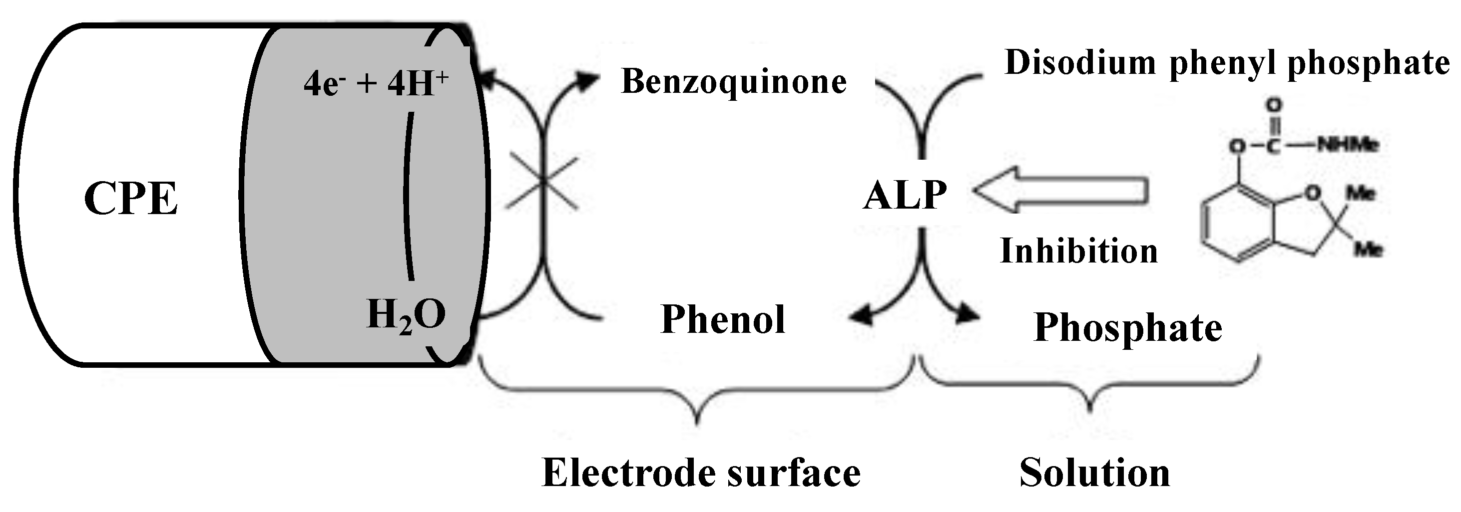
2.4. Alkaline Phosphatase Inhibition
2.5. Other Enzymes Inhibition
2.6. Comparison of Different Types of Enzyme Inhibition-Based Biosensor
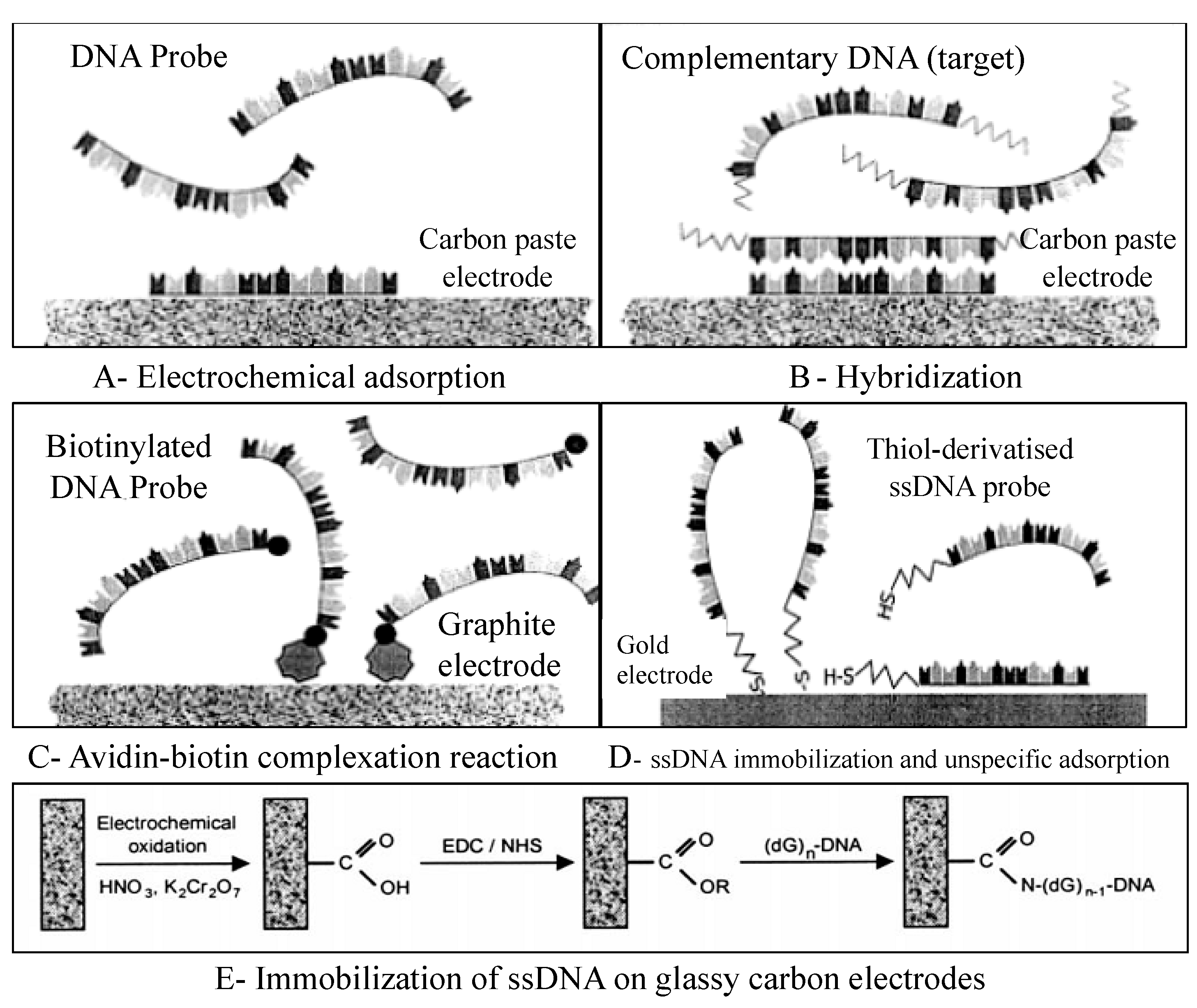
3. DNA-Based Biosensor
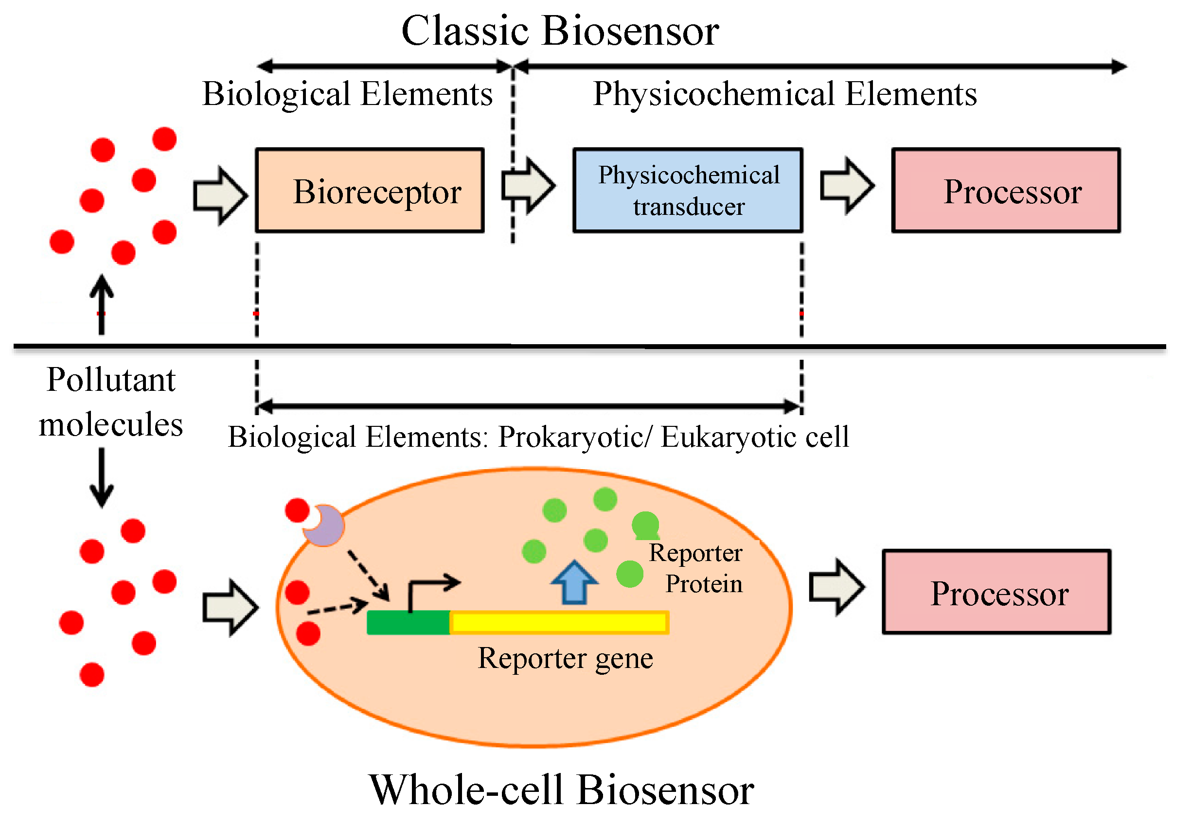
4. Whole-Cell Biosensor
4.1. Bacteria Based Whole-Cell Biosensor
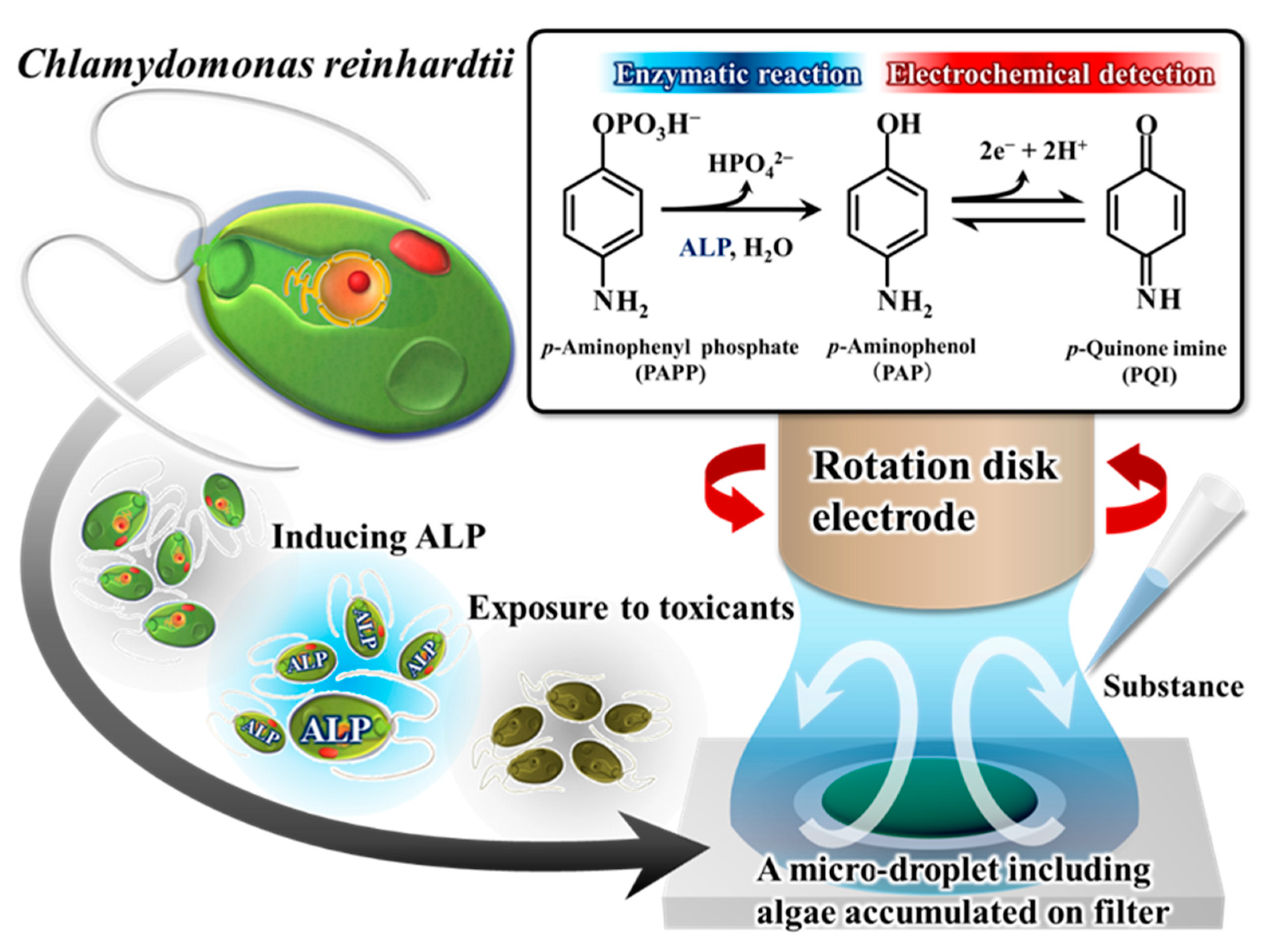
4.2. Algae-Based Whole-Cell Biosensor
4.3. Other Types of Whole-Cell Biosensors
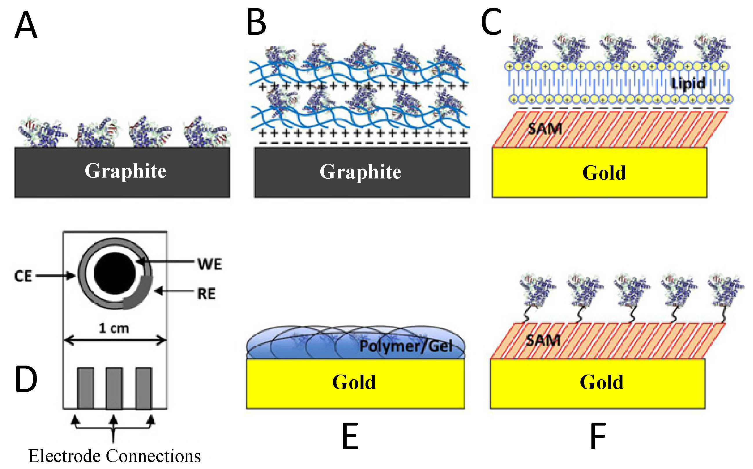
5. Biosensors Based on Cytochrome P450
5.1. LbL Adsorption
5.2. Adsorption to Thin Films
5.3. Screen-Printed Electrodes
5.4. Encapsulation in Polymers or Gels
5.5. Covalent Attachment to Self-Assembled Monolayers on Gold
5.6. Recent Advancements in CYP Biosensing
6. Conclusions
Author Contributions
Funding
Data Availability Statement
Acknowledgments
Conflicts of Interest
References
- Royal Commission on Environmental Pollution. Chemicals in Products: Safeguarding the Environment and Human Health. 24th Report; The Stationery Office: Norwich, UK, 2003. [Google Scholar]
- Oh, S.E.; Hassan, S.H.A.; Van Ginkel, S.W. A novel biosensor for detecting toxicity in water using sulfur-oxidizing bacteria. Sens. Actuators B Chem. 2011, 154, 17–21. [Google Scholar] [CrossRef]
- Eltzov, E.; Yehuda, A.; Marks, R.S. Creation of a new portable biosensor for water toxicity determination. Sens. Actuators B Chem. 2015, 221, 1044–1054. [Google Scholar] [CrossRef]
- Liu, C.; Yong, D.; Yu, D.; Dong, S. Cell-based biosensor for measurement of phenol and nitrophenols toxicity. Talanta 2011, 84, 766–770. [Google Scholar] [CrossRef]
- Wang, X.; Wang, X.; Zhang, J.; Bu, Y.; Yan, X.; Chen, J.; Huang, J.; Zhao, J. Direct toxicity assessment of copper (II) ions to activated sludge process using a p-benzoquinone-mediated amperometric biosensor. Sens. Actuators B Chem. 2015, 208, 554–558. [Google Scholar] [CrossRef]
- Volpi Ghirardini, A.V.; Girardini, M.; Marchetto, D.; Pantani, C. Microtox® solid phase test: Effect of diluent used in toxicity test. Ecotoxicol. Environ. Saf. 2009, 72, 851–861. [Google Scholar] [CrossRef] [PubMed]
- Tizzard, A.; Webber, J.; Gooneratne, R.; John, R.; Hay, J.; Pasco, N. MICREDOX: Application for rapid biotoxicity assessment. Anal. Chim. Acta 2004, 522, 197–205. [Google Scholar] [CrossRef]
- Wang, X.; Liu, M.; Wang, X.; Wu, Z.; Yang, L.; Xia, S.; Chen, L.; Zhao, J. p-benzoquinone-mediated amperometric biosensor developed with Psychrobacter sp. for toxicity testing of heavy metals. Biosens. Bioelectron. 2013, 41, 557–562. [Google Scholar] [CrossRef] [PubMed]
- Davidov, Y.; Rozen, R.; Smulski, D.R.; Van Dyk, T.K.; Vollmer, A.C.; Elsemore, D.A.; LaRossa, R.A.; Belkin, S. Improved bacterial SOS promoter∷ lux fusions for genotoxicity detection. Mutat. Res. Genet. Toxicol. Environ. Mutagen. 2000, 466, 97–107. [Google Scholar] [CrossRef]
- Rodriguez-Mozaz, S.; de Alda, M.J.L.; Marco, M.P.; Barceló, D. Biosensors for environmental monitoring A global perspective. Talanta 2005, 65, 291–297. [Google Scholar] [CrossRef] [PubMed]
- Mattias, K.J.M.; Turner, A.P.F. Biosensors in air monitoring. J. Environ. Monit. 1999, 1, 293–298. [Google Scholar] [CrossRef] [PubMed]
- Soelberg, S.D.; Chinowsky, T.; Geiss, G.; Spinelli, C.B.; Stevens, R.; Near, S.; Kauffman, P.; Yee, S.; Furlong, C.E. A portable surface plasmon resonance sensor system for real-time monitoring of small to large analytes. J. Ind. Microbiol. Biotechnol. 2005, 32, 669–674. [Google Scholar] [CrossRef]
- Badihi-Mossberg, M.; Buchner, V.; Rishpon, J. Electrochemical Biosensors for Pollutants in the Environment. Electroanalysis 2007, 19, 2015–2028. [Google Scholar] [CrossRef]
- Power, A.C.; Morrin, A. Electroanalytical sensor technology. In Electrochemistry; Khalid, M.A.A., Ed.; IntechOpen: London, UK, 2013; pp. 141–177. [Google Scholar]
- Wang, J. Analytical Electrochemistry, 3rd ed.; John Wiley & Sons, Inc.: Hoboken, NJ, USA, 2006. [Google Scholar]
- Shao, Y.; Wang, J.; Wu, H.; Liu, J.; Aksay, I.; Lin, Y. Graphene based electrochemical sensors and biosensors: A review. Electroanalysis 2010, 22, 1027–1036. [Google Scholar] [CrossRef]
- Zhao, Z.; Lei, W.; Zhang, X.; Wang, B.; Jiang, H. ZnO-based amperometric enzyme biosensors. Sensors 2010, 10, 1216–1231. [Google Scholar] [CrossRef]
- Siqueira, J.R.; Caseli, L.; Crespilho, F.N.; Zucolotto, V.; Oliveira, O.N. Immobilization of biomolecules on nanostructured films for biosensing. Biosens. Bioelectron. 2010, 25, 1254–1263. [Google Scholar] [CrossRef] [PubMed]
- Harper, A.; Anderson, M.R. Electrochemical glucose sensors—Developments using electrostatic assembly and carbon nanotubes for biosensor construction. Sensors 2010, 10, 8248–8274. [Google Scholar] [CrossRef]
- Singh, R.P.; Oh, B.K.; Choi, J.W. Application of peptide nucleic acid towards development of nanobiosensor arrays. Bioelectrochemistry 2010, 79, 153–161. [Google Scholar] [CrossRef]
- Park, B.W.; Yoon, D.Y.; Kim, D.S. Recent progress in bio-sensing techniques with encapsulated enzymes. Biosens. Bioelectron. 2010, 26, 1–10. [Google Scholar] [CrossRef]
- Su, L.; Jia, W.; Hou, C.; Lei, Y. Microbial biosensors: A review. Biosens. Bioelectron. 2011, 26, 1788–1799. [Google Scholar] [CrossRef]
- Grieshaber, D.; MacKenzie, R.; Vörös, J.; Reimhult, E. Electrochemical biosensors–Sensor principles and architectures. Sensors 2008, 8, 1400–1458. [Google Scholar] [CrossRef]
- Amine, A.; Mohammadi, H.; Bourais, I.; Palleschi, G. Enzyme inhibition-based biosensors for food safety and environmental monitoring. Biosens. Bioelectron. 2006, 21, 1405–1423. [Google Scholar] [CrossRef]
- Bachan Upadhyay, L.S.; Verma, N. Enzyme inhibition based biosensors: A review. Anal. Lett. 2013, 46, 225–241. [Google Scholar] [CrossRef]
- Singh, M.; Verma, N.; Garg, A.K.; Redhu, N. Urea biosensors. Sens. Actuators B Chem. 2008, 134, 345–351. [Google Scholar] [CrossRef]
- Zejli, H.; Hidalgo-Hidalgo de Cisneros, J.L.; Naranjo-Rodriguez, I.; Liu, B.; Temsamani, K.R.; Marty, J.L. Alumina sol-gel/sonogel-carbon electrode based on acetylcholinesterase for detection of organophosphorus pesticides. Talanta 2008, 77, 217–221. [Google Scholar] [CrossRef]
- Du, D.; Ding, J.; Cai, J.; Zhang, J.; Liu, L. In situ electrodeposited nanoparticles for facilitating electron transfer across self-assembled monolayers in biosensor design. Talanta 2008, 74, 1337–1343. [Google Scholar] [CrossRef]
- Anitha, K.; Mohan, S.V.; Reddy, S. Development of acetylcholinesterase silica sol–gel immobilized biosensor—An application towards oxydemeton methyl detection. Biosens. Bioelectron. 2004, 20, 848–856. [Google Scholar] [CrossRef] [PubMed]
- Andreescu, S.; Bucur, B.; Marty, J.L. Affinity immobilization of tagged enzymes. In Immobilization of Enzymes and Cells, 2nd ed.; Guisan, J.M., Ed.; Humana Press: Totowa, NJ, USA, 2006; pp. 97–106. [Google Scholar]
- Bucur, B.; Danet, A.F.; Marty, J.L. Cholinesterase immobilisation on the surface of screen-printed electrodes based on concanavalin A affinity. Anal. Chim. Acta 2005, 530, 1–6. [Google Scholar] [CrossRef]
- Ronkainen, N.J.; Halsall, H.B.; Heineman, W.R. Electrochemical biosensors. Chem. Soc. Rev. 2010, 39, 1747–1763. [Google Scholar] [CrossRef]
- Guerrieri, A.; Monaci, L.; Quinto, M.; Palmisano, F. A disposable amperometric biosensor for rapid screening of anticholinesterase activity in soil extracts. Analyst 2002, 127, 5–7. [Google Scholar] [CrossRef]
- Ivanov, A.; Evtugyn, G.; Budnikov, H.; Ricci, F.; Moscone, D.; Palleschi, G. Cholinesterase sensors based on screen-printed electrodes for detection of organophosphorus and carbamic pesticides. Anal. Bioanal. Chem. 2003, 377, 624–631. [Google Scholar] [CrossRef]
- Ghica, M.E.; Brett, C.M.A. Glucose oxidase inhibition in poly(neutral red) mediated enzyme biosensors for heavy metal determination. Microchim. Acta 2008, 163, 185–193. [Google Scholar] [CrossRef]
- Guascito, M.R.; Malitesta, C.; Mazzotta, E.; Turco, A. Inhibitive determination of metal ions by an amperometric glucose oxidase biosensor. Sens. Actuators B Chem. 2008, 131, 394–402. [Google Scholar] [CrossRef]
- Liu, J.X.; Xu, X.; Tang, L.; Zeng, G. Determination of trace mercury in compost extract by inhibition based glucose oxidase biosensor. Tran. Nonferrous Metal Soc. 2009, 19, 235–240. [Google Scholar] [CrossRef]
- Samphao, A.; Rerkchai, H.; Jitcharoen, J.; Nacapricha, D.; Kalcher, K. Indirect determination of mercury by inhibition of glucose oxidase immobilized on a carbon paste electrode. Int. J. Electrochem. Sci. 2012, 7, 1001–1010. [Google Scholar]
- Balasubramanian, A.; Ponnuraj, K. Crystal structure of the first plant urease from jack bean: 83 years of journey from its first crystal to molecular structure. J. Mol. Biol. 2010, 400, 274–283. [Google Scholar] [CrossRef]
- Yang, Y.; Wang, Z.; Yang, M.; Guo, M.; Wu, Z.; Shen, G.; Yu, R. Inhibitive determination of mercury ion using a renewable urea biosensor based on self-assembled gold nanoparticles. Sens. Actuators B Chem. 2006, 114, 1–8. [Google Scholar] [CrossRef]
- Do, J.S.; Lin, K.H.; Ohara, R. Preparation of urease/nano-structured polyaniline-Nafion®/Au/Al2O3 electrode for inhibitive detection of mercury ion. J. Taiwan Inst. Chem. Eng. 2011, 4, 662–668. [Google Scholar] [CrossRef]
- Gani, A.A.; Ashari, M.R.; Kuswandi, B. An optical fiber biosensor for heavy metal ions based on a modified single sol–gel film of urease and chlorophenol red in flow system. Sens. Lett. 2010, 8, 320–327. [Google Scholar] [CrossRef]
- Ilangovan, R.; Daniel, D.; Krastanov, A.; Zachariah, C.; Elizabeth, R. Enzyme based biosensor for heavy metal ions determination. Biotechnol. Biotechnol. Equip. 2006, 20, 184–189. [Google Scholar] [CrossRef]
- Arduini, F.; Ricci, F.; Tuta, C.S.; Moscone, D.; Amine, A.; Palleschi, G. Detection of carbamic and organophosphorous pesticides in water samples using a cholinesterase biosensor based on Prussian Blue-modified screen-printed electrode. Anal. Chim. Acta 2006, 580, 155–162. [Google Scholar] [CrossRef]
- Liu, G.; Lin, Y. Biosensor based on self-assembling acetylcholinesterase on carbon nanotubes for flow injection/amperometric detection of organophosphate pesticides and nerve agents. Anal. Chem. 2006, 78, 835–843. [Google Scholar] [CrossRef] [PubMed]
- Pohanka, M.; Jun, D.; Kuca, K. Amperometric biosensors for real time assays of organophosphates. Sensors 2008, 8, 5303–5312. [Google Scholar] [CrossRef]
- Sinha, R.; Ganesana, M.; Andreescu, S.; Stanciu, L. AChE biosensor based on zinc oxide sol-gel for the detection of pesticides. Anal. Chim. Acta 2010, 661, 195–199. [Google Scholar] [CrossRef] [PubMed]
- Alvarado-Gámez, A.L.; Alonso-Lomillo, M.A.; Domínguez-Renedo, O.; Arcos-Martínez, M.J. A disposable alkaline phosphatase-based biosensor for vanadium chronoamperometric determination. Sensors 2014, 14, 3756–3767. [Google Scholar] [CrossRef] [PubMed]
- Samphao, A.; Suebsanoh, P.; Wongsa, Y.; Pekec, B.; Jitchareon, J.; Kalcher, K. Alkaline phosphatase inhibition based amperometric biosensor for the detection of carbofuran. Int. J. Electrochem. Sci. 2013, 8, 3254–3264. [Google Scholar]
- Tekaya, N.; Saiapina, O.; Ben Ouada, H.; Lagarde, F.; Ben Ouada, H.; Jaffrezic-Renault, N. Ultrasensitive conductometric detection of heavy metals based on inhibition of alkaline phosphatase activity from Arthrospira platensis. Bioelectrochemistry 2013, 90, 24–29. [Google Scholar] [CrossRef]
- Islam, M.S.; Sazawa, K.; Sugawara, K.; Kuramitz, H. Micro-droplet hydrodynamic voltammetry for the determination of microcystin-LR based on protein phosphatase. J. Water Environ. Technol. 2019, 17, 18–26. [Google Scholar] [CrossRef]
- Abu-Salah, K.M.; Alrokyan, S.A.; Khan, M.N.; Ansari, A.A. Nanomaterials as analytical tools for genosensors. Sensors 2010, 10, 963–993. [Google Scholar] [CrossRef]
- Wang, J. Real time electrochemical monitoring toward green analytical chemistry. Acc. Chem. Res. 2002, 35, 811–816. [Google Scholar] [CrossRef]
- Mehrvar, M.; Abdi, M. Recent developments, characteristics, and potential applications of electrochemical biosensors. Anal. Sci. 2004, 20, 1113–1126. [Google Scholar] [CrossRef]
- Chiti, G.; Marrazza, G.; Mascini, M. Electrochemical DNA biosensor for environmental monitoring. Anal. Chim. Acta 2001, 427, 155–164. [Google Scholar] [CrossRef]
- Lucarelli, F.; Kicela, A.; Palchetti, I.; Marrazza, G.; Mascini, M. Electrochemical DNA biosensor for analysis of wastewater samples. Bioelectrochemistry 2002, 58, 113–118. [Google Scholar] [CrossRef]
- Lucarelli, F.; Palchetti, I.; Marrazza, G.; Mascini, M. Electrochemical DNA biosensor as screening tool for the detection of toxicant in water and wastewater samples. Talanta 2002, 56, 949–957. [Google Scholar] [CrossRef]
- Lucarelli, F.; Marrazza, G.; Turner, A.P.; Mascini, M. Carbon and gold electrodes as electrochemical transducers for DNA hybridization sensors. Biosens. Bioelectron. 2004, 19, 515–530. [Google Scholar] [CrossRef] [PubMed]
- Pividori, M.I.; Merkoçi, A.; Alegret, S. Electrochemical genosensor design: Immobilisation of oligonucleotides onto transducer surfaces and detection methods. Biosens. Bioelectron. 2000, 15, 291–303. [Google Scholar] [CrossRef]
- de-los-Santos-Alvarez, P.; Lobo-Castañón, M.J.; Miranda-Ordieres, A.J.; Tuñón-Blanco, P. Current strategies for electrochemical detection of DNA with solid electrodes. Anal. Bioanal. Chem. 2004, 378, 104–118. [Google Scholar] [CrossRef] [PubMed]
- Sun, X.; He, P.; Liu, S.; Ye, J.; Fang, Y. Immobilization of single-stranded deoxyribonucleic acid on gold electrode with self-assembled aminoethanethiol monolayer for DNA electrochemical sensor applications. Talanta 1998, 47, 487–495. [Google Scholar] [CrossRef]
- Chaki, N.K.; Vijayamohanan, K. Self-assembled monolayers as a tunable platform for biosensor applications. Biosens. Bioelectron. 2002, 17, 1–12. [Google Scholar] [CrossRef]
- Kerman, K.; Ozkan, D.; Kara, P.; Meric, B.; Gooding, J.J.; Ozsoz, M. Voltammetric determination of DNA hybridization using methylene blue and self-assembled alkanethiol monolayer on gold electrodes. Anal. Chim. Acta 2002, 462, 39–47. [Google Scholar] [CrossRef]
- Smith, R.K.; Lewis, P.A.; Weiss, P.S. Patterning self-assembled monolayers. Prog. Surf. Sci. 2004, 75, 1–68. [Google Scholar] [CrossRef]
- Cagnin, S.; Caraballo, M.; Guiducci, C.; Martini, P.; Ross, M.; Santaana, M.; Danley, D.; West, T.; Lanfranchi, G. Overview of electrochemical DNA biosensors: New approaches to detect the expression of life. Sensors 2009, 9, 3122–3148. [Google Scholar] [CrossRef] [PubMed]
- Tencaliec, A.M.; Laschi, S.; Magearu, V.; Mascini, M. A comparison study between a disposable electrochemical DNA biosensor and a Vibrio fischeri-based luminescent sensor for the detection of toxicants in water samples. Talanta 2006, 69, 365–369. [Google Scholar] [CrossRef] [PubMed]
- Babkina, S.S.; Ulakhovich, N.A. Complexing of heavy metals with DNA and new bioaffinity method of their determination based on amperometric DNA based biosensor. Anal. Chem. 2005, 77, 5678–5685. [Google Scholar] [CrossRef]
- Malhotra, B.D.; Chaubey, A.; Singh, S.P. Prospects of conducting polymers in biosensors. Anal. Chim. Acta 2006, 578, 59–74. [Google Scholar] [CrossRef]
- Im, Y.; Vasquez, R.P.; Lee, C.; Myung, N.; Penner, R.; Yun, M. Single metal and conducting polymer nanowire sensors for chemical and DNA detections. J. Phys. Conf. Ser. 2006, 38, 61–64. [Google Scholar] [CrossRef]
- Park, S.J.; Taton, T.A.; Mirkin, C.A. Array-based electrical detection of DNA using nanoparticle probes. Science 2002, 295, 1503–1506. [Google Scholar] [CrossRef] [PubMed]
- Wang, J.; Polsky, R.; Xu, D. Silver-enhanced colloidal gold electrochemical stripping detection of DNA hybridization. Langmuir 2001, 17, 5739–5741. [Google Scholar] [CrossRef]
- Wang, J.; Xu, D.; Kawde, A.N.; Polsky, R. Metal nanoparticle-based electrochemical stripping potentiometric detection of DNA hybridization. Anal. Chem. 2001, 73, 5576–5581. [Google Scholar] [CrossRef]
- Wang, J. Nanomaterial-based electrochemical biosensors. Analyst 2005, 130, 421–426. [Google Scholar] [CrossRef]
- Lermo, A.; Campoy, S.; Barbé, J.; Hernández, S.; Alegret, S.; Pividori, M.I. In situ DNA amplification with magnetic primers for the electrochemical detection of food pathogens. Biosens. Bioelectron. 2007, 22, 2010–2017. [Google Scholar] [CrossRef]
- Rivas, G.A.; Rubianes, M.D.; Rodríguez, M.C.; Ferreyra, N.F.; Luque, G.L.; Pedano, M.L.; Miscoria, S.A.; Parrado, C. Carbon nanotubes for electrochemical biosensing. Talanta 2007, 74, 291–307. [Google Scholar] [CrossRef] [PubMed]
- Zhang, Y.; Zhang, K.; Ma, H. Electrochemical DNA biosensor based on silver nanoparticles/poly(3-(3-pyridyl) acrylic acid)/carbon nanotubes modified electrode. Anal. Biochem. 2009, 387, 13–19. [Google Scholar] [CrossRef]
- Galandova, J.; Ziyatdinova, G.; Labuda, J. Disposable electrochemical biosensor with multiwalled carbon nanotubes-chitosan composite layer for the detection of deep DNA damage. Anal. Sci. 2008, 24, 711–716. [Google Scholar] [CrossRef]
- Belkin, S. Microbial whole-cell sensing systems of environmental pollutants. Curr. Opin. Microbiol. 2003, 6, 206–212. [Google Scholar] [CrossRef] [PubMed]
- Van der Meer, J.R.; Belkin, S. Where microbiology meets microengineering: Design and applications of reporter bacteria. Nature Reviews. Microbiology 2010, 8, 511–522. [Google Scholar] [CrossRef] [PubMed]
- Gutiérrez, J.C.; Amaro, F.; Martín-González, A. Heavy metal whole-cell biosensors using eukaryotic microorganisms: An updated critical review. Front. Microbiol. 2015, 6, 48. [Google Scholar] [CrossRef]
- Yagi, K. Applications of whole-cell bacterial sensors in biotechnology and environmental science. Appl. Microbiol. Biotechnol. 2007, 73, 1251–1258. [Google Scholar] [CrossRef]
- Wong, L.S.; Lee, Y.H.; Surif, S. Whole-cell biosensor using Anabaena torulosa with optical transduction for environmental toxicity evaluation. J. Sens. 2013, 2013, 567272. [Google Scholar] [CrossRef]
- Singh, J.; Mittal, S.K. Chlorella sp. based biosensor for selective determination of mercury in presence of silver ions. Sens. Actuators B Chem. 2012, 165, 48–52. [Google Scholar] [CrossRef]
- Matsuura, H.; Yamamoto, Y.; Muraoka, M.; Akaishi, K.; Hori, Y.; Uemura, K.; Tsuji, N.; Harada, K.; Hirata, K.; Bamba, T.; et al. Development of surface engineered yeast cells displaying phytochelatin synthase and their application to cadmium biosensors by the combined use of pyrene excimer fluorescence. Biotechnol. Prog. 2013, 29, 1197–1202. [Google Scholar] [CrossRef]
- Choe, S.I.; Gravelat, F.N.; Al Abdallah, Q.; Lee, M.J.; Gibbs, B.F.; Sheppard, D.C. Role of Aspergillus niger acrA in arsenic resistance and its use as the basis for an arsenicbiosensor. Appl. Environ. Microbiol. 2012, 78, 3855–3863. [Google Scholar] [CrossRef] [PubMed]
- Wong, L.S.; Choong, C.W. Rapid detection of heavy metals with the response of carotenoids in Daucus carota. Int. J. Environ. Sci. Dev. 2014, 5, 270–273. [Google Scholar] [CrossRef]
- Teo, S.C.; Wong, L.S. Whole-cell-based biosensors for environmental heavy metals detection. Annu. Res. Rev. Biol. 2014, 4, 2663–2674. [Google Scholar] [CrossRef]
- Liu, C.; Sun, T.; Xu, X.; Dong, S. Direct toxicity assessment of toxic chemicals with electrochemical method. Anal. Chim. Acta 2009, 641, 59–63. [Google Scholar] [CrossRef]
- Mcquillan, J.S.; Shaw, A.M. Whole-cell Escherichia coli-based bio-sensor assay for dual zinc oxide nanoparticle toxicity mechanisms. Biosens. Bioelectron. 2014, 51, 274–279. [Google Scholar] [CrossRef]
- Catterall, K.; Robertson, D.; Hudson, S.; Teasdale, P.R.; Welsh, D.T.; John, R. A sensitive, rapid ferricyanide-mediated toxicity bioassay developed using Escherichia coli. Talanta 2010, 82, 751–757. [Google Scholar] [CrossRef] [PubMed]
- Yong, D.; Liu, C.; Yu, D.; Dong, S. A sensitive, rapid and inexpensive way to assay pesticide toxicity based on electrochemical biosensor. Talanta 2011, 84, 7–12. [Google Scholar] [CrossRef]
- Pokhrel, L.R.; Silva, T.; Dubey, B.; El Badawy, A.M.; Tolaymat, T.M.; Scheuerman, P.R. Rapid screening of aquatic toxicity of several metal-based nanoparticles using the MetPLATE™ bioassay. Sci. Total Environ. 2012, 426, 414–422. [Google Scholar] [CrossRef] [PubMed]
- Yong, D.; Liu, L.; Yu, D.; Dong, S. Development of a simple method for biotoxicity measurement using ultramicroelectrode array under non-deaerated condition. Anal. Chim. Acta 2011, 701, 164–168. [Google Scholar] [CrossRef] [PubMed]
- Wang, H.; Wang, X.J.; Zhao, J.F.; Chen, L. Toxicity assessment of heavy metals and organic compounds using CellSense biosensor with E. coli. Chin. Chem. Lett. 2008, 19, 211–214. [Google Scholar] [CrossRef]
- Ravikumar, S.; Ganesh, I.; Yoo, I.-k.; Hong, S.H. Construction of a bacterial biosensor for zinc and copper and its application to the development of multifunctional heavy metal adsorption bacteria. Process Biochem. 2012, 47, 758–765. [Google Scholar] [CrossRef]
- Verma, N.; Singh, M. A Bacillus sphaericus based biosensor for monitoring nickel ions in industrial effluents and foods. J. Autom. Methods Manag. Chem. 2006, 2006, 83427. [Google Scholar] [CrossRef]
- Sochor, J.; Zitka, O.; Hynek, D.; Jilkova, E.; Krejcova, L.; Trnkova, L.; Adam, V.; Hubalek, J.; Kynicky, J.; Vrba, R.; et al. Bio-sensing of cadmium (II) ions using Staphylococcus aureus. Sensors 2011, 11, 10638–10663. [Google Scholar] [CrossRef] [PubMed]
- Ferro, Y.; Perullini, M.; Jobbagy, M.; Bilmes, S.A.; Durrieu, C. Development of a biosensor for environmental monitoring based on microalgae immobilized in silica hydrogels. Sensors 2012, 12, 16879–16891. [Google Scholar] [CrossRef] [PubMed]
- Pham, T.P.T.; Cho, C.-W.; Yun, Y.-S. Algal biosensor-based measurement system for rapid toxicity detection. In Advances in Measurement; Sharma, M.K., Ed.; IntechOpen: London, UK, 2010; pp. 273–288. [Google Scholar]
- Chouteau, C.; Dzyadevych, S.; Durrieu, C.; Chovelon, J.M. A bi-enzymatic whole-cell conductometric biosensor for heavy metal ions and pesticides detection in water samples. Biosens. Bioelectron. 2005, 21, 273–281. [Google Scholar] [CrossRef]
- Guedri, H.; Durrieu, C. A self-assembled monolayers based conductometric algal whole-cell biosensor for water monitoring. Microchim. Acta 2008, 163, 179–184. [Google Scholar] [CrossRef]
- Chouteau, C.; Dzyadevych, S.; Chovelon, J.M.; Durrieu, C. Development of novel conductometric biosensors based on immobilized whole-cell Chlorella vulgaris microalgae. Biosens. Bioelectron. 2004, 19, 1089–1096. [Google Scholar] [CrossRef]
- Berezhetskyy, A.L.; Durrieu, C.; Nguyen-Ngoc, H.; Chovelon, J.-M.; Dzyadevych, S.V.; Tran-Minh, C. Conductometric biosensor based on whole-cell microalgae for assessment of heavy metals in wastewater. Biopolym. Cell 2007, 23, 511–518. [Google Scholar] [CrossRef]
- Shitanda, I.; Takada, K.; Sakai, Y.; Tatsuma, T. Compact amperometric algal biosensors for the evaluation of water toxicity. Anal. Chim. Acta 2005, 530, 191–197. [Google Scholar] [CrossRef]
- Shitanda, I.; Takamatsu, S.; Watanabe, K.; Itagaki, M. Amperometric screen-printed algal biosensor with flow injection analysis system for detection of environmental toxic compounds. Electrochim. Acta 2009, 54, 4933–4936. [Google Scholar] [CrossRef]
- Shitanda, I.; Takada, K.; Sakai, Y.; Tatsuma, T. Amperometric biosensing systems based on motility and gravitaxis of flagellate algae for aquatic risk assessment. Anal. Chem. 2005, 77, 6715–6718. [Google Scholar] [CrossRef]
- Islam, M.S.; Sazawa, K.; Hata, N.; Sugawara, K.; Kuramitz, H. Determination of heavy metal toxicity by using a micro-droplet hydrodynamic voltammetry for microalgal bioassay based on alkaline phosphatase. Chemosphere 2017, 188, 337–344. [Google Scholar] [CrossRef] [PubMed]
- Amaro, F.; Turkewitz, A.P.; Martín-González, A.; Gutiérrez, J.C. Whole-cell biosensors for detection of heavy metal ions in environmental samples based on metallothionein promoters from Tetrahymena thermophila. Microb. Biotechnol. 2011, 4, 513–522. [Google Scholar] [CrossRef] [PubMed]
- Shumyantseva, V.V.; Bulko, T.V.; Archakov, A.I. Electrochemical reduction of cytochrome P450 as an approach to the construction of biosensors and bioreactors. J. Inorg. Biochem. 2005, 99, 1051–1063. [Google Scholar] [CrossRef] [PubMed]
- Lamb, D.C.; Lei, L.; Warrilow, A.G.; Lepesheva, G.I.; Mullins, J.G.; Waterman, M.R.; Kelly, S.L. The first virally encoded cytochrome P450. J. Virol. 2009, 83, 8266–8269. [Google Scholar] [CrossRef]
- Snyder, M.J. Aquatic P450 Species. In The Ubiquitous Roles of Cytochrome P450 Proteins, Volume 3: Metal Ions in Life Sciences; Sigel, A., Sigel, H., Sigel, R.K.O., Eds.; John Wiley & Sons, Inc.: Hoboken, NJ, USA, 2007; pp. 97–126. [Google Scholar]
- Danielson, P.B. The cytochrome P450 superfamily: Biochemistry, evolution and drug metabolism in humans. Curr. Drug Metab. 2002, 3, 561–597. [Google Scholar] [CrossRef] [PubMed]
- Guengerich, F.P. Cytochrome P450 and chemical toxicology. Chem. Res. Toxicol. 2008, 21, 70–83. [Google Scholar] [CrossRef]
- Bistolas, N.; Wollenberger, U.; Jung, C.; Scheller, F.W. Cytochrome P450 biosensors—A review. Biosens. Bioelectron. 2005, 20, 2408–2423. [Google Scholar] [CrossRef]
- Fantuzzi, A.; Fairhead, M.; Gilardi, G. Direct electrochemistry of immobilized human cytochrome P450 2E1. J. Am. Chem. Soc. 2004, 126, 5040–5041. [Google Scholar] [CrossRef]
- Mhaske, S.D.; Ray, M.; Mazumdar, S. Covalent linkage of CYP101 with the electrode enhances the electrocatalytic activity of the enzyme: Vectorial electron transport from the electrode. Inorg. Chim. Acta 2010, 363, 2804–2811. [Google Scholar] [CrossRef]
- Joseph, S.; Rusling, J.F.; Lvov, Y.M.; Friedberg, T.; Fuhr, U. An amperometric biosensor with human CYP3A4 as a novel drug screening tool. Biochem. Pharmacol. 2003, 65, 1817–1826. [Google Scholar] [CrossRef]
- Sultana, N.; Schenkman, J.B.; Rusling, J.F. Protein film electrochemistry of microsomes genetically enriched in human cytochrome p450 monooxygenases. J. Am. Chem. Soc. 2005, 127, 13460–13461. [Google Scholar] [CrossRef]
- Krishnan, S.; Wasalathanthri, D.; Zhao, L.; Schenkman, J.B.; Rusling, J.F. Efficient bioelectronic actuation of the natural catalytic pathway of human metabolic cytochrome P450s. J. Am. Chem. Soc. 2011, 133, 1459–1465. [Google Scholar] [CrossRef]
- Dodhia, V.R.; Sassone, C.; Fantuzzi, A.; Di Nardo, G.D.; Sadeghi, S.J.; Gilardi, G. Modulating the coupling efficiency of human cytochrome P450 CYP3A4 at electrode surfaces through protein engineering. Electrochem. Commun. 2008, 10, 1744–1747. [Google Scholar] [CrossRef]
- Rhieu, S.Y.; Ludwig, D.R.; Siu, V.S.; Palmore, G.T.R. Direct electrochemistry of cytochrome P450 27B1 in surfactant films. Electrochem. Commun. 2009, 11, 1857–1860. [Google Scholar] [CrossRef]
- Rudakov, Y.O.; Shumyantseva, V.V.; Bulko, T.V.; Suprun, E.V.; Kuznetsova, G.P.; Samenkova, N.F.; Archakov, A.I. Stoichiometry of electrocatalytic cycle of cytochrome P450 2B4. J. Inorg. Biochem. 2008, 102, 2020–2025. [Google Scholar] [CrossRef]
- Shumyantseva, V.V.; Bulko, T.V.; Suprun, E.V.; Chalenko, Y.M.; Vagin, M.Y.; Rudakov, Y.O.; Shatskaya, M.A.; Archakov, A.I. Electrochemical investigations of cytochrome P450. Biochim. Biophys. Acta 2011, 1, 94–101. [Google Scholar] [CrossRef]
- Liu, S.; Peng, L.; Yang, X.; Wu, Y.; He, L. Electrochemistry of cytochrome P450 enzyme on nanoparticle-containing membrane-coated electrode and its applications for drug sensing. Anal. Biochem. 2008, 375, 209–216. [Google Scholar] [CrossRef]
- Alonso-Lomillo, M.A.; Gonzalo-Ruiz, J.; Domínguez-Renedo, O.; Muñoz, F.J.; Arcos-Martínez, M.J. CYP450 biosensors based on gold chips for antiepileptic drugs determination. Biosens. Bioelectron. 2008, 23, 1733–1737. [Google Scholar] [CrossRef]
- Holtmann, D.; Mangold, K.M.; Schrader, J. Entrapment of cytochrome P450 BM-3 in polypyrrole for electrochemically-driven biocatalysis. Biotechnol. Lett. 2009, 31, 765–770. [Google Scholar] [CrossRef]
- Yang, M.; Kabulski, J.L.; Wollenberg, L.; Chen, X.; Subramanian, M.; Tracy, T.S.; Lederman, D.; Gannett, P.M.; Wu, N. Electrocatalytic drug metabolism by CYP2C9 bonded to a self-assembled monolayer-modified electrode. Drug Metab. Dispos. 2009, 37, 892–899. [Google Scholar] [CrossRef]
- Fantuzzi, A.; Capria, E.; Mak, L.H.; Dodhia, V.R.; Sadeghi, S.J.; Collins, S.; Somers, G.; Huq, E.; Gilardi, G. An electrochemical microfluidic platform for human P450 drug metabolism profiling. Anal. Chem. 2010, 82, 10222–10227. [Google Scholar] [CrossRef]
- Mak, L.H.; Sadeghi, S.J.; Fantuzzi, A.; Gilardi, G. Control of human cytochrome P450 2E1 electrocatalytic response as a result of unique orientation on gold electrodes. Anal. Chem. 2010, 82, 5357–5362. [Google Scholar] [CrossRef] [PubMed]
- Schneider, E.; Clark, D.S. Cytochrome P450 (CYP) enzymes and the development of CYP biosensors. Biosens. Bioelectron. 2013, 39, 1–13. [Google Scholar] [CrossRef] [PubMed]
- Luo, G.; Cunningham, M.; Kim, S.; Burn, T.; Lin, J.; Sinz, M.; Hamilton, G.; Rizzo, C.; Jolley, S.; Gilbert, D.; et al. CYP3A4 induction by drugs: Correlation between a pregnane X receptor reporter gene assay and CYP3A4 expression in human hepatocytes. Drug Metab. Dispos. 2002, 30, 795–804. [Google Scholar] [CrossRef]
- Mie, Y.; Suzuki, M.; Komatsu, Y. Electrochemically driven drug metabolism by membranes containing human cytochrome P450. J. Am. Chem. Soc. 2009, 131, 6646–6647. [Google Scholar] [CrossRef]
- Xue, Q.; Kato, D.; Kamata, T.; Guo, Q.; You, T.; Niwa, O. Human cytochrome P450 3A4 and a carbon nanofiber modified film electrode as a platform for the simple evaluation of drug metabolism and inhibition reactions. Analyst 2013, 138, 6463–6468. [Google Scholar] [CrossRef] [PubMed]
- Yoshioka, K.; Kato, D.; Kamata, T.; Niwa, O. Cytochrome P450 Modified Polycrystalline indium tin oxide Film asa Drug Metabolizing Electrochemical Biosensor with a Simple Configuration. Anal. Chem. 2013, 85, 9996–9999. [Google Scholar] [CrossRef]
- Shumyantseva, V.V.; Ivanov, Y.D.; Bistolas, N.; Scheller, F.W.; Archakov, A.I.; Wollenberger, U. Direct electron transfer of cytochrome P450 2B4 at electrodes modified with nonionic detergent and colloidal clay nanoparticles. Anal. Chem. 2004, 76, 6046–6052. [Google Scholar] [CrossRef]
- Wu, Y.; Liu, X.; Zhang, L.; Wang, C. An amperometric biosensor based on rat cytochrome p450 1A1 for benzo[a]pyrene determination. Biosens. Bioelectron. 2011, 26, 2177–2182. [Google Scholar] [CrossRef]
- Antonini, M.; Ghisellini, P.; Pastorino, L.; Paternolli, C.; Nicolini, C. Preliminary electrochemical characterisation of cytochrome P4501A2-clozapine interaction. IEE Proc. Nanobiotechnol. 2003, 150, 31–34. [Google Scholar] [CrossRef]
- Shumyantseva, V.V.; Bulko, T.V.; Rudakov, Y.O.; Kuznetsova, G.P.; Samenkova, N.F.; Lisitsa, A.V.; Karuzina, I.I.; Archakov, A.I. Electrochemical properties of cytochroms P450 using nanostructured electrodes: Direct electron transfer and electro catalysis. J. Inorg. Biochem. 2007, 101, 859–865. [Google Scholar] [CrossRef]
- Sugihara, N.; Ogoma, Y.; Abe, K.; Kondo, Y.; Akaike, T. Immobilization of cytochrome P-450 and electrochemical control of its activity. Polym. Adv. Technol. 1998, 9, 307–313. [Google Scholar] [CrossRef]
- Dai, C.; Ding, Y.; Li, M.; Fei, J. Direct electrochemistry of cytochrome P450 in a biocompatible film composed of an epoxy polymer and acetylene black. Microchim. Acta 2012, 176, 397–404. [Google Scholar] [CrossRef]
- Carrara, S.; Cavallini, A.; Erokhin, V.; De Micheli, G. Multi-panel drugs detection in human serum for personalized therapy. Biosens. Bioelectron. 2011, 26, 3914–3919. [Google Scholar] [CrossRef]
- Mie, Y.; Ikegami, M.; Komatsu, Y. Gold sputtered electrode surfaces enhance direct electron transfer reactions of human cytochrome P450s. Electrochem. Commun. 2010, 12, 680–683. [Google Scholar] [CrossRef]
- Ikegami, M.; Mie, Y.; Hirano, Y.; Suzuki, M.; Komatsu, Y. Size-controlled fabrication of gold nanodome arrays and its application to enzyme electrodes. Colloids Surf. A Physicochem. Eng. Asp. 2011, 384, 388–392. [Google Scholar] [CrossRef]
- Ndangili, P.M.; Jijana, A.M.; Baker, P.G.L.; Iwuoha, E.I. 3-mercaptopropionic acid capped ZnSe quantum dot-cytochrome P450 3A4 enzyme biotransducer for 17β-estradiol. J. Electroanal. Chem. 2011, 653, 67–74. [Google Scholar] [CrossRef]
- Niwa, O.; Ohta, S.; Takahashi, S.; Zhang, Z.; Kamata, T.; Kato, D.; Shiba, S. Hybrid carbon film electrodes for electroanalysis. Anal. Sci. 2021, 37, 37–47. [Google Scholar] [CrossRef]
- Mostafa, I.M.; Meng, C.; Dong, Z.; Lou, B.; Xu, G. Potentiometric sensors for the determination of pharmaceutical drugs. Anal. Sci. 2022, 38, 23–37. [Google Scholar] [CrossRef]
- Liu, Y.; Xue, Q.; Chang, C.; Wang, R.; Liu, Z.; He, L. Recent progress regarding electrochemical sensors for the detection of typical pollutants in water environments. Anal. Sci. 2022, 38, 55–70. [Google Scholar] [CrossRef] [PubMed]
- Troudt, B.K.; Rousseau, C.R.; Dong, X.I.N.; Anderson, E.L.; Bühlmann, P. Recent progress in the development of improved reference electrodes for electrochemistry. Anal. Sci. 2022, 38, 71–83. [Google Scholar] [CrossRef]
- Si, Y.; Li, J.; Jhung, S.H.; Lee, H.J. Recent research trends in voltammetric sensing platforms for hormones and their applications to human serum analyses. Anal. Sci. 2022, 38, 11–21. [Google Scholar] [CrossRef] [PubMed]
- Zambrano-Intriago, L.A.; Amorim, C.G.; Rodríguez-Díaz, J.M.; Araújo, A.N.; Montenegro, M.C.B.S.M. Challenges in the design of electrochemical sensor for glyphosate-based on new materials and biological recognition. Sci. Total Environ. 2021, 793, 148496. [Google Scholar] [CrossRef]


| Enzyme Type | Inhibitors | Detection Limit | References |
|---|---|---|---|
| Glucose oxidase | Cd(II) | 1 mg/L | [35] |
| Cu(II) | 6 mg/L | [35] | |
| Pb(II) | 3 mg/L | [35] | |
| Zn(II) | 9 mg/L | [35] | |
| Hg(II) | 0.49 mg/L | [37] | |
| 0.5 mg/L | [38] | ||
| Urease | Hg(II) | 0.05 µmole/L | [40] |
| 0.01 mg/L | [41] | ||
| Cu(II) | 0.1–1 µmole/L | [43] | |
| Cd(II) | 0.1–10 µmole/L | [43] | |
| Pb(II) | 0.1–1 µmole/L | [43] | |
| Cholinesterase | Paraoxon | 2 µg/L | [44] |
| 0.4 × 10⁻¹² mole/L | [45] | ||
| 1.2 µg/L | [46] | ||
| 0.035–1.38 mg/L | [47] | ||
| Alkaline phosphatase | Vanadium | 0.39 ± 0.06 µM | [48] |
| Carbofuran | 10 µg/L | [49] | |
| Cd(II) | 10−20 M | [50] | |
| Hg(II) | 10−20 M | [50] |
| Modification Types | CYP Enzyme | Materials Used as Electrode | Modification of Electrode | Reference |
|---|---|---|---|---|
| Layer-by-Layer Adsorption | CYP3A4 | Gold | MPS SAM followed by PDDA | [117] |
| CYP1A2 and CYP3A4 | Pyrolytic Graphite | Multilayer films of PEI and PSS | [118] | |
| CYP1A2 and CYP2E1 | Basal-Plane Pyrolytic Graphite | Multilayer films of CYP and CPR /b5 | [119] | |
| Adsorption to Thin Films | CYP3A4 fusion protein | Glassy Carbon | PDDA | [120] |
| CYP27B1 | Edge-Plane Pyrolytic Graphite | DDAB | [121] | |
| CYP101 | Glassy Carbon | Covalent attachment to thin film of pyrene maleimide | [116] | |
| Screen-Printed Electrodes | CYP11A1 | Rhodium-graphite | Gold nanoparticles | [109] |
| CYP2B4 | Graphite | Mixture of gold nanoparticles and DDAB | [122] | |
| CYP2B4, 1A2, 3A4, 11A1, 51b1 | Graphite | Mixture of gold nanoparticles and DDAB | [123] | |
| Encapsulation in Polymers of Gels | CYP2B6 | Glassy Carbon | Mixture of chitosan and gold nanoparticles | [124] |
| CYP2B4 | Gold | Polypyrrole | [125] | |
| CYP102 (P450BM-3) Mutant | Platinum and Glassy Carbon | Polypyrrole | [126] | |
| Covalent Attachment to Self-Assembled Monolayers on Gold | CYP2C9 | Gold | Amine coupling via EDC/NHS to a mixed SAM of OT and MUA | [127] |
| CYP3A4 fusion protein | Gold | Amine coupling via EDC/NHS to a mixed SAM of 6HT and 7MHA | [128] | |
| CYP2E1 Single Cysteine Mutants | Gold | DTME SAM | [129] |
Disclaimer/Publisher’s Note: The statements, opinions and data contained in all publications are solely those of the individual author(s) and contributor(s) and not of MDPI and/or the editor(s). MDPI and/or the editor(s) disclaim responsibility for any injury to people or property resulting from any ideas, methods, instructions or products referred to in the content. |
© 2023 by the authors. Licensee MDPI, Basel, Switzerland. This article is an open access article distributed under the terms and conditions of the Creative Commons Attribution (CC BY) license (https://creativecommons.org/licenses/by/4.0/).
Share and Cite
Islam, M.S.; Sazawa, K.; Sugawara, K.; Kuramitz, H. Electrochemical Biosensor for Evaluation of Environmental Pollutants Toxicity. Environments 2023, 10, 63. https://doi.org/10.3390/environments10040063
Islam MS, Sazawa K, Sugawara K, Kuramitz H. Electrochemical Biosensor for Evaluation of Environmental Pollutants Toxicity. Environments. 2023; 10(4):63. https://doi.org/10.3390/environments10040063
Chicago/Turabian StyleIslam, Md. Saiful, Kazuto Sazawa, Kazuharu Sugawara, and Hideki Kuramitz. 2023. "Electrochemical Biosensor for Evaluation of Environmental Pollutants Toxicity" Environments 10, no. 4: 63. https://doi.org/10.3390/environments10040063
APA StyleIslam, M. S., Sazawa, K., Sugawara, K., & Kuramitz, H. (2023). Electrochemical Biosensor for Evaluation of Environmental Pollutants Toxicity. Environments, 10(4), 63. https://doi.org/10.3390/environments10040063