Article

Bacteria and Yeasts Isolated from the Environment in Biodegradation of PS and PVC Microplastics: Screening and Treatment Optimization

Faculty of Chemical Engineering and Technology, University of Zagreb, Trg Marka Marulića 19, 10000 Zagreb, Croatia
*
Author to whom correspondence should be addressed.
Environments 2023, 10(12), 207; https://doi.org/10.3390/environments10120207
Submission received: 15 October 2023 / Revised: 25 November 2023 / Accepted: 28 November 2023 / Published: 29 November 2023
(This article belongs to the Special Issue Advanced Technologies of Water and Wastewater Treatment)

Abstract

:
Biodegradation is the most environmentally friendly and, at the same time, economically acceptable approach to removing various pollutants from the environment. However, its efficiency in removing microplastics (MPs) from the environment is generally low. The successful biodegradation of MPs requires microorganisms capable of producing enzymes that degrade MP polymers into compounds that the microorganisms can use as a source of carbon and energy. Therefore, scientists are screening and characterizing microorganisms that can degrade MPs more efficiently. These microorganisms are often isolated from sites contaminated with MPs because the microorganisms living there are adapted to these pollutants and should be able to better degrade MPs. In this study, five bacterial strains and five yeast strains were isolated from various environmental samples including activated sludge, compost, river sediment, and biowaste. Among them, screening was performed for bacteria and yeasts with the highest potential for the biodegradation of polystyrene (PS) and polyvinyl chloride (PVC) MPs, and the bacterium Delftia acidovorans and the yeast Candida parapsilosis were identified as the best candidates. Optimization of biodegradation of the selected MPs by each of these two microorganisms was performed, focusing on the influence of cell density, agitation speed and pH of the medium. It was found that within the selected experimental ranges, high values of cell density, low agitation speed, and a slightly basic medium favored the biodegradation of PS and PVC MPs by Delftia acidovorans. In the case of Candida parapsilosis, favorable conditions also included high cell density followed by a slightly higher, but not maximum, agitation speed and a weakly acidic medium. Broad spectroscopic and imaging methods indicated that Delftia acidovorans and Candida parapsilosis better adapt to PVC MPs to use it as a carbon and energy source.

Graphical Abstract

1. Introduction

Today, it is impossible to imagine life without plastics: plastics have excellent application properties and are used in practically all areas of human life. Accordingly, the annual production of plastics is continuously increasing worldwide. In 2021, around 390.7 million tons were produced, which is 15.2 million tons more than in 2020 and 25.5 million tons more than in 2018 [1]. By type of plastic polymer, polyethylene (PE) with 36%, polypropylene (PP) with 21%, and polyvinyl chloride (PVC) with 12% have the largest share in the total production of plastics, followed by polyethylene terephthalate (PET), polyurethane (PUR), and polystyrene (PS) with shares below 10% [2].
Unfortunately, the extensive production and use of plastics generates large amounts of plastic waste, and a significant portion of this waste ends up in the environment. Thus, plastic waste is found in virtually all parts of the environment, even in deserts, on mountain tops, in the deepest parts of the oceans, and in the Arctic snow [3]. Because plastics decompose very slowly and have a high hazardous potential [4], the presence of plastics in the environment has become a global environmental problem and a serious threat to all organisms on Earth. The special focus is on the hazardous potential of plastic particles smaller than 5 mm, so-called microplastics (MPs), and their fate and behavior in the environment [5,6]. These particles may be intentionally produced in various industries to improve the quality of certain products, or they may be a product of the degradation of larger pieces of plastic due to use or exposure to various environmental conditions [7].
To date, the presence of plastics has been noted in numerous species [8,9,10]. One of the worrying facts is that MPs have been found in 17% of species on the International Union for Conservation of Nature’s Red List of Threatened Species [11]. Humans are also exposed to MPs, primarily through eating and inhalation. A 2019 U.S. study estimated that an adult human ingests 50,000 particles of MP each year through food and drink, and another 55,000 particles through inhalation [12]. Numerous studies have shown that MPs have harmful effects on organisms [11,13,14,15]. MPs can cause oxidative stress and various metabolic changes, negatively affect reproduction, the immune system, and the nervous system, and eventually lead to cancer and death. In addition to the polymer base, MP particles may contain other harmful substances that increase the overall hazardous potential of the particles. We refer here primarily to various additives that are added to plastic polymers to improve their properties. Commercially available plastics usually contain numerous additives (UV stabilizers, antioxidants, dyes, plasticizers, flame retardants, etc. [14,16]) that are often very dangerous. In most cases, these additives can be easily released from MP particles because they are rarely chemically bonded to the plastic polymer [14,17]. In addition, various hazardous pollutants or even pathogens can be adsorbed to MP particles and thus ingested by organisms along with MPs [18]. Considering all this, it is necessary to find an efficient and environmentally friendly method to remove MP particles from the environment.
Among the various approaches tested so far, biodegradation has proven to be by far the most environmentally friendly and also economically acceptable approach to remove various pollutants from the environment. The organisms most commonly involved in biodegradation processes are microorganisms such as bacteria and fungi [19].
Despite the fact that the most commonly used plastics to date are highly resistant to biodegradation, bioremediation has attracted the attention of researchers as an alternative method of minimizing the presence of MPs in the environment [20]. Indeed, some microorganisms have shown that they can colonize and biodegrade MPs under certain conditions [21,22]. Figure 1 shows the main factors affecting the biodegradation process.
The successful biodegradation of MPs primarily requires microorganisms capable of producing enzymes that can break down plastic polymers into smaller compounds that can pass through the semipermeable membrane into the microorganism’s cell and be used as a source of carbon and energy [19]. Therefore, to solve the complex problems of the biodegradation of MPs, it is necessary to perform screening and characterization of efficient microorganisms [20,23,24]. The microorganisms that can potentially degrade MPs are often isolated from contaminated sites because the microorganisms that inhabit such sites are thought to be naturally capable of degrading long-chain fatty acids. Therefore, it is very likely that they are also more efficient in the degradation of plastic polymers [23]. The enzymes known to be involved in the degradation of certain types of plastic polymers and the microorganisms capable of producing these enzymes are listed in Table 1.
In addition, the most favorable process conditions (pH, temperature, agitation speed, salinity, cell density, humidity, etc.) are preferred. In biodegradation experiments with aerobic microorganisms, agitation must be continuous to ensure good aeration. The decision on the agitation speed depends on the experimental setup, the organism used, the nutrient source, and other parameters [25]. The pH of the medium is also important for cell activity. For example, most bacteria tolerate a pH between 5 and 8 [26] (i.e., the neutral range), while most fungi grow well in the pH range of 3–7 [27]. Also, the chemistry and morphology of the surface of the MP particles must be such that microorganisms can adhere to them and form a biofilm [19]. One of the most important factors affecting the adhesion of microorganisms to different surfaces is the hydrophobicity of the cells, as hydrophobic cells adhere more strongly to hydrophobic surfaces, while hydrophilic cells adhere more strongly to hydrophilic surfaces [28]. In the case of the biodegradation of MPs, adhesion is complicated by the fact that plastics are a hydrophobic material, while microorganisms are mostly hydrophilic. Bacteria, for example, have hydrophilic cells, with a few exceptions. When we talk about fungi, only the aerial hyphae and conidia are hydrophobic, while yeast cells and vegetative hyphae of filamentous fungi that live in humid environments are generally hydrophilic [29].
Table 1. Enzymes involved in the biodegradation of certain types of plastics.
MicroorganismsEnzymesPlasticsReference
FungiTrametes versicolor, Pleurotus ostreatus,
Pleurotus ostreatus, and Trametes pubescen
LaccasePE[21]
Trichoderma harzianumLaccase and manganese peroxidase[30]
Pleurotus ostreatusLignin peroxidase, manganese peroxidase, and laccase[31]
Fusarium graminearumPeroxidase[32]
Aspergillus flavusLaccase and laccase-like
multicopper oxidase
[33]
Bjerkandera adustaLaccase[34]
Phanerocheate chrysosporiumLignin peroxidasePVC[35]
Cochliobolus sp.Laccase[36]
Penicillium citrinumPolyesterasePET[37]
Candida antarcticaLipase[38]
Lentinus tigrinusEsterasePS[39]
Candida antarcticaLipasePUR[40]
Aspergillus flavusEsterase[41]
Aspergillus tubingenesisEsterase[42]
BacteriaRhodococcus ruberLaccasePE[22]
Pseudomonas sp.Alkane hydroxylase[43]
Pseudomonas aeruginosaAlkane hydroxylase and
rubredoxin reductase
[44]
Ideonella sakaiensisPETasePET[45]
Streptomyces scabiesGlycosyl hydrolase and esterase[46]
Thermobifida fuscaCutinase-like hydrolase[47]
Delftia acidovoransEsterasePUR[48]
Although it is estimated that more than 400 microorganisms have the potential to biodegrade plastic polymers, there is a general impression that the biodegradation of plastics has only been superficially studied [49,50]. From the literature, it appears that among bacteria, the genera Pseudomonas and Bacillus, and among fungi, the genera Aspergillus and Penicillium are most commonly used for the biodegradation of plastics, and that the biodegradation of plastics including MPs is not very efficient [51,52,53,54,55,56,57]. Of course, the efficiency of biodegradation largely depends on the duration of treatment, but generally rarely exceeds 10% [58,59]. Thus, Bacillus sp. was reported to be able to degrade 10.7% of PE films after 60 days [56], while Bacillus cereus and Sporosarcina globispora degraded 12% and 11%, respectively, of PP granules after 40 days of exposure [60]. Among the fungi, Sangeetha Devi et al. reported that the mold Aspergillus flavus degraded 8.5% of PE films after 30 days [61]. Surprisingly high values for the biodegradation of PE films by the genus Aspergillus were reported by Sáenz et al. [62]. The percentage of biodegradation reached 35.3% for Aspergillus niger and 22.14% for Aspergillus terreus, but the process took 77 days. Chaudhary et al. [63] incubated PS with Cephalosporium sp. and Mucor sp. for 8 weeks and reported a weight loss of 2.17% and 1.81%, respectively. Some other molds have been studied for the biodegradation of MPs, but the potential of yeasts is almost unexplored in this field.
In this study, the biodegradation of PS and PVC MPs was investigated using different microorganisms isolated from various environmental samples including activated sludge, compost, river sediment, and biowaste. The main objective was to screen the isolated microorganisms to find the bacteria and yeasts that have the greatest potential to utilize PS and PVC MPs as a carbon and energy source for growth and development (i.e., the greatest potential for the biodegradation of PS and PVC MPs). The optimal conditions for biodegradation within the experimental area were determined for the selection of the best bacterial culture and the best yeast culture.

2. Materials and Methods

2.1. Preparation of Microplastics

The plastic material was purchased in the form of granules as DOKI® POLISTIREN 472 (Dioki d.d., Zagreb, Croatia) and GS-28 (Drvoplast d.d., Buzet, Croatia) for PS and PVC, respectively. These materials were ground in a cryo-mill (Retsch, Haan, Germany) and dried for 48 h at room temperature (25.0 ± 0.2 °C). The ground material was sieved using stainless steel sieves (AS 200 jet, Retsch, Hann, Germany) to obtain MPs in the size range of 25–100 µm. Subsequently, the sieved particles were stored in glass bottles. Before the experiments, the MP particles were sterilized in a 100-mL flask containing 70% ethanol for 10 min on a rotary shaker (Incubator 1000 with Unimax 1010 platform shaker, Heidolph Instruments GmbH & Co., Schwabach, Germany) at 160 rpm. Particles were separated from ethanol by vacuum membrane filtration through a sterile 0.45-µm membrane filter made of cellulose nitrate (ReliaDiscTM membrane filter, Ahlstrom, Helsinki, Finland) and additionally washed with sterile deionized water.

2.2. Selection of Bacteria and Yeasts Strains

Five bacterial and five yeast strains were selected as microorganisms for which their potential to biodegrade PS and PVC MPs was tested. For bacteria, Bacillus cereus isolated from activated sludge (municipal wastewater treatment plant in Vrgorac, Croatia), Bacillus subtilis isolated from compost, and Pseudomonas alcaligenes, Delftia acidovorans, and Bacillus licheniformis isolated from river sediment (Kupa, Croatia) were selected. The yeast strains tested were Candida parapsilosis isolated from river sediment (Kupa, Croatia), and Saccharomyces cerevisiae, Rhodotorula glutinis, Geotrichum candidum, and Trichosporon sp. isolated from biowaste. The isolation procedure was explained in detail in the paper previously published by Kučić Grgić et al. [64]. Bacteria were cultivated on nutrient agar (Nutrient Broth, Biolife Italiana, Milano, Italy) for 24 h at 37 °C, and yeasts were cultivated on malt agar (Malt Agar, Biolife Italiana, Milano, Italy) for 3–5 days at 28 °C. The cell density of the bacterial or yeast suspensions was measured using a spectrophotometer (DR/2400 Portable Spectrophotometer, Hach, Loveland, CO, USA) at λ = 600 nm, and the CFU (colony forming units) value was determined using the decimal plate method [65].

2.3. Biodegradation Experiments

All biodegradation experiments were performed in 200-mL Erlenmeyer flasks with a working volume of 80 mL for a period of 30 days in a thermostatic rotary shaker (Incubator 1000 with Unimax 1010 platform shaker, Heidolph Instruments GmbH & Co., Schwabach, Germany) at 25.0 ± 0.2 °C. The flasks contained a mineral medium, as described in Table 2, a bacterial or yeast suspension, and PS or PVC microparticles. The mineral medium contained all the nutrients necessary for the growth of the microorganisms, except for a carbon source. The compositions of the mineral media used in our study were chosen in accordance with the reports of Kyaw et al. [53] and Gong et al. [66], for bacteria and yeasts, respectively. Control flasks with the mineral medium and microbial suspension (without addition of MPs) were also used. MP particles in the size range of 25–100 μm and at an amount of 500 mg L−1 were used in all experiments. The agitation speed of a rotary shaker was set according to the experimental design. All experiments were performed in duplicate.
During these experiments, the CFU was determined to monitor the growth of the microorganisms used. This is an indirect indicator of the biodegradation of MPs, since all organisms require carbon and energy for growth and the only source of carbon and energy in the system studied was the MP particles. At the end of each experiment, the MPs were separated from the aqueous phase by vacuum membrane filtration and washed in three washing steps to remove the biomass from the surface of the MPs. Each of the washing steps lasted 30 min and was performed in a 100-mL flask on a rotary shaker (Incubator 1000 with Unimax 1010 platform shaker, Heidolph Instruments GmbH & Co., Schwabach, Germany) at 160 rpm. Two percent sodium dodecyl sulfate, 70% ethanol, and sterile deionized water were used as wash solutions during the first, second, and third steps, respectively.

2.4. Modeling of the Optimal Conditions

The optimal conditions for the biodegradation of PS and PVC MPs by the best bacterium and yeast were determined from the results of the main experiments using response surface modeling (RSM), with the final log CFU value (obtained after 30 days of biodegradation) serving as the response parameter. The goodness-of-fit of four regression models described by Equations (1)–(4) was tested. For later discussion, we denote these models by the Roman numerals I through IV.
log CFU = a0 + a1x1 + a2x2 + a3x3
log CFU = a0 + a1x1 + a2x2 + a3x3 + a4x1x2 + a5x1x3 + a6x2x3
log CFU = a0 + a1x1 + a2x2 + a3x3 + a7x12 + a8x22 + a9x32
log CFU = a0 + a1x1 + a2x2 + a3x3 + a4x1x2 + a5x1x3 + a6x2x3 + a7x12 + a8x22 + a9x32
The coefficients of the models (Equations (1)–(4)) are denoted by a, while x1–3 represent the values of the three tested process parameters: pH, agitation speed, and cell density. To determine the optimal biodegradation conditions for a particular microorganism/MP combination, the model that best described the experimentally obtained data for that combination was used.
All calculations including statistical analyses of the results were performed using Design-Expert 10.0 software (StatEase, Minneapolis, Minnesota, MN, USA).

2.5. Surface Analysis of MP Particles

For additional confirmation of biodegradation, the untreated and treated MP particles were analyzed by attenuated total reflectance-Fourier transform infrared spectroscopy (ATR-FTIR; spectrometer Spectrum One, PerkinElmer, Waltham, MA, USA) and scanning electron microscopy (SEM; electron microscope Tescan Vega Easyprobe 3, Brno, Czech Republic). SEM imaging was performed in secondary electron mode (SE) and backscattered electron mode (BSE) at an accelerating voltage of 10 kV and a working distance less than 10 mm. The powdered samples were attached to the stubs with double-sided adhesive carbon tape. Prior to imaging, gold was sputtered onto the samples using a Quorum Technologies SC7620 sputter coater (Lewes, UK) at 17 mA for 30 s.

3. Results and Discussion

3.1. Screening of the Bacterial and Yeast Strains for Their Potential for PS and PVC Biodegradation

Five bacterial strains (Bacillus cereus, Bacillus subtilis, Pseudomonas alcaligenes, Delftia acidovorans, and Bacillus licheniformis) and five yeast strains (Candida parapsilosis, Saccharomyces cerevisiae, Rhodotorula glutinis, Geotrichum candidum, and Trichosporon sp.) were isolated from environmental samples potentially containing PS and PVC MPs and cultivated on agar (Figure 2).
After cultivation, the isolated microorganisms were screened to find the bacterium and yeast with the highest potential for the biodegradation of the two MPs mentioned. The selection was based on the criterion of the highest CFU value achieved. The process conditions for the preliminary experiments including pH, temperature, cell density (OD), and agitation speed (AS) were selected based on information available in the literature [66,67,68]; the conditions are listed in Table 3. Since the optical density of the suspensions is an indicator of the cell density in the systems, we refer to the cell density as OD.
Figure 3 shows the change in log CFU values during the biodegradation experiments with isolated bacterial cultures. The relatively high log CFU values observed for the control samples are not unusual, as microorganisms under conditions of nutrient deprivation may exhibit some unusual metabolic pathways that are otherwise suppressed in the cells and contribute to maintaining their minimal growth rate [69]. Metabolic pathways that are activated by nutrient deficiency include those that produce secondary metabolites that are not required for normal growth but can be of great importance when the growth conditions become very restrictive. Secondary metabolites can initiate a phase in which the microorganisms try to adapt to the non-ideal environmental conditions in order to survive. In a batch experiment such as the experiments we performed in this study, this phase is generally characterized by zero net growth. Some cells may die, while the others use the nutrients released by cell lysis to continue growing at a limited rate.
For cultures that showed a positive deviation in log CFU values compared to the control sample, we assumed that they were able to use PS or PVC MP as a carbon and energy source for growth and development. All three Bacillus strains tested—Bacillus cereus (Figure 3A), Bacillus subtilis (Figure 3C), and Bacillus licheniformis (Figure 3E)—showed similar log CFU values to the control sample throughout the experiment. Compared to the control sample, Pseudomonas alcaligenes (Figure 3B) consistently exhibited lower log CFU values in the PVC experiment, while higher log CFU values were observed in the PS experiment starting on day 14. Among the five bacteria tested, Delftia acidovorans (Figure 3D) exhibited the highest log CFU value compared to the control sample and was therefore selected for the main experiment as the bacterium with the highest potential for the biodegradation of PS and PVC MPs. This is consistent with some previous reports that bacteria of the genus Delftia have high adaptability to different environmental conditions and can be used for the biodegradation of numerous organic compounds including PE [70], PS [71], PET [72], and PUR [73].
Zhang et al. [74] reported several potential pathways for the microbial biodegradation of PS, as the exact degradation pathways and enzymes involved in the microbial biodegradation of PS are still unknown. Among the proposed pathways, in the context of the selection of Delftia acidovorans, the pathway focusing on the aromatic ring in the structure of PS is very interesting because it involves the action of aromatic ring hydrolases, and it is known that Delftia acidovorans is able to generate such enzymes [75]. Considering the potential of Delftia acidovorans for PVC biodegradation, it is interesting to note that Delftia acidovorans is capable of producing dehalogenases [76], indicating the chloride atom in the PVC structure as a potential degradation point.
The change in log CFU values during biodegradation with yeasts is shown in Figure 4. For both MPs tested, Geotrichum candidum (Figure 4E) had lower log CFU values than the control sample throughout the experiment, while Trichosporon sp. (Figure 4A) and Saccharomyces cerevisiae (Figure 4D) had very similar values to the control. In the case of Rhodotorula glutinis (Figure 4C), the log CFU values for PS were also very similar to the control and lower than the control for PVC. Finally, Candida parapsilosis (Figure 4B) showed an adaptation phase to MPs, and after day 14, a positive deviation in log CFU was observed for both the PS and PVC MPs compared to the control. Accordingly, Candida parapsilosis was selected for the main experiment as the yeast with the highest potential for the biodegradation of the MPs studied. In general, Candida sp. is known to be able to degrade various aliphatic and aromatic hydrocarbons [77] including plastics like PET [78] and PUR [79]. In addition, Candida parapsilosis is known to be capable of producing esterases [80], which are considered important enzymes for the biodegradation of PS (Table 1).

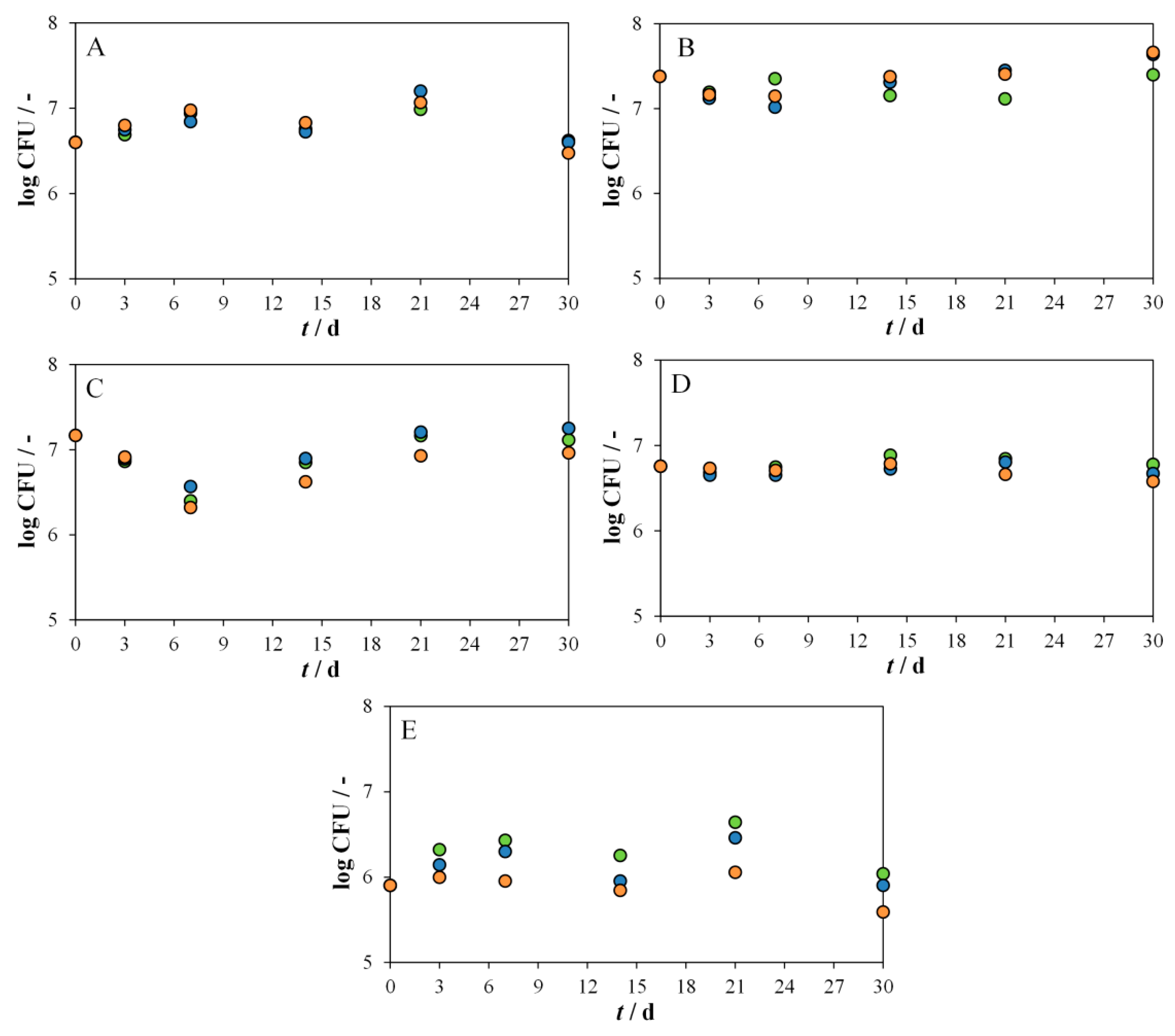
3.2. Determination of Optimal Conditions for the Biodegradation of PS and PVC MPs by Delftia acidovorans and Candida parapsilosis

It is well-known that process conditions have a strong influence on biodegradability [81,82]. In cases where biotreatment is performed on-site, there are numerous factors that may limit microbial activity and thus reduce the efficiency of biodegradation [83]. Therefore, for Delftia acidovorans and Candida parapsilosis, selected in the previous step as the bacterium and yeast with the highest potential to biodegrade PS and PVC MPs, the optimal biodegradation conditions were determined. These conditions were determined by considering the application of selected microorganisms in bioreactors. The experiments were conducted using a full factorial methodology combining three factors (pH of the mineral medium, agitation speed, and cell density of the bacterial or yeast suspension) at three levels (Supplementary Table S1). The effect of pH on biodegradation by bacteria was tested in the range of 6–8, since bacteria generally tolerate a neutral pH range [26]. Since yeasts generally grow in the acidic to neutral pH range [84], pH conditions in the range of 3–7 were tested for biodegradation by yeasts.
The results of the suitability of four regression models (Equations (1)–(4)) used to describe the behavior of the experimentally obtained data are presented in Table S2 (Supplementary Materials). These models varied in complexity: linear dependence on the parameters studied (model I; Equation (1)), linear dependence with the interaction effect included (model II; Equation (2)), quadratic dependence (model III; Equation (3)), and quadratic dependence with the interaction effect included (model IV; Equation (4)). All four models were found to be significant with 95% confidence (p-value was less than 0.05). However, model IV had the highest values of the adjusted coefficient of determination, R2adj, for all four microorganism/MP combinations and was thus the best model for determining the optimal conditions for the biodegradation processes tested.
The response surfaces at different agitation speeds are shown in Figure 5 and Figure 6. The red areas indicate the most favorable conditions for biodegradation, while the blue areas represent the least favorable.
In the biodegradation of both PS and PVC MPs by Delftia acidovorans (Figure 5), such red areas were observed at the lowest agitation speed (120 rpm) and the highest applied cell densities (Figure 5(A1,B1)). Agitation plays an important role in biodegradation by improving oxygen transfer to the process mixture and providing homogeneous chemical and physical conditions [85]. Since Delftia acidovorans is an aerobic microorganism, agitation should be beneficial to its activity. However, when we analyzed our response surfaces at different agitation speeds (Figure 5(A1–A3)), we found that the maximum log CFU values decreased with increasing agitation speed. The cause of this behavior was not investigated in this work, but we can assume that too intense agitation could damage the bacterial cells or perhaps inhibit the adsorption of bacteria on the surface of MPs and the formation of a biofilm. For example, it is known that intensive agitation can change the morphology of the bacterium Escherichia coli [86]. In the biodegradation of PVC by Delftia acidovorans, the unfavorable effect of the basic pH of the medium was observed at all agitation speeds (Figure 5(B1–B3)), while in the biodegradation of PS, this effect was observed at the highest agitation speed (Figure 5(A3)) and became imperceptible as the agitation speed decreased (Figure 5(A1,A2)). In agreement with what has been written, the optimal process values for the biodegradation of MPs by Delftia acidovorans (Table 4) within the selected experimental range included an agitation speed of 120 rpm with high cell densities (0.9 and 1.0 for PS and PVC, respectively) and a slightly basic medium (7.95 and 7.45 for PS and PVC, respectively). The higher log CFU achieved in PVC biodegradation suggests that Delftia acidovorans adapts better to PVC MPs than to PS MPs, using these particles more successfully as an energy and carbon source for its development.
Figure 6 shows the response surfaces for the biodegradation of PS and PVC MPs by the yeast Candida parapsilosis at different agitation speeds. As in the case of Delftia acidovorans, more efficient biodegradation was associated with higher values of cell density. Candida parapsilosis achieved the highest log CFU at pH values of 5.67 and 4.94 for PS and PVC, respectively (Table 4). These results are consistent with reports in the literature that the optimal pH for yeasts should be between 5.5 and 6.0 [84]. The unfavorable effect of more acidic and neutral media on the biodegradation of PS or PVC MPs by Candida parapsilosis can be clearly seen in Figure 6. Candida parapsilosis, like Delftia acidovorans, is an aerobic microorganism, which means that more intensive agitation should have a positive effect on the activity of this yeast. However, it was again found that the maximum agitation speed was not the optimal speed for biodegradation: the results showed an optimal agitation speed of 156 and 136 rpm for PS and PVC, respectively. Agitation speeds above these values were found to be less beneficial, probably for reasons similar to those discussed in the case of Delftia acidovorans. We found reports on the negative effects of agitation on the activity of another representative of fungi: molds. Thus, high agitation speed affected the intercellular enzyme activity of Aspergillus luchuensis [87] and Aspergillus niger [88] and reduced their enzyme production. A comparison of log CFU values at the optimal process conditions (Table 4) showed that Candida parapsilosis could also adapt better to PVC MPs than to PS MPs. One of the reasons for this behavior is probably the molecular structure of these two polymers, since PS, unlike PVC, contains an aromatic ring, and it is known that the presence of aromatic rings in the molecular structure increases the toxic potential of a substance [89,90]. Of course, this cannot be reliably asserted without a detailed analysis of the toxicity mechanisms of these two MPs. For example, we recently tested the toxicity of PS and PVC microparticles with different microorganisms and found different orders of toxicity: with applied 200–600 μm particles at an amount of 50–1000 mg L−1, PVC was more toxic to the microalga Scenedesmus sp., less toxic to the bacterium Pseudomonas putida, and equally toxic to the yeast Saccharomyces cerevisiae [91].

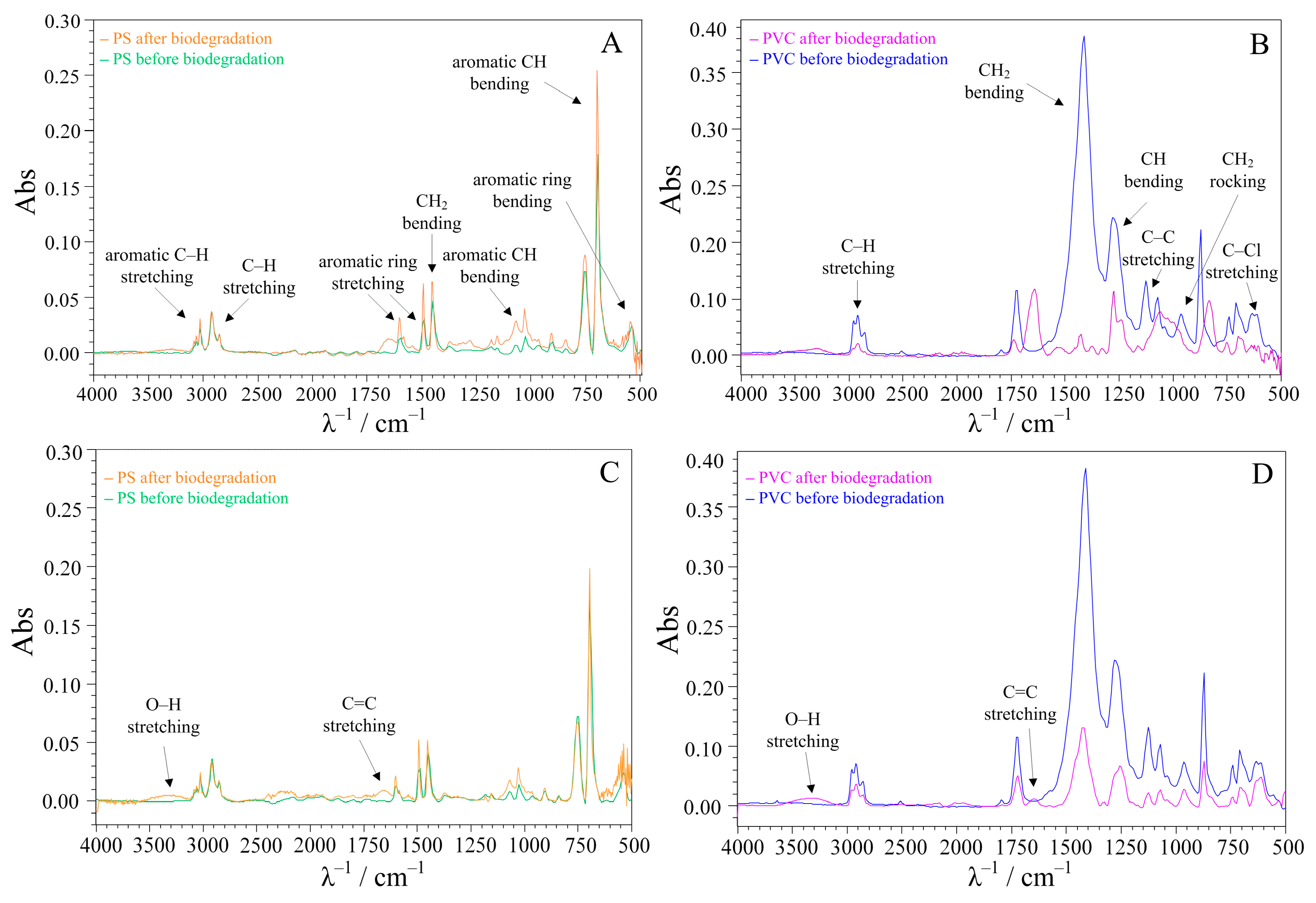
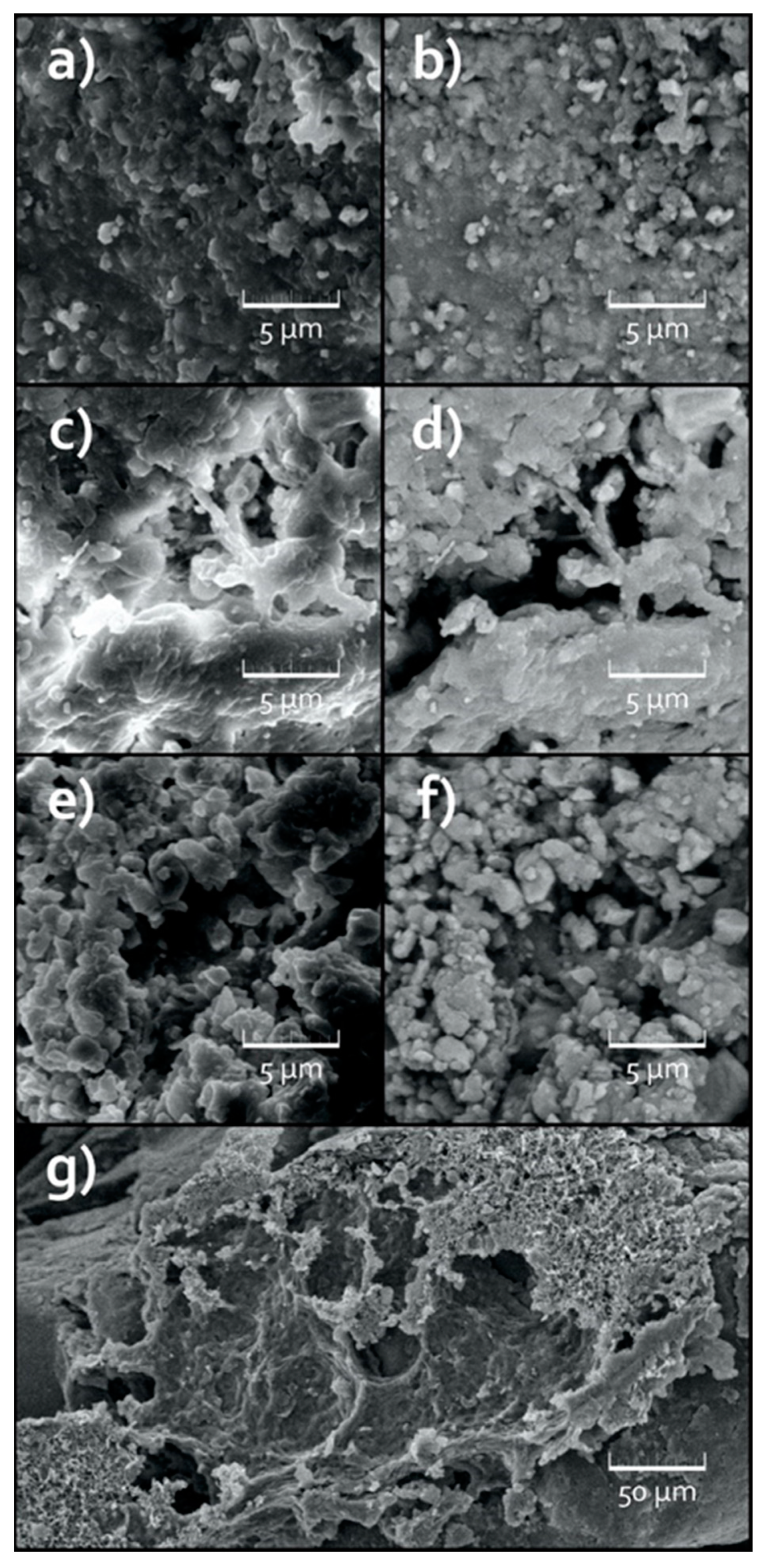
3.3. Surface Analysis of the Treated MPs

It is generally known that the biodegradation of synthetic plastics is a very slow process. Therefore, unless the experiments are conducted over an extremely long period of time (much longer than 30 days, as in this experiment), the changes associated with biodegradation mainly occur on the surface of the MP particles, which is insignificant compared to the total volume of the MP sample. Moreover, these effects are generally limited to topographic differences and minor chemical changes. Therefore, to confirm these changes, the untreated and treated MP particles were analyzed by ATR-FTIR and SEM.
FTIR is the perfect method for studying chemical changes at the level of the different bonding of the elemental C, H, and O constituents. The ATR-FTIR spectra recorded before and after treatment are compared in Figure 7. The spectra of the PS and PVC microparticles before the treatment are marked with green and blue lines, respectively. The characteristic peaks of the PS and PVC polymers are marked in the spectra [92,93]. The orange and pink lines represent the spectra of PS and PVC, respectively, recorded after the treatment.
No significant change in the intensities of the characteristic PS peaks was observed (Figure 7A,C). However, two new peaks appeared after both treatments: by bacteria and by yeast. The first was a broad peak in the 2500–3300 cm−1 range, corresponding to the stretching of the OH group. The appearance of this peak could indicate that biodegradation has occurred. In addition, a new peak appeared at 1645 cm−1. The peaks in this range mostly relate to the C=C stretching of the alkene [94,95,96,97], which could indicate the breaking of the aromatic benzene ring in the PS structure.
In the case of PVC treatment, comparison of the FTIR spectra recorded before and after treatment showed a decrease in the intensity of the characteristic PVC peaks at 616 cm−1 (C–Cl stretching), 966 cm−1 (CH2 rocking), 1255 cm−1 (CH bending), and 1427 cm−1 (CH2 bending), most likely due to the dehydrochlorination process (Figure 7B,D) [98]. This was much more pronounced in the bacterial treatment of PVC (Figure 7B). The dehydrochlorination of PVC generally leads to the formation of unsaturated C=C bonds [99], which corresponded to the appearance of a new FTIR peak at 1640 cm−1. This peak could be observed after both treatments. Its intensity was much more pronounced in the case of the bacterial treatment (Figure 7B), confirming our assumption that it is closely related to the dehydrochlorination process. It was expected that the biodegradation of PVC should also lead to the formation of the carbonyl group, a group in which the carbon atom is double-bonded to an oxygen atom. The carbonyl group gives a peak in the spectral region 1720–1740 cm−1 [100,101]. However, the FTIR spectrum of the untreated PVC already contained such a peak, which obviously represents an additive present in the tested PVC sample, since pure PVC has no carbonyl groups in its structure. The intensity of this peak decreased in both treatments, indicating that Delftia acidovorans and Candida parapsilosis may have attacked the additive, or that the additive was released from the surface of the MP particles due to biodegradation of the PVC polymer, as later confirmed by LC-MS analysis of the filtrate solution (Section 3.4). This also made it impossible to detect the formation of new carbonyl groups and use this as evidence of biodegradation of the PVC polymer.
To visualize the degradation effect, the samples were imaged with the SEM in secondary electron mode (SE) and in backscattered electron mode (BSE). SE SEM microscopy provides information about the textural changes on the surface of MPs, which is useful because these changes are the most relevant. BSE SEM is sensitive to chemical composition in terms of atomic mass. Therefore, in the unlikely event that the chemical environment has changed significantly, BSE SEM could visually qualify the areas where the respective changes took place. Untreated PS microparticles (Figure 8a,b) and untreated PVC microparticles (Figure 9a,b) were used as the reference. For all samples scanned, the bulk appearance resembled micrometric polydisperse particles with a wide size distribution in aggregates of several hundreds of microns.
In the case of the untreated PS MPs, the surface topography revealed by SE showed low roughness and a general absence of morphological defects (i.e., the morphological defects were confined to those mechanically induced by grinding plastics into powders) (Figure 8a). BSE imaging also showed that the phase composition was homogeneous in depth beyond the sample surface (Figure 8b).
PS microparticles treated with bacteria or yeast showed only a weak difference in surface topography compared to the reference PS sample (i.e., the presence of morphological defects and roughness could be sporadically indicated) (Figure 8c,e). However, it was not possible to quantify any degradation difference between the bacteria and yeast treated PS samples. From the BSE imaging (Figure 8d,f), it was not possible to observe any in depth compositional phase changes for the treated PS samples with respect to the reference. This is consistent with the results of the FTIR analysis discussed previously, which also did not indicate the biodegradation of PS by bacteria or yeast.
Just like in the case of the untreated PS MPs, the surface topography of the untreated PVC microparticles, scanned in SE mode (Figure 9a), showed low roughness and a general absence of morphological defects, while BSE imaging (Figure 9b) indicated a homogeneous phase composition.
However, the consequences of the degradation were more obvious for the case of the treated PVC samples. Specifically, both degraded samples showed the presence of areas characterized with higher surface roughness or higher surface porosity, or an increased presence of morphological defects, or in general, just a more damaged surface. Comparing the PVC MP samples treated with Delftia acidovorans with those treated with Candida parapsilosis, the apparent presence of degradation phenomena increased from sporadic in the yeast-treated sample to considerable in the bacteria-treated sample, confirming the conclusion from the analysis of the FTIR spectra that PVC MPs are more susceptible to biodegradation by Delftia acidovorans (Figure 9c,e). Some faint differences in the BSE images of the treated PVC samples could be observed, pointing out possible in depth compositional phase changes. However, the comparison of BSE between the bacteria and yeast treated PVC samples was not relevant (Figure 9d,f). Figure 9g depicts one of the apparent bacteria degradation areas.

3.4. Analysis of the Aqueous Phases in Contact with MPs

The MS analyses of the aqueous phases in contact with the MP samples were performed using an LC-MS 2020 (Shimadzu, Japan) to estimate the released additives, as the biodegradation of the surface of the MP particles can lead to additive release [102]. Comparison of the MS spectra obtained before and after the biodegradation experiments of the PS samples showed the appearance of a new peak at m/z ratios of 328 for the experiment with Delftia acidovorans (Figure S1, case A2) (i.e., 327 for the experiment with Candida parapsilosis (Figure S1, case B2)). This peak could represent tryphenyl phosphate [103], which is commonly used as a flame retardant [104]. In the experiments on the biodegradation of PVC samples, the analysis of the MS spectra showed the appearance of new peaks at m/z ratios of 391 and 447. The peak at 391 could represent bis(2-ethylhexyl)phthalate [105], while the peak at 447 could be associated with diisodecyl phthalate [106]. These two substances are among the most commonly used PVC plasticizers in Europe [107]. In addition, both additives contain a carbonyl group in their structure, so their release from the surface of the PVC microparticles is consistent with the observed decrease in the intensity of the FTIR peak in the range of 1720–1740 cm−1 (Figure 7B,D).

4. Conclusions

Although the degradation of microplastics by microorganisms is considered as one of the most acceptable approaches to solving the problem of microplastics in the environment [23], there are still numerous obstacles that need to be overcome before such treatments can be successfully implemented. Foremost among these are the considerably long duration of the treatments and the lack of information on specific enzymes that can successfully degrade some of the plastic polymers including PS and PVC [108].
Five bacterial strains including Bacillus cereus, Bacillus licheniformis, Bacillus subtilis, Delftia acidovorans, and Pseudomonas alcaligenes and five yeast strains, namely Candida parapsilosis, Geotrichum candidum, Rhodotorula glutinis, Saccharomyces cerevisiae, and Trichosporon sp. were isolated from environmental samples. Among them, Delftia acidovorans was identified as the bacterium with the highest potential to biodegrade PS and PVC MPs, while Candida parapsilosis showed the highest potential among the yeasts. The two microorganisms adapted better to the PVC MPs than to the PS MPs to use it as a carbon and energy source. A positive effect of increased cell density and a negative effect of high agitation speed on the biodegradation process were observed.
Based on the results obtained, it would be desirable to study the metabolic processes of Delftia acidovorans and Candida parapsilosis in the hope of identifying a new enzyme that efficiently degrades PS or PVC MPs.

Supplementary Materials

The following supporting information can be downloaded at: https://www.mdpi.com/article/10.3390/environments10120207/s1, Figure S1: The MS spectra of solutions in contact with MPs for biodegradation experiments with Delftia acidovorans (A) and Candida parapsilosis (B). The following cases are presented: the solutions at the beginning of the biodegradation experiments (A1 and B1), the solutions after 30 days of exposure to PS (A2 and B2), and the solutions after 30 days of exposure to PVC (A3 and B3); Table S1: Experimental design to determine the optimal conditions for the biodegradation of PS and PVC MPs by bacterium and yeast selected as the best in preliminary experiments. The design included the pH value of the medium, agitation speed (AS), and cell density (OD); Table S2: The suitability of four regression models (Equations (1)–(4)) to fit the experimental data of log CFU for the biodegradation of PS and PVC MPs by Delftia acidovorans and Candida parapsilosis. The influence of three factors: medium pH, agitation speed (AS) and cell density (OD) was considered.

Author Contributions

Conceptualization, V.O.B., Š.U., D.K.G. and T.B.; Methodology, M.M., V.M. (Viktorija Martinjak) and K.B.M.; Formal analysis, K.B.M., V.M. (Vilko Mandić), A.B. and M.M.; Investigation, K.B.M., M.M., Š.U., M.C., V.O.B. and D.K.G.; Writing—original draft preparation, K.B.M. and M.M.; Writing—review and editing, Š.U., V.O.B. and D.K.G.; Visualization, K.B.M. and Š.U. All authors have read and agreed to the published version of the manuscript.

Funding

The authors would like to acknowledge the financial support of the Croatian Science Foundation through the project Advanced Water Treatment Technologies for Microplastics Removal (AdWaTMiR; IP-2019-04-9661).

Data Availability Statement

Data are contained within the article.

Conflicts of Interest

The authors declare no conflict of interest.

References

  1. Plastics—The Facts. 2022. Available online: https://plasticseurope.org/wp-content/uploads/2022/10/PE-PLASTICS-THE-FACTS_V7-Tue_19-10-1.pdf (accessed on 20 March 2023).
  2. Geyer, R.; Jambeck, J.R.; Law, K.L. Production, use, and fate of all plastics ever made. Sci. Adv. 2017, 3, e1700782. [Google Scholar] [CrossRef]
  3. MacLeod, M.; Arp, H.P.H.; Tekman, M.B.; Jahnke, A. The global threat from plastic pollution. Science 2021, 373, 61–65. [Google Scholar] [CrossRef]
  4. Ostle, C.; Thompson, R.C.; Broughton, D.; Gregory, L.; Wootton, M.; Johns, D.G. The rise in ocean plastics evidenced from a 60-year time series. Nat. Commun. 2019, 10, 1622–1628. [Google Scholar] [CrossRef]
  5. Chen, J.; Wu, J.; Sherrell, P.C.; Chen, J.; Wang, H.; Zhang, W.-X.; Yang, J. How to Build a Microplastics-Free Environment: Strategies for Microplastics Degradation and Plastics Recycling. Adv. Sci. 2022, 9, 2103764. [Google Scholar] [CrossRef]
  6. Erceg, M.; Tutman, P.; Vojanić Varezić, D.; Bobanović, A. Karakterizacija mikroplastike u sedimentu plaže Prapratno. Kem. Ind. 2020, 69, 253–260. [Google Scholar] [CrossRef]
  7. Westphalen, H.; Abdelrasoul, A. Challenges and Treatment of Microplastics in Water. In Water Challenges of an Urbanizing World; Glavan, M., Ed.; InTech: Rijeka, Croatia, 2018; pp. 71–84. [Google Scholar] [CrossRef]
  8. Ozturk, R.C.; Altinok, I. Interaction of Plastics with Marine Species. Turk. J. Fish. Aquat. Sci. 2020, 20, 647–658. [Google Scholar] [CrossRef]
  9. Cho, Y.; Shim, W.J.; Jang, M.; Han, G.M.; Hong, S.H. Abundance and characteristics of microplastics in market bivalves from South Korea. Environ. Pollut. 2019, 245, 1107–1116. [Google Scholar] [CrossRef]
  10. Wang, L.; Nabi, G.; Yin, L.; Wang, Y.; Li, S.; Hao, Z.; Li, D. Birds and plastic pollution: Recent advances. Avian Res. 2021, 12, 59. [Google Scholar] [CrossRef]
  11. Hardesty, B.D.; Good, T.P.; Wilcox, C. Novel methods, new results and science-based solutions to tackle marine debris impacts on wildlife. Ocean Coast. Manag. 2015, 115, 4–9. [Google Scholar] [CrossRef]
  12. Cox, K.D.; Covernton, G.A.; Davies, H.L.; Dower, J.F.; Juanes, F.; Dudas, S.E. Human Consumption of Microplastics. Environ. Sci. Technol. 2019, 53, 7068–7074. [Google Scholar] [CrossRef] [PubMed]
  13. Yeung, C.W.S.; Teo, J.Y.Q.; Loh, X.J.; Lim, J.Y.C. Polyolefins and Polystyrene as Chemical Resources for a Sustainable Future: Challenges, Advances, and Prospects. ACS Mater. Lett. 2021, 3, 1660–1676. [Google Scholar] [CrossRef]
  14. Zolotova, N.; Kosyreva, A.; Dzhalilova, D.; Fokichev, N.; Makarova, O. Harmful effects of the microplastic pollution on animal health: A literature review. PeerJ 2022, 10, e13503. [Google Scholar] [CrossRef]
  15. Bhuyan, M.S. Effects of Microplastics on Fish and in Human Health. Front. Environ. Sci. 2022, 10, 827289. [Google Scholar] [CrossRef]
  16. Ugwu, K.; Herrera, A.; Gómez, M. Microplastics in marine biota: A review. Mar. Pollut. Bull. 2021, 169, 112540. [Google Scholar] [CrossRef]
  17. Barrick, A.; Champeau, O.; Chatel, A.; Manier, N.; Northcott, G.; Tremblay, L.A. Plastic additives: Challenges in ecotox hazard assessment. PeerJ 2021, 9, e11300. [Google Scholar] [CrossRef]
  18. Gkoutselis, G.; Rohrbach, S.; Harjes, J.; Obst, M.; Brachmann, A.; Horn, M.A.; Rambold, G. Microplastics accumulate fungal pathogens in terrestrial ecosystems. Sci. Rep. 2021, 11, 13214. [Google Scholar] [CrossRef]
  19. Bahl, S.; Dolma, J.; Singh, J.J.; Sehgal, S. Biodegradation of plastics: A state of the art review. Mater. Today Proc. 2020, 39, 31–34. [Google Scholar] [CrossRef]
  20. Miri, S.; Saini, R.; Mohammadreza Davoodi, S.; Pulicharla, R.; Kaur Brar, S.; Mag-douli, S. Biodegradation of microplastics: Better late than never. Chemosphere 2022, 286, 131670. [Google Scholar] [CrossRef]
  21. Osma, J.F.; Toca-Herrera, J.L.; Rodríguez-Couto, S. Uses of laccases in the food industry. Enzyme. Res. 2010, 2010, 918761. [Google Scholar] [CrossRef] [PubMed]
  22. Santo, M.; Weitsman, R.; Sivan, A. The Role of the Copper-Binding Enzyme—Laccase—In the Biodegradation of Polyethylene by the Actinomycete Rhodococcus Ruber. Int. Biodeterior. Biodegrad. 2013, 84, 204–210. [Google Scholar] [CrossRef]
  23. Cai, Z.; Li, M.; Zhu, Z.; Wang, X.; Huang, Y.; Li, T.; Gong, H.; Yan, M. Biological Degradation of Plastics and Microplastics: A Recent Perspective on Associated Mechanisms and Influencing Factors. Microorganisms 2023, 11, 1661. [Google Scholar] [CrossRef] [PubMed]
  24. Yang, X.-G.; Wen, P.-P.; Yang, Y.-F.; Jia, P.-P.; Li, W.-G.; Pei, D.-S. Plastic biodegradation by in vitro environmental microorganisms and in vivo gut microorganisms of insects. Front. Microbiol. 2023, 13, 1001750. [Google Scholar] [CrossRef] [PubMed]
  25. Hall, B.G.; Acar, H.; Nandipati, A.; Barlow, M. Growth Rates Made Easy. Mol. Biol. Evol. 2013, 31, 232–238. [Google Scholar] [CrossRef] [PubMed]
  26. Doelle, H.W.; Fiechter, A.; Schlegel, G.; Sell, D.; Shimizu, S.; Ulber, R.; Yamada, H. Biotechnology, 1. General. Ullmann’s Encycl. Ind. Chem. 2009, 5, 689–712. [Google Scholar] [CrossRef]
  27. Ali, S.R.M.; Fradi, A.J.; Al-araji, A.M. Effect of some physical factors on growth of five fungal species. Eur. Acad. Res. 2017, 2, 1069–1078. [Google Scholar]
  28. Krasowska, A.; Sigler, K. How microorganisms use hydrophobicity and what does this mean for human needs? Front. Cell. Infect. Microbiol. 2014, 19, 112. [Google Scholar] [CrossRef]
  29. Bayry, J.; Aimanianda, V.; Guijarro, J.I.; Sunde, M.; Latgé, J.-P. Hydrophobins—Unique Fungal Proteins. PLoS Pathog. 2012, 8, e1002700. [Google Scholar] [CrossRef]
  30. Sowmya, H.V.; Krishnappa, M.; Thippeswamy, B. Degradation of polyethylene by Trichoderma harzianum—SEM, FTIR, and NMR analyses. Environ. Monit. Assess. 2014, 186, 577–6586. [Google Scholar] [CrossRef]
  31. Gómez-Méndez, L.D.; Moreno-Bayona, D.A.; Poutou-Piñales, R.A.; Salcedo-Reyes, J.C.; Pedroza-Rodríguez, A.M.; Vargas, A.; Bogoya, J.M. Biodeterioration of plasma pretreated LDPE sheets by Pleurotus ostreatus. PLoS ONE 2018, 13, e0203786. [Google Scholar] [CrossRef]
  32. Ganesh, P.; Dineshrajn, D.; Yoganathan, K. Production and screening of depolymerising enzymes by potential bacteria and fungi isolated from plastic waste dump yard sites. Int. J. Appl. Res. 2017, 3, 693–695. [Google Scholar]
  33. Zhang, J.; Gao, D.; Li, Q.; Zhao, Y.; Li, L.; Lin, H.; Zhao, Y. Biodegradation of polyethylene microplastic particles by the fungus Aspergillus flavus from the guts of wax moth Galleria mellonella. Sci. Total Environ. 2020, 704, 135931. [Google Scholar] [CrossRef]
  34. Kang, B.R.; Kim, S.B.; Song, H.A.; Lee, T.K. Accelerating the Biodegradation of High-Density Polyethylene (HDPE) Using Bjerkandera adusta TBB-03 and Lignocellulose Substrates. Microorganisms 2019, 7, 304. [Google Scholar] [CrossRef]
  35. Khatoon, N.; Jamal, A.; Ali, M.I. Lignin peroxidase isoenzyme: A novel approach to biodegrade the toxic synthetic polymer waste. Environ. Technol. 2019, 40, 1366–1375. [Google Scholar] [CrossRef]
  36. Sumathi, T.; Viswanath, B.; Sri Lakshmi, A.; SaiGopal, D.V.R. Production of Laccase by Cochliobolus sp. Isolated from Plastic Dumped Soils and Their Ability to Degrade Low Molecular Weight PVC. Biochem. Res. Int. 2016, 2016, 9519527. [Google Scholar] [CrossRef]
  37. Liebminger, S.; Eberl, A.; Sousa, F.; Heumann, S.; Fischer-Colbrie, G.; Cavaco-Paulo, A.; Guebitz, G.M. Hydrolysis of PET and bis-(benzoyloxyethyl) terephthalate with a new polyesterase from Penicillium citrinum. Biocatal. Biotransform. 2007, 25, 171–177. [Google Scholar] [CrossRef]
  38. Carniel, A.; Valoni, É.; Nicomedes, J.; da Conceição Gomes, A.; de Castro, A.M. Lipase from Candida antarctica (CALB) and cutinase from Humicola insolens act synergistically for PET hydrolysis to terephthalic acid. Process Biochem. 2017, 59, 84–90. [Google Scholar] [CrossRef]
  39. Tahir, L.; Ali, M.I.; Zia, M.; Atiq, N.; Hasan, F.; Ahmed, S. Production and Characterization of Esterase in Lantinus tigrinus for Degradation of Polystyrene. Pol. J. Microbiol. 2013, 62, 101–108. [Google Scholar] [CrossRef]
  40. Takamoto, T.; Shirasaka, H.; Uyama, H.; Kobayashi, S. Lipase-catalyzed hydrolytic degradation of polyurethane in organic solvent. Chem. Lett. 2001, 30, 492–493. [Google Scholar] [CrossRef]
  41. Mathur, G.; Prasad, R. Degradation of Polyurethane by Aspergillus flavus (ITCC 6051) Isolated from Soil. Appl. Biochem. Biotechnol. 2012, 167, 1595–1602. [Google Scholar] [CrossRef] [PubMed]
  42. Khan, S.; Nadir, S.; Shah, Z.U.; Shah, A.A.; Karunarathna, S.C.; Xu, J.; Khan, A.; Munir, S.; Hasan, F. Biodegradation of polyester polyurethane by Aspergillus tubingensis. Environ. Pollut. 2017, 225, 469–480. [Google Scholar] [CrossRef] [PubMed]
  43. Yoon, M.G.; Jeon, H.J.; Kim, M.N. Biodegradation of polyethylene by a soil bacterium and AlkB cloned recombinant cell. J. Bioremed. Biodegrad. 2012, 3, 145. [Google Scholar] [CrossRef]
  44. Jeon, H.J.; Kim, M.N. Functional analysis of alkane hydroxylase system derived from Pseudomonas aeruginosa E7 for low molecular weight polyethylene biodegradation. Int. Biodeterior. Biodegrad. 2015, 103, 141–146. [Google Scholar] [CrossRef]
  45. Yoshida, S.; Hiraga, K.; Taniguchi, I.; Oda, K. Ideonella sakaiensis, PETase, and MHETase: From identification of microbial PET degradation to enzyme characterization. Meth. Enzymol. 2021, 648, 187–205. [Google Scholar] [CrossRef]
  46. Jabloune, R.; Khalil, M.; Ben Moussa, I.E.; Simao-Beaunoir, A.-M.; Lerat, S.; Brzezinski, R.; Beaulieu, C. Enzymatic Degradation of p-Nitrophenyl Esters, Polyethylene Terephthalate, Cutin, and Suberin by Sub1, a Suberinase Encoded by the Plant Pathogen Streptomyces scabies. Microbes Environ. 2020, 35, ME19086. [Google Scholar] [CrossRef] [PubMed]
  47. Müller, R.-J.; Schrader, H.; Profe, J.; Dresler, K.; Deckwer, W.-D. Enzymatic Degradation of Poly(Ethylene Terephthalate): Rapid Hydrolyse Using a Hydrolase from T. Fusca. Macromol. Rapid Commun. 2005, 26, 1400–1405. [Google Scholar] [CrossRef]
  48. Nakajima-Kambe, T.; Onuma, F.; Kimpara, N.; Nakahara, T. Isolation and Characterization of a Bacterium Which Utilizes Polyester Polyurethane as a Sole Carbon and Nitrogen Source. FEMS Microbiol. Lett. 1995, 129, 39–42. [Google Scholar] [CrossRef]
  49. Ekanayaka, A.H.; Tibpromma, S.; Dai, D.; Xu, R.; Suwannarach, N.; Stephenson, S.L.; Dao, C.; Karunarathna, S.C. A Review of the Fungi That Degrade Plastic. J. Fungi 2022, 8, 772. [Google Scholar] [CrossRef]
  50. Lear, G.; Kingsbury, J.; Franchini, S.; Gambarini, V.; Maday, S.; Wallbank, J.; Weaver, L.; Pantos, O. Plastics and the Microbiome: Impacts and Solutions. Environ. Microbiome 2021, 16, 2. [Google Scholar] [CrossRef]
  51. Ahmed, T.; Shahid, M.; Azeem, F.; Rasul, I.; Shah, A.A.; Noman, M.; Hameed, A.; Manzoor, N.; Manzoor, I.; Muhammad, S. Biodegradation of plastics: Current scenario and future prospects for environmental safety. Environ. Sci. Pollut. Res. 2018, 25, 7287–7298. [Google Scholar] [CrossRef]
  52. DSouza, G.C.; Sheriff, R.S.; Ullanat, V.; Shrikrishna, A.; Joshi, A.V.; Hiremath, L.; Entoori, K. Fungal biodegradation of low-density polyethylene using consortium of Aspergillus species under controlled conditions. Heliyon 2021, 7, e07008. [Google Scholar] [CrossRef]
  53. Kyaw, B.M.; Champakalakshmi, R.; Kishore Sakharkar, M.; Lim, C.S.; Sakharkar, K.R. Biodegradation of low density poly-ethene (LDPE) by Pseudomonas species. Indian J. Microbiol. 2012, 52, 411–419. [Google Scholar] [CrossRef] [PubMed]
  54. Harshvardhan, K.; Jha, B. Biodegradation of low-density polyethylene by marine bacteria from pelagic waters, Arabian Sea, India. Mar. Pollut. Bull. 2013, 77, 100–106. [Google Scholar] [CrossRef] [PubMed]
  55. Rogers, K.L.; Carreres-Calabuig, J.A.; Gorokhova, E.; Posth, N.R. Micro-by-micro interactions: How microorganisms influence the fate of marine microplastics. Limnol. Oceanogr. Lett. 2020, 5, 18–36. [Google Scholar] [CrossRef]
  56. Yang, J.; Yang, Y.; Wu, W.-M.; Zhao, J.; Jiang, L. Evidence of polyethylene biodegradation by bacterial strains from the guts of plastic-eating waxworms. Environ. Sci. Technol. 2014, 48, 13776–13784. [Google Scholar] [CrossRef]
  57. Tokiwa, Y.; Calabia, B.; Ugwu, C.; Aiba, S. Biodegradability of plastics. Int. J. Mol. Sci. 2009, 10, 3722–3742. [Google Scholar] [CrossRef] [PubMed]
  58. Lee, H.M.; Kim, H.R.; Jeon, E.; Yu, H.C.; Lee, S.; Li, J.; Kim, D.-H. Evaluation of the biodegradation efficiency of four various types of plastics by Pseudomonas aeruginosa isolated from the gut extract of superworms. Microorganisms 2020, 8, 1341. [Google Scholar] [CrossRef] [PubMed]
  59. Auta, H.S.; Emenike, C.U.; Jayanthi, B.; Fauziah, S.H. Growth kinetics and biodeterioration of polypropylene microplastics by Bacillus sp. and Rhodococcus sp. isolated from mangrove sediment. Mar. Pollut. Bull. 2018, 127, 15–21. [Google Scholar] [CrossRef]
  60. Auta, H.S.; Emenike, C.U.; Jayanthi, B.; Fauziah, S.H. Screening for polypropylene degradation potential of bacteria isolated from Mangrove ecosystems in Peninsular Malaysia. Int. J. Biosci. Biochem. Bioinform. 2017, 7, 245–251. [Google Scholar] [CrossRef]
  61. Sangeetha Devi, R.; Rajesh Kannan, V.; Nivas, D.; Kannan, K.; Chandru, S.; Robert Antony, A. Biodegradation of HDPE by Aspergillus spp. from marine ecosystem of Gulf of Mannar, India. Mar. Pollut. Bull. 2015, 96, 32–40. [Google Scholar] [CrossRef]
  62. Sáenz, M.; Borodulina, T.; Diaz, L.; Banchon, C. Minimal Conditions to Degrade Low Density Polyethylene by Aspergillus terreus and niger. J. Ecol. Eng. 2019, 20, 44–51. [Google Scholar] [CrossRef]
  63. Chaudhary, A.K.; Vijayakumar, R.P. Studies on biological degradation of polystyrene by pure fungal cultures. Environ. Dev. Sustain. 2020, 22, 4495–4508. [Google Scholar] [CrossRef]
  64. Kučić Grgić, D.; Miloloža, M.; Ocelić Bulatović, V.; Ukić, Š.; Slouf, M.; Gajdosova, V. Screening the Efficacy of a Microbial Consortium of Bacteria and Fungi Isolated from Different Environmental Samples for the Degradation of LDPE/TPS Films. Separations 2023, 10, 79. [Google Scholar] [CrossRef]
  65. Black, J.G. Microbiology: Principles and Explorations, 8th ed.; John Wiley & Sons: Hoboken, NJ, USA, 2012; p. 975. [Google Scholar]
  66. Gong, Y.; Ding, P.; Xu, M.-J.; Zhang, C.-M.; Xing, K.; Qin, S. Biodegradation of phenol by a halotolerant versatile yeast Candida tropicalis SDP-1 in wastewater and soil under high salinity conditions. J. Environ. Manag. 2021, 289, 112525. [Google Scholar] [CrossRef] [PubMed]
  67. Auta, H.S.; Emenike, C.U.; Fauziah, S.H. Screening of Bacillus strains isolated from mangrove ecosystems in Peninsular Malaysia for microplastic degradation. Environ. Pollut. 2017, 231, 1552–1559. [Google Scholar] [CrossRef] [PubMed]
  68. Wolski, E.A.; Murialdo, S.E.; Gonzalez, J.F. Effect of pH and inoculum size on pentachlorophenol degradation by Pseudomonas sp. Water SA 2006, 32, 93–98. [Google Scholar] [CrossRef]
  69. Gonzalez, J.M.; Aranda, B. Microbial Growth under Limiting Conditions-Future Perspectives. Microorganisms 2023, 11, 1641. [Google Scholar] [CrossRef]
  70. Peixoto, J.; Silva, L.P.; Krüger, R.H. Brazilian Cerrado soil reveals an untapped microbial potential for unpretreated polyethylene biodegradation. J. Hazard. Mater. 2017, 324, 634–644. [Google Scholar] [CrossRef]
  71. Lou, Y.; Li, Y.; Lu, B.; Liu, Q.; Yang, S.-S.; Liu, B.; Ren, N.; Wu, W.-M.; Xiang, D. Response of the yellow mealworm (Tenebrio molitor) gut microbiome to diet shifts during polystyrene and polyethylene biodegradation. J. Hazard. Mater. 2021, 416, 126222. [Google Scholar] [CrossRef]
  72. Liu, J.; Xu, G.; Dong, W.; Xu, N.; Xian, F.; Ma, J.; Fang, Y.; Zhou, J.; Jiang, M. Biodegradation of diethyl terephthalate (DET) and polyethylene terephthalate (PET) by a novel identified degrader Delftia sp. WL-3 and its proposed metabolic pathway. Lett. Appl. Microbiol. 2018, 67, 254–261. [Google Scholar] [CrossRef]
  73. Akutsu, Y.; Nakajima-Kambe, T.; Nomura, N.; Nakahara, T. Purification and Properties of a Polyester Polyurethane-Degrading Enzyme from Comamonas acidovorans TB-35. Appl. Environ. Microbiol. 1998, 64, 62–67. [Google Scholar] [CrossRef]
  74. Zhang, Y.; Pedersen, J.N.; Eser, B.E.; Guo, Z. Biodegradation of polyethylene and polystyrene: From microbial deterioration to enzyme discovery. Biotechnol. Adv. 2022, 60, 107991. [Google Scholar] [CrossRef] [PubMed]
  75. Vásquez-Piñeros, M.A.; Martínez-Lavanchy, P.M.; Jehmlich, N.; Pieper, D.H.; Rincón, C.A.; Harms, H.; Junca, H.; Heipieper, H.J. Delftia sp. LCW, a strain isolated from a constructed wetland shows novel properties for dimethylphenol isomers degradation. BMC Microbiol. 2018, 18, 108. [Google Scholar] [CrossRef] [PubMed]
  76. Harris, J.D.; Coon, C.M.; Doherty, M.E.; McHugh, E.A.; Warner, M.C.; Walters, C.L.; Orahood, O.M.; Loesch, A.E.; Hatfield, D.C.; Sitko, J.C.; et al. Engineering and characterization of dehalogenase enzymes from Delftia acidovorans in bioremediation of perfluorinated compounds. Synth. Syst. Biotechnol. 2022, 7, 671–676. [Google Scholar] [CrossRef] [PubMed]
  77. Hashem, M.; Alamri, S.A.; Al-Zomyh, S.S.A.A.; Alrumman, S.A. Biodegradation and detoxification of aliphatic and aromatic hydrocarbons by new yeast strains. Ecotoxicol. Environ. Saf. 2018, 151, 28–34. [Google Scholar] [CrossRef] [PubMed]
  78. Khoddami, A.; Morshed, M.; Tavani, H. Effects of Enzymatic Hydrolysis on Drawn Polyester Filament Yarns. Iran. Polym. J. 2001, 10, 363–370. [Google Scholar]
  79. Zafar, U.; Houlden, A.; Robson, G.D. Fungal communities associated with the biodegradation of polyester polyurethane buried under compost at different temperatures. Appl. Environ. Microbiol. 2013, 79, 7313–7324. [Google Scholar] [CrossRef] [PubMed]
  80. Mroczyńska, M.; Brillowska-Dąbrowska, A. Virulence of Clinical Candida Isolates. Pathogens 2021, 10, 466. [Google Scholar] [CrossRef] [PubMed]
  81. Bher, A.; Mayekar, P.C.; Auras, R.A.; Schvezov, C.E. Biodegradation of Biodegradable Polymers in Mesophilic Aerobic Environments. Int. J. Mol. Sci. 2022, 23, 12165. [Google Scholar] [CrossRef]
  82. Devi, R.; Kannan, V.; Natarajan, K.; Nivas, D.; Kannan, K.; Chandru, S.; Antony, A. The Role of Microbes in Plastic Degradation. In Environmental Waste Management; Chandra, R., Ed.; CRC Press: Boca Raton, FL, USA, 2016; pp. 355–384. [Google Scholar] [CrossRef]
  83. Singh, B.; Christina, E. Chapter 13—Indigenous microorganisms as an effective tool for in situ bioremediation. In Relationship between Microbes and the Environment for Sustainable Ecosystem Services; Samuel, J., Kumar, A., Singh, J., Eds.; Elsevier: Amsterdam, The Netherlands, 2022; Volume 2, pp. 273–295. [Google Scholar] [CrossRef]
  84. Magan, N. Fungi in Extreme Environments. In Environmental and Microbial Relationships; Kubicek, C.P., Druzhinina, I.S., Eds.; Springer: Berlin/Heidelberg, Germany, 2007; pp. 85–103. [Google Scholar] [CrossRef]
  85. Zhou, Y.; Han, L.R.; He, H.W.; Sang, B.; Yu, D.L.; Feng, J.T.; Zhang, X. Effects of Agitation, Aeration and Temperature on Production of a Novel Glycoprotein GP-1 by Streptomyces kanasenisi ZX01 and Scale-Up Based on Volumetric Oxygen Transfer Coefficient. Molecules 2018, 23, 125. [Google Scholar] [CrossRef]
  86. Valdez-Cruz, N.A.; Reynoso-Cereceda, G.I.; Pérez-Rodriguez, S.; Restrepo-Pineda, S.; González-Santana, J.; Olvera, A.; Zavala, G.; Alagón, A.; Trujillo-Roldán, M.A. Production of a recombinant phospholipase A2 in Escherichia coli using resonant acoustic mixing that improves oxygen transfer in shake flasks. Microb. Cell Fact. 2017, 16, 129. [Google Scholar] [CrossRef]
  87. Seth, M.; Chand, S. Biosynthesis of tannase and hydrolysis of tannins to gallic acid by Aspergillus awamori—Optimisation of process parameters. Process Biochem. 2000, 36, 39–44. [Google Scholar] [CrossRef]
  88. Ibrahim, D.; Weloosamy, H.; Lim, S.-H. Effect of agitation speed on the morphology of Aspergillus niger HFD5A-1 hyphae and its pectinase production in submerged fermentation. World. J. Biol. Chem. 2015, 6, 265–271. [Google Scholar] [CrossRef] [PubMed]
  89. Al-Sahhaf, Z.Y. Hematological and biochemical effects of an air freshener in rabbits. J. Am. Sci. 2012, 8, 70–73. [Google Scholar]
  90. Philibert, D.; Parketon, T.; Marteinson, S.; de Jourdan, B. Assessing the Toxicity of Individual Aromatic Compounds and Mixtures to American Lobster (Homarus americanus) Larvae Using a Passive Dosing System. Environ. Toxicol. Chem. 2021, 40, 1379–1388. [Google Scholar] [CrossRef]
  91. Miloloža, M.; Bule, K.; Prevarić, V.; Cvetnić, M.; Ukić, Š.; Bolanča, T.; Kučić Grgić, D. Assessment of the Influence of Size and Concentration on the Ecotoxicity of Microplastics to Microalgae Scenedesmus sp., Bacterium Pseudomonas putida and Yeast Saccharomyces cerevisiae. Polymers 2022, 14, 1246. [Google Scholar] [CrossRef]
  92. Jung, M.R.; Horgen, F.D.; Orski, S.V.; Rodriguez, V.; Beers, K.L.; Balazs, G.H.; Jones, T.T.; Work, T.M.; Brignac, K.C.; Royer, S.-J.; et al. Validation of ATR FT-IR to identify polymers of plastic marine debris, including those ingested by marine organisms. Mar. Pollut. Bull. 2008, 127, 704–716. [Google Scholar] [CrossRef]
  93. Ul-Hamid, A.; Soufi, K.Y.; Al-Hadhrami, L.M.; Shemsi, A.M. Failure investigation of an underground low voltage XLPE insulated cable. Anti-Corros. Methods Mater. 2015, 62, 281–287. [Google Scholar] [CrossRef]
  94. Smith, B.C. The Infrared Spectroscopy of Alkenes. Spectroscopy 2016, 31, 28–34. Available online: https://www.spectroscopyonline.com/view/infrared-spectroscopy-alkenes (accessed on 5 June 2023).
  95. Smith, B.C. The Infrared Spectra of Polymers IV: Rubbers. Spectroscopy 2022, 37, 8–12. [Google Scholar] [CrossRef]
  96. IR Spectrum Table & Chart. Available online: https://www.sigmaaldrich.com/HR/en/technical-documents/technical-article/analytical-chemistry/photometry-and-reflectometry/ir-spectrum-table (accessed on 5 June 2023).
  97. IR Spectroscopy Tutorial: Alkenes. Available online: https://orgchemboulder.com/Spectroscopy/irtutor/alkenesir.shtml (accessed on 5 June 2023).
  98. Amar, Z.H.; Chabira, S.F.; Sebaa, M.; Ahmed, B. Structural changes undergone during thermal aging and/or processing of Unstabilized, dry-blend and rigid PVC, investigated by FTIR-ATR and curve fitting. Ann. Chim. Sci. Mater. 2019, 43, 59–68. [Google Scholar] [CrossRef]
  99. Ma, D.; Liang, L.; Hu, E.; Chen, H.; Wang, D.; He, C.; Feng, Q. Dechlorination of polyvinyl chloride by hydrothermal treatment with cupric ion. Process Saf. Environ. Prot. 2021, 146, 108–117. [Google Scholar] [CrossRef]
  100. Kumari, A.; Chaudhary, D.R.; Jha, B. Destabilization of polyethylene and polyvinylchloride structure by marine bacterial strain. Environ. Sci. Pollut. Res. 2019, 26, 1507–1516. [Google Scholar] [CrossRef] [PubMed]
  101. Subramani, M.; Sepperumal, U. FTIR analysis of bacterial mediated chemical changes in Polystyrene foam. Ann. Biol. Res. 2016, 7, 55–61. [Google Scholar]
  102. Sutkar, P.R.; Gadewar, R.D.; Dhulap, V.P. Recent trends in degradation of microplastics in the environment: A state-of-the-art review. J. Hazard. Mater. Adv. 2023, 11, 100343. [Google Scholar] [CrossRef]
  103. MassBank Record: MSBNK-Eawag-EQ363802. Available online: https://massbank.eu/MassBank/RecordDisplay?id=MSBNK-Eawag-EQ363802 (accessed on 23 November 2023).
  104. Pawlowski, K.H.; Schartel, B. Flame retardancy mechanisms of triphenyl phosphate, resorcinol bis(diphenyl phosphate) and bisphenol A bis(diphenyl phosphate) in polycarbonate/acrylonitrile–butadiene–styrene blends. Polym. Int. 2007, 56, 1404–1414. [Google Scholar] [CrossRef]
  105. MassBank Record: MSBNK-BGC_Munich-RP019801. Available online: https://massbank.eu/MassBank/RecordDisplay?id=MSBNK-BGC_Munich-RP019801&dsn=BGC_Munich (accessed on 23 November 2023).
  106. MassBank Record: MSBNK-Antwerp_Univ-AN112602. Available online: https://massbank.eu/MassBank/RecordDisplay?id=MSBNK-Antwerp_Univ-AN112602&dsn=Antwerp_Univ (accessed on 23 November 2023).
  107. Plasticisers. Available online: http://www.seepvcforum.com/en/content/16-plasticisers (accessed on 23 November 2023).
  108. Thew, X.E.C.; Lo, S.C.; Ramanan, R.N.; Tey, B.T.; Huy, N.D.; Wei, O.C. Enhancing plastic biodegradation process: Strategies and opportunities. Crit. Rev. Biotechnol. 2023. Online ahead of print. [Google Scholar] [CrossRef]
Figure 1. The main factors affecting the biodegradation of MPs.
Environments 10 00207 g001
Figure 2. Bacteria Bacillus cereus (A1), Bacillus subtilis (B1), Bacillus licheniformis (C1), Pseudomonas alcaligenes (D1), and Delftia acidovorans (E1) cultivated on nutrient agar by the streak plate method, with associated microphotographs of bacterial Gram-stained smears ((A2E2); 1000× magnification). Yeasts Saccharomyces cerevisiae (F1), Rhodotorula glutinis (G1), Candida parapsilosis (H1), Geotrichum candidum (I1), and Trichosporon sp. (J1) cultivated on malt agar by the streak plate method, with associated microphotographs of cultivated yeasts ((F2J2); 400× magnification).
Environments 10 00207 g002
Figure 3. Changes in log CFU values determined during the preliminary biodegradation experiments using bacterial cultures: (A) Bacillus cereus, (B) Pseudomonas alcaligenes, (C) Bacillus subtilis, (D) Delftia acidovorans, and (E) Bacillus licheniformis. The blue, orange, and green circles represent PS, PVC, and the control samples, respectively.
Environments 10 00207 g003
Figure 4. Changes in log CFU values determined during the preliminary biodegradation experiments using yeast cultures: (A) Trichosporon sp., (B) Candida parapsilosis, (C) Rhodotorula glutinis, (D) Saccharomyces cerevisiae, and (E) Geotrichum candidum. The blue, orange, and green circles represent PS, PVC, and the control samples, respectively.
Environments 10 00207 g004
Figure 5. Response surfaces for the biodegradation of the PS (A1A3) and PVC (B1B3) microparticles by Delftia acidovorans.
Environments 10 00207 g005
Figure 6. Response surfaces for the biodegradation of the PS (A1A3) and PVC (B1B3) microparticles by Candida parapsilosis.
Environments 10 00207 g006
Figure 7. Comparison of the FTIR spectra of the studied MPs before and after biodegradation: (A) Delftia acidovorans and PS, (B) Delftia acidovorans and PVC, (C) Candida parapsilosis and PS, and (D) Candida parapsilosis and PVC. The biodegradation of the PS MPs was negligible in both treatments. However, for the PVC MPs, there was a significant decrease in the intensity of the characteristic peaks, indicating that both Delftia acidovorans and Candida parapsilosis initiated biodegradation.
Environments 10 00207 g007
Figure 8. SEM analysis of the PS MPs: (a) SE micrograph of the untreated PS MP sample, (b) BSE micrograph of the untreated PS MP sample, (c) SE micrograph of the PS MP sample treated with the bacterium Delftia acidovorans, (d) BSE micrograph of the PS MP sample treated with the bacterium Delftia acidovorans, (e) SE micrograph of the PS MP sample treated with the yeast Candida parapsilosis, and (f) BSE micrograph of the PS MP sample treated with the yeast Candida parapsilosis.
Environments 10 00207 g008
Figure 9. SEM analysis of the PVC MPs: (a) SE micrograph of the untreated PVC MP sample, (b) BSE micrograph of the untreated PVC MP sample, (c) SE micrograph of PVC MP sample treated with the bacterium Delftia acidovorans, (d) BSE micrograph of PVC MP sample treated with the bacterium Delftia acidovorans, (e) SE micrograph of the PVC MP sample treated with the yeast Candida parapsilosis, (f) BSE micrograph of the PVC MP sample treated with the yeast Candida parapsilosis, (g) SE micrograph of the throughout “surface damage” of the bacteria-treated PVC MP sample.
Environments 10 00207 g009
Table 2. Composition of the mineral media used in the biodegradation experiments.
For BacteriaFor Yeasts
SubstanceConc./g L−1SubstanceConc./g L−1
K2HPO412.500K2HPO41.000
KH2PO43.800KH2PO41.000
(NH4)2SO41.000NH4NO31.000
H3BO30.232NaCl0.500
ZnSO4·7H2O0.174MgSO4·7H2O0.200
FeSO4(NH4)2SO4·6H2O0.116CaCl20.020
MgSO4·7H2O0.100FeCl3·6H2O0.005
CoSO4·7H2O0.096
(NH4)6Mo7O24·4H2O0.022
CuSO4·5H2O0.008
MnSO4·4H2O0.008
Table 3. Process conditions used for the preliminary biodegradation experiments of PS and PVC MPs.
ConditionFor BacteriaFor Yeasts
pH7.0 ± 0.26.0 ± 0.2
T/°C25.0 ± 0.225.0 ± 0.2
ODinitial0.51.0
AS/rpm150160
Table 4. Optimal conditions for the biodegradation of PS and PVC MPs by Delftia acidovorans and Candida parapsilosis and the corresponding log CFU values.
MicroorganismType of MPsODinitialAS/rpmpHlog CFU *
Delftia acidovoransPS0.91207.958.10
PVC1.01207.458.40
Candida parapsilosisPS1.01565.677.24
PVC1.01364.947.75
* According to the optimal model.
Disclaimer/Publisher’s Note: The statements, opinions and data contained in all publications are solely those of the individual author(s) and contributor(s) and not of MDPI and/or the editor(s). MDPI and/or the editor(s) disclaim responsibility for any injury to people or property resulting from any ideas, methods, instructions or products referred to in the content.

