Article

Investigating Neural Reward Sensitivity in the School Grade Incentive Delay Task and Its Relation to Academic Buoyancy

by
Myrthe J. B. Vel Tromp
1,*,
Hilde M. Huizenga
1,
Brenda R. J. Jansen
1,
Anna C. K. van Duijvenvoorde
2 and
Ilya M. Veer
1
1
Department of Developmental Psychology, Faculty of Social and Behavioural Sciences, University of Amsterdam, Nieuwe Achtergracht 129, 1018 VZ Amsterdam, The Netherlands
2
Department of Developmental and Educational Psychology, Faculty of Social and Behavioural Sciences, Leiden University, Wassenaarseweg 52, 2333 AK Leiden, The Netherlands
*
Author to whom correspondence should be addressed.
Behav. Sci. 2025, 15(10), 1321; https://doi.org/10.3390/bs15101321
Submission received: 4 June 2025 / Revised: 12 September 2025 / Accepted: 20 September 2025 / Published: 26 September 2025

Abstract

Understanding the mechanisms behind academic buoyancy, the ability to effectively cope with everyday academic challenges, is essential for identifying the factors and mechanisms that help students maintain their motivation and cope with routine academic pressures. One potential underlying mechanism is reward sensitivity, or the capacity to experience pleasure both in anticipating and receiving reward-related stimuli. We hypothesized that individuals with higher sensitivity to anticipated reward would exhibit greater academic buoyancy. To test this in an academic context, we modified the Monetary Incentive Delay (MID) task into a School Grade Incentive Delay (SGID) task, where participants work towards a fictitious school grade by winning or losing points on each of the trials. In this study, we investigated whether the SGID activates the neural reward circuitry similar to the traditional MID and whether this is associated with academic buoyancy. The SGID task activated key brain regions associated with reward anticipation, validating its use for studying reward processing in academic contexts. Importantly, we found a negative association between academic buoyancy and right amygdala activation during reward anticipation, suggesting that buoyant students may benefit from reduced emotional reactivity when anticipating rewards. Further research in larger samples is needed to capture the full complexity of reward processing in relation to academic buoyancy.

1. Introduction

Understanding why students remain motivated to achieve good academic outcomes despite challenges at home or school is a key question in educational psychology. This phenomenon is often described in two related but distinct ways: academic resilience, which refers to positive adaptation in the face of chronic or major adversities, and academic buoyancy, which refers to students’ ability to successfully deal with the more common, everyday academic setbacks and pressures (Martin & Marsh, 2008a). While only a minority of students face chronic adversities, almost all students face daily academic hassles (Martin & Marsh, 2009). Because not all students are equally affected by these hassles, research should focus on identifying factors and mechanisms that could explain why some students adapt more easily than others (Kalisch et al., 2017), in order to help students maintain their motivation and cope with routine academic pressures.
One promising factor is reward sensitivity, or the capacity to experience pleasure both in anticipating and receiving reward-related stimuli (Gray, 1987). At the self-report and behavioral level, reduced reward sensitivity is impaired in disorders such as posttraumatic stress disorder (Ben-Zion et al., 2022; Elman et al., 2009; Kasparek et al., 2020) and major depressive disorder (Knutson et al., 2008; Pizzagalli et al., 2009). Not surprisingly, anhedonia, the diminished capacity to feel enjoyment or reward satisfaction, is commonly found in depressive disorders (Must et al., 2006). Stressors may play a significant role in the onset of anhedonic symptoms and associated mental disorders (Corral-Frías et al., 2015). Research suggests that individuals with higher reward sensitivity display higher levels of positive affect following stressor exposure (Corral-Frías et al., 2016) and may better maintain their mental health, compared to individuals with lower reward sensitivity (Geschwind et al., 2010). These findings suggest that behavioral reward sensitivity can buffer against the negative effects of stress.
At the neural level, reward sensitivity refers to the responsiveness of fronto-striatal and limbic circuits, particularly the ventral striatum, nucleus accumbens, and amygdala, during reward anticipation and outcome (Schultz et al., 1997). Studies converge on the idea that reward processing supports resilience. For instance, stronger nucleus accumbens (Nacc) activation in response to a reward task predicts fewer depressive symptoms following stress exposure (Tashjian & Galván, 2018), and heightened ventral striatal reward reactivity has been associated with decreases in depressive symptoms over time (Telzer et al., 2014). During reward anticipation specifically, amygdala and Nacc activation moderated the association between the number of life stressors and depressive symptoms (Fassett-Carman et al., 2023). These findings are consistent with the reward prediction error model, where dopaminergic neurons code deviations between expected and actual outcomes (Schultz et al., 1997), and with reinforcement learning research showing that striatal activity tracks prediction errors across paradigms (Garrison et al., 2013). Reward sensitivity supports adaptive decision-making under uncertainty, shaped by individual differences in reinforcement learning parameters (Daw et al., 2006; Cohen, 2007). Importantly, acute stress can dampen reward sensitivity, reducing striatal and orbitofrontal responses to rewards (Porcelli et al., 2012), and psychosocial stress reduces neural reward responsivity (Ethridge et al., 2020). Together, this suggests that neural sensitivity to reward anticipation may be particularly important for resilient outcomes (Vidal-Ribas et al., 2019).
Although it has been suggested that individual differences in responses to both the anticipation and the receipt of reward may be important mechanisms underlying resilience (Dutcher & Creswell, 2018), there is evidence to suggest that specifically the anticipation phase is important (Vidal-Ribas et al., 2019). For example, stress has been shown to potentiate motivation and “wanting” during the anticipatory phase but reduce reward responsiveness during the consummatory phase (Kumar et al., 2014). Importantly, recent neuroimaging work indicates that activation in the bilateral amygdala and the Nacc during reward processing can buffer the impact of life stressors on depressive symptoms (Fassett-Carman et al., 2023). Notably, this buffering effect was observed during the anticipation of rewards, not during the receipt of rewards, highlighting the particular relevance of reward anticipation in achieving a resilient outcome. In the context of academic stressors, this may explain why some students remain motivated and engaged despite encountering daily academic hassles. Accordingly, we hypothesize that individual differences in reward sensitivity, specifically during the anticipation of rewards, may play a key role in students’ academic buoyancy.
Reward sensitivity can be measured at the neural level using the Monetary Incentive Delay (MID) task (Knutson et al., 2001). The task differentiates between the neural response to (a) the anticipation of gain or loss and (b) the outcome where participants receive said monetary gain or loss (Knutson et al., 2000). This task triggers robust neural responses during the anticipation of monetary gains and losses (Wu et al., 2014), including in the striatum, insula, amygdala, and thalamus (Oldham et al., 2018), which are core brain areas for reward processing (Liu et al., 2011). Moreover, during the receipt of reward, the task activates the medial orbitofrontal cortex (mOFC) and dorsal anterior cingulate cortex (dACC) for both gains and losses (Chen et al., 2022). However, in an academic context, the main source of reward comes from school grades, and these grades act as an important source of external motivation for students (Koenka et al., 2021; Stan, 2012). Therefore, to obtain a more ecologically valid measure of reward in educational contexts, we modified the MID task to a School Grade Incentive Delay (SGID) task.
In the MID, participants can win or lose money in every trial and build toward a final amount of money at the end of the task. In the SGID, participants can win or lose points that are added or subtracted towards a final grade which is given at the end of the task. One major difference between the two tasks is that the money is paid out at the end of the MID, while the grade obtained in the SGID does not affect the participant’s actual academic career. These differing real-life consequences may influence the (strength of) neural responses, so it is important to examine the validity of the SGID. We hypothesized that the neural activations observed during the SGID would be similar to the activations evoked by the MID for gain and avoiding loss in both the anticipation and outcome phase. Beyond this validation, we also explored individual differences in neural responses. We expected that individuals who display a stronger neural response to the anticipation of rewards would be more academically buoyant. Finally, as regions associated with win and loss anticipation largely overlap (Chen et al., 2022), we tested whether stronger neural responses to the anticipation of losses also were associated with academic buoyancy.

2. Materials and Methods

2.1. Participants

The present study was conducted as a pilot study for a larger study embedded in the Growing Up Together in Society (GUTS) consortium (https://gutsproject.com; see Crone et al., 2024 for an overview paper). We aimed to recruit 30 participants to obtain a solid estimate of SGID activation, as comparable studies found robust activation on the MID with similar sample sizes (e.g., Bjork et al., 2004). Participants were recruited through online participant pool management systems (SONA systems; https://sona-systems.com) of the Vrije Universiteit Amsterdam and the Spinoza Centre for Neuroimaging, Amsterdam, The Netherlands. To participate in the study, participants had to be between 18 and 30 years old, speak Dutch fluently, have no MRI contraindications, have normal or corrected-to-normal vision, and not have a (history of) psychiatric illnesses. In total, we recruited 37 participants (23 female, 12 male, 1 non-binary, 1 unknown), with a mean age of 23.64 (range = 19–29, sd = 2.46). All participants were currently enrolled as a higher education student or completed a higher education degree (higher vocational education (N = 3) or university (N = 33); one unknown higher education). Participants signed informed consent prior to inclusion. This study was approved by the institutional review board of the Faculty of Behavioural and Movement Sciences of the Vrije Universiteit Amsterdam (VCWE) under file number VCWE-2023-114.

2.2. Measures

2.2.1. School Grade Incentive Delay Task

We modified the MID used in two recent resilience studies (MARP: Kampa et al., 2018; DynaMORE: Wackerhagen et al., 2023) to evoke neural responses of anticipating a potential gain or loss of points towards a fictitious school grade, and neural responses during the outcome phase. In the outcome phase, participants received feedback on whether they obtained points or managed to avert losing points. We followed the Dutch academic system, in which students receive a grade between 1 and 10 (1 = very poor, 10 = excellent). Participants started the task with an initial grade of 5, which is deemed ‘just insufficient’, to allow most students to obtain a final grade between 7 and 9 (deemed sufficient to good).
The task was programmed and presented using Presentation (Neurobehavioralsystems; https://neurobs.com). Participants were first presented with a fixation cross (jittered duration between 2 and 6 s), followed by the target cue (2 s). These cues encompassed five conditions, with 9 trials per condition: a substantial gain (+1 point), a minor gain (+0.5 point), a neutral condition (no gains or losses), a minor loss (−0.5 point), and a substantial loss (−1 point), mirroring the MID task used by Kampa et al. (2018). The gain cues were accompanied by a circular shape, the loss cues by a square shape, and the neutral condition by a diamond shape. After a second delay marked by another fixation cross (jittered duration between 2 and 2.5 s), participants were presented with a white star (the target) and required to respond with a button press as fast as possible. The target duration varied from 150 to 500 ms based on previous task performance, which was adapted to obtain a 66% hit rate (explained in more detail below). Finally, participants were presented with feedback (2 s) regarding the success of their response, with green text representing a timely button press and red text indicating a response that took too long, as well as a mention of the points gained or subtracted on that trial and the current ‘final grade’ (see Figure 1 for a graphic representation of trials). Pressing the button in time resulted in obtaining the reward in the gain condition, averting loss in the loss condition, and no gains or losses in the neutral condition. Pressing the button too late or not at all resulted in a missed reward in the gain condition, losing the points in the loss condition, and no gains or losses in the neutral condition. As in Kampa et al. (2018), we used 9 trials per condition, leading to a total of 45 trials. We used two quasi-randomized trial orders, in which a maximum of two consecutive trials with the same condition was allowed.
To ensure that the reward experience remained consistent across participants, and consistently with earlier MID studies (e.g., Kampa et al., 2018), we aimed to achieve a 66% hit rate within participants and conditions. To this end, an adaptive algorithm that dynamically adjusted the target duration for each participant was used, incorporating performance on the previous trials. If the participant’s hit rate in the past three trials of a certain condition dropped below 66%, the target duration was increased by 25 ms, whereas if the hit rate exceeded 66%, the target duration was decreased by 25 ms.

2.2.2. Academic Buoyancy Scale

We measured academic buoyancy using the Academic Buoyancy Scale (ABS; Martin & Marsh, 2008a). The ABS consists of four items (e.g., “I’m good at dealing with setbacks at school, such as negative feedback on my work or a poor result”) that were translated into Dutch by the first author. Participants could indicate how much they agreed with the statement on a seven-point Likert scale where 1 indicated “strongly disagree” and 7 indicated “strongly agree”, yielding a sum score between 4 and 28 with higher scores indicating stronger buoyancy. Previous studies have demonstrated that the ABS is validated, unidimensional, consistent across key sociodemographic factors (such as age, ethnicity, and gender), roughly follows a normal distribution, and is reliable (Martin & Marsh, 2008a, 2008b). In the present study, Cronbach’s alpha was acceptable (α = 0.74).

2.3. Procedure

Upon arrival at the research facility, the participant provided informed consent and filled out the MRI contraindication form, which was reviewed by the MRI certified researcher present. Next, we explained the tasks and provided information on how an MRI scanner operates and asked participants to fill in a short questionnaire relevant to another task not described in this paper. Participants were then positioned in the MRI scanner for testing, which took roughly 45 min. We obtained a structural scan, fMRI data of a task unrelated to this paper, and fMRI data of the SGID task. After the scan session, participants filled out an exit questionnaire on an iPad, including measures of task engagement and fatigue as well as the Academic Buoyancy Scale. In this exit questionnaire, participants could indicate whether they found the task (a) enjoyable and (b) convincing, that is, that the task felt like they were achieving a ‘real’ school grade, in two open questions. Participants were rewarded 25 euros or research credits for participation and received reimbursement for travel costs.

2.4. Data Acquisition

2.4.1. Behavioral Data

We collected response data (hits, misses, reaction times) via MRI-compatible button boxes (Current Designs; https://www.curdes.com) during the SGID.

2.4.2. MRI Data

Data were collected on a 3.0 Tesla Philips Ingenia CX MRI scanner, using a 32-channel-phased array head coil (Philips Medical Systems, Best, The Netherlands). We utilized foam pads to limit head movement and instructed participants to keep as still as possible. Visual stimuli were presented on a screen behind the scanner bore, which the participant could see through a mirror attached to the head coil. Since the SGID is self-paced, the duration of the task differed slightly per participant (between 243 and 247 volumes). We used a two-dimensional echo planar imaging-sensitivity encoding sequence, with a 3 mm isotropic voxel size and the following parameters: repetition time (TR): 2000 ms; echo time (TE): 27.63 ms; flip angle = 76.1°; field of view 240 × 240; 37 slices with a 0.3 mm gap. We additionally acquired a T1-weighted structural image for registration purposes (voxel size = 1 mm isotropic, TR = 8.2 ms, TE = 3.7 ms, flip angle = 8°, field of view 240 × 188; 220 slices).

2.5. Data Analysis

MRI data. MRI data were visually inspected for artifacts (none were found) and pre-processed and analyzed using FSL (Jenkinson et al., 2012). Preprocessing steps included skull stripping, motion correction, spatial smoothing (FWHM = 6 mm), grand mean scaling by a single multiplicative factor, and Gaussian high-pass temporal filtering (cutoff of 125 s). Functional images were first co-registered to the participant’s T1-weighted image and then normalized to the MNI-152 standard space template.
In the first level analysis we included 17 explanatory variables (EVs), comprising the following events: Five EVs for the anticipation period ([1] no win or loss, [2] low or [3] high gain, and [4] low or [5] high loss) spanning cue offset to target onset, ten EVs for the feedback period (hits and misses for [6,7] no win or loss, [8,9] low or [10,11] high win, and [12,13] low or [14,15] high loss) spanning the two seconds after feedback onset, and two EVs for [16] onsets of all cues (two-second duration) and for [17] all targets (duration equal to participant’s reaction times). Additionally, the six rigid-body motion parameters estimated during preprocessing were added as EVs of no interest to account for head motion in the analysis. Each EV was convolved with a double gamma hemodynamic response function. Next, primary contrasts of interest were specified: anticipation of a win > anticipation of no win or loss (“Anticipation: Gain > Zero”), anticipation of a loss > anticipation of no win or loss (“Anticipation: Loss > Zero”), feedback on hits on all gain trials > feedback on misses on all gain trials (“Outcome: Gain > No Gain”), and feedback on hits on loss trials > feedback on misses on loss trials (“Outcome: No Loss > Loss”).
For the second-level analyses, we performed permutation tests on the individual z-values of the contrasts of interest using FSL’s Randomise (Winkler et al., 2014), with 1000 random samples to generate a null distribution against which the true finding was tested. First, the main task effect (intercept) across all participants was assessed for each contrast of interest. The significance threshold was set at p < 0.05, Threshold-Free-Cluster-Enhancement-corrected for multiple comparisons (TFCE; Smith & Nichols, 2009). Next, associations with the ABS were assessed for the contrast “Anticipation: Gain > Zero”, and exploratively for the contrast “Anticipation: Loss > Zero”. Here, we applied a small-volume correction, separately for three regions of interest (ROIs) that were identified as relevant for this brain-behavior association: bilateral Nacc, bilateral amygdala, and ventromedial PFC (vmPFC). The ROI masks were independently created using the Harvard-Oxford cortical and subcortical atlases, using the versions with a 25% probability threshold in FSL. A Bonferroni correction was applied for testing three separate ROIs, thus setting the significance threshold at p < 0.0167, TFCE-corrected for multiple comparisons.

3. Results

3.1. Descriptive Statistics

Two participants were excluded from the analyses: One participant did not understand the task correctly and therefore had no correct trials, while the other participant admitted ‘cheating’ on the task and therefore obtained an artificially high final grade. The final sample thus consisted of 35 participants (23 female, 10 male, 1 non-binary, 1 unknown) with a mean age of 24.13 (SD = 2.54). Of these participants, three did not fill out the ABS and were thus excluded from analyses pertaining to the ABS.
We calculated descriptive statistics for the Academic Buoyancy Scale (ABS). An average sum score of 16.88 was found (SD = 4.62, range: 7–25), indicating moderate levels of academic buoyancy in the sample. The distribution was examined for normality: skewness = −0.35 and kurtosis = 2.38, indicating a nearly normal distribution with a slight negative skew and marginally lighter tails.

3.2. Behavioral Findings

The SGID algorithm aims for a target hit rate of 66%, which was roughly achieved with an observed average 63.5% hit rate (SD = 4.7%; range = 51.11–71.11%). A repeated-measures ANOVA with incentive magnitude as within-subject factor revealed a small but significant effect (p < 0.001), with post hoc tests indicating differences between 0 and +0.5 as well as between –0.5 and –1, but including RTs in the fMRI analyses did not affect the results. The mean final grade was 8.8 (SD = 1.76, range = 3.5–12). Notably, the final grade was higher than 10 for eight participants, which is impossible in the Dutch school system and thus a limitation in the present study. Most participants thought that the task was both fun and somewhat frustrating, based on responses to the open questions. The task also appeared to feel convincing, with 70% of participants indicating that the school grade paradigm felt ‘real’ and that they aimed to ‘achieve a good grade’.

3.3. fMRI Findings

The fMRI results described in this article are available on Neurovault (see https://neurovault.org/collections/ZVTIRUHL). Please find an overview of all local maxima in Appendix A.

3.3.1. Anticipation Phase

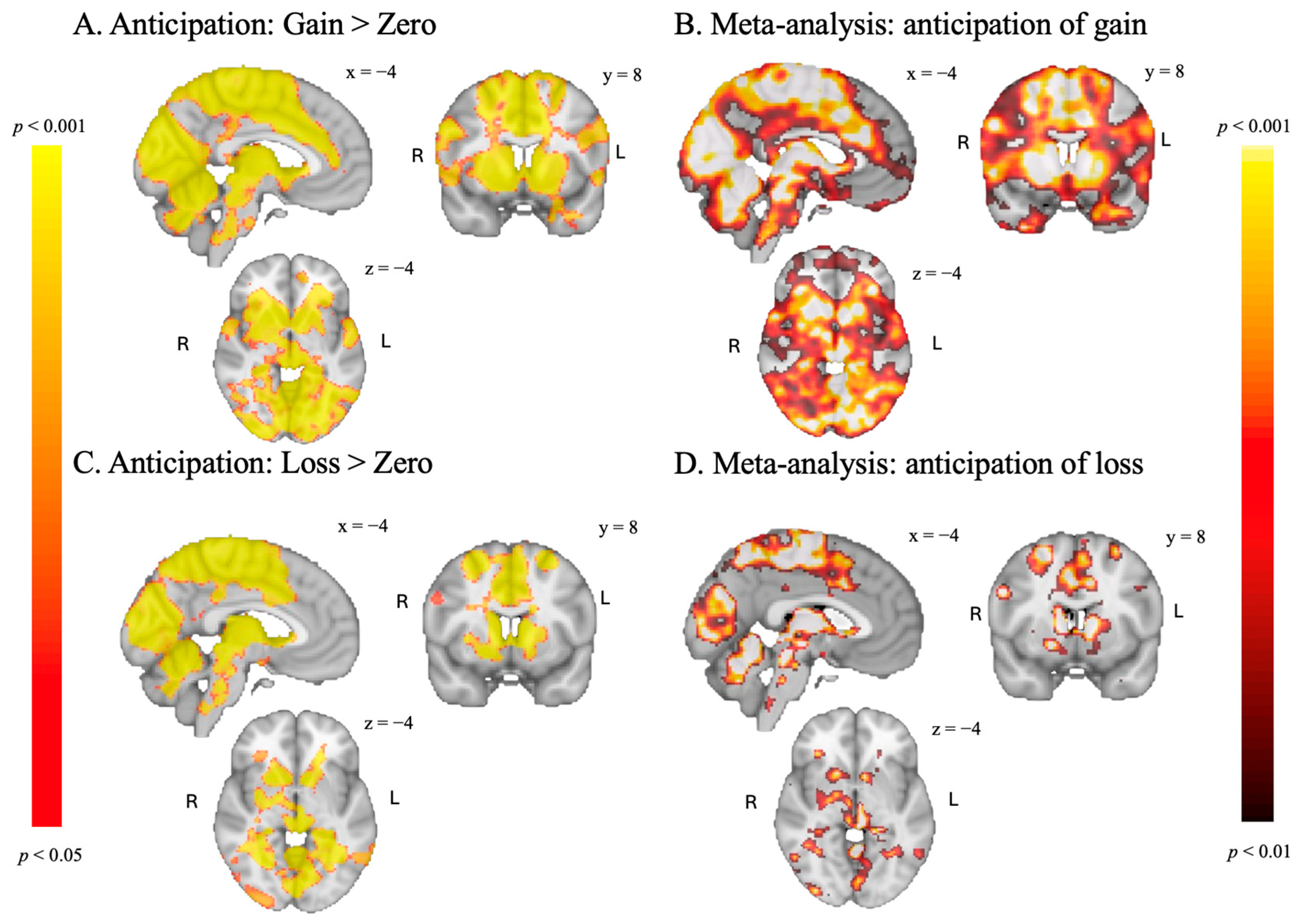
The SGID evoked significant activation related to reward anticipation in the bilateral NAcc and the anterior insula (AI) for both anticipation contrasts, consistent with several meta-analyses of reward processing (Oldham et al., 2018; Chen et al., 2022) and the original MID task by Kampa et al. (2018). Additionally, as seen in panel A and B in Figure 2, the results of the “Anticipation of Gain > Zero” contrast closely match activations from a meta-analysis of roughly 7000 participants on reward tasks (Waller et al., 2023), including in the precuneus and the insula. While not as strong, the “Anticipation of Loss > Zero” contrast also engages areas found in the Waller et al. meta-analysis, including in the left hippocampus (see Figure 2, panel C and D). These results indicate that our task successfully evokes activation in brain regions associated with anticipatory reward processing, as observed in traditional MID tasks.
To investigate the incentive magnitude manipulation (1 vs. 0.5 points), we examined activation across the five incentive magnitudes separately for left and right nucleus accumbens (NAcc). Figure 3 displays the mean activation (with SEM) for each magnitude. A clear parametric modulation of incentive magnitude was observed in both left and right NAcc activation, consistent with previous findings using the MID task (Knutson et al., 2001; Carter et al., 2009) and with Kampa et al. (2018) employing the same task with monetary rewards.
Post hoc contrasts did not reveal significant differences between anticipation of high versus low reward in either hemisphere (both p > 0.1), nor between high versus low loss in the left (p = 0.0153). In the right NAcc, however, activation was greater during anticipation of a high compared to a low loss (p = 0.01).

3.3.2. Feedback Phase

Consistent with the original MID task by Kampa et al. (2018), we found activation in the bilateral nucleus accumbens for the Gain > No Gain contrast. Additionally, as seen in panel A and B in Figure 4, the results of the Gain > No gain contrast overlap with the Waller et al. (2023) meta-analysis. In contrast to the original task by Kampa et al. (2018) and our expectations, we did not find activation in the nucleus accumbens for the No loss > Loss contrasts (see Figure 4). Parts of our activation appear to match the Waller meta-analysis, although we found less activation (see panel C and D in Figure 4).

3.3.3. Association Between Academic Buoyancy and Anticipation of Reward and Loss

We found a negative association between academic buoyancy and activation in the right amygdala when anticipating reward (see Figure 5), which was not found when anticipating loss. No associations with academic buoyancy were found for either contrast within the Nacc or vmPFC ROIs.

4. Discussion

In this study, we modified the traditional Monetary Incentive Delay (MID) task to a School Grade Incentive Delay (SGID) task, aiming to provide a more ecologically valid measure of reward sensitivity in academic contexts and to test the association between reward sensitivity and academic buoyancy. We found that the SGID task activated brain regions associated with reward anticipation, including the bilateral nucleus accumbens and anterior insula, similar to findings from previous MID studies (Bartra et al., 2013; Kampa et al., 2018) and a large meta-analysis of neural reward anticipation (Waller et al., 2023). Moreover, nucleus accumbens activation was also found when receiving a reward during the feedback phase, which is in concurrence with previous results (Kampa et al., 2018; Wu et al., 2014). In contrast, we did not find activation of the nucleus accumbens or other reward-related brain regions for having averted a loss. As we are specifically interested in reward sensitivity, we can conclude that our task effectively evokes brain activation related to reward processing and can therefore be used to assess reward sensitivity in an educational context in an ecologically more valid way.
Beyond validating the SGID, we examined associations between neural activation and academic buoyancy. After applying small-volume correction within our a priori regions of interest (bilateral Nacc, bilateral amygdala, and vmPFC), we found a negative association between academic buoyancy and activation in the right amygdala during reward anticipation. Contrary to our expectations, we did not find an association between academic buoyancy and ventral striatal and vmPFC activation.
These results appear to diverge from resilience research showing that stronger amygdala activation during reward anticipation buffered against the impact of life stressors (Fassett-Carman et al., 2023). Other studies similarly highlight that stress reduces neural reward responsibility, while stronger baseline reward responsiveness predicts lower stress reactivity (Pegg & Kujawa, 2024). Together, these findings support the idea that reward sensitivity plays a protective role under conditions of academic or emotional strain. Tentatively, the negative correlation between academic buoyancy and amygdala activation during reward anticipation may point to an alternative mechanism. Rather than heightened emotional responses, buoyant students may benefit from regulating affective reactivity to maintain focus and motivation under pressure. Given the exploratory nature of this finding and its divergence from prior work, further research is needed to clarify the role of the amygdala in academic stress contexts.
In conclusion, our findings suggest that the SGID task effectively activates brain regions implicated in reward processing that are observed in MID tasks, providing a promising tool for examining reward sensitivity in academic contexts. Our finding of a negative association between academic buoyancy and right amygdala activation during reward anticipation provides a first indication that buoyancy may be linked to affective downregulation rather than heightened reactivity and warrants further investigation. As part of the GUTS consortium (Crone et al., 2024), we are presently conducting a large-scale study in which we administer the SGID in a large sample of 10–14-year-old children and assess both trait and state measures of academic buoyancy to probe its dynamic, fluctuating nature. In this manner, we aim to provide clearer insights into how reward sensitivity supports students’ buoyancy in the face of everyday academic pressures.

Author Contributions

Conceptualization, A.C.K.v.D., B.R.J.J., H.M.H. and I.M.V.; Methodology, I.M.V. and M.J.B.V.T.; Formal Analysis, I.M.V. and M.J.B.V.T.; Investigation, M.J.B.V.T.; Resources, I.M.V.; Data Curation, M.J.B.V.T. and I.M.V.; Writing—Original Draft Preparation, B.R.J.J., H.M.H., I.M.V. and M.J.B.V.T.; Writing—Review and Editing, A.C.K.v.D., B.R.J.J., H.M.H., I.M.V. and M.J.B.V.T.; Visualization, M.J.B.V.T. and I.M.V.; Supervision, A.C.K.v.D., B.R.J.J., H.M.H. and I.M.V.; Project Administration, M.J.B.V.T.; Funding Acquisition, H.M.H. and A.C.K.v.D. All authors have read and agreed to the published version of the manuscript.

Funding

The GUTS program is funded by an NWO Gravitation program supported by the Dutch Ministry of Education, Culture and Science of the government of the Netherlands, Grant nr 024.005.011.

Institutional Review Board Statement

The study was conducted according to the guidelines with the Declaration of Helsinki and approved by the Institutional Review Board of the Faculty of Behavioural and Movement Sciences of the Vrije Universiteit Amsterdam (protocol code: VCWE-2023-114, 20 July 2023).

Informed Consent Statement

Informed consent was obtained from all subjects involved in the study.

Data Availability Statement

The fMRI results described in this article are available on Neurovault (see https://neurovault.org/collections/ZVTIRUHL).

Conflicts of Interest

The authors declare no conflicts of interest.

Abbreviations

The following abbreviations are used in this manuscript:
SGIDSchool Grade Incentive Delay (task)
MIDMonetary Incentive Delay (task)
NaccNucleus accumbens
dlPFCDorsolateral prefrontal cortex
dACCDorsal anterior cingulate cortex
GUTSGrowing Up Together in Society
ABSAcademic Buoyancy Scale
EVExplanatory variable
TFCEThreshold-Free Cluster Enhancement
AIAnterior insula

Appendix A

Table A1. Table of Local Maxima.
ContrastRegion of Local MaximumMNI (X Y Z)Corrected p-Value
Anticipation
Gain > Zero
Precuneus cortex−8 −70 220.001
Central Opercular Cortex−52 −22 140.003
Brain stem6 −38 −400.004
Superior Temporal Gyrus, anterior division62 4 00.009
Right Hippocampus28 −38 −20.003
Supramarginal Gyrus, anterior division−50 −30 380.002
Superior Temporal Gyrus, anterior division−54 −8 −100.003
Insular Cortex−34 −8 120.003
Middle Temporal Gyrus, temporooccipital part42 −48 100.009
Lateral Occipital Cortex, inferior division54 −62 −60.003
Precentral Gyrus−60 6 360.008
Cingulate Gyrus, posterior division14 −18 380.002
Inferior Frontal Gyrus, pars opercularis−42 12 220.008
Precentral Gyrus−42 4 300.008
Temporal Pole−54 6 −80.004
Temporal Pole−32 8 −320.011
Cingulate Gyrus, anterior division−4 42 80.002
Loss > Zero
Precentral Gyrus30 −18 50<0.001
Cerebellum0 −62 −22<0.001
Occipital Pole34 −92 −60.014
Supramarginal Gyrus, posterior division36 −42 360.004
Cerebellum−26 −48 −480.030
Lateral Occipital Cortex, superior division38 −74 340.003
Brain-Stem−2 −32 −340.003
Cerebellum−42 −44 −320.023
Right Cerebral White Matter30 28 200.008
Precentral Gyrus54 8 300.039
Cingulate Gyrus, posterior division14 −18 400.002
Cerebellum16 −54 −340.012
Left Cerebral White Matter−28 −52 26<0.001
Inferior Temporal Gyrus, temporooccipital part−46 −54 −200.003
Frontal Orbital Cortex34 32 −20.018
Left Cerebral White Matter−16 26 360.009
Lateral Occipital Cortex, inferior division54 −60 −40.031
Middle Temporal Gyrus, temporooccipital part−64 −60 −20.003
Left Hippocampus−26 −24 −160.003
(no structure available in Harvard-Oxford or MNI atlases)4 −4 −14<0.001
Feedback
Gain > No Gain
Precentral Gyrus−12 −32 680.004
Frontal Medial Cortex−14 46 −40.010
Middle Frontal Gyrus−28 32 460.018
Cerebellum34 −60 −420.018
Left Putamen−14 6 −120.010
Temporal Fusiform Cortex, posterior division−38 −26 −140.021
Cerebellum22 −68 −500.020
Left Cerebral White Matter−22 38 −60.014
Cerebellum−6 −66 −180.020
Juxtapositional Lobule Cortex (formerly known as Supplementary Motor Cortex)16 4 440.006
Left Hippocampus−20 −10 −200.029
Frontal Pole18 64 00.018
Postcentral Gyrus−54 −20 420.017
Inferior Temporal Gyrus, posterior division−42 −12 −240.031
Right Cerebral White Matter30 26 240.020
Left Cerebral White Matter−16 40 280.026
Superior Frontal Gyrus28 6 560.048
Lateral Occipital Cortex, superior division−24 −62 560.021
Interior Temporal Gyrus, temporooccipital part−52 −46 −320.030
Postcentral Gyrus−52 −16 280.018
Left Cerebral White Matter−20 −36 40.050
No Loss > Loss
Right Cerebral White Matter18 −8 280.011
Precentral Gyrus16 −26 620.011
Left Cerebral White Matter55 60 570.031
Left Cerebral White Matter−20 −6 420.050
Postcentral Gyrus42 −32 440.032
Precentral Gyrus32 −16 660.021
Right Cerebral White Matter18 20 200.022
Right Cerebral White Matter18 −42 200.047
Postcentral Gyrus44 −22 380.044

References

  1. Bartra, O., McGuire, J. T., & Kable, J. W. (2013). The valuation system: A coordinate-based meta-analysis of BOLD fMRI experiments examining neural correlates of subjective value. NeuroImage, 76, 412–427. [Google Scholar] [CrossRef]
  2. Ben-Zion, Z., Shany, O., Admon, R., Keynan, N. J., Avisdris, N., Balter, S. R., Shalev, A. Y., Liberzon, I., & Hendler, T. (2022). Neural responsivity to reward versus punishment shortly after trauma predicts long-term development of posttraumatic stress symptoms. Biological Psychiatry: Cognitive Neuroscience and Neuroimaging, 7(2), 150–161. [Google Scholar] [CrossRef]
  3. Bjork, J. M., Knutson, B., Fong, G. W., Caggiano, D. M., Bennett, S. M., & Hommer, D. W. (2004). Incentive-elicited brain activation in adolescents: Similarities and differences from young adults. The Journal of Neuroscience, 24(8), 1793–1802. [Google Scholar] [CrossRef]
  4. Carter, R. M., Macinnes, J. J., Huettel, S. A., & Adcock, R. A. (2009). Activation in the VTA and nucleus accumbens increases in anticipation of both gains and losses. Frontiers in Behavioral Neuroscience, 3, 21. [Google Scholar] [CrossRef]
  5. Chen, Y., Chaudhary, S., & Li, C.-S. R. (2022). Shared and distinct neural activity during anticipation and outcome of win and loss: A meta-analysis of the monetary incentive delay task. NeuroImage, 264, 119764. [Google Scholar] [CrossRef]
  6. Cohen, M. X. (2007). Individual differences and the neural representations of reward expectation and reward prediction error. Social Cognitive and Affective Neuroscience, 2(1), 20–30. [Google Scholar] [CrossRef]
  7. Corral-Frías, N. S., Nadel, L., Fellous, J.-M., & Jacobs, W. J. (2016). Behavioral and self-reported sensitivity to reward are linked to stress-related differences in positive affect. Psychoneuroendocrinology, 66, 205–213. [Google Scholar] [CrossRef] [PubMed]
  8. Corral-Frías, N. S., Nikolova, Y. S., Michalski, L. J., Baranger, D. A. A., Hariri, A. R., & Bogdan, R. (2015). Stress-related anhedonia is associated with ventral striatum reactivity to reward and transdiagnostic psychiatric symptomatology. Psychological Medicine, 45(12), 2605–2617. [Google Scholar] [CrossRef] [PubMed]
  9. Crone, E. A., Bol, T., Braams, B. R., De Rooij, M., Franke, B., Franken, I., Gazzola, V., Güroğlu, B., Huizenga, H., Hulshoff Pol, H., Keijsers, L., Keysers, C., Krabbendam, L., Jansen, L., Popma, A., Stulp, G., Van Atteveldt, N., Van Duijvenvoorde, A., & Veenstra, R. (2024). Growing Up Together in Society (GUTS): A team science effort to predict societal trajectories in adolescence and young adulthood. Developmental Cognitive Neuroscience, 67, 101403. [Google Scholar] [CrossRef]
  10. Daw, N. D., O’Doherty, J. P., Dayan, P., Seymour, B., & Dolan, R. J. (2006). Cortical substrates for exploratory decisions in humans. Nature, 441(7095), 876–879. [Google Scholar] [CrossRef] [PubMed]
  11. Dutcher, J. M., & Creswell, J. D. (2018). The role of brain reward pathways in stress resilience and health. Neuroscience and Biobehavioral Reviews, 95, 559–567. [Google Scholar] [CrossRef]
  12. Elman, I., Lowen, S., Frederick, B. B., Chi, W., Becerra, L., & Pitman, R. K. (2009). Functional neuroimaging of reward circuitry responsivity to monetary gains and losses in Posttraumatic Stress Disorder. Biological Psychiatry, 66(12), 1083–1090. [Google Scholar] [CrossRef]
  13. Ethridge, P., Ali, N., Racine, S. E., Pruessner, J. C., & Weinberg, A. (2020). Risk and resilience in an acute stress paradigm: Evidence from salivary cortisol and time-frequency analysis of the reward positivity. Clinical Psychological Science, 8(5), 872–889. [Google Scholar] [CrossRef]
  14. Fassett-Carman, A. N., Moser, A. D., Ruzic, L., Neilson, C., Jones, J., Barnes-Horowitz, S., Schneck, C. D., & Kaiser, R. H. (2023). Amygdala and nucleus accumbens activation during reward anticipation moderates the association between life stressor frequency and depressive symptoms. Journal of Affective Disorders, 330, 309–318. [Google Scholar] [CrossRef]
  15. Garrison, J., Erdeniz, B., & Done, J. (2013). Prediction error in reinforcement learning: A meta-analysis of neuroimaging studies. Neuroscience & Biobehavioral Reviews, 37(7), 1297–1310. [Google Scholar] [CrossRef] [PubMed]
  16. Geschwind, N., Peeters, F., Jacobs, N., Delespaul, P., Derom, C., Thiery, E., van Os, J., & Wichers, M. (2010). Meeting risk with resilience: High daily life reward experience preserves mental health. Acta Psychiatrica Scandinavica, 122(2), 129–138. [Google Scholar] [CrossRef]
  17. Gray, J. A. (1987). The psychology of fear and stress (2nd ed.). Cambridge University Press. [Google Scholar]
  18. Jenkinson, M., Beckmann, C. F., Behrens, T. E. J., Woolrich, M. W., & Smith, S. M. (2012). FSL. NeuroImage, 62(2), 782–790. [Google Scholar] [CrossRef] [PubMed]
  19. Kalisch, R., Baker, D. G., Basten, U., Boks, M. P., Bonanno, G. A., Brummelman, E., Chmitorz, A., Fernàndez, G., Fiebach, C. J., Galatzer-Levy, I., Geuze, E., Groppa, S., Helmreich, I., Hendler, T., Hermans, E. J., Jovanovic, T., Kubiak, T., Lieb, K., Lutz, B., … Kleim, B. (2017). The resilience framework as a strategy to combat stress-related disorders. Nature Human Behaviour, 1(11), 784–790. [Google Scholar] [CrossRef]
  20. Kampa, M., Schick, A., Yuen, K., Sebastian, A., Chmitorz, A., Saase, V., Wessa, M., Tüscher, O., & Kalisch, R. (2018). A combined behavioral and neuroimaging battery to test positive appraisal style theory of resilience in longitudinal studies. BioRxiv. [Google Scholar] [CrossRef]
  21. Kasparek, S. W., Jenness, J. L., & McLaughlin, K. A. (2020). Reward processing modulates the association between trauma exposure and externalizing psychopathology. Clinical Psychological Science, 8(6), 989–1006. [Google Scholar] [CrossRef]
  22. Knutson, B., Adams, C. M., Fong, G. W., & Hommer, D. (2001). Anticipation of increasing monetary reward selectively recruits nucleus accumbens. The Journal of Neuroscience, 21(16), RC159. [Google Scholar] [CrossRef]
  23. Knutson, B., Bhanji, J. P., Cooney, R. E., Atlas, L. Y., & Gotlib, I. H. (2008). Neural responses to monetary incentives in major depression. Biological Psychiatry, 63(7), 686–692. [Google Scholar] [CrossRef]
  24. Knutson, B., Westdorp, A., Kaiser, E., & Hommer, D. (2000). FMRI visualization of brain activity during a Monetary Incentive Delay task. NeuroImage, 12(1), 20–27. [Google Scholar] [CrossRef] [PubMed]
  25. Koenka, A. C., Linnenbrink-Garcia, L., Moshontz, H., Atkinson, K. M., Sanchez, C. E., & Cooper, H. (2021). A meta-analysis on the impact of grades and comments on academic motivation and achievement: A case for written feedback. Educational Psychology, 41(7), 922–947. [Google Scholar] [CrossRef]
  26. Kumar, P., Berghorst, L. H., Nickerson, L. D., Dutra, S. J., Goer, F. K., Greve, D. N., & Pizzagalli, D. A. (2014). Differential effects of acute stress on anticipatory and consummatory phases of reward processing. Neuroscience, 266, 1–12. [Google Scholar] [CrossRef]
  27. Liu, X., Hairston, J., Schrier, M., & Fan, J. (2011). Common and distinct networks underlying reward valence and processing stages: A meta-analysis of functional neuroimaging studies. Neuroscience and Biobehavioral Reviews, 35(5), 1219–1236. [Google Scholar] [CrossRef] [PubMed]
  28. Martin, A. J., & Marsh, H. W. (2008a). Academic buoyancy: Towards an understanding of students’ everyday academic resilience. Journal of School Psychology, 46(1), 53–83. [Google Scholar] [CrossRef]
  29. Martin, A. J., & Marsh, H. W. (2008b). Workplace and academic buoyancy: Psychometric assessment and construct validity amongst school personnel and students. Journal of Psychoeducational Assessment, 26, 168–184. [Google Scholar] [CrossRef]
  30. Martin, A. J., & Marsh, H. W. (2009). Academic resilience and academic buoyancy: Multidimensional and hierarchical conceptual framing of causes, correlates and cognate constructs. Oxford Review of Education, 35(3), 353–370. [Google Scholar] [CrossRef]
  31. Must, A., Szabó, Z., Bódi, N., Szász, A., Janka, Z., & Kéri, S. (2006). Sensitivity to reward and punishment and the prefrontal cortex in major depression. Journal of Affective Disorders, 90(2), 209–215. [Google Scholar] [CrossRef]
  32. Oldham, S., Murawski, C., Fornito, A., Youssef, G., Yücel, M., & Lorenzetti, V. (2018). The anticipation and outcome phases of reward and loss processing: A neuroimaging meta-analysis of the monetary incentive delay task. Human Brain Mapping, 39, 3398–3418. [Google Scholar] [CrossRef]
  33. Pegg, S., & Kujawa, A. (2024). The effects of stress on reward responsiveness: A systematic review and preliminary meta-analysis of the event-related potential literature. Cognitive, Affective & Behavioral Neuroscience, 24(1), 42–59. [Google Scholar] [CrossRef]
  34. Pizzagalli, D. A., Holmes, A. J., Dillon, D. G., Goetz, E. L., Birk, J. L., Bogdan, R., Dougherty, D. D., Iosifescu, D. V., Rauch, S. L., & Fava, M. (2009). Reduced caudate and nucleus accumbens response to rewards in unmedicated individuals with Major Depressive Disorder. The American Journal of Psychiatry, 166(6), 702–710. [Google Scholar] [CrossRef] [PubMed]
  35. Porcelli, A. J., Lewis, A. H., & Delgado, M. R. (2012). Acute stress influences neural circuits of reward processing. Frontiers in Neuroscience, 6, 2–9. [Google Scholar] [CrossRef]
  36. Schultz, W., Dayan, P., & Read Montague, P. (1997). A neural substrate of prediction and reward. Science, 275(5306), 1593–1599. [Google Scholar] [CrossRef]
  37. Smith, S. M., & Nichols, T. E. (2009). Threshold-free cluster enhancement: Addressing problems of smoothing, threshold dependence and localisation in cluster inference. NeuroImage, 44(1), 83–98. [Google Scholar] [CrossRef]
  38. Stan, E. (2012). The role of grades in motivating students to learn. Procedia, Social and Behavioral Sciences, 69, 1998–2003. [Google Scholar] [CrossRef]
  39. Tashjian, S. M., & Galván, A. (2018). The role of mesolimbic circuitry in buffering election-related distress. Journal of Neuroscience, 38(11), 2887–2898. [Google Scholar] [CrossRef]
  40. Telzer, E. H., Fuligni, A. J., Lieberman, M. D., & Galván, A. (2014). Neural sensitivity to eudaimonic and hedonic rewards differentially predict adolescent depressive symptoms over time. Proceedings of the National Academy of Sciences of the United States of America, 111(18), 6600–6605. [Google Scholar] [CrossRef]
  41. Vidal-Ribas, P., Benson, B., Vitale, A. D., Keren, H., Harrewijn, A., Fox, N. A., Pine, D. S., & Stringaris, A. (2019). Bidirectional associations between stress and reward processing in children and adolescents: A longitudinal neuroimaging study. Biological Psychiatry: Cognitive Neuroscience and Neuroimaging, 4(10), 893–901. [Google Scholar] [CrossRef]
  42. Wackerhagen, C., Veer, I. M., van Leeuwen, J. M. C., Reppmann, Z., Riepenhausen, A., Bögemann, S. A., Mor, N., Puhlmann, L. M. C., Uściƚko, A., Zerban, M., Mituniewicz, J., Lerner, A., Yuen, K. S. L., Köber, G., Marciniak, M. A., Pooseh, S., Weermeijer, J., Arias-Vásquez, A., Binder, H., … Walter, H. (2023). Dynamic modelling of mental resilience in young adults: Protocol for a longitudinal observational study (DynaM-OBS). JMIR Research Protocols, 12, e39817. [Google Scholar] [CrossRef] [PubMed]
  43. Waller, L., Veer, I. M., Pizzagalli, F., Han, L. K. M., Penninx, B. W. J. H., Liao, Z., Paus, T., Strike, L. T., Quidé, Y., Green, M. J., Harrison, B., Jamieson, A. J., Davey, C., Knodt, A., Hariri, A., Hermesdorf, M., Berger, K., Dannlowski, U., Kircher, T., … Walter, H. (2023, July 22–26). Meta-analytic results for task-based fMRI in the ENIGMA consortium [Conference abstract]. OHBM 2023 Annual Meeting, Montréal, QC, Canada. [Google Scholar]
  44. Winkler, A. M., Ridgway, G. R., Webster, M. A., Smith, S. M., & Nichols, T. E. (2014). Permutation inference for the general linear model. NeuroImage, 92, 381–397. [Google Scholar] [CrossRef] [PubMed]
  45. Wu, C. C., Samanez-Larkin, G. R., Katovich, K., & Knutson, B. (2014). Affective traits link to reliable neural markers of incentive anticipation. NeuroImage, 84, 279–289. [Google Scholar] [CrossRef] [PubMed]
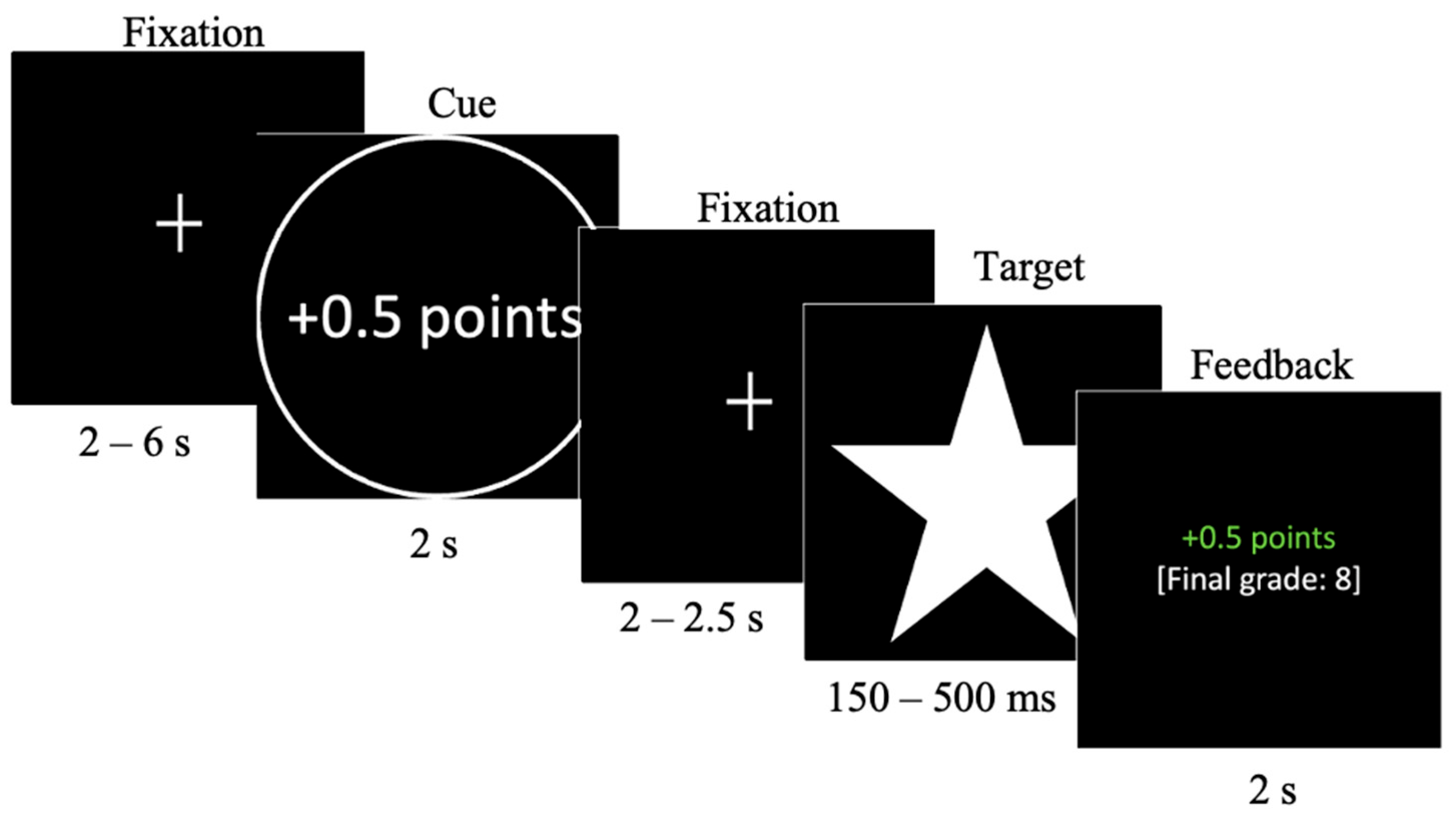
Figure 1. Structure of a gain trial during the SGID task.
Behavsci 15 01321 g001
Figure 2. (A,C): results of the anticipation phase of the SGID, TFCE-corrected at p < 0.05. (B,D): results of a reward task meta-analysis (Waller et al., 2023), p < 0.01, uncorrected.
Behavsci 15 01321 g002
Figure 3. Parametric modulation of nucleus accumbens activation by incentive magnitude during reward anticipation. Bars represent mean activation (with SEM) for the five incentive magnitudes (−1, −0.5, 0, +0.5, +1 points).
Behavsci 15 01321 g003
Figure 4. (A,C): results of the feedback phase of the SGID, TFCE-corrected at p < 0.05. (B,D): results of a reward task meta-analysis (Waller et al., 2023), p < 0.01, uncorrected.
Behavsci 15 01321 g004
Figure 5. (A): results of the association between ABS scores and activation for the anticipation of reward, small-volume TFCE-corrected at p < 0.0167. (B): scatterplot illustrating the association (blue line) and 95% confidence interval (grey area) between ABS scores and right amygdala activity when anticipating a re-ward.
Behavsci 15 01321 g005
Disclaimer/Publisher’s Note: The statements, opinions and data contained in all publications are solely those of the individual author(s) and contributor(s) and not of MDPI and/or the editor(s). MDPI and/or the editor(s) disclaim responsibility for any injury to people or property resulting from any ideas, methods, instructions or products referred to in the content.