Modeling Hemorrhagic Shock in Male SD Rats by Fixed Volume Blood Withdrawal Followed by Partial Resuscitation and Long-Term Outcome Assessment
 , , , , , , , and
Abstract
1. Introduction
2. Materials and Methods
2.1. Animals
2.2. Design Description
2.3. Drugs in the Study
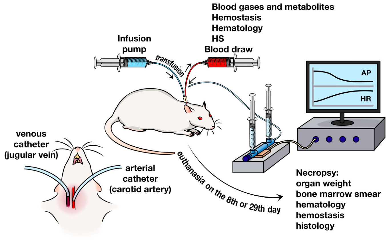
2.4. HS Modeling and VRT
2.5. VRT Procedure
2.6. VRT Issues Preparation
2.6.1. Preservative/Anticoagulant Preparation
2.6.2. Preparation of Autologous Blood for TF
2.7. AP and HR Registration
2.8. Blood Sample Collection
2.9. Removal of Catheters 48 h After HS
2.10. Blood Gas and Metabolite Analysis
2.11. Body Temperature Registration
2.12. Hematology Analysis
| Red blood cell count (RBC) |
| Hemoglobin level (Hb) |
| Hematocrit (HCT) |
| White blood cell count (WBC) |
| Mean corpuscular hemoglobin content (MCH) |
| Mean corpuscular hemoglobin concentration (MCHC) |
| Mean corpuscular volume (MCV) |
| Red cell distribution width—variation coefficient (RDW) |
| Red cell distribution width-standard deviation (RDW-SD) |
| Platelet count (PLT) |
| Mean platelet volume—variation coefficient (MPV) |
| Plateletcrit (PCT) |
| Platelet distribution width—variation coefficient (PDW) |
| Lymphocytes (LYM) |
| Monocytes (MON) |
| Granulocytes (GRA) |
2.13. Hemostasis Analysis
2.14. Registration of Mortality and Assessment of Animal Welfare
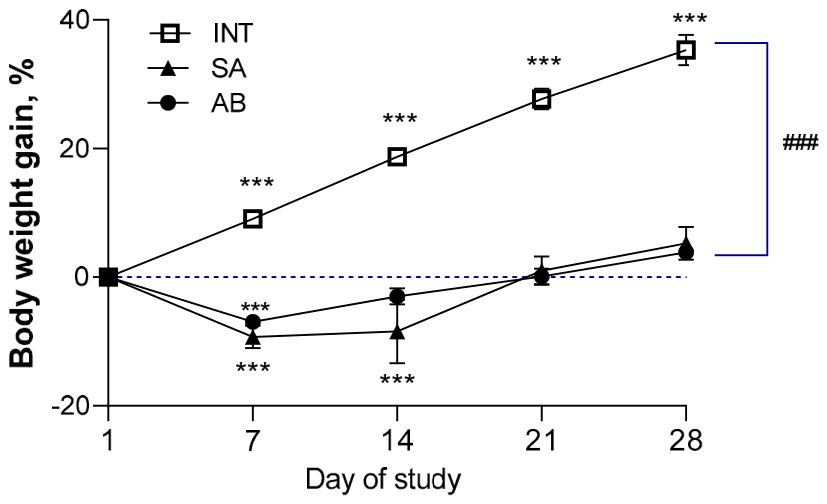
2.15. Registration of Body Weight Gain
2.16. Euthanasia
2.17. Necropsy and Organ Weighing
2.18. Bone Marrow Smear and Myelogram Analysis
2.19. Histology
2.20. Statistics
3. Results
3.1. Mortality
3.2. Weight Gain
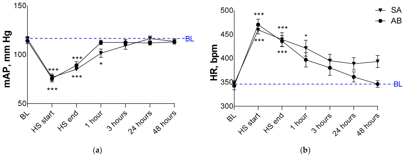
3.3. AP and HR
3.4. Body Temperature
3.5. Registration of Blood Gases and Metabolites with a Gas Analyzer (Oximetry)
3.6. Hematology
3.7. Myelogram
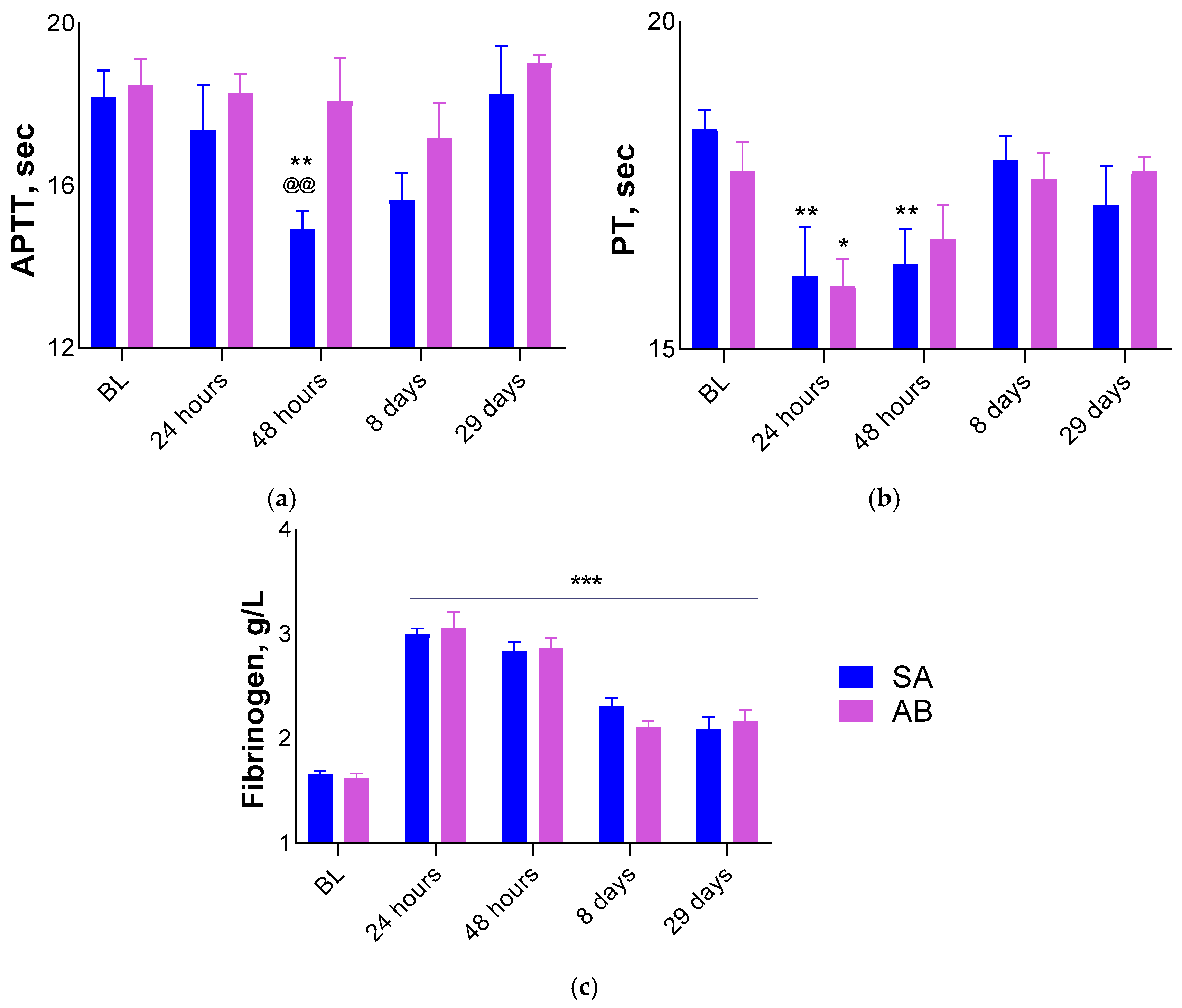
3.8. Hemostasis
3.9. Organ Weight
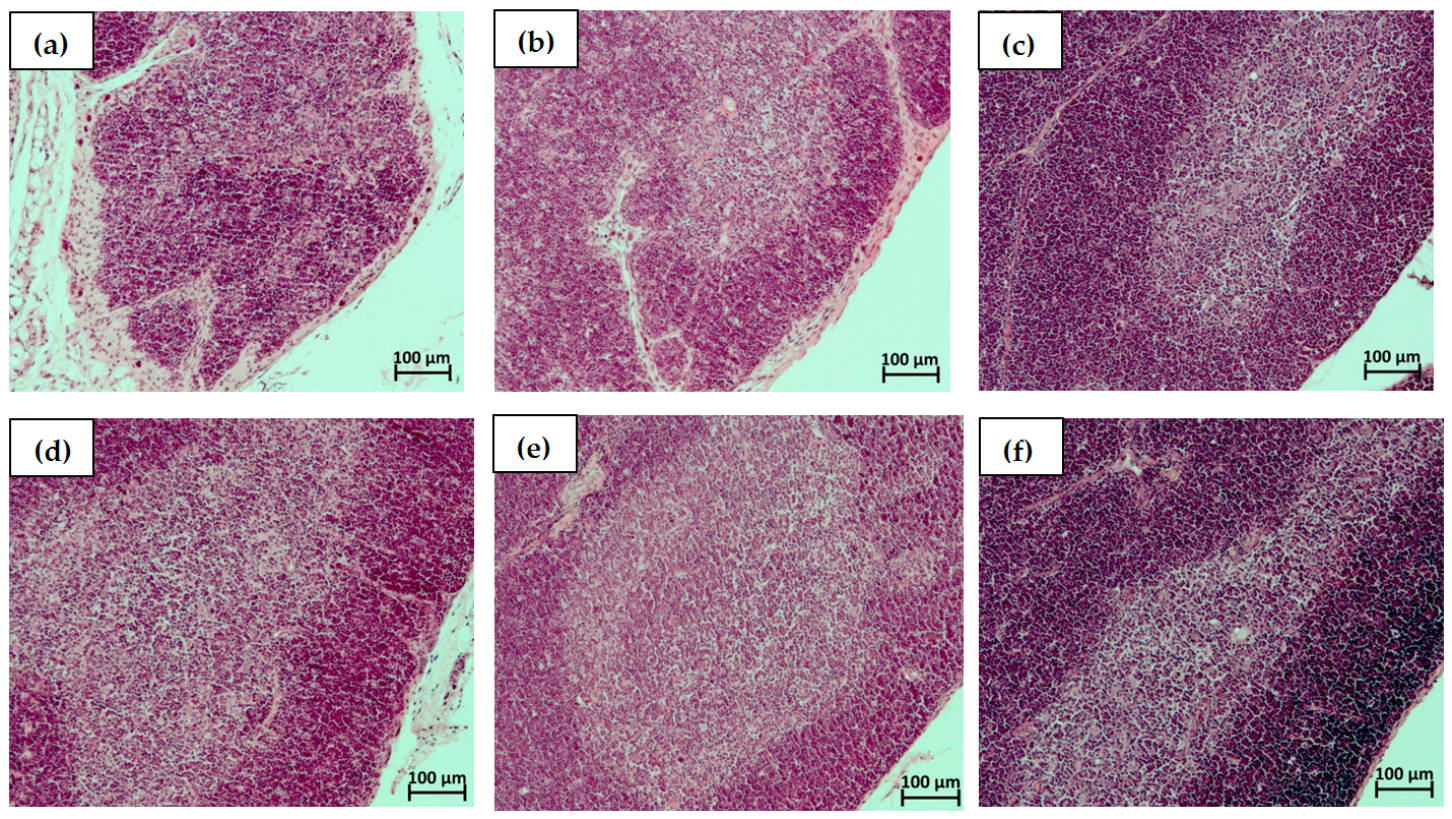
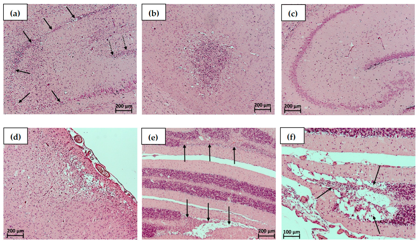
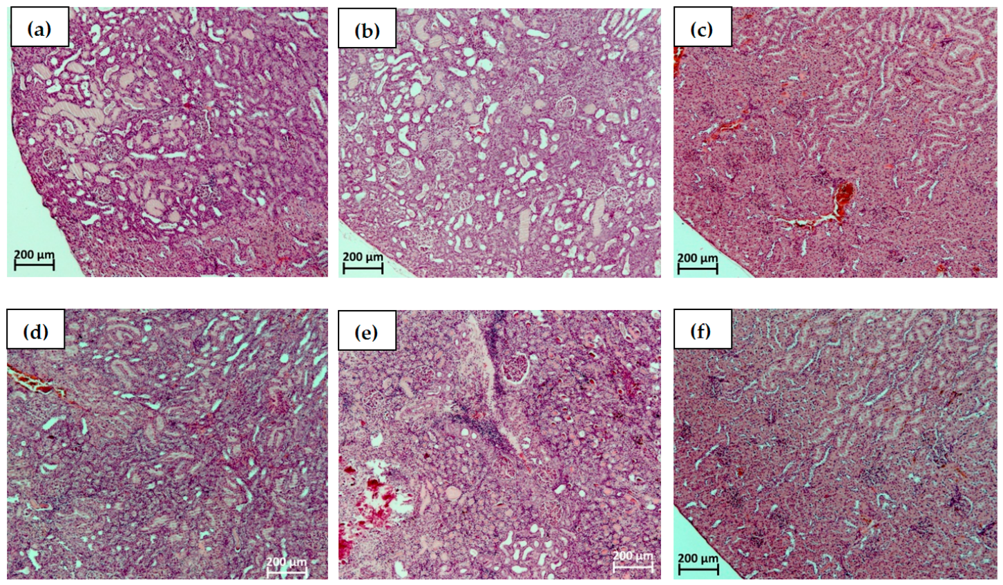
3.10. Histology
4. Discussion
5. Conclusions
Author Contributions
Funding
Institutional Review Board Statement
Informed Consent Statement
Data Availability Statement
Conflicts of Interest
Abbreviations
| HS | hemorrhagic shock |
| CBV | circulating blood volume |
| AP | arterial pressure |
| HR | heart rate |
| TF | transfusion |
| VRT | volume resuscitation therapy |
| AB | autologous blood |
| AS | albumin-containing saline (NaCl 0.9% + Albumin 20% (7:1)) |
| INT | intact |
| SD rat | Sprague Dawley rat |
| SPF | specific pathogen free |
| cBase (Ecf) | calculated Base, Extracellular fluid |
| pCO2 | carbon dioxide partial pressure |
| pO2 | oxygen partial pressure |
| ctHb | total hemoglobin concentration |
| cGlu | total glucose concentration |
| cLac | total lactate concentration |
| CaO2 | total amount of oxygen transported in arterial blood |
| APTT | activated partial thromboplastin time |
| PT | prothrombin time |
| Fg | fibrinogen |
| K3EDTA | tripotassium edetate |
| RBC | red blood cell |
| Hb | hemoglobin |
| HCT | hematocrit |
| WBC | white blood cell |
| MCH | mean corpuscular haemoglobin |
| MCHC | mean cell haemoglobin concentration |
| MCV | mean corpuscular volume |
| RDW | red cell distribution width—variation coefficient |
| RDW-SD | red cell distribution width-standard deviation |
| PLT | platelets |
| MPV | mean platelet volume—variation coefficient |
| PCT | plateletcrit |
| PDW | platelet distribution width—variation coefficient |
| LYM | lymphocytes |
| MON | monocytes |
| GRA | granulocytes |
| PBS | phosphate-buffer saline |
| HPCs | hematopoietic progenitor cells |
| SIRS | systemic inflammatory response syndrome |
| IL | interleukin |
| ATP | adenosine triphosphate |
| IFN | interferon |
| Mip | macrophage inflammatory protein |
| NF | nuclear factor |
References
- Meza Monge, K.; Rosa, C.; Sublette, C.; Pratap, A.; Kovacs, E.J.; Idrovo, J.-P. Navigating Hemorrhagic Shock: Biomarkers, Therapies, and Challenges in Clinical Care. Biomedicines 2024, 12, 2864. [Google Scholar] [CrossRef] [Scilit] [PubMed]
- Pigneri, D.A.; Beldowicz, B.; Jurkovich, G.J. Trauma systems: Origins, evolution, and current challenges. Surg. Clin. N. Am. 2017, 97, 947–959. [Google Scholar] [PubMed]
- GBD 2017 Causes of Death Collaborators. Global, regional, and national age-sex-specific mortality for 282 causes of death in 195 countries and territories, 1980–2017: A systematic analysis for the Global Burden of Disease Study 2017. Lancet 2018, 392, 1736–1788. [CrossRef] [Scilit] [PubMed]
- Tisherman, S.A.; Schmicker, R.H.; Brasel, K.J.; Bulger, E.M.; Kerby, J.D.; Minei, J.P.; Powell, J.L.; Reiff, D.A.; Rizoli, S.B.; Schreiber, M.A. Detailed description of all deaths in both the shock and traumatic brain injury hypertonic saline trials of the Resuscitation Outcomes Consortium. Ann. Surg. 2015, 261, 586–590. [Google Scholar] [CrossRef] [Scilit] [PubMed]
- Latif, R.K.; Clifford, S.P.; Baker, J.A.; Lenhardt, R.; Haq, M.Z.; Huang, J.; Farah, I.; Businger, J.R. Traumatic hemorrhage and chain of survival. Scand. J. Trauma Resusc. Emerg. Med. 2023, 31, 25. [Google Scholar] [CrossRef] [Scilit] [PubMed]
- Cannon, J.W. Hemorrhagic Shock. N. Engl. J. Med. 2018, 378, 370–379. [Google Scholar] [CrossRef] [Scilit] [PubMed]
- Chaudry, I.H. Cellular mechanisms in shock and ischemia and their correction. Am. J. Physiol. 1983, 245, R117–R134. [Google Scholar] [CrossRef] [Scilit] [PubMed]
- Cosgriff, N.; Moore, E.E.; Sauaia, A.; KennyMoynihan, M.; Burch, J.M.; Galloway, B. Predicting life-threatening coagulopathy in the massively transfused trauma patient: Hypothermia and acidoses revisited. J. Trauma 1997, 42, 857–861. [Google Scholar] [PubMed]
- Iwata, S.; Tanaka, A.; Ozawa, K. Alterations in the proton ATPase activity of rat liver mitochondria after hemorrhagic shock. J. Lab. Clin. Med. 1992, 120, 420–427. [Google Scholar] [PubMed]
- White, N.J.; Ward, K.R.; Pati, S.; Strandenes, G.; Cap, A.P. Hemorrhagic blood failure: Oxygen debt, coagulopathy, and endothelial damage. J. Trauma Acute Care Surg. 2017, 82, S41–S49. [Google Scholar] [PubMed]
- Moore, H.B.; Moore, E.E.; Liras, I.N.; Gonzalez, E.; Harvin, J.A.; Holcomb, J.B.; Sauaia, A.; Cotton, B.A. Acute fibrinolysis shutdown after injury occurs frequently and increases mortality: A multicenter evaluation of 2,540 severely injured patients. J. Am. Coll. Surg. 2016, 222, 347–355. [Google Scholar] [CrossRef] [Scilit] [PubMed]
- Lier, H.; Krep, H.; Schroeder, S.; Stuber, F. Preconditions of hemostasis in trauma: A review. The influence of acidosis, hypocalcemia, anemia, and hypothermia on functional hemostasis in trauma. J. Trauma 2008, 65, 951–960. [Google Scholar] [CrossRef] [Scilit] [PubMed]
- Tompkins, R.G. Genomics of injury: The Glue Grant experience. J. Trauma Acute Care Surg. 2015, 78, 671–686. [Google Scholar] [CrossRef] [Scilit] [PubMed]
- Xiao, W.; Mindrinos, M.N.; Seok, J.; Cuschieri, J.; Cuenca, A.G.; Gao, H.; Hayden, D.L.; Hennessy, L.; Moore, E.E.; Minei, J.P.; et al. Inflammation, Host Response to Injury Large-Scale Collaborative Research P. A genomic storm in critically injured humans. J. Exp. Med. 2011, 208, 2581–2590. [Google Scholar] [CrossRef] [Scilit] [PubMed]
- Nathan, C.; Ding, A. Nonresolving inflammation. Cell 2010, 140, 871–882. [Google Scholar] [CrossRef] [Scilit] [PubMed]
- Hierholzer, C.; Billiar, T.R. Molecular mechanisms in the early phase of hemorrhagic shock. Langenbecks Arch. Surg. 2001, 386, 302–308. [Google Scholar] [CrossRef] [Scilit] [PubMed]
- Maegele, M.; Lefering, R.; Yucel, N.; Tjardes, T.; Rixen, D.; Paffrath, T.; Simanski, C.; Neugebauer, E.; Bouillon, B.; AG Polytrauma of the German Trauma Society. Early coagulopathy in multiple injury: An analysis from the German Trauma Registry on 8724 patients. Injury 2007, 38, 298–304. [Google Scholar] [CrossRef] [Scilit] [PubMed]
- Balvers, K.; Van der Horst, M.; Graumans, M.; Boer, C.; Binnekade, J.M.; Goslings, J.C.; Juffermans, N.P. Hypothermia as a predictor for mortality in trauma patients at admittance to the Intensive Care Unit. J. Emerg. Trauma Shock. 2016, 9, 97–102. [Google Scholar] [CrossRef] [Scilit] [PubMed]
- Gale, S.C.; Kocik, J.F.; Creath, R.; Crystal, J.S.; Dombrovskiy, V.Y. A comparison of initial lactate and initial base deficit as predictors of mortality after severe blunt trauma. J. Surg. Res. 2016, 205, 446–455. [Google Scholar] [CrossRef] [Scilit] [PubMed]
- Martin, M.; Oh, J.; Currier, H.; Tai, N.; Beekley, A.; Eckert, M.; Holcomb, J. An analysis of in-hospital deaths at a modern combat support hospital. J. Trauma. 2009, 66, S51–S60; discussion S60–S61. [Google Scholar] [CrossRef] [Scilit] [PubMed]
- Smith, W.; Williams, A.; Agudelo, J.; Shannon, M.; Morgan, S.; Stahel, P.; Moore, E. Early predictors of mortality in hemodynamically unstable pelvis fractures. J. Orthop. Trauma. 2007, 21, 31–37. [Google Scholar] [CrossRef] [Scilit] [PubMed]
- National Academies of Sciences, Engineering, and Medicine. A National Trauma Care System: Integrating Military and Civilian Trauma Systems to Achieve Zero Preventable Deaths After Injury; National Academies Press: Washington, DC, USA, 2016. [Google Scholar]
- Hussmann, B.; Lefering, R.; Waydhas, C.; Touma, A.; Kauther, M.D.; Ruchholtz, S.; Lendemans, S. Does increased prehospital replacement volume lead to a poor clinical course and an increased mortality? A matched-pair analysis of 1896 patients of the Trauma Registry of the German Society for Trauma Surgery who were managed by an emergency doctor at the accident site. Injury 2013, 44, 611–617. [Google Scholar] [CrossRef] [Scilit] [PubMed]
- Haut, E.R.; Kalish, B.T.; Cotton, B.A.; Efron, D.T.; Haider, A.H.; Stevens, K.A.; Kieninger, A.N.; Cornwell, E.E.; Chang, D.C. Prehospital intravenous fluid administration is associated with higher mortality in trauma patients: A National Trauma Data Bank analysis. Ann. Surg. 2011, 253, 371–377. [Google Scholar] [PubMed]
- Cannon, J.W.; Khan, M.A.; Raja, A.S.; Cohen, M.J.; Como, J.J.; Cotton, B.A.; Dubose, J.J.; Fox, E.E.; Inaba, K.; Rodriguez, C.J.; et al. Damage control resuscitation in patients with severe traumatic hemorrhage: A practice management guideline from the Eastern Association for the Surgery of Trauma. J. Trauma Acute Care Surg. 2017, 82, 605–617. [Google Scholar] [CrossRef] [Scilit] [PubMed]
- Yunos, N.M.; Bellomo, R.; Hegarty, C.; Story, D.; Ho, L.; Bailey, M. Association between a chloride-liberal vs chloride-restrictive intravenous fluid administration strategy and kidney injury in critically ill adults. JAMA 2012, 308, 1566–1572. [Google Scholar] [CrossRef] [Scilit] [PubMed]
- Chowdhury, A.H.; Cox, E.F.; Francis, S.T.; Lobo, D.N. A randomized, controlled, double-blind crossover study on the effects of 2-L infusions of 0.9% saline and plasma-lyte(R) 148 on renal blood flow velocity and renal cortical tissue perfusion in healthy volunteers. Ann. Surg. 2012, 256, 18–24, Erratum in Ann Surg. 2013, 258, 1118. [Google Scholar] [CrossRef] [Scilit] [PubMed]
- Young, J.B.; Utter, G.H.; Schermer, C.R.; Galante, J.M.; Phan, H.H.; Yang, Y.; Anderson, B.A.; Scherer, L.A. Saline versus Plasma-Lyte A in initial resuscitation of trauma patients: A randomized trial. Ann. Surg. 2014, 259, 255–262. [Google Scholar] [CrossRef] [Scilit] [PubMed]
- Holcomb, J.B.; Tilley, B.C.; Baraniuk, S.; Fox, E.E.; Wade, C.E.; Podbielski, J.M.; del Junco, D.J.; Brasel, K.J.; Bulger, E.M.; Callcut, R.A.; et al. Transfusion of plasma, platelets, and red blood cells in a 1:1:1 vs a 1:1:2 ratio and mortality in patients with severe trauma: The PROPPR randomized clinical trial. JAMA 2015, 313, 471–482. [Google Scholar] [CrossRef] [Scilit] [PubMed]
- Kornblith, L.Z.; Howard, B.M.; Cheung, C.K.; Dayter, Y.; Pandey, S.; Busch, M.P.; Pati, S.; Callcut, R.A.; Vilardi, R.F.; Redick, B.J.; et al. The whole is greater than the sum of its parts: Hemostatic profles of whole blood variants. J. Trauma Acute Care Surg. 2014, 77, 818–827. [Google Scholar] [CrossRef] [Scilit] [PubMed]
- Shea, S.M.; Staudt, A.M.; Thomas, K.A.; Schuerer, D.; Mielke, J.E.; Folkerts, D.; Lowder, E.; Martin, C.; Bochicchio, G.V.; Spinella, P.C. The use of low-titer group O whole blood is independently associated with improved survival compared to component therapy in adults with severe traumatic hemorrhage. Transfusion 2020, 60, S2–S9. [Google Scholar] [CrossRef] [Scilit] [PubMed]
- Costantini, T.W.; Kornblith, L.Z.; Pritts, T.; Coimbra, R. The intersection of coagulation activation and inflammation after injury What you need to know. J. Trauma Acute Care Surg. 2024, 96, 347–356. [Google Scholar] [CrossRef] [Scilit] [PubMed]
- Tyburski, J.G.; Collinge, J.D.; Wilson, R.F.; Carlin, A.M.; Albaran, R.G.; Stefes, C.P. End-tidal CO 2-derived values during emergency trauma surgery correlated with outcome: A prospective study. J. Trauma 2002, 53, 738–743. [Google Scholar] [CrossRef] [Scilit] [PubMed]
- Caputo, N.; Fraser, R.; Paliga, A.; Kanter, M.; Hosford, K.; Madlinger, R. Triage vital signs do not correlate with serum lactate or base deficit, and are less predictive of operative intervention in penetrating trauma patients: A prospective cohort study. Emerg. Med. J. 2013, 30, 546–550. [Google Scholar] [CrossRef] [Scilit] [PubMed]
- Herbert, H.K.; Dechert, T.A.; Wolfe, L.; Aboutanos, M.B.; Malhotra, A.K.; Ivatury, R.R.; Duane, T.M. Lactate in trauma: A poor predictor of mortality in the setting of alcohol ingestion. Am. Surg. 2011, 77, 1576–1579. [Google Scholar] [CrossRef] [Scilit]
- Mutschler, M.; Nienaber, U.; Brockamp, T.; Wafaisade, A.; Fabian, T.; Paffrath, T.; Bouillon, B.; Maegele, M. Renaissance of base deficit for the initial assessment of trauma patients: A base deficitbased classification for hypovolemic shock developed on data from 16,305 patients derived from the TraumaRegister DGU(R). Crit. Care 2013, 17, R42. [Google Scholar] [CrossRef] [Scilit] [PubMed]
- Johansson, P.I.; Stensballe, J. Effect of haemostatic control resuscitation on mortality in massively bleeding patients: A before and after study. Vox Sang. 2009, 96, 111–118. [Google Scholar] [CrossRef] [Scilit] [PubMed]
- Luo, X.; Chen, G.; You, G.; Wang, B.; Lu, M.; Zhao, J.; Wang, Y.; Yin, Y.; Zhao, L.; Zhou, H. Gradually increased oxygen administration promoted survival after hemorrhagic shock. Exp. Biol. Med. 2016, 241, 1603–1610. [Google Scholar] [CrossRef] [Scilit] [PubMed]
- Hof, S.; Krüll, L.; Stepanyan, L.; Jeikowski, D.; Marcus, C.; Kuebart, A.; Relja, B.; Herminghaus, A.; Vollmer, C.; Bauer, I.; et al. Remote ischemic preconditioning improves ileal microvascular oxygenation during rodent hemorrhagic shock without improving variables of microcirculation and mitochondrial respiration. Sci. Rep. 2025, 15, 43230. [Google Scholar] [CrossRef] [Scilit] [PubMed]
- Bonanno, F.G. Management of hemorrhagic shock: Physiology approach, timing and strategies. J. Clin. Med. 2022, 12, 260. [Google Scholar] [CrossRef] [Scilit] [PubMed]
- Warren, M.; Subramani, K.; Schwartz, R.; Raju, R. Mitochondrial dysfunction in rat splenocytes following hemorrhagic shock. Biochim. Biophys. Acta Mol. Basis Dis. 2017, 1863, 2526–2533. [Google Scholar] [CrossRef] [Scilit] [PubMed]
- Denk, S.; Weckbach, S.; Eisele, P.; Braun, C.K.; Wiegner, R.; Ohmann, J.J.; Wrba, L.; Hoenes, F.M.; Kellermann, P.; Radermacher, P.; et al. Role of hemorrhagic shock in experimental polytrauma. Shock 2018, 49, 154–163. [Google Scholar] [CrossRef] [Scilit] [PubMed]
- Wu, X.; Dubick, M.A.; Schwacha, M.G.; Cap, A.P.; Darlington, D.N. Tranexamic acid attenuates the loss of lung barrier function in a rat model of polytrauma and hemorrhage with resuscitation. Shock 2017, 47, 500–505. [Google Scholar] [CrossRef] [Scilit] [PubMed]
- Chen, J.; Wu, X.; Keesee, J.; Liu, B.; Darlington, D.N.; Cap, A.P. Limited resuscitation with fresh or stored whole blood corrects cardio-vascular and metabolic function in a rat model of polytrauma and hemorrhage. Shock 2017, 47, 208–216. [Google Scholar] [CrossRef] [Scilit] [PubMed]
- Wichmann, M.W.; Ayala, A.; Chaudry, I.H. Severe depression of host immune functions following closed- bone fracture, soft-tissue trauma, and hemorrhagic shock. Crit. Care Med. 1998, 26, 1372–1378. [Google Scholar] [CrossRef] [Scilit] [PubMed]
- GV-SOLAS, Committee for Animal Welfare Officers, and TVT, Blood Sampling in Laboratory Animals, July 2017. Available online: https://www.gv-solas.de/wp-content/uploads/2017/03/tie_blutentnahme17_e.pdf (accessed on 4 May 2026).
- Guidelines for Survival Bleeding of Mice and Rats. NIH-ARAC Guidelines [Online]. 12 January 2005. Available online: http://oacu.od.nih.gov/ARAC/Bleeding.pdf (accessed on 4 May 2026).
- Roberts, I.; Blackhall, K.; Alderson, P.; Bunn, F.; Schierhout, G.; Cochrane Injuries Group. Human albumin solution for resuscitation and volume expansion in critically ill patients. Cochrane Database Syst. Rev. 2000, 2, CD001208. [Google Scholar]
- Wettstein, R.; Wilkins, R. Interpretation of blood gases. In Clinical Assessment in Respiratory Care, 6th ed.; Mosby: St. Louis, MO, USA, 2010. [Google Scholar]
- Gutierrez, G.; Reines, H.D.; Wulf-Gutierrez, M.E. Clinical review: Hemorrhagic shock. Crit. Care 2004, 8, 373–381. [Google Scholar] [CrossRef] [Scilit] [PubMed]
- Mallat, J.; Vallet, B. Ratio of venous-to-arterial PCO2 to arteriovenous oxygen content difference during regional ischemic or hypoxic hypoxia. Sci. Rep. 2021, 11, 10172. [Google Scholar] [CrossRef] [Scilit] [PubMed]
- Piaton, E.; Fabre, M.; Goubin-Versini, I.; Bretz-Grenier, M.-F.; Courtade-Saïdi, M.; Vincent, S.; Belleannée, G.; Thivolet, F.; Boutonnat, J.; Debaque, H.; et al. Guidelines for May-Grünwald–Giemsa staining in haematology and non-gynaecological cytopathology: Recommendations of the French Society of Clinical Cytology (SFCC) and of the French Association for Quality Assurance in Anatomic and Cytologic Pathology (AFAQAP). Cytopathology 2016, 27, 359–368. [Google Scholar] [CrossRef] [Scilit] [PubMed]
- Gallimore, E. Clinical features and management of haemorrhagic shock. Nurs. Stand. 2015, 30, 51–60. [Google Scholar] [CrossRef] [Scilit] [PubMed]
- Copotoiu, R.; Cinca, E.; Collange, O.; Levy, F.; Mertes, P.-M. Pathophysiology of hemorragic shock. Transfus. Clin. Biol. 2016, 23, 222–228. [Google Scholar] [PubMed]
- Gann, D.S.; Drucker, W.R. Hemorrhagic shock. J. Trauma Acute Care Surg. 2013, 75, 888–895. [Google Scholar] [CrossRef] [Scilit] [PubMed]
- Li, T.; Zhu, Y.; Tian, K.; Xue, M.; Peng, X.; Lan, D.; Liu, L. Ideal resuscitation pressure for uncontrolled hemorrhagic shock in different ages and sexes of rats. Crit. Care 2013, 17, R194. [Google Scholar] [CrossRef] [Scilit] [PubMed]
- Wanot, B.; Jasikowska, K.; Niewiadomska, E.; Biskupek-Wanot, A. Cardiovascular effects of H3 histamine receptor inverse agonist/H4 histamine receptor agonist, clobenpropit, in hemorrhage-shocked rats. PLoS ONE 2018, 13, e0201519. [Google Scholar] [CrossRef] [Scilit] [PubMed]
- Shen, T.; Baker, K. Venous return and clinical hemodynamics: How the body works during acute hemorrhage. Adv. Physiol. Educ. 2015, 39, 267–271. [Google Scholar] [CrossRef] [Scilit] [PubMed]
- Fage, N.; Asfar, P.; Radermacher, P.; Demiselle, J. Norepinephrine and Vasopressin in Hemorrhagic Shock: A Focus on Renal Hemodynamics. Int. J. Mol. Sci. 2023, 24, 4103. [Google Scholar] [CrossRef] [Scilit] [PubMed]
- Khazaei, M.; Barmaki, B. Role of exogenous nitric oxide donor in treatment of decompensated hemorrhagic shock in normotensive and hypertensive rats. J. BioMed Biotechnol. 2012, 2012, 365195. [Google Scholar] [CrossRef] [Scilit] [PubMed]
- van Veelen, M.J.; Brodmann Maeder, M. Hypothermia in Trauma. Int. J. Environ. Res. Public Health 2021, 18, 8719. [Google Scholar] [CrossRef] [Scilit] [PubMed]
- Kutcher, M.E.; Howard, B.M.; Sperry, J.L.; Hubbard, A.E.; Decker, A.L.; Cuschieri, J.; Minei, J.P.; Moore, E.E.; Brownstein, B.H.; Maier, R.V.; et al. Evolving beyond the vicious triad: Differential mediation of traumatic coagulopathy by injury, shock, and resuscitation. J. Trauma Acute Care Surg. 2015, 78, 516–523. [Google Scholar] [PubMed]
- Nardi, G.; Agostini, V.; Rondinelli, B.; Russo, E.; Bastianini, B.; Bini, G.; Bulgarelli, S.; Cingolani, E.; Donato, A.; Gambale, G.; et al. Traumainduced coagulopathy: Impact of the early coagulation support protocol on blood product consumption, mortality and costs. Crit. Care 2015, 19, 83. [Google Scholar] [CrossRef] [Scilit] [PubMed]
- Paydar, S.; Dalfardi, B.; Shayan, Z.; Shayan, L.; Saem, J.; Bolandparvaz, S. Early Predictive Factors of Hypofibrinogenemia in Acute Trauma Patients. J. Emerg. Trauma Shock 2018, 11, 38–41. [Google Scholar] [CrossRef] [Scilit] [PubMed]
- Pierce, A.; Pittet, J.F. Inflammatory response to trauma: Implications for coagulation and resuscitation. Curr. Opin. Anaesthesiol. 2014, 27, 246–252. [Google Scholar] [CrossRef] [Scilit] [PubMed]
- Morrison, C.A.; Carrick, M.M.; Norman, M.A.; Scott, B.G.; Welsh, F.J.; Tsai, P.; Liscum, K.R.; Wall, M.J.; Mattox, K.L. Hypotensive resuscitation strategy reduces transfusion requirements and severe postoperative coagulopathy in trauma patients with hemorrhagic shock: Preliminary results of a randomized controlled trial. J. Trauma 2011, 70, 652–663. [Google Scholar] [CrossRef] [Scilit] [PubMed]
- Bommhardt, U.; Chang, K.C.; Swanson, P.E.; Wagner, T.H.; Tinsley, K.W.; Karl, I.E.; Hotchkiss, R.S. Akt decreases lymphocyte apoptosis and improves survival in sepsis. J. Immunol. 2004, 172, 583–591. [Google Scholar] [CrossRef] [Scilit] [PubMed]
- Giamarellos-Bourboulis, E.; Routsi, C.; Plachouras, D.; Markaki, V.; Raftogiannis, M.; Zervakis, D.; Koussoulas, V.; Orfanos, S.; Kotanidou, A.; Armaganidis, A.; et al. Early apoptosis of blood monocytes in the septic host: Is it a mechanism of protection in the event of septic shock? Crit. Care 2006, 10, 76. [Google Scholar] [CrossRef] [Scilit] [PubMed]
- Hotchkiss, R.; Tinsley, K.; Karl, I. Role of apoptotic cell death in sepsis. Scand. J. Infect. Dis. 2003, 35, 585–592. [Google Scholar] [CrossRef] [Scilit] [PubMed]
- Papathanassoglou, E.; Moynihan, J.; Ackerman, M. Does programmed cell death (apoptosis) play role in the development of multiple organ dysfunction in critically ill patients? A review and a theoretical framework. Crit. Care Med. 2000, 28, 537–549. [Google Scholar] [CrossRef] [Scilit] [PubMed]
- Zhang, Q.; Raoof, M.; Chen, Y.; Sumi, Y.; Sursal, T.; Junger, W.; Brohi, K.; Itagaki, K.; Hauser, C.J. Circulating mitochondrial DAMPs cause inflammatory responses to injury. Nature 2010, 464, 104–107. [Google Scholar] [CrossRef] [Scilit] [PubMed]
- Bhoi, S.; Tiwari, S.; Kumar, M. Estrogen: Is it a new therapeutic paradigm for trauma hemorrhagic shock? Int. J. Crit. Illn. Inj. Sci. 2016, 6, 53. [Google Scholar] [CrossRef] [Scilit] [PubMed]
- Kumar, M.; Bhoi, S. Impaired hematopoietic progenitor cells in trauma hemorrhagic shock. J. Clin. Orthop. Trauma 2016, 7, 282–285. [Google Scholar] [CrossRef] [Scilit] [PubMed]
- Spinella, P.C.; Perkins, J.G.; Grathwohl, K.W.; Beekley, A.C.; Holcomb, J.B. Warm fresh whole blood is independently associated with improved survival for patients with combat-related traumatic injuries. J. Trauma 2009, 66, S69–S76. [Google Scholar] [CrossRef] [Scilit] [PubMed]
- Shapiro, M.J.; Gettinger, A.; Corwin, H.L.; Napolitano, L.; Levy, M.; Abraham, E.; Fink, M.P.; MacIntyre, N.; Pearl, R.G.; Shabot, M.M. Anemia and blood transfusion in trauma patients admitted to the intensive care unit. J. Trauma 2003, 55, 269–273; discussion 273–274. [Google Scholar] [CrossRef] [Scilit] [PubMed]
- Kelly, L.S.; Munley, J.A.; Pons, E.E.; Kannan, K.B.; Apple, C.G.; Thompson, C.W.; Efron, P.A.; Mohr, A.M. Mechanisms of improved erythroid progenitor growth with removal of chronic stress after trauma. Surgery 2022, 172, 759–765. [Google Scholar] [CrossRef] [Scilit] [PubMed]
- Athar, M.K.; Puri, N.; Gerber, D.R. Anemia and blood transfusions in critically ill patients. J. Blood Transfus. 2012, 2012, 629204. [Google Scholar] [CrossRef] [Scilit] [PubMed]
- Bateman, A.P.; McArdle, F.; Walsh, T.S. Time course of anemia during six months follow up following intensive care discharge and factors associated with impaired recovery of erythropoiesis. Crit. Care Med. 2009, 37, 1906–1912. [Google Scholar] [CrossRef] [Scilit] [PubMed]
- Gillies, G.S.; Munley, J.A.; Kelly, L.S.; Pons, E.E.; Kannan, K.B.; Bible, L.E.; Efron, P.A.; Mohr, A.M. Anemia Recovery After Lung Contusion, Hemorrhagic Shock, and Chronic Stress Is Gender-Specific in a Rat Model. Surg. Infect. 2023, 24, 773–781. [Google Scholar] [CrossRef] [Scilit] [PubMed]
- Pillay, J.; Hietbrink, F.; Koenderman, L.; Leenen, L.P.H. The Systemic Inflammatory Response Induced by Trauma is Reflected by Multiple Phenotypes of Blood Neutrophils. Injury 2007, 38, 1365–1372. [Google Scholar] [CrossRef] [Scilit] [PubMed]
- Botha, A.J.; Moore, F.A.; Moore, E.E.; Peterson, V.M.; Silliman, C.C.; Goode, A.W. Sequential Systemic Platelet-Activating Factor and Interleukin 8 Primes Neutrophils in Patients With Trauma at Risk of Multiple Organ Failure. Br. J. Surg. 1996, 83, 1407–1412. [Google Scholar] [CrossRef] [Scilit] [PubMed]
- Okubo, K.; Kurosawa, M.; Kamiya, M.; Urano, Y.; Suzuki, A.; Yamamoto, K.; Hase, K.; Homma, K.; Sasaki, J.; Miyauchi, H.; et al. Macrophage Extracellular Trap Formation Promoted by Platelet Activation is a Key Mediator of Rhabdomyolysis-Induced Acute Kidney Injury. Nat. Med. 2018, 24, 232–238. [Google Scholar] [CrossRef] [Scilit] [PubMed]
- Lee, S.; Huen, S.; Nishio, H.; Nishio, S.; Lee, H.K.; Choi, B.-S.; Ruhrberg, C.; Cantley, L.G. Distinct Macrophage Phenotypes Contribute to Kidney Injury and Repair. J. Am. Soc. Nephrol. 2011, 22, 317–326. [Google Scholar] [CrossRef] [Scilit] [PubMed]
- Malone, D.L.; Dunne, J.; Tracy, J.K.; Putnam, A.T.; Scalea, T.M.; Napolitano, L.M. Blood transfusion, independent of shock severity, is associated with worse outcome in trauma. J. Trauma 2003, 54, 898–905. [Google Scholar] [CrossRef] [Scilit] [PubMed]
- Kumar, M.; Bhoi, S.; Mohanty, S.; Kamal, V.K.; Rao, D.N.; Mishra, P.; Galwankar, S. Bone marrow hematopoietic stem cells behavior with or without growth factors in trauma hemorrhagic shock. Int. J. Crit. Illn. Inj. Sci. 2016, 6, 119–126. [Google Scholar] [CrossRef] [Scilit] [PubMed]
- Francis, W.R.; Ireland, R.E.; Spear, A.M.; Jenner, D.; Watts, S.A.; Kirkman, E.; Pallister, I. Flow Cytometric Analysis of Hematopoietic Populations in Rat Bone Marrow. Impact of Trauma and Hemorrhagic Shock. Cytom. A 2019, 95, 1167–1177. [Google Scholar] [CrossRef] [Scilit] [PubMed]
- Darden, D.B.; Mira, J.C.; Lopez, M.C.; Stortz, J.A.; Fenner, B.P.; Kelly, L.S.; Nacionales, D.C.; Budharaju, A.; Loftus, T.J.; Baker, H.V.; et al. Identification of unique microRNA expression patterns in bone marrow hematopoietic stem and progenitor cells after hemorrhagic shock and multiple injuries in young and old adult mice. J. Trauma Acute Care Surg. 2021, 91, 692–699. [Google Scholar] [CrossRef] [Scilit] [PubMed]
- Schlichtig, R.; Kramer, D.J.; Pinsky, M.R. Flow distribution during progressive hemorrhage isa determinat of critical O2 delivery. J. Appl. Physiol. 1991, 70, 169–178. [Google Scholar] [PubMed]
- Oropello, J.M.; Manasia, A.; Hannon, E.; Leibowitz, A.; Benjamin, E. Continuous Fiberoptic Arterial and Venous Blood Gas Monitoring in Hemorrhagic Shock. Chest 1996, 109, 1049–1055. [Google Scholar] [CrossRef] [Scilit] [PubMed]
- Williams, K.B.; Christmas, A.B.; Heniford, B.T.; Sing, R.F.; Messick, J. Arterial vs venous blood gas differences during hemorrhagic shock. World J. Crit. Care Med. 2014, 3, 55–60. [Google Scholar] [CrossRef] [Scilit] [PubMed]
- Schadt, J.C.; Ludbrook, J. Hemodynamic and neurohumoral responses to acute hypovolemia in conscious mammals. Am. J. Physiol. Circ. Physiol. 1991, 260, H305–H318. [Google Scholar] [CrossRef] [Scilit] [PubMed]
- Treml, B.; Kleinsasser, A.; Knotzer, J.; Breitkopf, R.; Velik-Salchner, C.; Rajsic, S. Hemorrhagic Shock: Blood Marker Sequencing and Pulmonary Gas Exchange. Diagnostics 2023, 13, 639. [Google Scholar] [CrossRef] [Scilit] [PubMed]
- Lopes, L.R.; Curi, R.; Lopes, O.U. Blood glucose and lactate levels during hemorrhagic shock reversion by hypertonic NaCl solution. Braz. J. Med. Biol. Res. 1994, 27, 1255–1267. [Google Scholar] [PubMed]
- Canton, S.P.; Lutfi, W.; Daley, B.J.; Miller, R.S.; Harbrecht, B.G.; Claridge, J.A.; Phelan, H.A.; Guyette, F.X.; Sperry, J.L.; Brown, J.B. Lactate as a mediator of prehospital plasma mortality reduction in hemorrhagic shock. J. Trauma Acute Care Surg. 2021, 91, 186–191. [Google Scholar] [CrossRef] [Scilit] [PubMed]
- Kincaid, E.H.; Miller, P.R.; Meredith, W.J.; Rahman, N.; Chang, M.C. Elevated Arterial Base Deficit in Trauma Patients: A Marker of Impaired Oxygen Utilization. J. Am. Coll. Surg. 1998, 187, 384–392. [Google Scholar] [CrossRef] [Scilit] [PubMed]
- Berend, K. Diagnostic use of base excess in acid-base disorders. N. Engl. J. Med. 2018, 378, 1419–1428. [Google Scholar] [CrossRef] [Scilit] [PubMed]
- Heldeweg, M.L.A.; Langer, T.; Duška, F. Guiding resuscitation in shock: Base excess or lactate? Crit. Care 2024, 28, 249. [Google Scholar] [CrossRef] [Scilit] [PubMed]
- Kietaibl, S.; Ahmed, A.; Afshari, A.; Albaladejo, P.; Aldecoa, C.; Barauskas, G.; De Robertis, E.; Faraoni, D.; Filipescu, D.C.; Fries, D.; et al. Management of severe perioperative bleeding: Guidelines from the European Society of Anaesthesiology and Intensive Care: Second update 2022. Eur. J. Anaesthesiol. 2023, 40, 226–304. [Google Scholar] [CrossRef] [Scilit] [PubMed]
- Bisbe, E.; Munoz, M. Management of preoperative anemia: The NATA consensus statements. ISBT Sci. Ser. 2012, 7, 5. [Google Scholar] [CrossRef] [Scilit]
- Bruce, W.; Campbell, D.; Daly, D.; Isbister, J. Practical recommendations for patient blood management and the reduction of perioperative transfusion in joint replacement surgery. ANZ J. Surg. 2013, 83, 222–229. [Google Scholar] [CrossRef] [Scilit] [PubMed]
- Lin, D.M.; Lin, E.S.; Tran, M.H. Efficacy and safety of erythropoietin and intravenous iron in perioperative blood management: A systematic review. Transfus. Med. Rev. 2013, 27, 221–234. [Google Scholar] [CrossRef] [Scilit] [PubMed]
- Chapman, M. Hemorrhagic shock: A review. Crit. Care 2004, 8, 396. [Google Scholar] [CrossRef] [Scilit] [PubMed][Green Version]
- Owattanapanich, N.; Chittawatanarat, K.; Benyakorn, T.; Sirikun, J. Risks and benefits of hypotensive resuscitation in patients with traumatic hemorrhagic shock: A meta-analysis. Scand. J. Trauma Resusc. Emerg. Med. 2018, 26, 107. [Google Scholar] [CrossRef] [Scilit] [PubMed]
- Freedman, B.S. Physiology assays in human kidney organoids. Am. J. Physiol. Ren. Physiol. 2022, 322, F625–F638. [Google Scholar] [CrossRef] [Scilit] [PubMed]
- Maleki, M.; Nematbakhsh, M. Renal blood flow response to angiotensin 1–7 versus hypertonic sodium chloride 7.5% administration after acute hemorrhagic shock in rats. Int. J. Vasc. Med. 2016, 2016, 6562017. [Google Scholar] [CrossRef] [Scilit] [PubMed]
- Yang, X.; Li, J.; Liang, F.; Qu, J.; Dong, X.; Liu, J. Mechanism of secondary renal injury in traumatic hemorrhagic shock model under a dry and heat desert environment. Sci. Rep. 2025, 15, 14833. [Google Scholar] [CrossRef] [Scilit] [PubMed]
- Dutton, R.P. Current concepts in hemorrhagic shock. Anesthesiol. Clin. 2007, 25, 23–34. [Google Scholar] [CrossRef] [Scilit] [PubMed]
- Millot, S.; Andrieu, V.; Letteron, P.; Lyoumi, S.; Hurtado-Nedelec, M.; Karim, Z.; Thibaudeau, O.; Bennada, S.; Charrier, J.L.; Lasocki, S.; et al. Erythropoietin stimulates spleen BMP4-dependent stress erythropoiesis and partially corrects anemia in a mouse model of generalized inflammation. Blood 2010, 116, 6072–6081. [Google Scholar] [CrossRef] [Scilit] [PubMed]
- Porayette, P.; Paulson, R.F. BMP4/Smad5 dependent stress erythropoiesis is required for the expansion of erythroid progenitors during fetal development. Dev. Biol. 2008, 317, 24–35. [Google Scholar] [CrossRef] [Scilit] [PubMed]
- Alamo, I.G.; Kannan, K.B.; Loftus, T.J.; Ramos, H.; Efron, P.A.; Mohr, A.M. Severe trauma and chronic stress activates extramedullary erythropoiesis. J. Trauma Acute Care Surg. 2017, 83, 144–150. [Google Scholar] [CrossRef] [Scilit] [PubMed]
- Bennett, M.; Pinkerton, P.H.; Cudkowicz, G.; Bannerman, R.M. Hemopoietic progenitor cells in marrow and spleen of mice with hereditary iron deficiency anemia. Blood 1968, 32, 908–921. [Google Scholar] [CrossRef] [Scilit]
- Paulson, R.F.; Shi, L.; Wu, D.C. Stress erythropoiesis: New signals and new stress progenitor cells. Curr. Opin. Hematol. 2011, 18, 139–145. [Google Scholar] [CrossRef] [Scilit] [PubMed]
- Hiraiwa, H.; Yura, Y.; Okumura, T.; Murohara, T. Interplay of the heart, spleen, and bone marrow in heart failure: The role of splenic extramedullary hematopoiesis. Heart Fail. Rev. 2024, 29, 1049–1063. [Google Scholar] [CrossRef] [Scilit] [PubMed]
- Yin, Y.; Li, Y.; Mariuzza, R.A. Structural basis for selfrecognition by autoimmune T-cell receptors. Immunol. Rev. 2012, 250, 32–48. [Google Scholar] [CrossRef] [Scilit] [PubMed]
- Geenen, V. The appearance of the thymus and the integrated evolution of adaptive immune and neuroendocrine systems. Acta Clin. Belg. 2012, 67, 209–213. [Google Scholar] [PubMed]
- Geenen, V. Presentation of neuroendocrine self in the thymus: A necessity for integrated evolution of the immune and neuroendocrine systems. Ann. N. Y. Acad. Sci. 2012, 1261, 42–48. [Google Scholar] [CrossRef] [Scilit] [PubMed]
- Angele, M.K.; Xu, Y.X.; Ayala, A.; Schwacha, M.G.; Catania, R.K.; Cioffi, W.G.; Bland, K.I.; Chaudry, I.H. Gender dimorphism in trauma-hemorrhage-induced thymocyte apoptosis. Shock 1999, 12, 316–322. [Google Scholar] [CrossRef] [Scilit] [PubMed]
- Liu, H.; Zhao, Z.G.; Xing, L.Q.; Zhang, L.M.; Niu, C.Y. Post-shock Mesenteric Lymph Drainage Ameliorates Cellular Immune Function in Rats Following Hemorrhagic Shock. Inflammation 2014, 38, 584–594. [Google Scholar] [CrossRef] [Scilit] [PubMed]
- Kotyolkina, A.A.; Merkulova, L.M.; Kostrova, O.; Struchko, G. The morphology of the accidental involution of the thymus at the experimental cancerogenesis. Morfol. Vedom. Newsl. 2019, 27, 30–35. [Google Scholar] [CrossRef] [Scilit]
- Munley, J.A.; Kelly, L.S.; Gillies, G.S.; Pons, E.E.; Kannan, K.B.; Whitley, E.M.; Bible, L.E.; Efron, P.A.; Mohr, A.M. Multicompartmental Trauma Induces Persistent Inflammation and Organ Injury. J. Surg. Res. 2024, 293, 266–273. [Google Scholar] [CrossRef] [Scilit] [PubMed]
- Tiesi, G.; Reino, D.; Mason, L.; Palange, D.; Tomaio, J.N.; Deitch, E.A. Early trauma-hemorrhage-induced splenic and thymic apoptosis is gut-mediated and toll-like receptor 4-dependent. Shock 2013, 39, 507–513. [Google Scholar] [CrossRef] [Scilit] [PubMed]
- Klacanova, K.; Kovalska, M.; Chomova, M.; Pilchova, I.; Tatarkova, Z.; Kaplan, P.; Racay, P. Global brain ischemia in rats is associated with mitochondrial release and downregulation of Mfn2 in the cerebral cortex, but not the hippocampus. Int. J. Mol. Med. 2019, 43, 2420–2428. [Google Scholar] [CrossRef] [Scilit] [PubMed]
- Kovalska, M.; Hnilicova, P.; Kalenska, D.; Tomascova, A.; Adamkov, M.; Lehotsky, J. Effect of Methionine Diet on Time-Related Metabolic and Histopathological Changes of Rat Hippocampus in the Model of Global Brain Ischemia. Biomolecules 2020, 10, 1128. [Google Scholar] [CrossRef] [Scilit] [PubMed]
- Kumar, R.; Bukowski, M.J.; Wider, J.M.; Reynolds, C.A.; Calo, L.; Lepore, B.; Tousignant, R.; Jones, M.; Przyklenk, K.; Sanderson, T.H. Mitochondrial dynamics following global cerebral ischemia. Mol. Cell Neurosci. 2016, 76, 68–75. [Google Scholar] [CrossRef] [Scilit] [PubMed]
- Niizuma, K.; Yoshioka, H.; Chen, H.; Kim, G.S.; Jung, J.E.; Katsu, M.; Okami, N.; Chan, P.H. Mitochondrial and apoptotic neuronal death signaling pathways in cerebral ischemia. Biochim. Biophys. Acta 2010, 1802, 92–99. [Google Scholar] [CrossRef] [Scilit] [PubMed]






| Group Number | Drug/Code Name in Study | Administration Scheme | Animal Numbers | |
|---|---|---|---|---|
| Euthanasia on 8th Day After HS | Euthanasia on 29th Day After HS | |||
| 1 | Saline (NaCl 0.9%) + Albumin 20% (7:1)/SA | HS (blood withdrawal 40% of CBV) + VRT 50% of withdrawn volume | 1–5 | 6–10 |
| 2 | Autologous blood/AB | 11–15 | 16–20 | |
| 3 | Intact/INT | No impact | 21–25 | 26–30 |
| Time Point | Volume, µL | ||
|---|---|---|---|
| Hematology | Hemostasis | Gas Analyzer | |
| Before HS | 200 | 500 | 75 |
| Before VRT (60 min after HS start) | 200 | - | 75 |
| 3 h after VRT | 200 | - | 75 |
| 24 h after HS | 200 | 500 | 75 |
| 48 h after HS | 200 | 500 | 75 |
| 8 days after HS | 200 | 500 | - |
| 29 days after HS | 200 | 500 | - |
| Parameter | Description |
|---|---|
| pCO2 (mmHg) | Partial pressure of carbon dioxide (the respiratory component of acid-base balance, reflecting the adequacy of pulmonary ventilation) |
| pO2 (mmHg) | Partial pressure of oxygen (reflects only a small fraction (1–2%) of the total oxygen in the blood dissolved in the blood plasma). The remaining 98–99% of the oxygen present in the blood is bound to hemoglobin in red blood cells. pO2 primarily reflects the absorption of oxygen by the lungs [49]. |
| ctHb (g/dL) | Total hemoglobin concentration is concentration of all forms of hemoglobin in the blood. |
| cGlu (mmol/L) | Blood glucose concentration (glucose is the most abundant carbohydrate in mammalian metabolism and serves as a major source of intracellular energy) |
| cLac (mmol/L) | Blood lactate anion concentration (lactate anion is the result of lactic acid dissociation). |
| cBase (Ecf) (mmol/L) | An indicator of standard excess (or deficit) of bases in all extracellular fluids of the body. It reflects the metabolic component of acid-base imbalances. Positive values indicate an excess of bases (metabolic alkalosis), while negative values indicate a deficit (metabolic acidosis). |
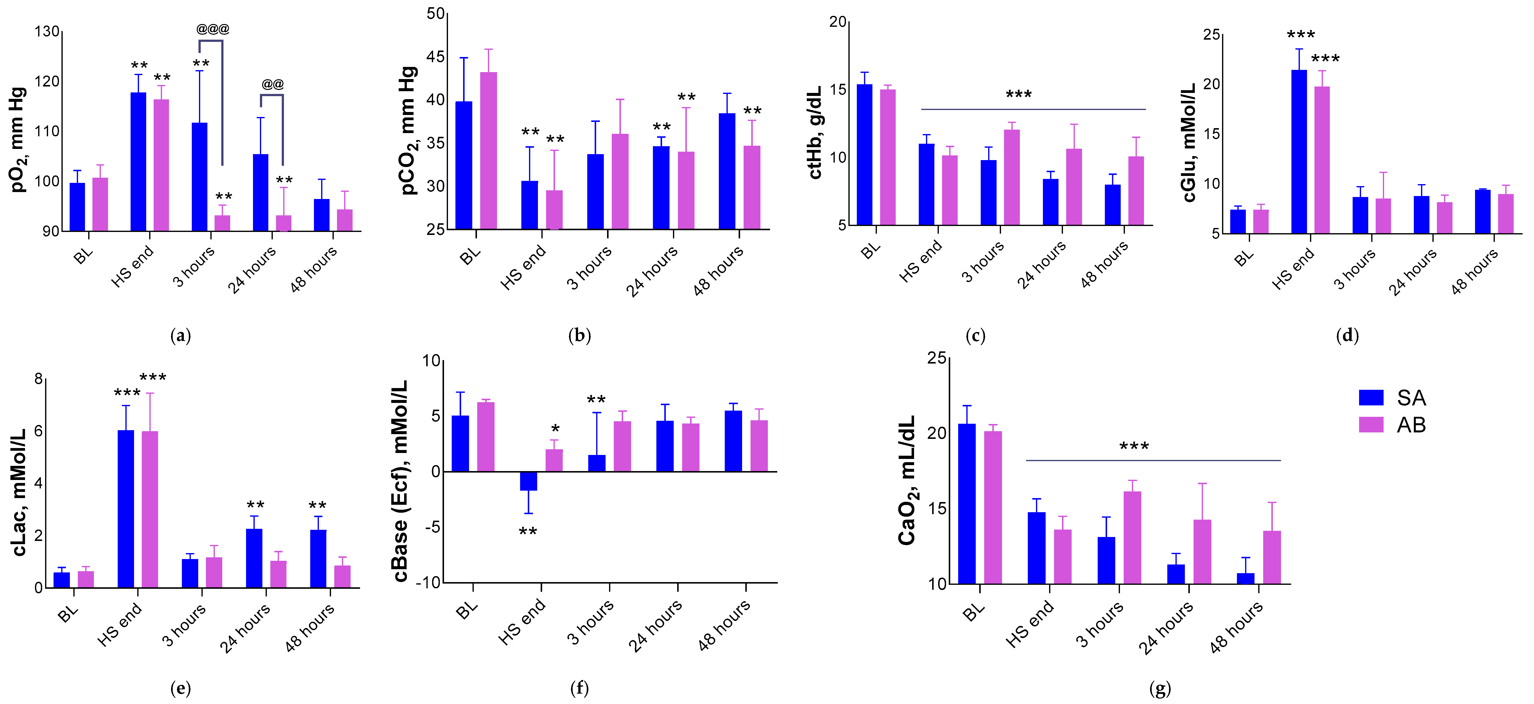
| CaO2 (mL) | Total amount of oxygen transported in arterial blood, combining oxygen bound to hemoglobin and oxygen dissolved in plasma. Arterial oxygen content was calculated as CaO2 (mL) = 1.34 × Hb (g/dL) × SO2 + 0.0031 × PO2 (mmHg) [50,51]. |
| Score | Severity | Description |
|---|---|---|
| 0 | Within normal range | The organ tissue appears normal, consistent with the conditions of the study, age, sex, and strain of the animal. Minor deviations may be present, which are considered normal variations. |
| 1 | Minimal | Observed change in tissue barely deviates from the normal state. |
| 2 | Slight | Tissue damage is easily identified, but the severity is minor. |
| 3 | Moderate | Moderate tissue change. |
| 4 | Marked | Tissue change is noticeable, but there is potential for increased severity. |
| 5 | Severe | The degree of change is maximum (and/or occupies most of the organ). |
| P | Present | It is set when it is impossible to express a feature on a point scale (only its presence is recorded). |
| Group 1 SA | Group 2 AB | Group 3 INT | ||||
|---|---|---|---|---|---|---|
| Day of the Study | Mean ± SD | N | Mean ± SD | N | Mean ± SD | N |
| Day 7 | −9.3 ± 5.0 *** ### | 8 | −6.9 ± 2.9 *** ### | 10 | 9.1 ± 1.7 | 10 |
| Day 14 | −1.0 ± 5.1 *** ### | 4 | −3.0 ± 2.8 ### | 5 | 18.7 ± 2.0 *** | 5 |
| Day 21 | 1.0 ± 3.8 *** ### | 3 | 0.1 ± 2.7 ### | 5 | 27.7 ± 3.6 *** | 5 |
| Day 28 | 5.3 ± 4.4 ### | 3 | 3.9 ± 2.0 ### | 5 | 35.3 ± 5.3 *** | 5 |
| BL | HS Before VRT | 3 h After HS | 24 h After HS | 48 h After HS | 8 Days After HS | 29 Days After HS | ||||||||
|---|---|---|---|---|---|---|---|---|---|---|---|---|---|---|
| SA | AB | SA | AB | SA | AB | SA | AB | SA | AB | SA | AB | SA | AB | |
| N = 10 | N = 10 | N = 10 | N = 10 | N = 8 | N = 10 | N = 8 | N = 10 | N = 8 | N = 10 | N = 4 | N = 5 | N = 3 | N = 5 | |
| WBC, 109/L | 5.7 ± 1.4 | 5.2 ± 0.5 | 6.4 ± 0.8 | 6.9 ± 0.8 | 9.0 ± 1.6 | ** ↑ 9.5 ± 1.2 | *** ↑ 11.6 ± 3.3 | * ↑ @ ↓ 10.7 ± 3.2 | *** ↑ 15.3 ± 5.4 | @ ↓ 8.7 ± 2.9 | *** ↑ 15.3 ± 5.5 | ** ↑ @ ↓ 9.4 ± 2.3 | 6.9 ± 0.8 | 7.4 ± 1.2 |
| LYM, 109/L | 4.1 ± 1.1 | 3.8 ± 0.5 | 4.7 ± 0.6 | 5.0 ± 0.8 | * ↓ 2.3 ± 0.4 | * ↓ 2.4 ± 0.8 | 5.9 ± 1.8 | 6.2 ± 2.4 | *** ↑ 10.4 ± 3.3 | @ ↓ 5.9 ± 1.9 | *** ↑ 10.8 ± 3.6 | ** ↑ 6.4 ± 1.6 | 5.0 ± 0.7 | * ↑ @ ↑ 5.7 ± 1.2 |
| MON, 109/L | 0.5 ± 0.2 | 0.4 ± 0.2 | 0.8 ± 0.4 | 0.9 ± 0.5 | * ↑ 1.3 ± 0.7 | @ ↓ 0.9 ± 0.5 | ** ↑ 1.5 ± 0.8 | @@ ↓ 1.3 ± 1.0 | 1.1 ± 0.6 | @ ↓ 0.7 ± 0.6 | *** ↑ 2.3 ± 2.0 | @@@ ↓ 0.8 ± 0.2 | 0.5 ± 0.2 | 0.4 ± 0.1 |
| GRA, 109/L | 1.0 ± 0.4 | 1.0 ± 0.4 | 1.0 ± 0.5 | 1.1 ± 0.6 | ** ↑ 5.3 ± 1.3 | *** ↑ 6.2 ± 1.1 | *** ↑ 4.2 ± 1.8 | ** ↑ 3.2 ± 1.7↑ | *** ↑ 3.8 ± 2.5 | @ ↓ 2.1 ± 0.7 | 2.3 ± 1.1 | 2.3 ± 1.2 | 1.3 ± 0.2 | 1.3 ± 0.3 |
| LYM, % | 73.8 ± 4.2 | 73.7 ± 5.8 | 73.7 ± 4.0 | 71.2 ± 5.5 | *** ↓ 26.5 ± 4.3 | *** ↓ 25.5 ± 6.8 | *** ↓ 52.1 ± 11.6 | *** ↓ 57.4 ± 16.2 | 70.1 ± 12.7 | 70.6 ± 5.0 | 71.8 ± 11.3 | 70.8 ± 5.9 | 72.8 ± 1.8 | 76.4 ± 6.2 |
| MON, % | 9.4 ± 3.4 | 7.5 ± 3.0 | 11.4 ± 5.6 | 12.2 ± 6.3 | *** ↑ 14.7 ± 7.4 | @ ↓ 9.6 ± 5.4 | 11.8 ± 4.7 | 11.3 ± 5.7 | 7.0 ± 2.1 | 7.1 ± 3.7 | *** ↑ 13.0 ± 8.5 | @ ↓ 8.0 ± 0.9 | 7.7 ± 1.4 | 6.0 ± 0.8 |
| GRA, % | 16.8 ± 4.8 | 18.7 ± 7.4 | 15.0 ± 7.7 | 16.6 ± 10.5 | *** ↑ 58.8 ± 7.4 | *** ↑ 64.9 ± 11.0 | *** ↑ 36.2 ± 11.0 | *** ↑ 31.2 ± 16.8 | 22.9 ± 11.3 | 22.4 ± 4.5 | 15.3 ± 4.5 | 21.2 ± 5.9 | 19.6 ± 2.8 | 17.6 ± 6.0 |
| RBC, 1012/L | 8.4 ± 0.3 | 8.0 ± 0.2 | *** ↓ 5.3 ± 0.6 | *** ↓ 5.3 ± 0.3 | *** ↓ 4.6 ± 0.7 | *** ↓ @@ ↑ 6.1 ± 0.4 | *** ↓ 4.5 ± 0.2 | *** ↓ @@ ↑ 6.2 ± 0.5 | *** ↓ 4.2 ± 0.4 | *** ↓ @@ ↑ 5.7 ± 0.8 | *** ↓ 5.6 ± 0.2 | *** ↓ @ ↑ 6.4 ± 0.2 | *** ↓ 7.1 ± 0.5 | * ↓ 7.6 ± 0.3 |
| HGB, g/L | 147 ± 4 | 143 ± 4 | *** ↓ 95 ± 8 | *** ↓ 93 ± 5 | *** ↓ 81 ± 12 | *** ↓ @@ ↑ 107 ± 7 | *** ↓ 80 ± 4 | *** ↓ @@ ↑ 111 ± 10 | *** ↓ 78 ± 8 | *** ↓ @@ ↑ 104 ± 14 | *** ↓ 121 ± 6 | *** ↓ 128 ± 1 | *** ↓ 141 ± 6 | 147 ± 5 |
| HCT, L/L | 0.51 ± 0.02 | 0.49 ± 0.04 | *** ↓ 0.33 ± 0.03 | *** ↓ 0.33 ± 0.02 | *** ↓ 0.28 ± 0.04 | *** ↓ @@@ ↑ 0.37 ± 0.03 | *** ↓ 0.28 ± 0.01 | *** ↓ @@@ ↑ 0.38 ± 0.03 | *** ↓ 0.27 ± 0.02 | *** ↓ @@@ ↑ 0.35 ± 0.04 | *** ↓ 0.44 ± 0.02 | *** ↓ 0.43 ± 0.01 | 0.47 ± 0.02 | 0.49 ± 0.02 |
| MCV, fL | 60.2 ± 1.7 | 61.0 ± 0.9 | 60.8 ± 1.9 | 61.6 ± 1.2 | 60.6 ± 1.6 | 61.4 ± 1.0 | 61.1 ± 1.1 | 61.5 ± 1.2 | ** ↑ 64.2 ± 1.5 | 62.5 ± 1.6 | *** ↑ 77.2 ± 4.4 | *** ↑ @@ 66.5 ± 2.8 | *** ↑ 66.9 ± 2.7 | *** ↑ 64.3 ± 1.3 |
| MCH, pg | 17.6 ± 0.5 | 17.9 ± 0.4 | 17.5 ± 0.7 | 17.7 ± 0.3 | 17.5 ± 0.7 | 17.7 ± 0.3 | 17.7 ± 0.4 | 17.9 ± 0.3 | ** ↑ 17.6 ± 2.2 | 18.4 ± 0.4 | *** ↑ 21.5 ± 0.3 | *** ↑ @ ↓ 19.9 ± 0.6 | *** ↑ 20.0 ± 0.5 | *** ↑ 19.4 ± 0.1 |
| MCHC, g/L | 292 ± 8 | 293 ± 7 | 288 ± 10 | 287 ± 6 | 289 ± 8 | 288 ± 7 | 289 ± 5 | 292 ± 10 | 287 ± 9 | 294 ± 8 | *** ↓ 279 ± 16 | @@ ↑ 299 ± 6 | 299 ± 6 | 302 ± 5 |
| RDW, % | 11.9 ± 0.5 | 11.7 ± 0.4 | 11.6 ± 0.5 | 11.8 ± 0.4 | 11.6 ± 0.4 | 11.9 ± 0.4 | 11.6 ± 0.4 | 11.8 ± 0.5 | 10.9 ± 0.6 | 11.5 ± 0.6 | *** ↑ 18.7 ± 1.6 | *** ↑ @@@ ↓ 14.3 ± 1.1 | *** ↑ 13.9 ± 0.3 | *** ↑ 13.0 ± 0.4 |
| RDW-SD, fL | 30.4 ± 1.5 | 30.9 ± 1.5 | 30.9 ± 2.0 | 31.3 ± 1.5 | 30.3 ± 1.5 | 30.9 ± 1.0 | 33.6 ± 1.8 | 32.3 ± 2.0 | 33.0 ± 1.9 | 32.8 ± 1.7 | *** ↑ 60.9 ± 10.7 | *** ↑ @@@ 40.8 ± 4.4 | *** ↑ 39.2 ± 1.2 | *** ↑ 36.4 ± 2.5 |
| PLT, 109/L | 779 ± 129 | 765 ± 88 | 823 ± 170 | 807 ± 118 | 640 ± 70 | * ↓ 655 ± 79 | 786 ± 112 | * ↓ @ ↓ 663 ± 151 | 834 ± 144 | * ↓ @@ ↓ 634 ± 93 | *** ↑ 1763 ± 397 | * ↑ @@@ ↓ 1467 ± 121 | 786 ± 113 | * ↑ 787 ± 80 |
| MPV, fL | 6.0 ± 0.2 | 5.8 ± 0.4 | 5.8 ± 0.3 | 5.8 ± 0.4 | 5.6 ± 0.2 | 5.6 ± 0.2 | 5.9 ± 0.2 | 5.8 ± 0.2 | 5.6 ± 0.1 | 5.6 ± 0.2 | 5.3 ± 0.3 | *** ↓ 5.1 ± 0.1 | 5.7 ± 0.4 | 5.7 ± 0.2 |
| PCT, cL/L | 0.5 ± 0.1 | 0.5 ± 0.0 | 0.5 ± 0.1 | * ↑ 0.7 ± 0.1 | 0.4 ± 0.0 | 0.4 ± 0.0 | 0.5 ± 0.1 | 0.4 ± 0.1 | 0.5 ± 0.1 | ** ↓ @ ↓ 0.4 ± 0.1 | *** ↑ 0.9 ± 0.3 | *** ↑ @@@ ↓ 0.8 ± 0.1 | 0.5 ± 0.0 | 0.5 ± 0.0 |
| PDW, % | 16.4 ± 1.7 | 15.5 ± 1.4 | * ↓ 14.7 ± 0.7 | * ↓ 14.0 ± 0.9 | ** ↓ 14.0 ± 1.1 | 14.1 ± 0.8 | * ↓ 14.5 ± 0.8 | 14.8 ± 1.1 | ** 13.2 ± 0.3 | 13.8 ± 0.8 | *** ↓ 12.3 ± 0.8 | *** ↓ 12.5 ± 0.3 | ** ↓ 14.1 ± 1.5 | * ↓ 14.0 ± 0.4 |
| P-LCR | 3.4 ± 1.2 | 5.2 ± 0.5 | 2.9 ± 1.3 | 3.0 ± 1.6 | 2.4 ± 0.6 | 2.2 ± 0.8 | 3.1 ± 0.5 | ** ↓ 2.8 ± 0.8 | 2.6 ± 0.3 | 2.7 ± 0.9 | 1.7 ± 0.6 | ** ↓ 1.1 ± 0.2 | 2.2 ± 0.8 | 2.2 ± 0.6 |
| Group 1 SA | Group 2 AB | Group 3 INT | ||||
|---|---|---|---|---|---|---|
| Day 8 N = 4 | Day 29 N = 3 | Day 8 N = 5 | Day 29 N = 5 | Day 8 N = 5 | Day 29 N = 5 | |
| Body weight, g | 305 ± 18 | 352 ± 7 | 301 ± 31 | 347 ± 15 | 304 ± 18 | 349 ± 19 |
| Absolute organ weight, g | ||||||
| Spleen | 1.17 ± 0.07 *** ↑ | 0.97± 0.04 ### ↓ | 1.00 ± 0.16 ** ↑ @ ↓ | 0.92 ± 0.13 | 0.79 ± 0.08 | 0.80 ± 0.08 |
| Adrenal glands | 0.049 ± 0.006 | 0.045 ± 0.001 * ↓ | 0.050 ± 0.005 | 0.048 ± 0.002 | 0.048 ± 0.006 | 0.054 ± 0.006 |
| Kidneys | 1.76 ± 0.06 *** ↓ | 1.88 ± 0.09 *** ↓ | 1.89 ± 0.22 *** ↓ | 1.91 ± 0.19 *** ↓ | 2.42 ± 0.22 | 2.59 ± 0.18 |
| Liver | 10.5 ± 0.6 ** ↓ | 12.4 ± 0.3 # ↑ | 10.5 ± 1.5 ** ↓ | 11.7 ± 0.7 | 12.3 ± 1.0 | 12.6 ± 1.3 |
| Submandibular lymph nodes | 0.033 ± 0.005 | 0.037 ± 0.006 | 0.025 ± 0.006 | 0.034 ± 0.010 | 0.037 ± 0.010 | 0.036 ± 0.011 |
| Thymus | 0.11 ± 0.02 *** ↓ | 0.25 ± 0.03 *** ↓ ## ↑ | 0.16 ± 0.04 *** ↓ | 0.26 ± 0.09 *** ↓ | 0.58 ± 0.06 | 0.60 ± 0.08 |
| Lungs | 1.7 ± 0.1 | 1.8 ± 0.1 | 1.7 ± 0.2 | 1.7 ± 0.1 | 1.9 ± 0.5 | 1.8 ± 0.3 |
| Brain | 1.7 ± 0.1 * ↓ | 1.8 ± 0.1 ** ↓ | 1.7 ± 0.1 * ↓ | 1.8 ± 0.1 | 1.9 ± 0.1 | 1.9 ± 0.07 |
| Relative organ weight (relative to body weight at necropsy), % | ||||||
| Spleen | 0.38 ± 0.02 *** ↑ | 0.27 ± 0.01 ### ↓ | 0.33 ± 0.04 ** ↑ @ ↑ | 0.27 ± 0.04 ## ↓ | 0.26 ± 0.03 | 0.23 ± 0.03 |
| Adrenal glands | 0.016 ± 0.002 | 0.013 ± 0.001 # ↓ | 0.017 ± 0.001 | 0.014 ± 0.001 # ↓ | 0.016 ± 0.002 | 0.016 ± 0.002 |
| Kidneys | 0.58 ± 0.04 *** ↓ | 0.53 ± 0.02 *** ↓ | 0.63 ± 0.05 *** ↓ | 0.55 ± 0.04 *** ↓ # ↓ | 0.80 ± 0.08 | 0.74 ± 0.05 |
| Liver | 3.4 ± 0.06 *** ↓ | 3.52 ± 0.14 | 3.48 ± 0.20 *** ↓ | 3.37 ± 0.15 | 4.05 ± 0.23 | 3.61 ± 0.27 |
| Submandibular lymph nodes | 0.011 ± 0.002 | 0.010 ± 0.002 | 0.008 ± 0.001 | 0.010 ± 0.003 | 0.012 ± 0.004 | 0.010 ± 0.004 |
| Thymus | 0.036 ± 0.008 *** ↓ | 0.072 ± 0.009 *** ↓ # ↑ | 0.054 ± 0.0146 *** ↓ | 0.074 ± 0.030 *** ↓ | 0.190 ± 0.017 | 0.172 ± 0.021 |
| Lungs | 0.558 ± 0.027 | 0.504 ± 0.012 | 0.546 ± 0.026 | 0.494 ± 0.021 | 0.618 ± 0.148 | 0.505 ± 0.110 |
| Brain | 0.562 ± 0.020 | 0.506 ± 0.020 | 0.564 ± 0.040 | 0.530 ± 0.049 | 0.611 ± 0.038 | 0.551 ± 0.025 |
| Group | SA | AB |
|---|---|---|
| Spleen (number of samples) | 4 | 5 |
| White pulp cell hyperplasia (individual score 0–5 (average score)) | 3/3/3/3 (3.0 ± 0.0) | 3/3/2/3/3 (2.8 ± 0.4) |
| Increased extramedullary hematopoiesis in red pulp (individual score 0–5 (average score)) | 3/3/3/3 (3.0 ± 0.0) | 3/3/3/3/3 (3.0 ± 0.0) |
| Kidneys (number of samples) | 4 | 5 |
| Acute focal ischemia (present “+”, absent “−” (“+” percentage, %)) | +/−/+/− (50.0%) | +/−/+/+/+ (80.0%) |
| Thymus (number of samples) | 4 | 5 |
| Accidental involution (individual score 0–5 (average score)) | 4/3/3/3 (3.3 ± 0.5) | 2/3/0/2/2 (1.8 ± 1.1) * |
| Decreased corticomedullary ratio (individual score 0–5 (average score)) | 4/4/4/4 (4.0 ± 0.0) | 3/4/2/3/3 (3.0 ± 0.7) * |
| Brain (number of samples) | 4 | 5 |
| Neuronal death in projection of CA1, CA2, CA3 and dentate gyrus of hippocampus (number of cases (“+” percentage, %)) | 1 (25.0%) | 1 (20.0%) |
| Foci of necrosis in projection of basal ganglia (number of cases (“+” percentage, %)) | 1 (25.0%) | 0 (0.0%) |
| Group | SA | AB |
|---|---|---|
| Spleen (number of samples) | 3 | 5 |
| White pulp cell hyperplasia (individual score 0–5 (average score)) | 2/2/2 (2.0 ± 0.0) | 2/2/2/2/2 (2.0 ± 0.0) |
| Kidneys (number of samples) | 3 | 5 |
| Focal post-ischemic nephrosclerosis (present “+”, absent “−” (“+” percentage, %)) | −/−/+ (33.3%) | −/+/+/−/− (40.0%) |
| Thymus (number of samples) | 3 | 5 |
| Decreased corticomedullary ratio (individual score 0–5 (average score)) | 2/2/2 (2.0 ± 0.0) | 2/2/2/2/2 (2.0 ± 0.0) |
| Brain (number of samples) | 3 | 5 |
| Gliomesodermal scars in projection of parietal cortex (number of cases (“+” percentage, %)) | 1 (33.3%) | 0 (0.0%) |
| Gliomesodermal scars in cerebellar cortex (number of cases (“+” percentage, %)) | 1 (33.3%) | 0 (0.0%) |
Disclaimer/Publisher’s Note: The statements, opinions and data contained in all publications are solely those of the individual author(s) and contributor(s) and not of MDPI and/or the editor(s). MDPI and/or the editor(s) disclaim responsibility for any injury to people or property resulting from any ideas, methods, instructions or products referred to in the content. |
© 2026 by the authors. Licensee MDPI, Basel, Switzerland. This article is an open access article distributed under the terms and conditions of the Creative Commons Attribution (CC BY) license.
Share and Cite
Ismailova, A.M.; Palikov, V.A.; Severyukhina, M.S.; Tukhovskaya, E.A.; Kholina, A.V.; Semushina, S.G.; Slashcheva, G.A.; Shaykhutdiniva, E.R.; Kravchenko, I.N.; Kazakov, V.A.; et al. Modeling Hemorrhagic Shock in Male SD Rats by Fixed Volume Blood Withdrawal Followed by Partial Resuscitation and Long-Term Outcome Assessment. Med. Sci. 2026, 14, 587. https://doi.org/10.3390/medsci14050587
Ismailova AM, Palikov VA, Severyukhina MS, Tukhovskaya EA, Kholina AV, Semushina SG, Slashcheva GA, Shaykhutdiniva ER, Kravchenko IN, Kazakov VA, et al. Modeling Hemorrhagic Shock in Male SD Rats by Fixed Volume Blood Withdrawal Followed by Partial Resuscitation and Long-Term Outcome Assessment. Medical Sciences. 2026; 14(5):587. https://doi.org/10.3390/medsci14050587
Chicago/Turabian StyleIsmailova, Alina M., Victor A. Palikov, Maria S. Severyukhina, Elena A. Tukhovskaya, Arina V. Kholina, Svetlana G. Semushina, Gulsara A. Slashcheva, Elvira R. Shaykhutdiniva, Irina N. Kravchenko, Vitaly A. Kazakov, and et al. 2026. "Modeling Hemorrhagic Shock in Male SD Rats by Fixed Volume Blood Withdrawal Followed by Partial Resuscitation and Long-Term Outcome Assessment" Medical Sciences 14, no. 5: 587. https://doi.org/10.3390/medsci14050587
APA StyleIsmailova, A. M., Palikov, V. A., Severyukhina, M. S., Tukhovskaya, E. A., Kholina, A. V., Semushina, S. G., Slashcheva, G. A., Shaykhutdiniva, E. R., Kravchenko, I. N., Kazakov, V. A., Kazakova, E. N., Kazakova, M. S., Timakina, A. B., Shinelev, M. V., Rykov, V. A., Patsap, O. I., Dyachenko, I. A., & Murashev, A. N. (2026). Modeling Hemorrhagic Shock in Male SD Rats by Fixed Volume Blood Withdrawal Followed by Partial Resuscitation and Long-Term Outcome Assessment. Medical Sciences, 14(5), 587. https://doi.org/10.3390/medsci14050587