Article

Effects of Oral Doxofylline and Procaterol on Chronic Obstructive Pulmonary Disease: A Randomized Crossover Study

by
Narongkorn Saiphoklang
*,
Sarawut Panichaporn
,
Thiravit Siriyothipun
and
Pitchayapa Ruchiwit
Division of Pulmonary and Critical Care Medicine, Department of Internal Medicine, Faculty of Medicine, Thammasat University, Pathum Thani 12120, Thailand
*
Author to whom correspondence should be addressed.
Med. Sci. 2025, 13(2), 49; https://doi.org/10.3390/medsci13020049
Submission received: 5 March 2025 / Revised: 20 April 2025 / Accepted: 24 April 2025 / Published: 29 April 2025
(This article belongs to the Section Pneumology and Respiratory Diseases)

Abstract

:
Background: Oral bronchodilators may serve as an adjunctive therapy in patients with chronic obstructive pulmonary disease (COPD). This study aimed to evaluate the effects of oral doxofylline and oral procaterol on lung function and clinical symptoms in COPD patients. Methods: A crossover randomized controlled trial was conducted in patients with clinically stable COPD. Participants first received either doxofylline or procaterol for 4 weeks, followed by a 1-week washout period. Assessments included the modified Medical Research Council (mMRC) dyspnea scale, COPD assessment test (CAT) scores, and 6-minute walking distance (6MWD). Pulmonary function was evaluated using spirometry with bronchodilator (BD) testing and all adverse events were recorded. Results: Twenty patients were randomly assigned to begin treatment with either doxofylline or procaterol. Their mean age was 71.7 ± 9.4 years. After four weeks of treatment, the doxofylline group showed significantly greater improvement in pulmonary function parameters (post-BD peak expiratory flow and post-BD forced expiratory flow 25–75) compared to the procaterol group. However, there were no significant differences in mMRC scores, CAT scores, or 6MWD between the two groups. More neurological adverse events were observed in the doxofylline group compared to the procaterol group (35% vs. 5%, p = 0.044). Conclusions: Doxofylline improved pulmonary function in COPD patients but did not provide superior functional performance compared to procaterol. Neurological adverse events were more frequently associated with doxofylline. Doxofylline may serve as an adjunctive therapy to enhance pulmonary function in COPD patients, but caution is advised due to its potential side effects.

1. Introduction

Chronic obstructive pulmonary disease (COPD) is a common respiratory disorder and the third leading cause of death worldwide [1]. It is a heterogeneous lung condition characterized by chronic respiratory symptoms (dyspnea, cough, sputum production, and/or exacerbations) that cause persistent and often progressive airflow obstruction [1,2]. Pharmacotherapy for stable COPD treatment currently focuses on managing symptoms and preventing exacerbation. Bronchodilator medications, particularly inhaled, long-acting bronchodilators (beta2-agonists and muscarinic antagonists), are the mainstay of therapy for symptomatic COPD patients [1]. Oral medications, including methylxanthines (e.g., theophylline, doxofylline) and beta2-agonists (e.g., salbutamol, procaterol), may also be effective in promoting bronchodilation in COPD treatment [1].
Methylxanthines have stimulant effects and promote bronchodilation. Theophylline relaxes the airway smooth muscle by inhibiting phosphodiesterase (PDE) activity (PDE3, PDE4, and PDE5), but relatively high concentrations are needed to achieve maximal relaxation [3]. Theophylline also activates histone deacetylase (HDAC) activity, thereby suppressing the expression of inflammatory genes [4], resulting in anti-inflammatory effects for COPD treatment. Additionally, it blocks the adenosine receptors, potentially causing side effects that affect the central nervous, cardiovascular, gastrointestinal, and urinary systems. These side effects can include seizures, tremors, nausea, frequent urination, and arrhythmia [5]. Theophylline is also associated with several adverse drug reactions, the most severe being its impact on the cardiovascular system [6]. In cases of overdose, it can cause severe arrhythmia or even cardiac arrest. Theophylline is primarily metabolized in the liver by one or more cytochrome P450 enzymes and has a narrow therapeutic window, with effective plasma concentrations ranging from 10 to 20 mcg/mL [7]. Due to these limitations, doxofylline was developed as an alternative medication. Doxofylline has a structure similar to theophylline but also includes a dioxalane group at position 7, which enhances its selectivity for the adenosine A2A receptor [8]. This selectivity reduces the occurrence of side effects and provides a wider therapeutic window than theophylline. Additionally, doxofylline is not metabolized by cytochrome P450 enzymes, which lowers the risk of drug interactions. Consequently, it has become a safer and increasingly preferred option for treating asthma and COPD [9]. Doxofylline also leads to improvement in pulmonary functions compared with baseline [10].
Procaterol is an oral bronchodilator belonging to the beta2-adrenergic receptor agonist class. Procaterol acts directly on bronchial cells by activating adenyl cyclase to stimulate β-receptors, resulting in the relaxation of airway smooth muscle and subsequent bronchodilation [11]. Additionally, procaterol reduces the aggregation of human fetal lung fibroblast-1 (HFL-1) cells, which affects fibroblast activity and may reduce pulmonary fibrosis [12]. Furthermore, procaterol enhances mucociliary clearance, improving the removal of airway secretions [13].
However, the bronchodilatory effects of doxofylline in comparison to procaterol have not yet been explored in patients with COPD. This study aimed to evaluate and compare their effectiveness in improving airway function and controlling symptoms in this population.

2. Materials and Methods

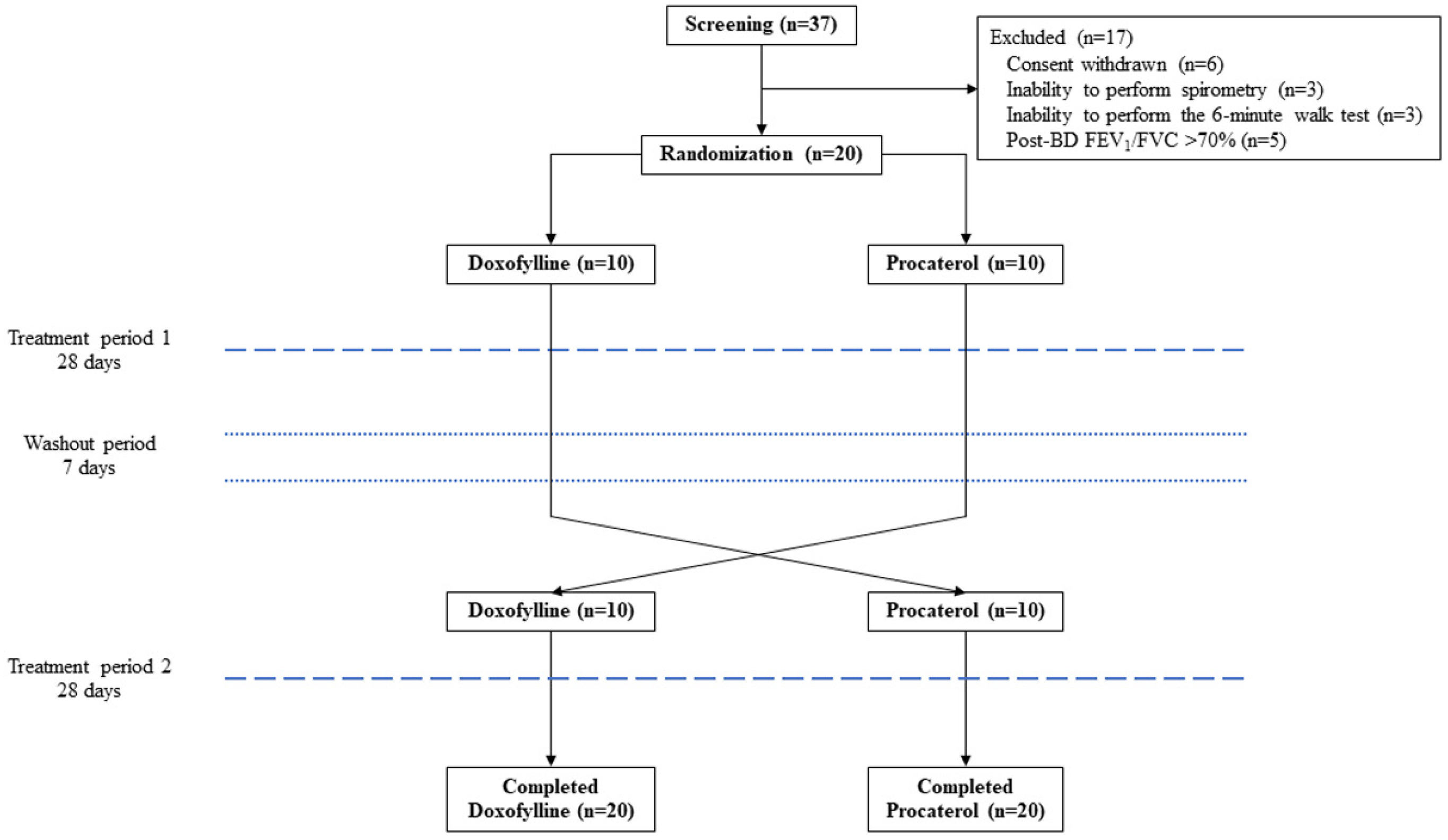
2.1. Study Design and Participants

A prospective, randomized crossover study was carried out at the pulmonary outpatient clinic of Thammasat University Hospital, Thailand from March 2024 to October 2024. The inclusion criteria included: (1) patients aged 40 years or older, (2) a smoking history of 10 pack-years or more, and (3) a diagnosis of COPD confirmed by a post-bronchodilator (BD) forced expiratory volume in 1 second (FEV1) to forced vital capacity (FVC) ratio of less than 0.7. Exclusion criteria included: (1) any pulmonary diseases, such as asthma, bronchiectasis, or pulmonary fibrosis, (2) an inability to perform spirometry or a 6-minute walk test (6MWT), (3) COPD exacerbation within the 12 weeks prior to study recruitment, (4) the use of oral methylxanthines (theophylline or doxofylline), oral roflumilast, oral salbutamol, or oral procaterol within 1 week prior to study recruitment, (5) any chronic disease, such as uncontrolled cardiac arrhythmia, uncontrolled hypertension (blood pressure > 180/110 mmHg), chronic kidney disease (glomerular filtration rate < 50 mL/min), chronic liver disease (liver enzymes > 1.5 times the upper limit of normal), ongoing cancer treatment, or active hyperthyroidism, (6) pregnancy or lactation, and (7) any condition or illness that, at the discretion of the researcher, could hinder the research or interfere with the study results.
Demographic data, respiratory symptoms, and functional capacity were collected using the modified Medical Research Council (mMRC) dyspnea scale [14], COPD assessment test (CAT) [15], 6-minute walking distance (6MWD), and spirometry data. Baseline medications, including inhaled corticosteroids (ICS), long-acting beta2-agonists (LABA), and long-acting muscarinic antagonists (LAMA), were also recorded.
The Global Initiative for Chronic Obstructive Lung Disease (GOLD) criteria for COPD severity were based on the FEV1 value: grade 1 indicates mild COPD (≥80% of predicted value), grade 2 indicates moderate COPD (50–79% of predicted value), and grades 3 and 4 indicate severe and very severe COPD (<50% and <30% of predicted values), respectively [1]. GOLD symptoms and risk were classified into three groups: A, B, and E, depending on the dyspnea score and history of exacerbation [1].
Ethical approval was obtained from the Human Research Ethics Committee of Thammasat University (Medicine), Thailand (IRB No. MTU-EC-IM-0-235/66, COA No.067/2024, date of approval: 29 February 2024), in full compliance with international guidelines, including the Declaration of Helsinki, the Belmont Report, CIOMS Guidelines, and the International Conference on Harmonization’s Good Clinical Practice (ICH-GCP). All methods were performed in accordance with these guidelines and regulations. Written informed consent was obtained from all participants. This study was registered on ClinicalTrials.gov with the number NCT06346691.

2.2. Randomization and Intervention

Patients were randomly assigned in a 1:1 ratio to receive either doxofylline or procaterol, with randomization performed using a computerized, web-based system. The study flowchart is shown in Figure 1.
Prior to randomization, COPD patients underwent a 1-week run-in period and were then screened for eligibility. Eligible participants were assigned to one of two groups: one received doxofylline at a dose of 400 mg twice daily (800 mg/day), while the other received procaterol at a dose of 50 mcg twice daily (100 mcg/day) for 4 weeks. Following a 1-week washout period, patients crossed over to the alternate treatment for an additional 4 weeks (Figure 1). Study medications were administered by the attending physicians. Throughout the study, all patients underwent spirometry with BD testing, as well as assessments using the mMRC dyspnea scale, CAT, and 6MWT. Adverse events were recorded at the following time points: baseline (visit 0), before the run-in period (visit 1), at the start (visit 2) and end (visit 3) of period 1 treatment, and at the start (visit 4) and end (visit 5) of period 2 treatment. BD testing was conducted using an inhalation of 400 mcg of salbutamol, followed by repeating spirometry after 15 min. Pulmonary function parameters measured by spirometry included FVC, FEV1, FEV1/FVC, peak expiratory flow (PEF), forced expiration flow rate at 25–75% of FVC (FEF25–75), and BD responsiveness. Spirometry was performed in accordance with the American Thoracic Society (ATS) and European Respiratory Society (ERS) guidelines [16,17] using a PC spirometer (Vyntus® SPIRO, Vyaire Medical, Inc., Mettawa, IL, USA).
Doxofylline has a half-life of 7 h [18], and procaterol has a half-life of 3.8 h [19]. Therefore, a washout period of 1 week was sufficient to eliminate the effects of both medications, as this duration exceeds five half-lives for each drug [20].
Before and during the study, patients were allowed to inhale long-acting bronchodilators (beta2-agonists and muscarinic antagonists) for COPD maintenance therapy and inhaled short-acting beta-agonists for symptom relief. On days when spirometry was performed, patients were instructed to take the oral study medications in the morning.

2.3. Outcome Assessment

The primary outcome was the difference in pulmonary function parameters between the doxofylline and the procaterol groups following COPD treatment. Secondary outcomes included changes in the pulmonary function parameters, mMRC scores, CAT scores, 6MWD, and the occurrence of adverse events within each group following treatment.

2.4. Statistical Analysis

In a study by Wang T and colleagues [21], oral administration of 400 mg of doxofylline per day in COPD patients increased FEV1 values by 16.0 ± 7.2% from baseline. In another study by Crowe MJ and colleagues, oral administration of 100 mcg of procaterol in asthmatic patients increased FEV1 values by 30.0 ± 10.0% from baseline [22]. We hypothesized that the effects of doxofylline and procaterol on FEV1 in COPD patients would align with those observed in previous studies. Based on this hypothesis, we determined that enrolling 20 patients (10 per group) would provide 90% power to detect a difference in pulmonary function, with a two-sided alpha of 0.05.
Categorical data were expressed as a number (%), and continuous data as the mean ± standard deviation (SD). The chi-squared test was used to compare categorical variables between the two groups. The unpaired t-test and paired t-test were used to compare continuous variables with normal distribution—pulmonary function parameters, mMRC scores, CAT scores, and 6MWD—between the two independent and dependent groups, respectively. The Mann–Whitney U test and Wilcoxon signed-rank test were used to compare continuous variables with non-normal distributions between two independent and dependent groups, respectively. A two-sided p-value of <0.05 was considered statistically significant. Statistical analyses were performed using SPSS version 24 software (IBM Corp., Armonk, NY, USA).

3. Results

3.1. Participants

A total of 37 COPD patients were screened, and 20 eligible patients were subsequently randomized into the study. No participants were lost to follow-up throughout the study duration (Figure 1). The twenty participants were predominantly male (95.0%), with a mean age of 71.7 ± 9.4 years (Table 1). Common comorbidities included hyperlipidemia (80%), hypertension (65%), and cardiovascular disease (40%). Most participants were classified as GOLD grade 2 (60%) and group A (70%). Triple therapy (ICS/LABA/LAMA) was the predominant treatment (45%). The mean mMRC score was 1.1 ± 0.9, the CAT score was 6.3 ± 5.6, and the 6MWD was 391.9 ± 107.7 m. Post-BD FEV1 performance was 59.34 ± 13.92% of the predicted value (Table 1).

3.2. Primary Outcome

Compared to the procaterol group, the doxofylline group showed significantly greater improvement in pulmonary function parameters: post-BD PEF value (mean difference 0.455, 95% CI 0.127 to 0.782 L/s, p = 0.008), post-BD FEF25–75 value (mean difference 0.083, 95% CI 0.023 to 0.143 L/s, p = 0.008), and post-BD FEF25–75% predicted (mean difference 4.115, 95% CI 0.917 to 7.313% predicted, p = 0.013). However, there were no significant differences in other pulmonary function parameters, mMRC scores, CAT scores, or 6MWD between the two groups (Table 2).

3.3. Secondary Outcomes

After the 4-week treatment, there were significant increases in post-BD FEF25–75 value of 0.041 ± 0.082 L/s (p = 0.040) and post-BD FEF25–75 percentage of 2.310 ± 4.268% predicted (p = 0.026) in the doxofylline group. Conversely, significant decreases were observed in the procaterol group, with a post-BD PEF value of 0.352 ± 0.568 L/s (p = 0.015) and a percentage of PEF change after BD test of 5.884 ± 11.900% (p = 0.045) (Table 3). However, there were no significant changes in other pulmonary function parameters, mMRC scores, CAT scores, or 6MWD in either group after the 4-week treatment (Table 3).
Neurological adverse events were observed more markedly in the doxofylline group compared to the procaterol group (35% vs. 5%, p = 0.044), particularly regarding dizziness (25% vs. 0%, p = 0.047) (Table 4). A serious adverse event, i.e., one death, was reported in the procaterol group.

4. Discussion

This study is the first crossover randomized trial comparing the bronchodilatory effects of these two oral bronchodilators in COPD. We found that patients in the doxofylline group showed significantly greater improvement in pulmonary function parameters (post-BD PEF and post-BD FEF25–75) compared to those in the procaterol group after the 4-week treatment. However, neurological side effects were significant in the doxofylline group, and one patient in the procaterol group died during the study.
According to GOLD recommendations [1], inhaled bronchodilators are the mainstay treatment of COPD to prevent exacerbation and control the respiratory symptoms. Oral bronchodilators may be an alternative option for COPD treatment, particularly in cases of inhalation technique error, poor hand–breath coordination, insufficient inspiratory effort, or uncontrolled COPD despite the use of maximum doses of inhaled medications. A meta-analysis by Cho-Reyes S et al. found that 86.7% of adults with obstructive lung diseases using metered dose inhalers exhibited at least one inhalation technique error, and 76.9% incorrectly performed ≥ 20% of the device use steps [23]. Critical errors in inhalation were associated with worse clinical outcomes in patients with asthma or COPD, as reviewed by Kocks JWH et al. [24]. In addition, suboptimal peak inspiratory flow and inhalation technique errors were related to higher COPD-related healthcare utilization and costs in COPD patients using dry powder inhalers for maintenance therapy, as found in a study by Leving MT et al. [25]. Oral bronchodilators improve COPD symptoms and quality of life in COPD patients.
Methylxanthines exhibit stimulatory effects, are able to cause bronchodilation, and possess anti-inflammatory properties [3,4,26]. Theophylline, a methylxanthine, has been shown to significantly reduce histone deacetylase-2 levels in macrophages and the peripheral lung tissue of COPD patients, leading to decreased inflammation and reduced steroid resistance [26]. In a randomized controlled trial by Murciano D et al. [27], theophylline improved respiratory function and alleviated dyspnea in patients with severe COPD. Additionally, a randomized, double-blind, placebo-controlled study by Zhou Y et al. [28] found that theophylline led to greater improvements in pre-BD FEV1, a lower frequency of COPD exacerbations, fewer days of exacerbation, less frequent clinical visits, and enhanced quality of life compared to the placebo group. Furthermore, a study by Bellia V et al. [29] demonstrated that theophylline improved FEV1, PEF, and symptoms in COPD patients, showing similar efficacy to inhaled oxitropium bromide or a combination of both drugs.
Doxofylline, which differs from theophylline by containing a dioxalane group at position 7, demonstrates comparable efficacy to theophylline in treating respiratory diseases. However, unlike theophylline, it offers an improved tolerability profile, a more favorable risk-to-benefit ratio, and no significant drug–drug interactions [8]. Therefore, doxofylline is considered a safer and more favorable option for the treatment of COPD and asthma [9]. Moreover, doxofylline exhibits significant anti-inflammatory activity in the lungs, which can lead to a notable steroid-sparing effect [30]. A study by Lal D et al. [31] showed that both theophylline and doxofylline improved pulmonary function (FEV1, FVC, and FEV1/FVC) at different time intervals in asthma and COPD patients. The maximum effects were observed at 6 weeks for asthma patients and at 8 weeks for COPD patients for both drugs. Doxofylline showed a trend toward fewer side effects compared to theophylliWang T et al. [21] reported that both oral doxofylline (200 mg twice daily) and inhaled tiotropium (18 μg/day) significantly improved pulmonary function (FEV1 and FEV1/FVC%) from baseline at 12 and 24 weeks in patients with moderate to severe but stable COPD. However, major adverse effects occurred in 12.9% of the doxofylline group, compared to 9.9% of the tiotropium group. Furthermore, a meta-analysis by Cazzola M et al. [10], which included 20 studies involving 820 COPD patients, found that doxofylline improved FEV1 by 8.2% and 317 mL over baseline. However, patients treated with doxofylline treatment experienced more side effects, such as epigastric discomfort, nausea, and headaches, compared to the placebo group.
Our study demonstrated that doxofylline significantly improved only the FEF25–75 value, indicating an effect on small airway disease, but not on FEV1 or FVC. This may be because most of our patients had baseline pulmonary function that was classified as mild to moderate rather than severe, making it difficult to observe significant changes from baseline. Similarly, the baseline COPD symptoms, assessed by the mMRC and CAT scores in our study, indicated mild symptoms (mean mMRC score of 1.1 and CAT score of 6.3), which also made it challenging to detect changes in symptoms from baseline. Moreover, the 4-week duration of doxofylline treatment in our study may have been too short to achieve the maximum therapeutic effects of treatment, which, in a study by Lal D et al. [31], required at least 8 weeks. Consequently, FEV1 and FVC, which reflect larger airway function, did not show significant changes after the 4-week treatment period.
A positive bronchodilator response is typically defined as an increase of at least 12% and an absolute value of at least 200 mL compared to the baseline in either FEV1 or FVC [32]. However, our results did not meet this criterion. Additionally, to accurately compare differences in bronchodilation—whether between the start and end of treatment or among different medications—it is essential to evaluate not only the statistical differences but also the minimal clinically important difference (MCID) in the clinical parameters, including lung function (an increase in FEV1 of 100 mL), exercise capacity (a 6MWD increase of 26 m), and respiratory symptoms (a two-point change in CAT) [33,34,35]. In our study, changes in these clinical outcomes did not reach their MCIDs.
Procaterol, a beta2-adrenergic receptor agonist, is a bronchodilator used in the treatment of COPD and asthma. It works by directly relaxing the airway smooth muscles, resulting in bronchodilation [36]. A study by Sukisaki T et al. [37] demonstrated that, in patients with moderate to severe COPD, the inhalation of 20 μg of procaterol significantly increased walking distance at 4 h post-inhalation compared to the no-treatment group (331 ± 119 m vs. 294 ± 113 m, p < 0.001), despite there being no significant difference in FEV1 between the groups. However, these findings were not observed in our study involving 4 weeks of oral procaterol treatment. A study by Shioya T et al. [38] revealed that, when compared to inhaled oxitropium (200 μg thrice daily), inhaled procaterol (20 μg thrice daily) significantly improved pulmonary function (FEV1, FVC, total lung capacity, functional residual capacity, residual volume, maximal inspiratory pressure, and maximal expiratory pressure), 6MWD, and the Borg dyspnea scale, as well as scores for dyspnea, fatigue, emotional function, mastery, total scores, and activities of daily living over 12, 24, and 52 weeks compared to baseline. However, no clinical studies have been published on the use of oral procaterol in COPD patients. Our study is the first clinical study of the effect of oral procaterol in COPD patients.
Although our COPD patients showed improvement in pulmonary function after 4 weeks of oral bronchodilator treatment, some adverse events were observed during the study. Neurological adverse events were more commonly observed in the doxofylline group (35%), particularly regarding dizziness (25%). These side effects may lead to the discontinuation of doxofylline in 20% of patients. A serious adverse event (one death) was reported in the procaterol group. The case involved an 88-year-old female with hypertension, atrial fibrillation, and COPD. She completed the first 4-week period of study with doxofylline but experienced a cardiac arrest at night after taking oral procaterol for 2 days. The cause of death could not be determined, as her family member did not request an autopsy to identify the cause. Therefore, we suggest that elderly COPD patients with underlying cardiovascular disease, particularly arrhythmia, should either avoid using or be closely monitored for serious side effects when using oral bronchodilators for COPD treatment.
Additionally, our study found newly emerging COPD exacerbation during the study period in both groups, and treatment was discontinued in the doxofylline group. This may have resulted from the lower effectiveness of medications in reducing COPD exacerbation, along with a higher rate of side effects.
This study has several limitations. Firstly, the sample size was small, with a mean age of 71 years and only 5% of participants being female. This limited sample may not be representative of the broader clinical population. Secondly, our patients had well-controlled COPD symptoms at the time of study enrollment. As a result, the findings may not show significant differences in other pulmonary function parameters, CAT scores, mMRC scores, or 6MWD after oral bronchodilator treatments. Thirdly, a single- or double-blind design could not be implemented due to differences in the packaging of both medications. Consequently, both physicians and patients could identify the study drugs, potentially leading to bias in the clinical outcomes. To mitigate this, we employed a cross-over design. Lastly, the 4-week follow-up period may have been too brief to capture meaningful changes in pulmonary function or clinical outcomes, especially COPD exacerbation. To more thoroughly assess the effectiveness of oral bronchodilators, larger-scale studies with longer follow-up durations are needed.

5. Conclusions

Doxofylline treatment demonstrated improvements in pulmonary function in COPD patients; however, it did not result in superior functional performance compared to procaterol. Notably, neurological adverse events, such as dizziness, were more frequently associated with doxofylline treatment, highlighting the potential risks of this medication. Despite these concerns, doxofylline may still serve as an adjunctive therapy to enhance pulmonary function in COPD patients. However, its use should be approached with caution, particularly in patients at higher risk of adverse events, and careful monitoring is recommended to ensure patient safety.

Author Contributions

Conceptualization, N.S., S.P., T.S. and P.R.; methodology, N.S. and S.P.; software, N.S.; validation, N.S., S.P. and P.R.; formal analysis, N.S. and S.P.; investigation, N.S., S.P. and T.S.; resources, N.S.; data curation, N.S., S.P. and T.S.; writing—original draft preparation, S.P.; writing—review and editing, N.S., S.P., T.S. and P.R.; visualization, N.S.; supervision, P.R.; project administration, S.P.; funding acquisition, S.P. All authors have read and agreed to the published version of the manuscript.

Funding

This research was funded by the Faculty of Medicine, Thammasat University, Thailand, grant number: 1-14/2567.

Institutional Review Board Statement

The study was conducted in accordance with the Declaration of Helsinki and approved by the Human Research Ethics Committee of Thammasat University (Medicine), Thailand (IRB No. MTU-EC-IM-0-235/66, COA No.067/2024, date of approval: 29 February 2024).

Informed Consent Statement

Informed consent was obtained from all subjects involved in the study.

Data Availability Statement

The data supporting the results of this study are available within the article.

Acknowledgments

The authors thank Michael Jan Everts, Faculty of Medicine, Thammasat University, for proofreading this manuscript. This work was supported by the Thammasat University Research Unit in Respiratory Medicine, Pathum Thani, Thailand.

Conflicts of Interest

The authors declare no conflicts of interest. The funders had no role in the design of the study; in the collection, analyses, or interpretation of data; in the writing of the manuscript; or in the decision to publish the results.

Abbreviations

The following abbreviations are used in this manuscript:
ATSAmerican Thoracic Society
BDbronchodilator
CATCOPD assessment test
COPDchronic obstructive pulmonary disease
ERSEuropean Respiratory Society
FEF25–75forced expiration flow rate at 25–75% of FVC
FEV1forced expiratory volume in 1 second
FVCforced vital capacity
GOLDGlobal Initiative for Chronic Obstructive Lung Disease
HDAChistone deacetylase
HFL-1human fetal lung fibroblasts-1
ICSinhaled corticosteroids
LABAlong-acting beta-2 agonists
LAMAlong-acting muscarinic antagonists
MCIDminimal clinically important difference
mMRCmodified Medical Research Council
PDEphosphodiesterase
PEFpeak expiratory flow
6MWD6-minute walking distance
6MWT6-minute walk test

References

  1. Global Initiative for Chronic Obstructive Lung Disease. Global Strategy for the Diagnosis, Management, and Prevention of Chronic Obstructive Pulmonary Disease 2024 Report. Available online: https://goldcopd.org/ (accessed on 3 January 2025).
  2. Celli, B.; Fabbri, L.; Criner, G.; Martinez, F.J.; Mannino, D.; Vogelmeier, C.; Montes de Oca, M.; Papi, A.; Sin, D.D.; Han, M.K.; et al. Definition and nomenclature of chronic obstructive pulmonary disease: Time for its revision. Am. J. Respir. Crit. Care Med. 2022, 206, 1317–1325. [Google Scholar] [CrossRef]
  3. Rabe, K.F.; Magnussen, H.; Dent, G. Theophylline and selective PDE inhibitors as bronchodilators and smooth muscle relaxants. Eur. Respir. J. 1995, 8, 637–642. [Google Scholar] [CrossRef] [PubMed]
  4. Ito, K.; Lim, S.; Caramori, G.; Cosio, B.; Chung, K.F.; Adcock, I.M.; Barnes, P.J. A molecular mechanism of action of theophylline: Induction of histone deacetylase activity to decrease inflammatory gene expression. Proc. Natl. Acad. Sci. USA 2002, 99, 8921–8926. [Google Scholar] [CrossRef] [PubMed]
  5. Barnes, P.J. Theophylline. Pharmaceuticals 2010, 3, 725–747. [Google Scholar] [CrossRef] [PubMed]
  6. Scurek, M.; Brat, K. A narrative review of theophylline: Is there still a place for an old friend? J. Thorac. Dis. 2024, 16, 3450–3460. [Google Scholar] [CrossRef]
  7. Weinberger, M.; Hendeles, L. Therapeutic effect and dosing strategies for theophylline in the treatment of chronic asthma. J. Allergy Clin. Immunol. 1986, 78, 762–768. [Google Scholar] [CrossRef]
  8. Matera, M.G.; Page, C.; Cazzola, M. Doxofylline is not just another theophylline! Int. J. Chron. Obstr. Pulm. Dis. 2017, 12, 3487–3493. [Google Scholar] [CrossRef]
  9. Cazzola, M.; Matera, M.G. The effect of doxofylline in asthma and COPD. Respir. Med. 2020, 164, 105904. [Google Scholar] [CrossRef]
  10. Cazzola, M.; Calzetta, L.; Rogliani, P.; Page, C.; Matera, M.G. Impact of doxofylline in COPD: A pairwise meta-analysis. Pulm. Pharmacol. Ther. 2018, 51, 1–9. [Google Scholar] [CrossRef]
  11. Tashimo, H.; Yamashita, N.; Ishida, H.; Nagase, H.; Adachi, T.; Nakano, J.; Yamamura, K.; Yano, T.; Yoshihara, H.; Ohta, K. Effect of procaterol, a beta(2) selective adrenergic receptor agonist, on airway inflammation and hyperresponsiveness. Allergol. Int. 2007, 56, 241–247. [Google Scholar] [CrossRef]
  12. Kohyama, T.; Yamauchi, Y.; Takizawa, H.; Itakura, S.; Kamitani, S.; Desaki, M.; Kawasaki, S.; Nagase, T. Procaterol inhibits lung fibroblast migration. Inflammation 2009, 32, 387–392. [Google Scholar] [CrossRef] [PubMed]
  13. Bennett, W.D. Effect of beta-adrenergic agonists on mucociliary clearance. J. Allergy Clin. Immunol. 2002, 110, S291–S297. [Google Scholar] [CrossRef] [PubMed]
  14. Mahler, D.A.; Wells, C.K. Evaluation of clinical methods for rating dyspnea. Chest 1988, 93, 580–586. [Google Scholar] [CrossRef]
  15. Jones, P.W.; Harding, G.; Berry, P.; Wiklund, I.; Chen, W.H.; Kline Leidy, N. Development and first validation of the COPD Assessment Test. Eur. Respir. J. 2009, 34, 648–654. [Google Scholar] [CrossRef]
  16. Miller, M.R.; Crapo, R.; Hankinson, J.; Brusasco, V.; Burgos, F.; Casaburi, R.; Coates, A.; Enright, P.; van der Grinten, C.P.; Gustafsson, P.; et al. General considerations for lung function testing. Eur. Respir. J. 2005, 26, 153–161. [Google Scholar] [CrossRef]
  17. Graham, B.L.; Steenbruggen, I.; Miller, M.R.; Barjaktarevic, I.Z.; Cooper, B.G.; Hall, G.L.; Hallstrand, T.S.; Kaminsky, D.A.; McCarthy, K.; McCormack, M.C.; et al. Standardization of spirometry 2019 update. An official American Thoracic Society and European Respiratory Society technical statement. Am. J. Respir. Crit. Care Med. 2019, 200, e70–e88. [Google Scholar] [CrossRef]
  18. Bologna, E.; Lagana, A.; Terracino, D.; Bolignari, P.; Biffignandi, P. Oral and intravenous pharmacokinetic profiles of doxofylline in patients with chronic bronchitis. J. Int. Med. Res. 1990, 18, 282–288. [Google Scholar] [CrossRef]
  19. Kobayashi, H.; Masuda, M.; Kashiyama, E.; Koga, N.; Yasuda, Y.; Shibutani, T.; Endo, K.; Funaki, T.; Miyamoto, T. Pharmacokinetic study of the oral administration of procaterol hydrochloride hydrate 50 microg in healthy adult Japanese men. Int. J. Clin. Pharmacol. Ther. 2010, 48, 744–750. [Google Scholar] [CrossRef] [PubMed]
  20. Dhariwal, K.; Jackson, A. Effect of length of sampling schedule and washout interval on magnitude of drug carryover from period 1 to period 2 in two-period, two-treatment bioequivalence studies and its attendant effects on determination of bioequivalence. Biopharm. Drug Dispos. 2003, 24, 219–228. [Google Scholar] [CrossRef]
  21. Wang, T.; Luo, G.; Hu, Y.; Li, F.; Ma, J.; Wang, J.; Zuo, P.; Xiong, W.; Liu, X.; Zhao, J.; et al. Comparative study on the efficacy of tiotropium bromide inhalation and oral doxofylline treatment of moderate to severe stable chronic obstructive pulmonary disease. J. Huazhong Univ. Sci. Technol. Med. Sci. 2011, 31, 614. [Google Scholar] [CrossRef]
  22. Crowe, M.J.; Counihan, H.E.; O’Malley, K. A comparative study of a new selective beta 2-adrenoceptor agonist, procaterol and salbutamol in asthma. Br. J. Clin. Pharmacol. 1985, 19, 787–791. [Google Scholar] [CrossRef] [PubMed]
  23. Cho-Reyes, S.; Celli, B.R.; Dembek, C.; Yeh, K.; Navaie, M. Inhalation technique errors with metered-dose inhalers among patients with obstructive lung diseases: A systematic review and meta-analysis of U.S. studies. Chronic Obstr. Pulm. Dis. 2019, 6, 267–280. [Google Scholar] [CrossRef]
  24. Kocks, J.W.H.; Chrystyn, H.; van der Palen, J.; Thomas, M.; Yates, L.; Landis, S.H.; Driessen, M.T.; Gokhale, M.; Sharma, R.; Molimard, M. Systematic review of association between critical errors in inhalation and health outcomes in asthma and COPD. NPJ Prim. Care Respir. Med. 2018, 28, 43. [Google Scholar] [CrossRef] [PubMed]
  25. Leving, M.T.; van Boven, J.F.M.; Bosnic-Anticevich, S.Z.; van Cooten, J.; Correia de Sousa, J.; Cvetkovski, B.; Dekhuijzen, R.; Dijk, L.; Garcia Pardo, M.; Gardev, A.; et al. Suboptimal peak inspiratory flow and critical inhalation errors are associated with higher COPD-related healthcare costs. Int. J. Chron. Obstr. Pulm. Dis. 2022, 17, 2401–2415. [Google Scholar] [CrossRef]
  26. Barnes, P.J. Theophylline in chronic obstructive pulmonary disease: New horizons. Proc. Am. Thorac. Soc. 2005, 2, 334–339. [Google Scholar] [CrossRef]
  27. Murciano, D.; Auclair, M.H.; Pariente, R.; Aubier, M. A randomized, controlled trial of theophylline in patients with severe chronic obstructive pulmonary disease. N. Engl. J. Med. 1989, 320, 1521–1525. [Google Scholar] [CrossRef] [PubMed]
  28. Zhou, Y.; Wang, X.; Zeng, X.; Qiu, R.; Xie, J.; Liu, S.; Zheng, J.; Zhong, N.; Ran, P. Positive benefits of theophylline in a randomized, double-blind, parallel-group, placebo-controlled study of low-dose, slow-release theophylline in the treatment of COPD for 1 year. Respirology 2006, 11, 603–610. [Google Scholar] [CrossRef]
  29. Bellia, V.; Foresi, A.; Bianco, S.; Grassi, V.; Olivieri, D.; Bensi, G.; Volonte, M.; Group, B.I.S. Efficacy and safety of oxitropium bromide, theophylline and their combination in COPD patients: A double-blind, randomized, multicentre study (BREATH Trial). Respir. Med. 2002, 96, 881–889. [Google Scholar] [CrossRef]
  30. Riffo-Vasquez, Y.; Venkatasamy, R.; Page, C.P. Steroid sparing effects of doxofylline. Pulm. Pharmacol. Ther. 2018, 48, 1–4. [Google Scholar] [CrossRef]
  31. Lal, D.; Manocha, S.; Ray, A.; Vijayan, V.K.; Kumar, R. Comparative study of the efficacy and safety of theophylline and doxofylline in patients with bronchial asthma and chronic obstructive pulmonary disease. J. Basic Clin. Physiol. Pharmacol. 2015, 26, 443–451. [Google Scholar] [CrossRef]
  32. Pellegrino, R.; Viegi, G.; Brusasco, V.; Crapo, R.O.; Burgos, F.; Casaburi, R.; Coates, A.; van der Grinten, C.P.; Gustafsson, P.; Hankinson, J.; et al. Interpretative strategies for lung function tests. Eur. Respir. J. 2005, 26, 948–968. [Google Scholar] [CrossRef] [PubMed]
  33. Donohue, J.F. Minimal clinically important differences in COPD lung function. COPD 2005, 2, 111–124. [Google Scholar] [CrossRef]
  34. Jones, P.W.; Beeh, K.M.; Chapman, K.R.; Decramer, M.; Mahler, D.A.; Wedzicha, J.A. Minimal clinically important differences in pharmacological trials. Am. J. Respir. Crit. Care Med. 2014, 189, 250–255. [Google Scholar] [CrossRef] [PubMed]
  35. Kon, S.S.; Canavan, J.L.; Jones, S.E.; Nolan, C.M.; Clark, A.L.; Dickson, M.J.; Haselden, B.M.; Polkey, M.I.; Man, W.D. Minimum clinically important difference for the COPD Assessment Test: A prospective analysis. Lancet Respir. Med. 2014, 2, 195–203. [Google Scholar] [CrossRef] [PubMed]
  36. Liippo, K.; Silvasti, M.; Tukiainen, H. Inhaled procaterol versus salbutamol in bronchial asthma. Eur. J. Clin. Pharmacol. 1991, 40, 417–418. [Google Scholar] [CrossRef]
  37. Sukisaki, T.; Senjyu, H.; Oishi, K.; Rikitomi, N.; Ariyoshi, K. Single dose of inhaled procaterol has a prolonged effect on exercise performance of patients with COPD. Physiother. Theory Pract. 2008, 24, 255–263. [Google Scholar] [CrossRef]
  38. Shioya, T.; Satake, M.; Sato, K.; Sano, M.A.; Sugawara, K.; Takahashi, H.; Honma, M. Long-term effect of the beta2-receptor agonist procaterol on daily life performance and exercise capacity in patients with stable chronic obstructive pulmonary disease. Clinical study with special reference to health-related quality of life and activities of daily living. Arzneimittelforschung 2008, 58, 24–28. [Google Scholar] [CrossRef]
Figure 1. Flow diagram of the prospective, randomized, crossover trial in patients with chronic obstructive pulmonary disease. BD = bronchodilator, FEV1 = forced expiratory volume in 1 second, and FVC = forced vital capacity.
Medsci 13 00049 g001
Table 1. Baseline characteristics of COPD patients.
CharacteristicsTotal (n = 20)Doxofylline (n = 10)Procaterol (n = 10)p-Value
Age, years71.7 ± 9.474.1 ± 9.469.3 ± 9.10.263
Male/female19 (95)/1 (5)9 (90)/1 (10)10 (100)/0 (0)1.000
Body mass index, kg/m223.2 ± 4.222.5 ± 4.023.8 ± 4.50.487
Formerly smoking16 (80)9 (90)7 (70)0.453
Smoking, pack-years38.0 ± 22.834.6 ± 23.041.4 ± 23.20.519
Comorbidity
Dyslipidemia16 (80)9 (90)7 (70)0.582
Hypertension13 (65)8 (80)5 (50)0.350
Coronary artery disease8 (40)5 (50)3 (30)0.980
Diabetes mellitus7 (35)4 (40)3 (30)1.000
Obstructive sleep apnea4 (20)2 (20)2 (20)1.000
Chronic kidney disease4 (20)3 (30)1 (10)0.582
Allergic rhinitis7 (35)4 (40)3 (30)1.000
COPD grade 0.478
11 (5)1 (10.0)0 (0)
212 (60)5 (50)7 (70)
37 (35)4 (40)3 (30)
COPD group 0.549
A14 (70)7 (70)7 (70)
B5 (25)3 (30)2 (20)
E1 (5)0 (0)1 (10)
Medication 0.736
LAMA1 (5)1 (10)0 (0)
LABA/LAMA8 (40)4 (40)4 (40)
ICS/LABA2 (10)1 (10)1 (10)
ICS/LABA/LAMA9 (45)4 (40)5 (40)
Functional capacity
mMRC scores1.1 ± 0.91.3 ± 0.90.6 ± 1.00.119
CAT scores6.3 ± 5.66.2 ± 4.45.0 ± 8.30.689
6MWD, m391.9 ± 107.7357.0 ± 102.7432.9 ± 101.50.114
Laboratory data
Blood eosinophils, %3.57 ± 2.704.40 ± 3.512.73 ± 1.250.183
Blood eosinophil counts, cells/mm3250.20 ± 178.09276.9 ± 224.8223.5 ± 121.90.518
Blood eosinophil counts ≥ 300 cells/mm314 (35)7 (70)7 (70)1.000
Spirometry data
Pre-BD FVC, L2.68 ± 0.692.42 ± 0.802.98 ± 0.510.078
Pre-BD FVC, % predicted86.11 ± 16.7782.62 ± 20.6291.63 ± 16.670.297
Post-BD FVC, L2.84 ± 0.732.50 ± 0.823.07 ± 0.530.084
Post-BD FVC, % predicted91.33 ± 17.4585.61 ± 20.7094.35 ± 18.090.328
FVC change after BD test, %3.37 ± 4.543.92 ± 4.532.82 ± 4.720.601
Pre-BD FEV1, L1.35 ± 0.391.36 ± 0.461.34 ± 0.340.886
Pre-BD FEV1, % predicted55.56 ± 14.0659.74 ± 14.8052.40 ± 14.200.273
Post-BD FEV1, L1.41 ± 0.361.42 ± 0.461.38 ± 0.330.837
Post-BD FEV1, % predicted59.34 ± 13.9261.74 ± 14.1851.11 ± 18.370.165
FEV1 change after BD test, %3.72 ± 5.753.77 ± 6.353.66 ± 5.420.967
Pre-BD FEV1/FVC, %50.39 ± 11.3957.78 ± 11.2145.31 ± 10.570.020
Post-BD FEV1/FVC, %51.17 ± 12.4957.55 ± 10.6145.76 ± 11.080.026
FEV1/FVC change after BD test, %0.39 ± 4.41−0.08 ± 4.850.86 ± 4.110.646
Pre-BD PEF, L/s4.37 ± 1.264.93 ± 1.634.21 ± 0.990.249
Pre-BD PEF, % predicted59.96 ± 15.8670.88 ± 19.7756.87 ± 13.790.083
Post-BD PEF, L/s4.61 ± 1.254.99 ± 1.674.32 ± 0.870.276
Post-BD PEF, % predicted63.46 ± 15.3771.15 ± 19.8358.43 ± 12.810.106
PEF change after BD test, %2.15 ± 12.760.24 ± 10.884.05 ± 14.750.519
Pre-BD FEF25–75, L/s0.49 ± 0.250.58 ± 0.290.39 ± 0.170.115
Pre-BD FEF25–75, % predicted20.60 ± 10.2529.90 ± 14.7618.43 ± 8.460.047
Post-BD FEF25–75, L/s0.51 ± 0.240.52 ± 0.230.44 ± 0.180.397
Post-BD FEF25–75, % predicted22.83 ± 10.3725.94 ± 9.7120.50 ± 9.590.223
FEF25–75 change after BD test, %2.08 ± 20.31−7.08 ± 22.5411.23 ± 13.270.040
Data shown as n (%) or mean ± SD, 6MWD = 6-minute walking distance, BD = bronchodilator, CAT = COPD assessment test, COPD = chronic obstructive pulmonary disease, FEF25–75 = forced expiratory flow at 25–75% of FVC, PEF = peak expiratory flow, FEV1 = forced expiratory volume in 1 second, FVC = forced vital capacity, ICS = inhaled corticosteroids, kg = kilogram, L = liter, LABA = long-acting beta2-agonists, LAMA = long-acting muscarinic antagonists, m = meter, mm = millimeter, mMRC = modified Medical Research Council value, s = second.
Table 2. Changes in spirometry data and functional capacity of COPD patients after 4 weeks of doxofylline and procaterol treatment.
DataDoxofyllineProcaterolMean Difference (95% CI)p-Value
Spirometry data change from baseline
Pre-BD FVC, L−0.021 ± 0.224−0.021 ± 0.1960.000 (−0.137, 0.137)1.000
Pre-BD FVC, % predicted−0.365 ± 8.995−1.011 ± 6.8580.645 (−4.565, 5.856)0.803
Post-BD FVC, L−0.195 ± 0.254−0.073 ± 0.2220.054 (−0.101, 0.209)0.488
Post-BD FVC, % predicted−0.580 ± 10.126−2.874 ± 7.4902.294 (−3.510, 8.098)0.428
FVC change after BD test, %−0.435 ± 6.551−2.258 ± 9.4401.823 (−3.426, 7.072)0.486
Pre-BD FEV1, L−0.001 ± 0.128−0.015 ± 0.1120.724 (−0.065, 0.092)0.724
Pre-BD FEV1, % predicted0.160 ± 6.623−0.847 ± 4.7581.007 (−2.752, 4.766)0.590
Post-BD FEV1, L0.004 ± 0.116−0.031 ± 0.1500.035 (−0.052, 0.121)0.426
Post-BD FEV1, % predicted1.750 ± 7.643−1.642 ± 5.7623.392 (−1.018, 7.802)0.128
FEV1 change after BD test, %0.190 ± 5.624−1.421 ± 8.0881.611 (−2.889, 6.111)0.473
Pre-BD FEV1/FVC, %0.207 ± 3.074−0.343 ± 2.9270.550 (−1.399, 2.500)0.571
Post-BD FEV1/FVC, %0.565 ± 2.5940.381 ± 3.7540.183 (−1.901, 2.268)0.859
FEV1/FVC change after BD test, %0.710 ± 4.6701.089 ± 9.490−0.379 (−5.194, 4.435)0.874
Pre-BD PEF, L/s0.127 ± 0.495−0.070 ± 0.5630.197 (−0.147, 0.541)0.253
Pre-BD PEF, % predicted2.125 ± 7.948−4.979 ± 20.8307.104 (−3.026, 17.233)0.164
Post-BD PEF, L/s0.103 ± 0.436−0.352 ± 0.5680.455 (0.127, 0.782)0.008
Post-BD PEF, % predicted−0.215 ± 12.637−4.663 ± 7.9494.448 (−2.444, 11.340)0.199
PEF change after BD test, %−1.330 ± 15.503−5.884 ± 11.9004.554 (−4.448, 13.556)0.312
Pre-BD FEF25–75, L/s0.011 ± 0.1440.001 ± 0.0720.010 (−0.064, 0.085)0.778
Pre-BD FEF25–75, % predicted0.670 ± 8.2660.274 ± 3.5730.396 (−3.775, 4.568)0.848
Post-BD FEF25–75, L/s0.041 ± 0.082−0.426 ± 0.1020.083 (0.023, 0.143)0.008
Post-BD FEF25–75, % predicted2.310 ± 4.268−1.805 ± 5.5384.115 (0.917, 7.313)0.013
FEF25–75 change after BD test, %2.840 ± 22.101−9.021 ± 31.33611.861 (−5.659, 29.381)0.178
Functional capacity
mMRC scores−0.100 ± 0.447−0.105 ± 0.3150.005 (−0.247, 0.258)0.967
CAT scores−1.000 ± 4.316−0.473 ± 2.458−0.526 (−2.822, 1.769)0.645
6MWD, m7.450 ± 30.7059.053 ± 28.905−1.603 (−20.974, 17.769)0.868
Data shown as mean ± SD, 6MWD = 6-minute walking distance, BD = bronchodilator, CAT = COPD assessment test, COPD = chronic obstructive pulmonary disease, FEF25–75 = forced expiratory flow at 25–75% of FVC, PEF = peak expiratory flow, FEV1 = forced expiratory volume in 1 second, FVC = forced vital capacity, L = liters, m = meter, mMRC = modified Medical Research Council, s = second.
Table 3. Pulmonary function and functional performance of COPD patients after 4 weeks of doxofylline and procaterol treatment.
DataDoxofyllineProcaterol
Before TreatmentAfter TreatmentMean Change (95% CI)p-ValueBefore TreatmentAfter TreatmentMean Change (95% CI)p-Value
Pre-BD FVC, L2.699 ± 0.7172.679 ± 0.645−0.021 ± 0.224 (−0.125, 0.084)0.6872.678 ± 0.6432.658 ± 0.636−0.021 ± 0.196 (−0.115, 0.074)0.654
Pre-BD FVC, % predicted87.125 ± 18.82886.760 ± 17.225−0.365 ± 8.995 (−4.575, 3.845)0.85884.858 ± 20.15983.847 ± 19.200−1.011 ± 6.858 (−4.316, 2.295)0.529
Post-BD FVC, L2.784 ± 0.7332.764 ± 0.714−0.195 ± 0.254 (−0.138, 0.099)0.7352.806 ± 0.6492.733 ± 0.659−0.073 ± 0.222 (−0.180, 0.034)0.168
Post-BD FVC, % predicted89.980 ± 19.44789.400 ± 18.636−0.580 ± 10.126 (−5.319, 4.159)0.80188.779 ± 19.60385.905 ± 18.647−2.874 ± 7.490 (−6.484, 0.737)0.112
FVC change after BD test, %3.370 ± 4.5362.935 ± 5.318−0.435 ± 6.551 (−3.501, 2.631)0.7705.268 ± 8.1253.011 ± 8.398−2.258 ± 9.440 (−6.808, 2.292)0.311
Pre-BD FEV1, L1.351 ± 0.3901.350 ± 0.362−0.001 ± 0.128 (−0.061, 0.059)0.9731.336 ± 0.3811.322 ± 0.393−0.015 ± 0.112 (−0.069, 0.039)0.572
Pre-BD FEV1, % predicted56.070 ± 14.61156.230 ± 14.6430.160 ± 6.623 (−2.939, 3.259)0.91554.068 ± 15.81753.221 ± 15.756−0.847 ± 4.758 (−3.141, 1.446)0.448
Post-BD FEV1, L1.397 ± 0.3861.401 ± 0.3740.004 ± 0.116 (−0.050, 0.058)0.8791.435 ± 0.3561.404 ± 0.366−0.031 ± 0.150 (−0.103, 0.042)0.387
Post-BD FEV1, % predicted56.425 ± 16.87558.175 ± 14.1511.750 ± 7.643 (−1.827, 5.327)0.31958.116 ± 14.78256.474 ± 14.211−1.642 ± 5.762 (−4.419,1.135)0.230
FEV1 change after BD test, %3.715 ± 5.7483.905 ± 4.0690.190 ± 5.624 (−2.442, 2.822)0.8818.900 ± 9.4537.479 ± 9.161−1.421 ± 8.088 (−5.320, 2.477)0.454
Pre-BD FEV1/FVC, %51.546 ± 12.38651.753 ± 12.4870.207 ± 3.074 (−1.231, 1.645)0.76751.181 ± 13.69250.837 ± 12.927−0.343 ± 2.927 (−1.754, 1.068)0.616
Post-BD FEV1/FVC, %51.650 ± 12.16752.215 ± 12.4270.565 ± 2.594 (−0.650, 1.779)0.34352.658 ± 13.02653.040 ± 13.7000.381 ± 3.754 (−1.428, 2.191)0.663
FEV1/FVC change after BD test, %0.390 ± 4.4061.100 ± 4.7520.710 ± 4.670 (−1.476, 2.896)0.5053.542 ± 5.8474.632 ± 8.1471.089 ± 9.490 (−3.485, 5.663)0.623
Pre-BD PEF, L/s4.573 ± 1.3674.700 ± 1.1130.127 ± 0.495 (−0.105, 0.359)0.2664.620 ± 1.1824.550 ± 1.180−0.070 ± 0.563 (−0.342, 0.202)0.595
Pre-BD PEF, % predicted63.875 ± 18.07966.000 ± 14.4862.125 ± 7.948 (−1.595, 5.845)0.24763.274 ± 15.99958.295 ± 18.752−4.979 ± 20.830 (−15.019, 5.061)0.311
Post-BD PEF, L/s4.652 ± 1.3434.7545 ± 1.2580.103 ± 0.436 (−0.102, 0.307)0.3074.897 ± 1.0804.545 ± 0.958−0.352 ± 0.568 (−0.626, −0.078)0.015
Post-BD PEF, % predicted64.790 ± 17.51164.575 ± 15.294−0.215 ± 12.637 (−6.129, 5.699)0.94067.137 ± 14.74062.474 ± 12.719−4.663 ± 7.949 (−8.495, −0.832)0.020
PEF change after BD test, %2.145 ± 12.7620.815 ± 9.176−1.330 ± 15.503 (−8.586, 5.926)0.7057.321 ± 11.0701.437 ± 9.632−5.884 ± 11.900 (−11.620, −0.149)0.045
Pre-BD FEF25–75, L/s0.486 ± 0.2500.497 ± 0.2230.011 ± 0.144 (−0.057, 0.079)0.0740.466 ± 0.2370.466 ± 0.2520.001 ± 0.072 (−0.034, 0.035)0.975
Pre-BD FEF25–75, % predicted24.160 ± 13.10524.830 ± 11.5530.670 ± 8.266 (−3.199, 4.539)0.72122.268 ± 12.05922.542 ± 13.0560.274 ± 3.573 (−1.448, 1.996)0.742
Post-BD FEF25–75, L/s0.476 ± 0.2050.517 ± 0.2260.041 ± 0.082 (0.002, 0.079)0.0400.532 ± 0.2330.490 ± 0.238−0.426 ± 0.102 (−0.092, 0.007)0.086
Post-BD FEF25–75, % predicted23.220 ± 9.79525.530 ± 11.2182.310 ± 4.268 (0.313, 4.307)0.02625.374 ± 11.63823.568 ± 11.563−1.805 ± 5.538 (−4.474, 0.864)0.172
FEF25–75 change after BD test, %2.075 ± 20.3094.915 ± 14.5772.840 ± 22.101 (−7.504, 13.184)0.57218.700 ± 18.7529.679 ± 23.757−9.021 ± 31.336 (−24.124, 6.082)0.226
mMRC scores0.950 ± 0.9990.850 ± 0.875−0.100 ± 0.447 (−0.309, 0.109)0.3301.000 ± 0.8820.890 ± 0.937−0.105 ± 0.315 (−0.257, 0.047)0.163
CAT scores5.600 ± 6.4604.600 ± 5.605−1.000 ± 4.316 (−3.020, 1.020)0.3135.320 ± 5.2394.840 ± 6.423−0.474 ± 2.458 (−1.658, 0.711)0.412
6MWD, m394.950 ± 106.729402.400 ± 96.9607.450 ± 30.705 (−6.920, 21.820)0.291408.737 ± 108.588417.790 ± 112.7499.053 ± 28.905 (−4.879, 22.984)0.189
Data shown as mean ± SD, 6MWD = 6-minute walking distance, BD = bronchodilator, CAT = COPD assessment test, COPD = chronic obstructive pulmonary disease, FEF25–75 = forced expiratory flow at 25–75% of FVC, PEF = peak expiratory flow, FEV1 = forced expiratory volume in 1 second, FVC = forced vital capacity, L = liter, m = meter, mMRC = modified Medical Research Council, s = second.
Table 4. Adverse events of doxofylline and procaterol treatment in COPD patients.
Adverse EventsDoxofylline (n = 20)Procaterol (n = 20)p-Value
Serious adverse event01 (5)1.000
COPD exacerbation1 (5)2 (10)1.000
Stopping medication4 (20)00.106
Cardiovascular adverse events3 (15)1 (5)0.605
Palpitation3 (15)1 (5)0.605
Tachycardia00NA
Chest pain1 (5)01.000
Gastrointestinal adverse events5 (25)1 (5)0.182
Nausea5 (25)1 (5)0.182
Diarrhea1 (5)01.000
Abdominal pain1 (5)01.000
Anorexia3 (15)00.231
Neurological adverse events7 (35)1 (5)0.044
Headache00NA
Dizziness5 (25)00.047
Insomnia3 (15)1 (5)0.605
Respiratory adverse events2 (10)3 (15)1.000
Rhinitis01 (5)1.000
Pharyngitis00NA
Increased cough00NA
Dry mouth2 (10)00.487
Data shown as n (%), mean ± SD, COPD = chronic obstructive pulmonary disease, NA = not applicable.
Disclaimer/Publisher’s Note: The statements, opinions and data contained in all publications are solely those of the individual author(s) and contributor(s) and not of MDPI and/or the editor(s). MDPI and/or the editor(s) disclaim responsibility for any injury to people or property resulting from any ideas, methods, instructions or products referred to in the content.

