Review

Review of Nano- and Micro- Indentation Tests for Rocks

Geotechnical Institute, TU Bergakademie Freiberg, 09599 Freiberg, Germany
*
Authors to whom correspondence should be addressed.
Geosciences 2025, 15(10), 389; https://doi.org/10.3390/geosciences15100389
Submission received: 30 June 2025 / Revised: 24 September 2025 / Accepted: 30 September 2025 / Published: 7 October 2025
(This article belongs to the Section Geomechanics)

Abstract

Nano- and micro-indentation have become essential tools for quantifying the micromechanical behavior of rocks beyond traditional macroscopic tests. This review summarizes the historical evolution, experimental methodologies, and interpretation models (e.g., Oliver–Pharr, Doerner–Nix, energy-based methods, Hertz/ECM/Lawn), with a particular focus on rock-specific challenges such as heterogeneity, anisotropy, and surface roughness. A structured literature survey (1980–August 2025) covers representative studies on shale, limestone, marble, sandstone, claystone, and granite. The transition from classical hardness measurements to advanced instrumented indentation has enabled more reliable determination of localized properties, including hardness, elastic modulus, fracture toughness, and creep. Special attention is given to the applicability and limitations of different interpretation models when applied to heterogeneous and anisotropic rocks. Current challenges include high sensitivity to surface conditions and difficulties in capturing the full complexity of natural rock behavior. Looking forward, promising directions involve intelligent systems that integrate AI-driven data analytics, robotic automation, and multiscale modeling (from molecular dynamics to continuum FEM) to enable predictive material design. This review aims to provide geoscientists and engineers with a comprehensive foundation for the effective application and further development of indentation-based testing in rock mechanics and geotechnical engineering.

1. Introduction

1.1. Background

Indentation testing can be traced back to the late 1800s [1], when industrialization created an urgent demand for consistent measures of hardness and material quality. One of the earliest modern hardness testing techniques was the Brinell hardness test [2], introduced in 1900 by Swedish engineer Johan August Brinell. This method was invented to assess the consistency of steel. In the Brinell test, a hard steel ball is pressed into the surface of the material, and the hardness value is calculated by measuring the diameter of the indentation. Owing to its simplicity and reliability, the Brinell test rapidly became a standard for evaluating the hardness of metals. With the progress in science and technology and the diversification of industrial needs, indentation test methods developed further, and a variety of new test techniques have emerged subsequently, aiming to improve the test accuracy to adapt to different materials and application environments, such as the Vickers hardness test [3], the Knoop hardness test [4], the Rockwell hardness test [5], and so on. The introduction of instrumented indentation in the late 20th century represented a major breakthrough. Unlike classical hardness methods that rely only on residual impressions, instrumented indentation records the entire load–displacement curve during the test. This enables precise determination of multiple micromechanical properties, including hardness, elastic modulus, fracture toughness, and creep parameters. As a result, indentation evolved from a simple hardness test into a powerful tool for probing the local mechanical behavior of materials across multiple length scales applicable to the study of thin films [6], nanomaterials [7], or biomaterials [8,9] to mention just a few of the applications.

1.2. Importance of Indentation Testing

Researchers recognized long ago that the surface contact behavior of a material depends to a large extent on its mechanical properties [10]. For this reason, several indentation test methods with specific tip geometries have been developed to quantitatively assess mechanical properties. The importance of indentation testing is reflected by the following aspects:
Simplicity and Repeatability: Indentation testing is relatively simple and easy to standardize, so it is widely used in laboratories and the industry. By controlling the experimental parameters (e.g., load, time, indenter shape, etc.), highly reproducible results can be obtained.
Non-destructive testing: Indentation experiments are largely non-destructive, especially in the case of micro- and nanoindentation applications. Therefore, it can be used for quality control and on-site inspection of real engineering components. Nanoindentation was originally designed for metallic materials, which have smooth and highly ductile surfaces. Therefore, initially, scholars believed that when performing microindentation on rocks, the surface of the sample should be carefully polished to be as smooth as possible and cracks should be avoided as much as possible during the test [11,12]. However, researchers found through tests that cracks are almost inevitable during micro- and nanoindentation tests on rocks. Subsequently, a new method for further evaluating the brittleness of rocks based on micro- and nanoindentation fracture toughness was derived by analyzing the relationship between the energy of the new fracture surface and the total energy applied [13]. The fracture appearing on the rock surface during the indentation test provides an important reference for inferring the brittleness of the material [14].
Multiple property measurements: In addition to traditional hardness measurements, modern indentation experiments are able to provide a variety of mechanical parameters such as modulus of elasticity, plasticity parameters, fracture toughness, creep properties, and viscoelastic parameters [15,16,17,18,19,20]. By analyzing load–displacement curves, researchers can gain insight into the micro- and nanoscale mechanical behavior of materials.
Wide applicability: Indentation tests are applicable to a wide range of materials, including metals [21,22,23,24,25], ceramics, polymers [26], thin films, composites [27], and biomaterials [28,29,30,31]. Its applicability covers several length scales, making it an indispensable tool in materials science.

1.3. Application of Indentation Tests in Rock Mechanics

The crushing of rock—typically for rock drilling and cutting—can be considered as a kind of macroscopic indentation process. Therefore, understanding the indentation process can help to optimize rock disintegration processes, but it also provides deeper insight into the failure mechanisms of rocks in general. Distribution and mechanical properties of the minerals composing the rocks directly determine the macroscopic mechanical properties [32,33,34,35]. From a microscopic perspective, indentation tests provide insight into closure, formation, expansion, interconnection, and accumulation of intercrystalline and intracrystalline microcracks in rocks [36,37,38,39]. The in-depth study of the micro-mechanical properties, structure, and interaction between constituent minerals of rocks is crucial to understanding the macroscopic mechanical properties of rocks [40].
In recent years, indentation techniques have been increasingly applied in rock mechanical research, leading to a substantial body of high-quality studies. However, there remains a notable lack of systematic review articles focusing on the application of these techniques in the field of rock mechanics. In 2024, Xie et al. [41] reviewed macroscopic indentation methods such as drilling, tunneling, cutting, and sawtooth indentation, and discussed various numerical simulation approaches used for analyzing macroscopic rock indentation, and elaborated on their perspective and broad engineering applications. In the same year, Liu et al. [42] summarized recent advances in micro-scale rock mechanical evaluation systems (Micro-RMEs) and improved methods for extracting mechanical parameters, highlighting the potential of non-destructive mechanical testing for studying planetary rocks with unprecedented accuracy. In 2023, Puchi-Cabrera et al. [43] provided an overview of numerical and experimental data analysis from instrumented indentation tests—particularly using spherical and sharp indenters—with the assistance of machine learning, specifically addressing the extent to which nanoindentation can be used in parameter identification. Meanwhile, Ma et al. [44] systematized the mechanical parameters obtainable through nanoindentation, including not only conventional Young’s modulus and hardness but also fracture toughness, time-dependent creep, and tensile strength, while also outlining the advantages and limitations of previous nanoindentation studies. Building on these foundations, this review aims to provide a more systematic and comprehensive summary of indentation techniques specifically within the context of rock mechanics, bridging existing gaps in the literature and assisting researchers in better understanding the developments and applicability of indentation technology.

2. Technological Evolution of Indentation Testing

The ability of a material to resist permanent deformation of its surface when subjected to a constant compressive force is called hardness. The hardness of a material is closely related to other mechanical properties such as strength, ductility, and abrasion resistance. Therefore, hardness is also an important property to measure the resistance of a material to deformation, scratching, and cutting. Humans have perceived the hardness of objects since ancient times. Austrian mineralogist Friedrich Mohs established the first system for measuring the hardness of minerals in 1812, the so-called Mohs hardness. This scale ranks minerals by their relative ability to scratch each other, rather than directly measuring the resistance to scratching.
Brinell hardness was first introduced in 1900 by the Swedish engineer Johan August Brinell and has been widely adopted because it provides a direct indication of the material’s ability to resist deformation and because of the simplicity of the experimental method [45]. The Brinell hardness test is performed by pressing a hardened steel or tungsten carbide ball of diameter D into the surface of the material with a certain load P for a certain time. After unloading, the diameter of the indentation d is measured, and finally the Brinell hardness value HB is obtained as follows:
H B = P A c = 2 P π D ( D D 2 d 2 )
The Brinell hardness test is simple and easy to carry out. The diameter of the indentation is large and therefore easy to measure. However, this also limits its scope of application in case the material is thin, small, very hard, or should not be damaged.
In 1908, the German engineer Eugen Meyer proposed to consider the response under different indentation loads based on the material type [46]. By analyzing the change in indentation diameter under different loads, Meyers hardness reflects the response of materials under different states of stress, which allows more comprehensive analysis and is very suitable for the study of load sensitivity and plasticity properties.
In 1814, Stanley P. Rockwell and Hugh M. Rockwell (USA) developed a hardness measuring device that could quickly and easily measure the effect of heat treatment on bearing races or tracks and received a patent for the Rockwell Hardness Tester in 1919 [5]. Rockwell hardness is widely used in industry because hardness values can be read directly from the machine display.
In 1921, George Sandland and Robert L. Smith (UK) proposed a pyramid-shaped diamond indenter with a top angle of 136 degrees, applying a fixed load P and measuring the size of the indentation to calculate the material’s ability to resist permanent deformation under a specified load, or the hardness of the material [3].
Macroscopic indentation has been in use since the mid 20th century. In recent years, researchers have begun to use micro- and nanometer-scale force-displacement relations for characterization of rocks. Instrumented indentation or depth-sensing indentation testing has high displacement and load resolution, allows real-time monitoring, and high data acquisition rates [47].
In 1945, Richardson and Worner (USA) described a spring-loaded micro-hardness apparatus for micro-indentations with loads between 40 and 600 mg [48]. Microindentation uses a much smaller indentation load than conventional indentation testing, typically a few mN to a few N. Microindentation hardness values depend on a combination of surface and body material properties. With variations in indenter geometry and test loads (especially at loads below about 1 N), different combinations of surface and body properties may affect the net response of the material. The relationship between impression size and microstructural feature size (“size effect”) is important. In addition, at lower loads and with duller indenters (e.g., Knoop indenters), the depth of material penetration is less, thus placing higher technical demands on the roughness of the material surface.
Figure 1 Chronological overview of materials investigated by micro- and nano-indentation techniques from 1988 to 2025, demonstrating the methodological expansion from traditional ceramics and metals to geomaterials such as limestone, sandstone, and claystone, and reflecting the interdisciplinary integration of materials science, geotechnical engineering, and rock mechanics.
In 1983, Pethica et al. [68] made the first systematic measurement of hardness at the 20 nm scale, verified the reliability of the nanoindentation technique, and proposed a revised model by combining direct microscopic observations with load-depth curves. The advantages of nanoindentation, such as high resolution of force and displacement sensing, fast and accurate measurements, and fewer requirements on sample size and geometry, have made it an important nondestructive testing technique. Scholars have successfully used it to characterize the mechanical properties of various low-dimensional homogeneous and monolithic materials as well as multiphase heterogeneous composites [69]. However, it is this high resolution that leads to limited loading and displacement monitoring. Nanoindentation is often used to characterize individual components or minerals in a composite or rock [70,71], respectively, but is often limited in size for the successful characterization of fine-scaled or macroscopic homogeneous bulk materials. Table 1 lists previous studies.
Both micro- and nanoindentation are important techniques for measuring localized mechanical properties of materials (e.g., hardness, modulus of elasticity, etc.). They differ in loading force and indentation size, applicable material types, and experimental resolution. Table 2 illustrates their differences.
Compared to conventional indentation tests, micro- and nanoindentation tests are no longer limited to hardness and modulus of elasticity determination [22,92,93], but also have the possibility to obtain hardening indices [23,24,94,95,96], creep parameters [97,98,99,100,101], and residual stresses of materials [102,103,104,105].
Upscaling from indentation grids to bulk rock properties depends on the following: (i) microstructural representativeness (grid size vs. grain size/phase variance), (ii) choice of homogenization (Voigt/Reuss/Hill vs. Mori–Tanaka/self-consistent), and (iii) cross-validation against dynamic pulse/core tests. Recent evaluations show systematic gaps between nanoindentation-derived moduli and triaxial/core results when mineralogical weighting or crack density is mis-specified. Micro-indentation and VRH often come closer, yet may still over- or underestimate depending on bedding and porosity. We recommend reporting uncertainty bands from phase-fraction sensitivity and providing an independent macroscopic check when feasible.
Since indentation experiments at the micrometer or nanometer scale usually include a large number of individual tests, processing requires the analysis of a large amount of indentation data, which often relies on statistical methods. While traditional indentation technology focuses on the extraction of mechanical parameters from load–displacement curves, the new generation of technology is breaking through these limitations of a single mechanical characterization by combining in situ chemical analysis, multi-scale numerical simulation, and artificial intelligence and is evolving into a full-chain research paradigm of mechanism analysis, performance prediction, and material design.
It has been demonstrated that mechanical nano-test data can be utilized to identify and characterize the phase information and microstructure of materials by combining them with chemical tests [12,106,107,108,109]. For example, in the field of geomechanical research, researchers [110,111,112] often perform SEM-EDS on the sample region of a specimen that has undergone indentation testing and use Gaussian Mixture Modeling (GMM) to extract the mechanical properties of the material phases from the coupled chemo-mechanical data, which can be summarized in the following steps: firstly, the mechanical property data (E, H) of the specimen are obtained through indentation experiments, and then the indentation grids are located and photographed using SEM. The indentation grids are relying on EDS to identify the mechanical phases. Localized photographs are taken, and the mineral composition is determined relying on EDS and XRD analyses, and finally the material phases are determined by combining the elemental intensities of the individual indentations with the obtained mechanical data using GMM clustering.
Indentation technology is transforming from a single mechanical characterization tool to a core platform for multi-dimensional material performance analysis through chemical testing. Numerical simulation is used to enhance prediction capability, and neural networks are used to improve analysis efficiency. In the future, with the deepening of multi-technology integration and the establishment of a standardization system, indentation experiments will play a more critical role as a “data engine” in the development of new materials and the assessment of service in extreme environments, accelerating the leap of materials science to a “design-verification” intelligent closed loop.

3. Indentation Test Methodology

The indentation test methodology comprises rigorous experimental preparation, including sample surface polishing according to required roughness, test equipment calibration (indenter geometry verification and thermal drift compensation), and reasonable selection of load and loading rate to ensure data reliability. Among the core methods, the Oliver-Pharr method has become the gold standard for high-precision characterization of thin films and composite materials by analyzing contact stiffness and corrected contact depth through unloading curves. The Doerner-Nix method directly relates the maximum indentation depth to the contact area through empirical formulas, providing a fast hardness estimate for hard and brittle materials, but is limited by the error caused by ignoring unloading nonlinearity. The Energy law is used for comprehensive analysis of the elasto–plastic behavior of materials by quantifying the proportions of elastic and plastic energy during the loading-unloading process, which is especially suitable for complex systems such as biological tissues and polymers. The key experimental preparation steps for conducting an indentation test are:
  • Selection of suitable indenter
The type of indenter should be determined by the expected hardness of the material and the specific problem under investigation. Sharp-tip indenters are generally self-similar, which makes them more advantageous when analyzing ductile materials, thereby simplifying the process of extracting elastic properties. In contrast, blunt tips are more commonly used for brittle materials or more complex systems. In these systems, smaller elastic deformations are often preferred because it simplifies the analysis process and is used to extract the elasto–plastic properties of the material [113,114,115]. Commonly used indenter types are shown in Table 3.
2.
Sample preparation
There are no standards for sample preparation. However, the shape, size, roughness, and thickness of the sample have an influence on test results. Since both micro- and nanoindentation tests are performed under very small forces, they have higher requirements on the surface roughness of the specimen. In general, the lower the test indentation force and the lower the indentation depth, the smoother and flatter the specimen surface should be. In addition, the international standard ISO 14577 [116] specifies that the thickness of the indentation sample should be at least ten times the indentation depth or three times the indentation diameter in order to avoid the results being influenced by the installation method of the sample. To ensure that the results are not affected by the edges of the sample surface or any previously performed indentations, the spacing of the indentations should be approximately three to five times the diameter of the previous indentation.
3.
Fixation
To ensure that the sample will not move during the indentation test, the sample should be fixed. Fixation can be performed with a glue or clamps. The fixation should be long-term stable, especially in the case of creep experiments. In general, the experimental device is equipped with various clamps to meet the requirements of different materials.
4.
Appropriate indentation parameters
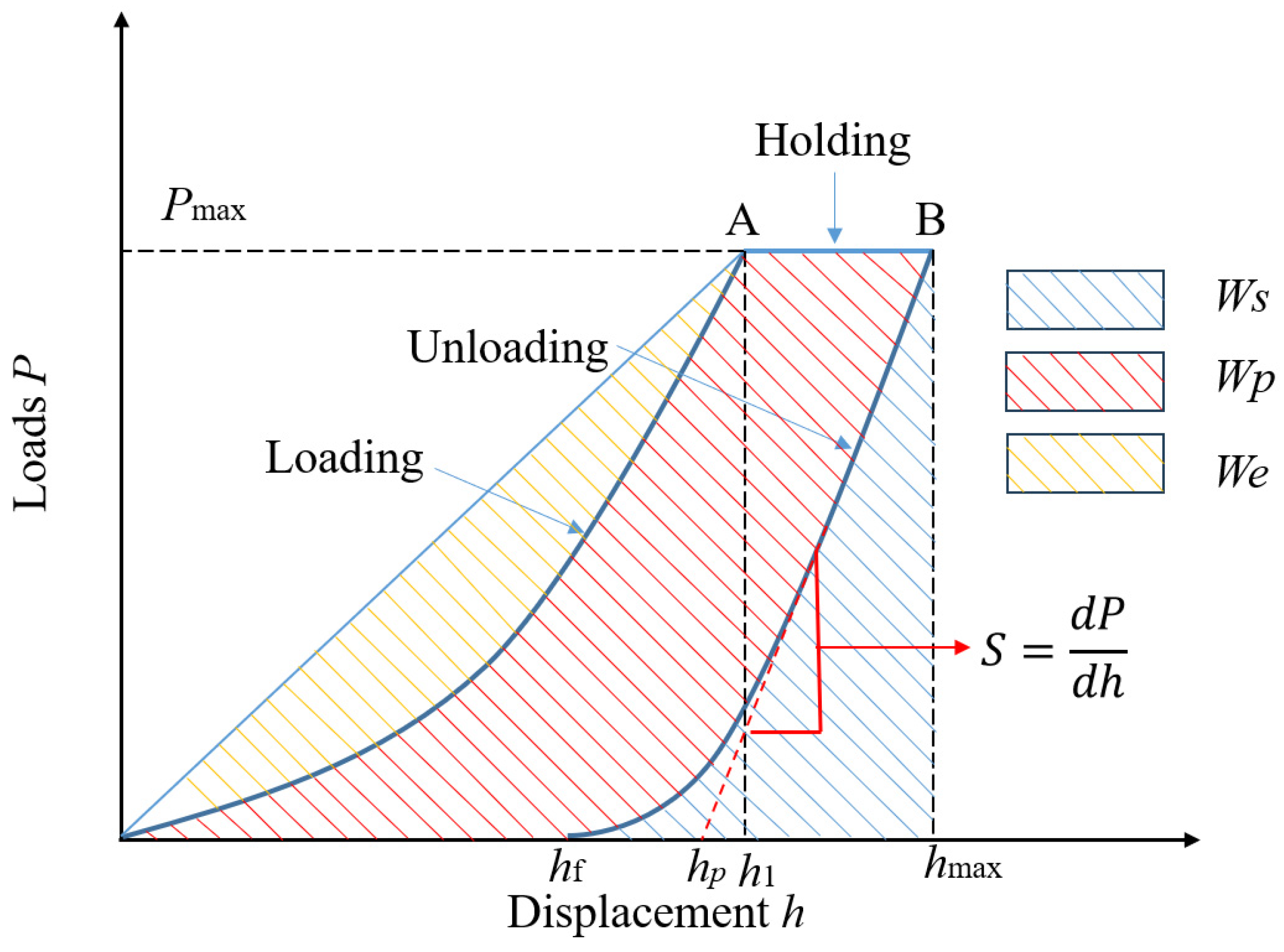
Maximum indentation load, loading rate and unloading rate, total number of indentations, and indentation spacing are the key test parameters. It is necessary to ensure that the different indentations do not affect each other, and at the same time, the position of the indentation in respect to the sample edge should be kept at a certain distance to ensure the safety and accuracy of the experimental results. All indentation tests are basically executed based on ASTM E384. A typical profile of a single indentation with corresponding key parameters is shown in Figure 2, and an obtained idealized load–displacement curve is shown in Figure 3.
The most popular indentation test methodologies are the following:
  • Oliver-Pharr method
In 1992, Oliver and Pharr [92] proposed a widely used algorithm for calculating the mechanical properties (modulus of elasticity and hardness) of a material, which is currently the standard calculation method used in commercial nanoindentation instruments on the market. The Oliver–Pharr method was originally developed for the measurement of hardness and modulus of elasticity of materials by means of an indentation load–depth curve when using a sharp indenter (e.g., Vickers indenter or Berkovich tip). It has been shown that the method can also be applied to any axisymmetric indenter geometry, including spheres [22]. The Oliver–Pharr method utilizes the elastic contact theory to fit the unloaded portion of the indentation load–depth data to the following power–law relationship:
P = β ( h h f ) m
where β and m are fitting parameters. The contact stiffness (S) is then determined from the slope of the unloaded P–h curve and calculated by differentiating Equation (2) at the maximum indentation depth as follows:
S = m β h m a x h f m 1
The contact depth ( h c ) between the indenter and the sample can be calculated as:
h c = h m a x h s = h m a x P m a x / S
where ∈ is a constant, depending on the geometry of the indenter. According to ASTM E2546, for Vickers indenter ∈ is 0.75. Different shapes of indenters produce different indentation areas, so there are different formulas for evaluation. For the Vickers indenter the following formula is valid:
A c = 26.43 h c 2
Indentation hardness and reduced modulus can be calculated by the following two equations:
H = P m a x A c
E r = S π 2 η A c
The reduced modulus E r is an equivalent parameter that describes the joint elastic deformation of the indenter and material in the indentation experiment. By quantifying the synergistic effect of the two, it provides a theoretical foundation for accurately extracting the elastic modulus of the material. The reduced modulus reflects indentation and deformation of the compound. The modulus of elasticity of the sample can be obtained by the following equation:
1 ν 2 E = 1 E r 1 ν i 2 E i
where E r is the reduced modulus, E i is the elastic modulus of indenter, and ν i is the Poisson’s ratio of indenter.
2.
Doerner–Nix method
The Doerner–Nix method is mainly used to study plastic and elastic properties of thin films. The hardness is calculated by subtracting the elastic displacement from the load–displacement data. The Young’s modulus of the material under test is still calculated from the slope of the linear part of the unloading curve. The influence of the elastic properties of the substrate can also be observed in film testing. The hardness can be calculated from the loading curve so that the depth dependence of the hardness can be obtained from a single indentation. The effect of strain rate on the measured hardness obtained in this way can be significant. Doerner and Nix fitted the tangent to the unloading curve at maximum load and extended it to the point when the load is zero to obtain the intercept on the depth axis and defined it as the plastic depth (i.e., h p in Figure 2).
d P d h = 2 π 1 2 D E r
1 E r = 1 ν 2 E + 1 ν i 2 E i
d P d H = 1 2 h p π 24.5 1 2 1 E r
In contrast to previous microhardness tests that require imaging techniques to measure the diagonal length of an indentation to determine the contact area, the Doerner-Nix method calculates the contact area directly by analyzing the load–displacement data, thus overcoming imaging difficulties and dimensional errors.
3.
Energy method
The energy method is a method to evaluate the mechanical properties of materials by analyzing the distribution and transformation of energy during the indentation process. As shown in Figure 2, the areas under the load–displacement curve correspond to energy values. The total energy can be divided into two parts: elastic and plastic deformation energy. The total energy W t is related to the work performed by the indenter on the material during loading as follows:
W t = 0 h m a x P d h
Elastic deformation energy W e is the energy recovered by the material during unloading as follows:
W e = h f h m a x P d h
Plastic deformation energy W p is the energy consumed by the permanent deformation of the material as follows:
W p = W t W e
Through the energy ratio (e.g., W e / W t ), the elastic resilience and plastic dissipation properties of materials can be further analyzed. The energy analysis method is widely used to characterize the mechanical properties of materials, which mainly includes the following: Assessing the fracture toughness by the energy required for crack extension, investigating the elastic resilience and plastic dissipation properties of materials as well as analyzing the influence of the substrate effect on the energy distribution.
In 2019, Su et al. [117] proposed an addition to the energy analysis method by adding absolute work ( W s ) and specifying the difference between total and elastic energies as W i r , which is further categorized into pure plastic energy ( W p p ) and crack energy ( W c r a c k ):
W i r = W t W e = 0 h m a x P d h h f h m a x P d h = W p p + W c r a c k
The critical stress intensity factor K I C of the specimen is finally obtained by the following:
K I C = W c r a c k · E r A m
where A m is the maximum crack area.
The energy analysis method of indentation testing comprehensively evaluates the mechanical properties of materials by analyzing the energy distribution during loading and unloading. Its main advantage lies in its ability to study elastic and plastic behaviors at the same time, and it is suitable for the micromechanical characterization of a wide range of materials. However, the method places high demands on experimental data and analytical models, which need to be optimized for specific materials and application scenarios. Through the energy analysis method, indentation hardness, and elastic modulus can be accurately calculated, providing an important basis for material design and performance optimization.
The method selection is linked to the test objectives: Oliver–Pharr is the first choice for high-precision nanoindentation, Doerner–Nix is used for rapid screening, and the energy law focuses on mechanism research, revealing the elasto–plastic nature of materials from an energy perspective. The future trend is to integrate the advantages of multiple methods, combining AI and multi-scale simulation, and will promote indentation technology from performance characterization to intelligent design and realize high-throughput evaluation and cross-scale correlation of mechanical properties.
4.
Comparison of methods
Oliver–Pharr (OP): Best for elastic modulus and hardness determination with sharp tips when pile-up/sink-in are corrected and contact area is well calibrated; sensitive to tip rounding and rough surfaces. Robust for brittle phases (quartz, calcite) provided h/d ≲ 0.1–0.2 and spacing ≥ 3–5× indenter size according to ISO 14577.
Doerner–Nix (DN): Useful for rapid hardness profiling and thin films/coatings; unloading linearization may bias Young’s modulus for heterogeneous rocks; recommended as a screening method, not a sole basis for modulus in coarse-grained or highly anisotropic rocks.
Energy–Method (EM): Complements OP/DN by partitioning elastic vs. plastic work and enables fracture/creep analysis; recommended when crack systems or time-dependent effects are central, with concurrent imaging (SEM/AFM) for crack morphology control.
For rock phases where anisotropy and roughness matter, we recommend spherical or blunt-tip elastic modulus checks alongside OP/EM analysis when fracture/creep are relevant and microstructure-aware homogenization (e.g., Voigt–Reuss–Hill/Mori–Tanaka) for upscaling. Cross-validate with dynamic pulse or core tests when available.

4. Interpretation Models

4.1. Classical Models

Interpretation models are used to extract the mechanical properties of the material (e.g., modulus of elasticity, hardness, yield strength, and fracture toughness, etc.) from the load–displacement curve. The most common ones are described below:
  • Hertz contact theory [118]
In 1881, the German Heinrich Hertz described stress distribution and deformation in the contact region by equations of elastomechanics. The Hertz contact theory is based on a series of idealized assumptions to ensure simplicity and applicability. First, it is assumed that the contact material is a linear elastomer, i.e., the material strictly follows Hooke’s law when subjected to a force, where stress is proportional to strain, and is able to completely recover to its original state after unloading without any plastic deformation. Secondly, the size of the contact area is much smaller than the radius of curvature of the contacting object, and the deformation is much smaller than the geometric size of the object, so that the contact problem can be simplified to a local elastic small deformation problem, ignoring the deformation effect of the overall structure. Therefore, the basic equations of linear elasticity can be used to describe the contact behavior. In addition, the theory assumes that the contact surface is ideally smooth, ignoring the effects of surface roughness at the contact, as well as disregarding the friction between the contact surfaces. Finally, Hertz contact theory applies only to static contact problems, ignoring dynamic effects such as inertial forces and impact loads. Together, these assumptions form the basis of the Hertz contact theory, allowing it to accurately describe the stress distribution and deformation behavior in the contact region under ideal conditions.
When extending the Hertz contact theory to indentation experiments, it is assumed that the indenter is a rigid sphere (radius R) and the material under test is an isotropic elastic half-space body with elastic modulus E and Poisson’s ratio ν. According to Hertz’s theory, the equivalent radius of curvature Reff is reduced to the radius of the indenter (since the radius of curvature of the plane is infinite), and the equivalent modulus of elasticity E is determined by the following formula:
1 E = 1 ν 2 E + 1 v i 2 E i
Since the indenter is assumed to be rigid, i.e., E i is infinite, the equivalent modulus is given by the following:
E = E 1 ν 2
In the elastic contact phase, the Hertz theory results in a nonlinear relationship between the load P and the depth of indentation h as follows:
P = 4 3 E R 1 / 2 h 2 / 3
Contact radius a and maximum load P m a x are as follows:
a = 3 P R 4 E 1 / 3
P m a x = 3 P 2 π a 2
The contact stiffness S can be expressed as follows:
S = d P d h = 2 E R h π
Considering the contact area A c = π a 2 provides an expression for the modulus of elasticity as follows:
E = S 2 π A c
However, its application is limited by idealized assumptions: The theory is strictly applicable only to purely elastic response under small deformation conditions and cannot describe the mechanical behavior after plastic deformation or material yielding. At the same time, it assumes that the contact surfaces are smooth and frictionless, which ignores the influence of actual material surface roughness and friction effects on the distribution of contact stresses. In addition, when the indentation depth is close to or exceeds the radius of curvature of the indenter, the geometrical and material nonlinearities caused by large deformation will significantly deviate from the predictions of Hertz theory. Nonetheless, Hertz’s theory lays the central framework for the elastic phase analysis of indentation experiments, and its simple mathematical form and physically meaningful parameter associations make it a starting point for understanding complex contact problems. For indentation problems involving plastic deformation, viscoelastic response, or dynamic loading, further comprehensive multi-scale and multi-physical field analyses combined with the reaming model, the Oliver-Pharr method, or dynamic contact theory are needed to compensate for the limitations of the Hertz theory under idealized assumptions.
2.
Elasto-plastic contact theory (Expanding Cavity Model) [119]
Johnson proposed the Expanding Cavity Model (ECM) in 1970 to describe the elasto–plastic contact behavior in indentation experiments. This theory considers the penetration of a rigid indenter into an elasto–plastic material. Plastic flow occurs in the material underneath the indenter due to the shear stress exceeding the yield strength, forming an approximately spherical plastic zone (dilatation zone), which is surrounded by an elastic deformation zone. This concept considers (a) the plastic dissipation energy, i.e., the energy required to yield the material in the plastic zone, and (b) the elastic confinement energy, i.e., the work performed by the peripheral elastic zone on the reversed stresses exerted in the plastic zone.
The theoretical framework of ECM also relies on a series of idealized assumptions to ensure mathematical simplicity and analytical feasibility. First, the model assumes that the material is an isotropic and incompressible continuous medium whose plastic deformation strictly follows the Mises yield criterion, and the volume of the material remains unchanged during deformation by default (Poisson’s ratio is 0.5). Therefore, only a pure shape distortion is assumed. Secondly, the model idealizes the geometry of the plastic zone formed under the indenter as strictly spherical and assumes that the radius c of the plastic zone is proportional to the contact radius a (ca), which simplifies the calculation of the energy of the expansion of the plastic zone through spherical symmetry. In addition, the model further assumes that the boundary between the elastic and plastic zones is clear and unambiguous, and the stress distribution at the interface of the two regions is continuous and without transition zones, which circumvents the possible existence of a complex asymptotic stress field in practice. In terms of loading conditions, similar to the Hertz model, it is limited to quasi-static processes, completely ignoring dynamic effects (e.g., inertial force, strain rate dependence) on plastic flow, and the default deformation process is dominated only by hydrostatic equilibrium. Finally, the model assumes that the material is an ideal elasto-plastic body, i.e., there is no strain-hardening effect during the plastic deformation phase and the yield strength σ y remains constant. Together, these assumptions lead to a correlation between indentation load, plastic zone dimension, and mechanical parameters under ideal conditions, but at the same time, they set clear boundary conditions for the model’s range of applicability.
Plastic zone radius c and contact radius a satisfy a linear relationship as follows:
c = k a
where k is a constant of proportionality, usually taken as k 2 3 , depending on the material constraints.
The relationship between indentation load P, volume of plastic zone and yield strength is as follows:
P = 4 3 π a 3 σ y l n c a + 2 3
Combined with Equation (23), this can be simplified to as follows:
P σ y a 3 ( l n k + 2 3 )
According to the cavity expansion theory, the relationship between indentation hardness H and material yield strength σ y is as follows:
H = C σ y ( E σ y ) n
where
-
C and n are dimensionless constants, with C 3 and n 0.1 0.2 for typical metallic materials.
-
E is the modulus of elasticity of the material.
The formula shows that the hardness depends not only on the yield strength but is also related to the modulus of elasticity, reflecting the restraining effect of the elastic zone on plastic deformation.
3.
Fracture mechanics model (Lawn crack extension model) [120]
In 1987, Lawn et al. proposed a procedure to assess the fracture toughness ( K I C ) of a material through the crack morphology produced by indentation experiments. The core idea of this model is that the energy required for crack extension is provided by the residual stress field, and the fracture toughness can be calculated by balancing the crack driving force with the material resistance. When a sharp indenter (e.g., a Vickers indenter) is pressed into the surface of a brittle material, a high stress field below the indenter initiates plastic deformation while generating residual stresses at the edges of the plastic zone. Upon unloading, the residual stresses drive radial cracks or median cracks to expand outward from the corner of the indentation.
The theoretical construction of Lawn’s crack propagation model is based on a series of strict idealized assumptions to ensure that the crack propagation process can be quantitatively described by analytical equations. First, the model assumes that the material is a homogeneous and isotropic continuous medium, thus simplifying the complex microstructural effects into a macroscopic uniform response. Secondly, a semi-infinite body is assumed, i.e., the size of the indentation region and crack is much smaller than the overall size of the material. Therefore, crack extension is dominated by the local stress field only. In terms of crack types, the model focuses on radial or median cracks driven by residual stresses after indentation unloading, ignores other possible crack morphologies (e.g., transverse cracks or annular cracks), and assumes that these cracks always follow the linear-elastic fracture mechanics (LEFM) framework during extension, i.e., the size of the plastic zone at the tip of the crack is much smaller than the length of the crack. In addition, the model emphasizes the central role of the residual stress field, arguing that the driving force for crack extension originates entirely from the residual stress field formed after unloading of the indentation, while the transient stresses during the loading process make a negligible contribution to crack initiation and extension. Together, these assumptions limit the applicability of the model to achieve high-precision assessment of fracture toughness in brittle materials, macroscopically homogeneous structures and typical indentation crack morphology. However, it provides a starting point for theoretical corrections introduced by material anisotropy, microscopic defects, or complex crack networks in practical applications.
The fracture toughness (mode I) according to the Lawn model is given by:
K I C = α ( E H ) 1 / 2 P c 3 / 2
where
  • α: dimensionless constant, depending on indenter geometry and crack type (usually α 0.016 0.035 );
  • E: modulus of elasticity of the material (GPa);
  • H: hardness of the material (GPa), calculated by H = P/Ac (Ac is the projected area of the indentation);
  • P: Maximum load of the indentation (N);
  • c: Average length from the center of the indentation to the tip of the crack (m).
The crack driving force is provided by the residual stress field σ r , which is expressed as:
σ r P c 3 / 2
When the residual stress reaches the critical value of the fracture toughness of the material, the crack undergoes destabilizing extension. The steady state crack length c is related to the load P by the following relation:
c P 2 / 3
Although the Lawn crack extension model has the advantages of high efficiency and convenience in the fracture toughness assessment of brittle materials, its application is still limited by the multiple constraints of the theoretical assumptions and experimental conditions. The model’s strict idealization of crack morphology (e.g., only radial/median cracks are considered) leads to significant deviations in the prediction of actual complex crack networks (e.g., bifurcation and annular cracks), and the empirical constant α in the formulation is highly dependent on the crack type and indenter geometry, which needs to be calibrated by a large number of experiments. The model is strictly limited to the framework of linear-elastic fracture mechanics and is therefore inappropriate for ductile materials (e.g., metals or polymers) or for micro- and nanoscale indentations with significant plastic deformation. In addition, the model is only applicable to residual stress fields induced by sharp indenters (e.g., Vickers indenters), lacking explanations for crack extension behavior under spherical indenters or complex load histories, and does not take into account the interference of surface roughness, machining defects, or environmental factors (e.g., humidity and temperature) on crack initiation.
Nevertheless, by quantitatively correlating indentation morphology with fracture toughness, the Lawn model provides a unique means of mechanically characterizing ceramics, glasses, and brittle coatings without the need for prefabricated cracks, greatly simplifying the experimental process. In order to break through the theoretical boundaries, the researchers have gradually overcome the inherent limitations of the model by introducing dynamic correction factors, coupling finite element simulations to analyze the complex stress field, and expanding to crystalline materials by integrating anisotropic constitutive equations. In the future, with the deep integration of micro- and nanoindentation technology and multi-scale fracture mechanics, the Lawn model is expected to play an important role in the reliability assessment of thin-film devices, optimization of bioceramics, and design of materials for extreme environments.
Existing studies have mainly focused on investigating the anisotropic mechanical behavior of rocks using micro-indentation or micro-compression techniques. For example, Ma et al. [121] conducted micro-indentation tests on three types of shales in directions normal (BPN) and parallel (BPP) to the bedding planes and measured hardness, elastic modulus, UCS, and fracture toughness. Their results showed that hardness, elastic modulus, and UCS in the BPP direction were higher than those in the BPN direction, whereas fracture toughness exhibited the opposite trend. Zhang et al. [57] performed micro-indentation and micro-compression tests on claystone specimens under different relative humidity conditions, both parallel and perpendicular to the bedding planes. They found that elastic modulus, peak stress, and axial strain at peak stress all displayed clear anisotropy. Using the explicit contact finite element method, Dong and Chen [122] simulated the failure of rock elements and demonstrated that shale crushing efficiency is highly sensitive to the dip angle once it exceeds 30°.
However, although these studies have revealed significant anisotropic behaviors of rocks in respect to bedding directions, no research has yet directly applied the Hertz or Lawn models to interpret such anisotropic indentation responses. We believe that using and further developing these classical contact mechanics models for investigating the anisotropic characteristics of rocks could represent a promising way for providing new insights into the understanding and prediction of rock failure mechanisms.

4.2. Simulation and Verification for Rocks

With the increasing use of indentation technology in the field of rock mechanics, more and more scholars combine indentation tests with numerical simulations. This strategy is used, for instance, to find out the relationship between the elastic modulus at different scales or to investigate the relationship between microscopic indentation hardness, strength parameters, and bulk density through inverse analysis and comparing them with macroscopic test results.
First studies were conducted with shale to understand the micromechanical properties. Bobko and Ulm [69] revealed through large-scale indentation experiments that the porous clay phase of shale has nanomechanical morphology that is different from the macroscopic visible structure. They found that a permeability threshold appears when the packing density of clay particles reaches η0 ≥ 0.5 and constructed a nanoparticle morphology model characterized by elastic anisotropy and strength isotropy. This model breaks through the limitations of mineral composition and provides a universal explanatory framework for the multi-scale mechanical behavior of shale. Kumar et al. [123] used scanning electron microscopy and nanoindentation technology to reveal the mechanical properties of shale organic matter: The Young’s modulus of kerogen (1.9–15 GPa) is jointly regulated by porosity and maturity, and the increase in organic matter content significantly reduces Young’s modulus and hardness. The nanoindentation test results based on small samples at the millimeter level are consistent with the dynamic modulus of the core, providing a microscale solution for hydraulic fracturing optimization and unstable rock mechanical evaluation in shale gas development. Veytskin et al. [12] used nanoindentation and SEM-EDS technology, combined with mineral volume fraction statistics and strength homogenization analysis, to reveal the mechanical morphological characteristics of shale as a clay–silicate–sulfide nanoparticle composite material. They clarified the mechanical relationship between high clay content in soft shale and high silicate/sulfide content in hard shale. They also developed a cross-scale response law of microscopic clay packing density and macroscopic porosity and proposed a multi-scale research path to analyze mineral phase distribution and clay–quartz bonding mechanism by using three-dimensional X-ray microscopy technology. Liu et al. [71] used grid nanoindentation technology combined with the Mori–Tanaka homogenization method to achieve statistical deconvolution of elastic parameters of micromechanical phases and macro-modulus prediction of the Bakken shale (with core test error < 15%), verifying the reliability of nanoindentation technology for cross-scale mechanical characterization of heterogeneous and anisotropic shales and providing an innovative solution for micro–macro linkage for geomechanical research in the oil industry in the absence of core samples. Xu et al. [124] revealed the multi-scale mechanical degradation mechanism of weakly interlayered shales caused by softening of clay minerals, attenuation of cohesion and decrease in elastic modulus under water–rock interaction through nanoindentation experiments and numerical simulations, providing a new method for micro–macro linkage for rock slope instability analysis. Based on large volume statistical nanoindentation technology, Lu et al. [125] revealed the cross-scale softening mechanism of shale (decrease in Young’s modulus obtained by micro–macro linkage) through the cumulative distribution function deconvolution and confining pressure effect model. They also developed a softening rate law of rock intrinsic permeability regulation and provided a new cross-scale experimental paradigm for the optimization of hydraulic fracturing chemicals and studied the shale dynamic mechanical response. Yang et al. [126] used dynamic nanoindentation combined with optical microscopy to reveal the multi-scale mechanism of shale mineral mechanical response: silicate minerals (quartz/feldspar) are mainly highly elastic, while carbonate and clay minerals show significant plasticity (clay undergoes large deformation due to sliding of flaky particles). They investigated the coupling between the indentation size effect (which disappears at 200–300 nm) and the substrate effect (the depth needs to be controlled within d/50), proposed an optimized test range of 200 nm < h < d/50, and measured the intrinsic modulus and hardness of minerals such as clay (30 GPa/1.5 GPa) and quartz (105–110 GPa/14–16 GPa), providing a depth-sensitive experimental optimization scheme for the cross-scale mechanical characterization of shale minerals.
Some scholars have conducted indentation studies on other rocks or mineral components. Zhang et al. [127] studied the deformation and failure behavior of Sichuan sandstone by combining a two-dimensional indentation device with DIC non-destructive technology. The results showed that sandstone mainly exhibited a splitting failure mechanism under cylindrical indentation, and DIC could effectively capture the crack initiation and slip regions, providing a basis for further high-resolution research. Song et al. [128] proposed a new method combining microindentation experiments and numerical simulations to quantitatively obtain the initial and residual strength of brittle rocks with known constitutive models and pore–scale structures at the microscale. Ma et al. [129] revealed the nanomechanical properties of α-quartz through nanoindentation experiments and molecular dynamics simulations: The choice of contact area function plays a decisive role in the MD simulation hardness calculation. Elastic modulus and hardness in shallow indentation increase with depth (consistent with the indentation size effect). The deeper layers show a non-Hertzian response due to the dominant plastic deformation. The radius increase in the indenter tip leads to a decrease in Er and H, and the shear strain analysis shows that the plastic zone expands with the depth of indentation, which verifies the reliability of the multi-scale method and provides a theoretical basis for the nanomechanical characterization of quartz materials.

5. Summary and Outlook

The indentation technology is widely applied in shale, but other rocks still need exploration. This may be attributed to the historical emphasis on the macroscopic mechanical properties of rocks in the past. However, as shale is used as an unconventional oil and gas reservoir, indentation tests have been introduced to determine the microscopic and even nanoscopic mechanical properties of various phases of shale, but less attention has been paid to other types of common rocks. So far, only very limited research has been conducted on anisotropic crystalline rocks. Through traditional mechanical tests such as uniaxial and triaxial compression tests, the average mechanical properties of relatively large intact rock samples can be measured at the macroscopic scale, but the anisotropy of a single component cannot be measured. At the same time, some imaging analysis techniques are used to qualitatively and quantitatively analyze the microstructure, but the impact of these on the macroscopic mechanical properties cannot be accurately evaluated. This leads to the lack of a bridge connecting the research on mechanical properties of rocks at the micro- and macroscale and the rock structure.
In the past few decades, indentation testing has developed from macroscopic to microscopic and even nanoscopic scale, and the scope of application has developed from single-phase homogeneous materials to multiphase composite materials. In the future, the development of indentation experiments in the field of rock mechanics will move towards intelligent systems, multi-scale, and engineering practicality. The rise in intelligent indentation systems, such as AI-driven real-time analysis, robotic automated testing platforms, and virtual experiments assisted by digital twins, will significantly improve experimental efficiency and accuracy and enable indentation technology to move from laboratory to on-site in situ testing. The deep integration of multi-scale models, combined with molecular dynamics, discrete element, and finite element methods, is expected to establish cross-scale mechanical correlations from mineral particles to macroscopic rock masses, while the fusion of high-resolution imaging technology (such as μ-CT and FIB-SEM) and indentation data will more accurately reveal the influence of rock microstructure on mechanical behavior. In addition, indentation research under extreme environments (high temperature, high pressure, dynamic impact) will provide key parameters for deep resource development, earthquake mechanism and impact as well as geothermal reservoir optimization. In the future, the establishment of standardized databases and the fusion of multimodal data will promote the transformation of rock mechanics from empirical science to predictive science and ultimately realize a wider application of indentation technology in energy exploration, geological disaster prediction, and deep earth engineering.

Author Contributions

Conceptualization, Q.H. and H.K.; methodology, Q.H. and H.K.; software, Q.H.; validation, Q.H. and H.K.; formal analysis, Q.H.; investigation, Q.H.; resources, Q.H. and H.K.; data curation, Q.H.; writing—original draft preparation, Q.H.; writing—review and editing, H.K.; visualization, Q.H.; supervision, H.K.; project administration, H.K.; funding acquisition, Q.H. and H.K. All authors have read and agreed to the published version of the manuscript.

Funding

This research was funded by China Scholarship Council, grant number 202210220002.

Acknowledgments

Qingqing He would like to thank the China Scholarship Council (CSC) for the financial support (grant No. 202210220002).

Conflicts of Interest

The authors declare no conflicts of interest.

References

  1. Walley, S.M. Historical origins of indentation hardness testing. Mater. Sci. Technol. 2012, 28, 1028–1044. [Google Scholar] [CrossRef]
  2. Wright, J. Jewelry alloys: Hardness and hardenability. In Proceedings of the Santa Fe Symposium on Jewelry Manufacturing, Albuquerque, NM, USA, 4 December 2009; pp. 453–472. [Google Scholar]
  3. Smith, R.L.; Sandly, G.E. An accurate method of determining the hardness of metals, with particular reference to those of a high degree of hardness. Proc. Inst. Mech. Eng. 1922, 102, 623–641. [Google Scholar] [CrossRef]
  4. Hays, C.; Kendall, E.G. An analysis of Knoop microhardness. Metallography 1973, 6, 275–282. [Google Scholar] [CrossRef]
  5. Barbato, G.; Galetto, M.; Germak, A.; Fabrizio, M. Influence of the indenter shape in Rockwell hardness test. In Proceedings of the HARDMEKO 98, Beijing, China, 21–23 September 1998; pp. 21–23. [Google Scholar]
  6. Chang, Y.-H.; Lin, S.-H.; Chen, H.-L.; Chen, Y.-T.; Huang, X.-C. Annealing effects on grain size, surface roughness, nanoindentation, and magnetic properties of Fe80Ce20 films deposited on PET and PMMA substrates. Materialia 2025, 39, 102310. [Google Scholar] [CrossRef]
  7. Ban, Y.; Yang, X.; Mi, C. On the nanoindentation of a couple-stress half-plane with Steigmann–Ogden surface effects. Mech. Adv. Mater. Struct. 2025, 1–17. [Google Scholar] [CrossRef]
  8. Adams, D.S.; Boyce, B.L.; Hooks, D.E.; Garber, K.W.; Klitsner, B.; Price, S.A.; Blob, R. A brief introductory guide to nanoindentation for comparative and evolutionary biologists, with a case study of bone material property diversity across artiodactyl skulls. Integr. Org. Biol. 2025, 7, obaf010. [Google Scholar] [CrossRef]
  9. Efremenko, B.; Chabak, Y.; Petryshynets, I.; Efremenko, V.; Wu, K.; Arshad, S.; Kromka, F. Microstructure Evolution, Tensile/Nanoindentation Response, and Work-Hardening Behaviour of Prestrained and Subsequently Annealed LPBF 316L Stainless Steel. Materials 2025, 18, 1102. [Google Scholar] [CrossRef]
  10. Timoshenko, S. History of Strength of Materials; Mcgraw-Hill Book Company Inc.: New York, NY, USA, 1953. [Google Scholar]
  11. Boulenouar, A.; Mighani, S.; Pourpak, H.; Bernabé, Y.; Evans, B. Mechanical Properties of Vaca Muerta Shales from NanoIndentation Tests. In Proceedings of the 51st U.S. Rock Mechanics/Geomechanics Symposium, San Francisco, CA, USA, 25–28 June 2017. [Google Scholar]
  12. Veytskin, Y.B.; Tammina, V.K.; Bobko, C.P.; Hartley, P.G.; Clennell, M.B.; Dewhurst, D.N.; Dagastine, R.R. Micromechanical characterization of shales through nanoindentation and energy dispersive x-ray spectrometry. Geomech. Energy Environ. 2017, 9, 21–35. [Google Scholar] [CrossRef]
  13. Gupta, I.; Sondergeld, C.; Rai, C. Fracture toughness in shales using nano-indentation. J. Pet. Sci. Eng. 2020, 191, 107222. [Google Scholar] [CrossRef]
  14. Fan, M.; Jin, Y.; Chen, M.; Geng, Z. Mechanical characterization of shale through instrumented indentation test. J. Pet. Sci. Eng. 2019, 174, 607–616. [Google Scholar] [CrossRef]
  15. Cheng, P.; Zhang, C.; Ma, Z.; Zhou, J.; Zhang, D.; Liu, X.; Chen, H.; Ranjith, P. Experimental study of micromechanical properties alterations of shale matrix treated by ScCO2-Water saturation using nanoindentation tests. Energy 2022, 242, 122965. [Google Scholar] [CrossRef]
  16. Ma, Z.; Zhang, C.; Gamage, R.P.; Zhang, G. Uncovering the creep deformation mechanism of rock-forming minerals using nanoindentation. Int. J. Min. Sci. Technol. 2022, 32, 283–294. [Google Scholar] [CrossRef]
  17. Sun, C.; Li, G.; Gomah, M.E.; Xu, J.; Sun, Y. Creep characteristics of coal and rock investigated by nanoindentation. Int. J. Min. Sci. Technol. 2020, 30, 769–776. [Google Scholar] [CrossRef]
  18. Ma, G.; Zhou, W.; Ng, T.-T.; Cheng, Y.-G.; Chang, X.-L. Microscopic modeling of the creep behavior of rockfills with a delayed particle breakage model. Acta Geotech. 2015, 10, 481–496. [Google Scholar] [CrossRef]
  19. Nastic, A.; Merati, A.; Bielawski, M.; Bolduc, M.; Fakolujo, O.; Nganbe, M. Instrumented and Vickers indentation for the characterization of stiffness, hardness and toughness of zirconia toughened Al2O3 and SiC armor. J. Mater. Sci. Technol. 2015, 31, 773–783. [Google Scholar] [CrossRef]
  20. Liu, K.; Ostadhassan, M.; Bubach, B. Applications of nano-indentation methods to estimate nanoscale mechanical properties of shale reservoir rocks. J. Nat. Gas Sci. Eng. 2016, 35, 1310–1319. [Google Scholar] [CrossRef]
  21. Chen, W.; Li, M.; Zhang, T.; Cheng, Y.-T.; Cheng, C.-M. Influence of indenter tip roundness on hardness behavior in nanoindentation. Mater. Sci. Eng. A 2007, 445, 323–327. [Google Scholar] [CrossRef]
  22. Oliver, W.C.; Pharr, G.M. Measurement of hardness and elastic modulus by instrumented indentation: Advances in understanding and refinements to methodology. J. Mater. Res. 2004, 19, 3–20. [Google Scholar] [CrossRef]
  23. Bucaille, J.; Stauss, S.; Felder, E.; Michler, J. Determination of plastic properties of metals by instrumented indentation using different sharp indenters. Acta Mater. 2003, 51, 1663–1678. [Google Scholar] [CrossRef]
  24. Chollacoop, N.; Dao, M.; Suresh, S. Depth-sensing instrumented indentation with dual sharp indenters. Acta Mater. 2003, 51, 3713–3729. [Google Scholar] [CrossRef]
  25. Karimzadeh, A.; Ayatollahi, M.R.; Alizadeh, M. Finite element simulation of nanoindentation experiment on aluminum 1100. Comput. Mater. Sci. 2014, 81, 595–600. [Google Scholar] [CrossRef]
  26. Xie, A.; Ji, X.; Chen, Y.; Zhang, M.; Inoue, S.-I. Nanoindentation measurements of mechanical properties of polyurethane elastomers which crosslinked by β-cyclodextrin. Open J. Org. Polym. Mater. 2016, 6, 112–118. [Google Scholar] [CrossRef]
  27. Ayatollahi, M.; Alishahi, E.; Doagou-R, S.; Shadlou, S. Tribological and mechanical properties of low content nanodiamond/epoxy nanocomposites. Compos. Part B Eng. 2012, 43, 3425–3430. [Google Scholar] [CrossRef]
  28. Arnold, M.; Zhao, S.; Ma, S.; Giuliani, F.; Hansen, U.; Cobb, J.P.; Abel, R.L.; Boughton, O. Microindentation—A tool for measuring cortical bone stiffness? A systematic review. Bone Jt. Res. 2017, 6, 542–549. [Google Scholar] [CrossRef] [PubMed]
  29. Ishimoto, T.; Nakano, T.; Yamamoto, M.; Tabata, Y. Biomechanical evaluation of regenerating long bone by nanoindentation. J. Mater. Sci. Mater. Med. 2011, 22, 969–976. [Google Scholar] [CrossRef]
  30. Cuy, J.L.; Mann, A.B.; Livi, K.J.; Teaford, M.F.; Weihs, T.P. Nanoindentation mapping of the mechanical properties of human molar tooth enamel. Arch. Oral Biol. 2002, 47, 281–291. [Google Scholar] [CrossRef]
  31. Karimzadeh, A.; Ayatollahi, M.R. Investigation of mechanical and tribological properties of bone cement by nano-indentation and nano-scratch experiments. Polym. Test. 2012, 31, 828–833. [Google Scholar] [CrossRef]
  32. Pan, C.; Li, X.; He, L.; Li, J. Study on the effect of micro-geometric heterogeneity on mechanical properties of brittle rock using a grain-based discrete element method coupling with the cohesive zone model. Int. J. Rock Mech. Min. Sci. 2021, 140, 104680. [Google Scholar] [CrossRef]
  33. Song, R.; Wang, Y.; Sun, S.; Liu, J. Characterization and microfabrication of natural porous rocks: From micro-CT imaging and digital rock modelling to micro-3D-printed rock analogs. J. Pet. Sci. Eng. 2021, 205, 108827. [Google Scholar] [CrossRef]
  34. Li, X.F.; Li, H.B.; Zhao, J. The role of transgranular capability in grain-based modelling of crystalline rocks. Comput. Geotech. 2019, 110, 161–183. [Google Scholar] [CrossRef]
  35. Wu, Z.; Xu, X.; Liu, Q.; Yang, Y. A zero-thickness cohesive element-based numerical manifold method for rock mechanical behavior with micro-Voronoi grains. Eng. Anal. Bound. Elem. 2018, 96, 94–108. [Google Scholar] [CrossRef]
  36. Martin, C.D.; Chandler, N.A. The progressive fracture of Lac du Bonnet granite. Int. J. Rock Mech. Min. Sci. Geomech. Abstr. 1994, 31, 643–659. [Google Scholar]
  37. Jia, L.; Chen, M.; Zhang, W.; Xu, T.; Zhou, Y.; Hou, B.; Jin, Y. Experimental study and numerical modeling of brittle fracture of carbonate rock under uniaxial compression. Mech. Res. Commun. 2013, 50, 58–62. [Google Scholar] [CrossRef]
  38. Lan, H.; Martin, C.D.; Hu, B. Effect of heterogeneity of brittle rock on micromechanical extensile behavior during compression loading. J. Geophys. Res. Solid Earth 2010, 115, B01202. [Google Scholar] [CrossRef]
  39. Wang, X.; Cai, M. Modeling of brittle rock failure considering inter-and intra-grain contact failures. Comput. Geotech. 2018, 101, 224–244. [Google Scholar] [CrossRef]
  40. Yang, Z.; Wang, L.; Zhang, G.; Ho, C. Micromechanical Characterization of Fluid-Shale Interactions via Nanoindentation. In Proceedings of the SPE Asia Pacific Hydraulic Fracturing Conference, Beijing, China, 24–26 August 2016; Volume 24, p. 181833. [Google Scholar]
  41. Xie, W.; Liu, X.; Zhang, X.; Yang, X.; Zhou, X. A review of rock macro-indentation: Theories, experiments, simulations, and applications. J. Rock Mech. Geotech. Eng. 2024, 16, 2351–2374. [Google Scholar] [CrossRef]
  42. Liu, Y.; Zhang, G.; Qiao, J.; Tang, X. Micromechanical testing and property upscaling of planetary rocks: A critical review. Int. J. Min. Sci. Technol. 2024, 34, 1217–1241. [Google Scholar] [CrossRef]
  43. Puchi-Cabrera, E.S.; Rossi, E.; Sansonetti, G.; Sebastiani, M.; Bemporad, E. Machine learning aided nanoindentation: A review of the current state and future perspectives. Curr. Opin. Solid State Mater. Sci. 2023, 27, 101091. [Google Scholar] [CrossRef]
  44. Ma, Z.; Pathegama Gamage, R.; Zhang, C. Application of nanoindentation technology in rocks: A review. Geomech. Geophys. Geo-Energy Geo-Resour. 2020, 6, 60. [Google Scholar] [CrossRef]
  45. Brinell, J. Sätt att bestämma kroppars hårdhet jämte några tillämpningar of detsamma. Tek. Tidskr. 1900, 30, 69–87. [Google Scholar]
  46. Meyer, E. Untersuchungen über härteprüfung und härte. Z. Vereines Dtsch. Ingenieure 1908, 52, 645–654. [Google Scholar]
  47. Huang, Y.; Liu, X.; Zhou, Y.; Ma, Z.; Lu, C. Mathematical analysis on the uniqueness of reverse algorithm for measuring elastic-plastic properties by sharp indentation. J. Mater. Sci. Technol. 2011, 27, 577–584. [Google Scholar] [CrossRef]
  48. Richardson, J.F.; Worner, H.K. A microhardness instrument for studying surface hardness. Aust. J. Dent. 1945, 49, 217. [Google Scholar] [PubMed]
  49. Lee, M.; Brun, M.K. Fracture toughness measurement by microindentation and three-point bend methods. Mater. Sci. Eng. A 1988, 105, 369–375. [Google Scholar] [CrossRef]
  50. Duncan-Hewitt, W.C.; Weatherly, G.C. Evaluating the fracture toughness of sucrose crystals using microindentation techniques. Pharm. Res. 1989, 6, 373–378. [Google Scholar] [CrossRef]
  51. Petzold, M.; Landgraf, J.; Füting, M.; Olaf, J. Application of atomic force microscopy for microindentation testing. Thin Solid Film. 1995, 264, 153–158. [Google Scholar] [CrossRef]
  52. Ringstad, C.; Lofthus, E.B.; Sonstebo, E.F.; Fjær, E.; Zausa, F.; Fuh, G.F. Prediction of rock parameters from micro-indentation measurements: The effect of sample size. In Proceedings of the SPE/ISRM Rock Mechanics in Petroleum Engineering, Trondheim, Norway, 8–10 July 1998; p. SPE-47313-MS. [Google Scholar]
  53. Zhu, W.; Bartos, P.J.M. Application of depth-sensing microindentation testing to study of interfacial transition zone in reinforced concrete. Cem. Concr. Res. 2000, 30, 1299–1304. [Google Scholar] [CrossRef]
  54. Wei, Y.; Wang, X.; Wu, X.; Bai, Y. Theoretical and experimental researches of size effect in micro-indentation test. Sci. China Ser. A Math. 2001, 44, 74–82. [Google Scholar] [CrossRef]
  55. Quinn, G.D.; Patel, P.J.; Lloyd, I. Effect of loading rate upon conventional ceramic microindentation hardness. J. Res. Natl. Inst. Stand. Technol. 2002, 107, 299. [Google Scholar] [CrossRef]
  56. Xue, Z.; Huang, Y.; Hwang, K.C.; Li, M. The influence of indenter tip radius on the micro-indentation hardness. J. Eng. Mater. Technol. 2002, 124, 371–379. [Google Scholar] [CrossRef]
  57. Yang, F.; Peng, L.; Okazaki, K. Microindentation of aluminum. Metall. Mater. Trans. A 2004, 35, 3323–3328. [Google Scholar] [CrossRef]
  58. Qin, J.; Huang, Y.; Hwang, K.; Song, J.; Pharr, G. The effect of indenter angle on the microindentation hardness. Acta Mater. 2007, 55, 6127–6132. [Google Scholar] [CrossRef]
  59. Wang, W.; Xu, W.Y.; Corn, S.; Ienny, P. Characterization of mechanical properties of rocks by microindentation test—A new method for testing rock properties. In Boundaries of Rock Mechanics; CRC Press: Boca Raton, FL, USA, 2008; pp. 211–216. [Google Scholar]
  60. Cai, J.; Li, F.; Liu, T.; Chen, B. Microindentation study of Ti–6Al–4V alloy. Mater. Des. 2011, 32, 2756–2762. [Google Scholar] [CrossRef]
  61. Wei, P.; Zheng, Y.-Y.; Xiong, Y.; Zhou, S.; Ali Zaoui, K.A. Effect of water content and structural anisotropy on mechanical property of claystone. Appl. Clay Sci. 2012, 69, 79–86. [Google Scholar] [CrossRef]
  62. Zhang, Q.; Le Roy, R.; Vandamme, M.; Zuber, B. Long-term creep properties of cementitious materials: Comparing microindentation testing with macroscopic uniaxial compressive testing. Cem. Concr. Res. 2014, 58, 89–98. [Google Scholar] [CrossRef]
  63. Baitsch, M.; Le, K.C.; Tran, T.M. Dislocation structure during microindentation. Int. J. Eng. Sci. 2015, 94, 195–211. [Google Scholar] [CrossRef]
  64. Auvray, C.; Lafrance, N.; Bartier, D. Elastic modulus of claystone evaluated by nano-/micro-indentation tests and meso-compression tests. J. Rock Mech. Geotech. Eng. 2017, 9, 84–91. [Google Scholar] [CrossRef]
  65. Mallick, S.; Anoop, M.B.; Rao, K.B. Creep of cement paste containing fly ash-an investigation using microindentation technique. Cem. Concr. Res. 2019, 121, 21–36. [Google Scholar] [CrossRef]
  66. Borc, J.; Sangwal, K.; Świetlicki, M.; Chocyk, D.; Gładyszewski, G. Vickers microindentation deformation of different cleavage faces of potassium bichromate single crystals: Anisotropy in microhardness and crack formation. Mater. Sci. Eng. A 2020, 790, 139642. [Google Scholar] [CrossRef]
  67. Xu, K.; Huang, M.; Cui, M.; Jin, G.; Li, S. Micromechanical properties and bonding fracture of EICP-reinforced sand analyzed using microindentation test. Acta Geotech. 2025, 20, 3543–3561. [Google Scholar]
  68. Pethicai, J.B.; Hutchings, R.; Oliver, W.C. Hardness measurement at penetration depths as small as 20 nm. Philos. Mag. A 1983, 48, 593–606. [Google Scholar] [CrossRef]
  69. Bobko, C.; Ulm, F.J. The nano-mechanical morphology of shale. Mech. Mater. 2008, 40, 318–337. [Google Scholar] [CrossRef]
  70. Kong, L.; Hadavimoghaddam, F.; Li, C.; Liu, K.; Liu, B.; Semnani, A.; Ostadhassan, M. AFM vs. Nanoindentation: Nanomechanical properties of organic-rich Shale. Mar. Pet. Geol. 2021, 132, 105229. [Google Scholar] [CrossRef]
  71. Liu, K.; Ostadhassan, M.; Bubach, B.; Ling, K.; Tokhmechi, B.; Robert, D. Statistical grid nanoindentation analysis to estimate macro-mechanical properties of the Bakken Shale. J. Nat. Gas Sci. Eng. 2018, 53, 181–190. [Google Scholar] [CrossRef]
  72. Kabir, P.; Akono, A.T. Fluid-rock reactions in Mt. Simon sandstone at microscopic length-scale. In Proceedings of the ARMA US Rock Mechanics/Geomechanics Symposium, Seattle, DC, USA, 17–20 June 2018; p. ARMA-2018-219. [Google Scholar]
  73. Bobko, C.P. Material Invariant Properties and Reconstruction of Microstructure of Sandstones by Nanoindentation and Microporoelastic Analysis. Master’s Thesis, Massachusetts Institute of Technology, Cambridge, MA, USA, 2005. [Google Scholar]
  74. Ante, M.A.; Lingareddy, M.G.; Aminzadeh, F.; Jha, B. Nano-and micro-scale deformation behavior of sandstone and shale. In Proceedings of the ARMA US Rock Mechanics/Geomechanics Symposium, Seattle, DC, USA, 17–20 June 2018; p. ARMA-2018-055. [Google Scholar]
  75. Tyurin, A.I.; Victorov, S.D.; Kochanov, A.N.; Shuklinov, A.V.; Pirozhkova, T.S. Methods of micro-and nanoindentation for characterization of local physical and mechanical properties of multiphase materials. AIP Conf. Proc. 2016, 1783, 020227. [Google Scholar] [CrossRef]
  76. Viktorov, S.D.; Golovin, Y.I.; Kochanov, A.N.; Tyurin, A.I.; Shuklinov, A.V.; Shuvarin, I.A.; Pirozhkova, T.S. Micro-and nano-indentation approach to strength and deformation characteristics of minerals. J. Min. Sci. 2014, 50, 652–659. [Google Scholar] [CrossRef]
  77. Golovin, Y.I.; Tyurin, A.I.; Victorov, S.D.; Kochanov, A.N.; Samodurov, A.A.; Pirozhkova, T.S. Physico-mechanical properties and micromechanisms of local deformation in thin near-surface layers of complex multiphase materials. Bull. Russ. Acad. Sci. Phys. 2017, 81, 360–364. [Google Scholar] [CrossRef]
  78. Golovin, Y.I.; Tyurin, A.I.; Victorov, S.D.; Kochanov, A.N.; Pirozhkova, T.S. Size effects and charting the physical and mechanical properties of individual phases and interphases in polycrystalline materials. Bull. Russ. Acad. Sci. Phys. 2018, 82, 856–859. [Google Scholar] [CrossRef]
  79. Vialle, S.; Lebedev, M. Heterogeneities in the elastic properties of microporous carbonate rocks at the microscale from nanoindentation tests. In Proceedings of the SEG International Exposition and Annual Meeting, New Orleans, LA, USA, 18–23 October 2015; p. SEG-2015-5885016. [Google Scholar]
  80. Shukla, P.; Taneja, S.; Sondergeld, C.; Rai, C.S. Nanoindentation measurements on rocks. In Proceedings of the 2014 Annual Conference on Experimental and Applied Mechanics, Greenville, SC, USA, 2–5 June 2014; pp. 99–105. [Google Scholar]
  81. Zhang, Y.; Lebedev, M.; Sarmadivaleh, M.; Barifcani, A. Change in geomechanical properties of limestone due to supercritical CO2 injection. In Proceedings of the SPE Asia Pacific Oil and Gas Conference and Exhibition, Perth, Australia, 25–27 October 2016; p. SPE-182285-MS. [Google Scholar]
  82. Zhang, Y.; Lebedev, M.; Al-Yaseri, A.; Yu, H.; Nwidee, L.N.; Sarmadivaleh, M.; Barifcani, A.; Iglauer, S. Morphological evaluation of heterogeneous oolitic limestone under pressure and fluid flow using X-ray microtomography. J. Appl. Geophys. 2018, 150, 172–181. [Google Scholar] [CrossRef]
  83. Lebedev, M.; Wilson, M.E.J.; Mikhaltsevitch, V. An experimental study of solid matrix weakening in water-saturated Savonnières limestone. Geophys. Prospect. 2014, 62, 1253–1265. [Google Scholar] [CrossRef]
  84. Bandini, A.; Berry, P.; Bemporad, E.; Sebastiani, M. Effects of intra-crystalline microcracks on the mechanical behavior of a marble under indentation. Int. J. Rock Mech. Min. Sci. 2012, 54, 47–55. [Google Scholar] [CrossRef]
  85. Bandini, A.; Berry, P.; Bemporad, E.; Sebastiani, M.; Chicot, D. Role of grain boundaries and micro-defects on the mechanical response of a crystalline rock at multiscale. Int. J. Rock Mech. Min. Sci. 2014, 71, 429–441. [Google Scholar] [CrossRef]
  86. Konstantinidis, A.A.; Frantziskonis, G.; Askes, H.; Aifantis, E.C. The use of nanoindentation for determining internal lengths and the constitutive response of monument materials: Models and experiments. J. Mech. Behav. Mater. 2016, 25, 57–60. [Google Scholar] [CrossRef]
  87. Brooks, Z.; Ulm, F.J.; Einstein, H.H.; Abousleiman, Y. A nanomechanical investigation of the crack tip process zone. In Proceedings of the ARMA US Rock Mechanics/Geomechanics Symposium, Salt Lake City, UT, USA, 27–30 June 2010; p. ARMA-10-309. [Google Scholar]
  88. Sly, M.K.; Thind, A.S.; Mishra, R.; Flores, K.M.; Skemer, P. Low-temperature rheology of calcite. Geophys. J. Int. 2020, 221, 129–141. [Google Scholar] [CrossRef]
  89. Zhang, F.; Guo, H.; Hu, D.; Shao, J.-F. Characterization of the mechanical properties of a claystone by nano-indentation and homogenization. Acta Geotech. 2018, 13, 1395–1404. [Google Scholar] [CrossRef]
  90. Magnenet, V.; Auvray, C.; Francius, G.; Giraud, A. Determination of the matrix indentation modulus of Meuse/Haute-Marne argillite. Appl. Clay Sci. 2011, 52, 266–269. [Google Scholar] [CrossRef]
  91. Bartier, D.; Auvray, C. Determination of elastic modulus of claystone: Nano-/micro-indentation and meso-compression tests used to investigate impact of alkaline fluid propagation over 18 years. J. Rock Mech. Geotech. Eng. 2017, 9, 511–518. [Google Scholar] [CrossRef]
  92. Oliver, W.C.; Pharr, G.M. An improved technique for determining hardness and elastic modulus using load and displacement sensing indentation experiments. J. Mater. Res. 1992, 7, 1564–1583. [Google Scholar] [CrossRef]
  93. Bushan, B. Handbook of Micro/Nano Tribology; CRC Press: Boca Raton, FL, USA, 2020. [Google Scholar]
  94. Dao, M.; Chollacoop, N.; Van Vliet, K.J.; Venkatesh, T.A.; Suresh, S. Computational modeling of the forward and reverse problems in instrumented sharp indentation. Acta Mater. 2001, 49, 3899–3918. [Google Scholar] [CrossRef]
  95. Kucharski, S.; Mroz, Z. Identification of plastic hardening parameters of metals from spherical indentation tests. Mater. Sci. Eng. A 2001, 318, 65–76. [Google Scholar] [CrossRef]
  96. Ma, D.; Ong, C.W.; Lu, J.; He, J. Methodology for the evaluation of yield strength and hardening behavior of metallic materials by indentation with spherical tip. J. Appl. Phys. 2003, 94, 288–294. [Google Scholar] [CrossRef]
  97. Fischer-Cripps, A.C. A simple phenomenological approach to nanoindentation creep. Mater. Sci. Eng. A 2004, 385, 74–82. [Google Scholar] [CrossRef]
  98. Oyen, M.L.; Cook, R.F. Load–displacement behavior during sharp indentation of viscous–elastic–plastic materials. J. Mater. Res. 2003, 18, 139–150. [Google Scholar] [CrossRef]
  99. Storåkers, B.; Larsson, P.L. On Brinell and Boussinesq indentation of creeping solids. J. Mech. Phys. Solids 1994, 42, 307–332. [Google Scholar] [CrossRef]
  100. Lucas, B.N.; Oliver, W.C. Indentation power-law creep of high-purity indium. Metall. Mater. Trans. A 1999, 30, 601–610. [Google Scholar] [CrossRef]
  101. Takagi, H.; Dao, M.; Fujiwara, M.; Otsuka, M. Experimental and computational creep characterization of Al–Mg solid-solution alloy through instrumented indentation. Philos. Mag. 2003, 83, 3959–3976. [Google Scholar] [CrossRef]
  102. Suresh, S.; Giannakopoulos, A.E. A new method for estimating residual stresses by instrumented sharp indentation. Acta Mater. 1998, 46, 5755–5767. [Google Scholar] [CrossRef]
  103. Carlsson, S.; Larsson, P.L. On the determination of residual stress and strain fields by sharp indentation testing.: Part I: Theoretical and numerical analysis. Acta Mater. 2001, 49, 2179–2191. [Google Scholar] [CrossRef]
  104. Carlsson, S.; Larsson, P.L. On the determination of residual stress and strain fields by sharp indentation testing.: Part II: Experimental investigation. Acta Mater. 2001, 49, 2193–2203. [Google Scholar] [CrossRef]
  105. Swadener, J.G.; Taljat, B.; Pharr, G.M. Measurement of residual stress by load and depth sensing indentation with spherical indenters. J. Mater. Res. 2001, 16, 2091–2102. [Google Scholar] [CrossRef]
  106. Abedi, S.; Slim, M.; Hofmann, R.; Bryndzia, T.; Ulm, F.-J. Nanochemo-mechanical signature of organic-rich shales: A coupled indentation–EDX analysis. Acta Geotech. 2016, 11, 559–572. [Google Scholar] [CrossRef]
  107. Deirieh, A.; Ortega, J.A.; Ulm, F.-J.; Abousleiman, Y. Nanochemomechanical assessment of shale: A coupled WDS-indentation analysis. Acta Geotech. 2012, 7, 271–295. [Google Scholar] [CrossRef]
  108. Prakash, R.; Nguene, P.C.K.; Benoit, D.; Henkel, K.; Abedi, S. Assessment of local phase to mechanical response link: Application to the chemo-mechanical identification of rock phases subjected to reactive environments. J. Nat. Gas Sci. Eng. 2021, 89, 103857. [Google Scholar] [CrossRef]
  109. Mashhadian, M.; Verde, A.; Sharma, P.; Abedi, S. Assessing mechanical properties of organic matter in shales: Results from coupled nanoindentation/SEM-EDX and micromechanical modeling. J. Pet. Sci. Eng. 2018, 165, 313–324. [Google Scholar] [CrossRef]
  110. Du, J.; Luo, S.; Hu, L.; Guo, B.; Guo, D.; Zhang, G. Multiscale mechanical properties of shales: Grid nanoindentation and statistical analytics. Acta Geotech. 2022, 17, 339–354. [Google Scholar] [CrossRef]
  111. Li, C.; Ostadhassan, M.; Kong, L.; Bubach, B. Multi-scale assessment of mechanical properties of organic-rich shales: A coupled nanoindentation, deconvolution analysis, and homogenization method. J. Pet. Sci. Eng. 2019, 174, 80–91. [Google Scholar] [CrossRef]
  112. Ulm, F.; Vandamme, M.; Bobko, C.; Ortega, J.A.; Tai, K.; Ortiz, C. Statistical indentation techniques for hydrated nanocomposites: Concrete, bone, and shale. J. Am. Ceram. Soc. 2007, 90, 2677–2692. [Google Scholar] [CrossRef]
  113. Ahn, J.-H.; Kwon, D. Derivation of plastic stress–strain relationship from ball indentations: Examination of strain definition and pileup effect. J. Mater. Res. 2001, 16, 3170–3178. [Google Scholar] [CrossRef]
  114. Cao, Y.P.; Lu, J. A new method to extract the plastic properties of metal materials from an instrumented spherical indentation loading curve. Acta Mater. 2004, 52, 4023–4032. [Google Scholar] [CrossRef]
  115. Zhao, M.; Ogasawara, N.; Chiba, N.; Chen, X. A new approach to measure the elastic–plastic properties of bulk materials using spherical indentation. Acta Mater. 2006, 54, 23–32. [Google Scholar] [CrossRef]
  116. ISO14577; Metallic Materials—Instrumented Indentation Test for Hardness and Materials Parameters. International Organization for Standardization: Geneva, Switzerland, 2002.
  117. Su, X.; Chen, P.; Ma, T. Evaluation of shale fracture toughness based on micrometer indentation test. Petroleum 2019, 5, 52–57. [Google Scholar] [CrossRef]
  118. Fu, G. An extension of Hertz’s theory in contact mechanics. J. Appl. Mech. 2006, 74, 373–374. [Google Scholar] [CrossRef]
  119. Johnson, K.L. The correlation of indentation experiments. J. Mech. Phys. Solids 1970, 18, 115–126. [Google Scholar] [CrossRef]
  120. Lawn, B.R.; Evans, A.G.; Marshall, D.B. Elastic/plastic indentation damage in ceramics: The median/radial crack system. J. Am. Ceram. Soc. 1980, 63, 574–581. [Google Scholar] [CrossRef]
  121. Ma, T.; Liu, K.; Su, X.; Chen, P.; Ranjith, P.G.; Martyushev, D.A. Investigation on the anisotropy of meso-mechanical properties of shale rock using micro-indentation. Bull. Eng. Geol. Environ. 2024, 83, 29. [Google Scholar] [CrossRef]
  122. Dong, G.; Chen, P. Anisotropy indentation fragmentation mechanism of shale under coupled loads and different deviation angles. Electron. J. Geotech. Eng. 2017, 22, 981–995. [Google Scholar]
  123. Kumar, V.; Curtis, M.E.; Gupta, N.; Sondergeld, C.H.; Rai, C.S. Estimation of elastic properties of organic matter and Woodford shale through nano-indentation measurements. In Proceedings of the SPE Canada Unconventional Resources Conference, Calgary, AB, Canada, 30 October–1 November 2012; p. SPE-162778-MS. [Google Scholar]
  124. Xu, J.; Tang, X.; Wang, Z.; Feng, Y.; Bian, K. Investigating the softening of weak interlayers during landslides using nanoindentation experiments and simulations. Eng. Geol. 2020, 277, 105801. [Google Scholar] [CrossRef]
  125. Lu, Y.; Li, Y.; Wu, Y.; Luo, S.; Jin, Y.; Zhang, G. Characterization of shale softening by large volume-based nanoindentation. Rock Mech. Rock Eng. 2020, 53, 1393–1409. [Google Scholar] [CrossRef]
  126. Yang, C.; Xiong, Y.; Wang, J.; Li, Y.; Jiang, W. Mechanical characterization of shale matrix minerals using phase-positioned nanoindentation and nano-dynamic mechanical analysis. Int. J. Coal Geol. 2020, 229, 103571. [Google Scholar] [CrossRef]
  127. Zhang, H.; Huang, G.; Song, H.; Kang, Y. Experimental investigation of deformation and failure mechanisms in rock under indentation by digital image correlation. Eng. Fract. Mech. 2012, 96, 667–675. [Google Scholar] [CrossRef]
  128. Song, R.; Wang, Y.; Sun, S.; Cui, M.; Liu, J. Evaluation of elastoplastic properties of brittle sandstone at microscale using micro-indentation test and simulation. Energy Sci. Eng. 2020, 8, 3490–3501. [Google Scholar] [CrossRef]
  129. Ma, Z.; Gamage, R.P.; Zhang, C. Mechanical properties of α-quartz using nanoindentation tests and molecular dynamics simulations. Int. J. Rock Mech. Min. Sci. 2021, 147, 104878. [Google Scholar] [CrossRef]
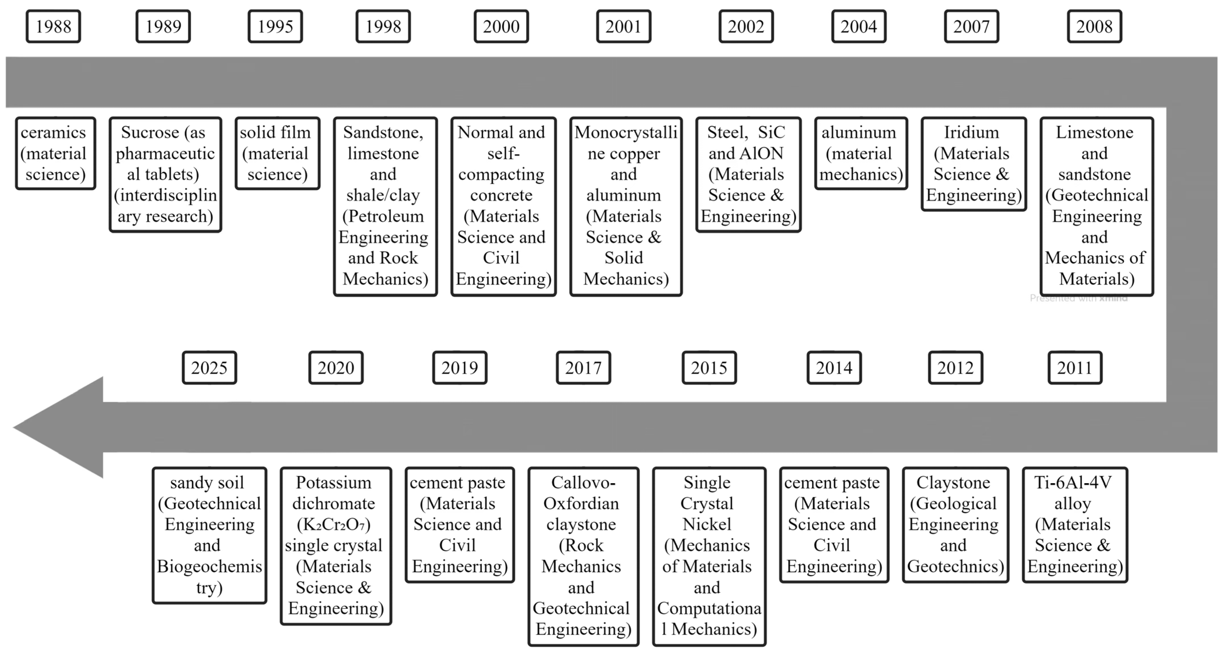
Figure 1. Summary of microindenter usages in different fields [49,50,51,52,53,54,55,56,57,58,59,60,61,62,63,64,65,66,67].
Geosciences 15 00389 g001
Figure 2. Indentation profile with key measurement data.
Geosciences 15 00389 g002
Figure 3. Schematic of a typical indentation curve (Ws = absolute work, Wp = plastic deformation energy, We = elastic deformation energy).
Geosciences 15 00389 g003
Table 1. References of nanoindentation test for several common rocks [64,72,73,74,75,76,77,78,79,80,81,82,83,84,85,86,87,88,89,90,91].
Authors and YearSignificance/Key ResultsMethod/ParametersType of Rocks
Kabir and Akono, 2018 [72]Used to reveal microseismic activity during geological sequestration of carbon dioxide.Combining scratch tests, nanoindentation tests, as well as atomic force microscopy.Sandstone
Bobko, 2005 [73]A new technology has been developed and validated to estimate the volume fraction of composite materials.A nanoindentation experiment was conducted based on environmental scanning electron microscopy (ESEM) images.
Ante et al., 2018 [74]Provides important insights into the particle-scale deformation behavior of shale and sandstone.Young’s modulus, hardness and stiffness of rocks quantified by nanoindentation.
Tyurin et al., 2016 [75]Size effects on physical and mechanical properties of individual phases and interfaces are investigated.Hardness in a wide range of rocks has been investigated using micro- and nanoindentation techniques.
Viktorov et al., 2014 [76]Validation of the operability of the indentation test in the field of rock mechanics.Young’s modulus, fracture toughness and hardness of rocks and minerals were obtained by micro- and nanoindentation.
Golovin et al., 2017 [77]Size effects in the local mechanical properties of multiphase materials are studied.Elasticity modulus, hardness, and crack resistance are determined by means of micro- and nanoindentation.
Golovin et al., 2018 [78]Size effects in hardness and correlations between the distribution of local physical and mechanical properties and the morphology of the studied samples are found.Hardness is obtained via micro- and nanoindentation.
Vialle and Lebedev, 2015 [79]It is possible to correlate the microstructure of mud crystals with their elastic properties.Young’s moduli were mapped on a microscopic scale using nanoindentation technique combined with scanning electron microscopy (SEM).Limestone
Shukla et al., 2015 [80]The measurements do not correspond to the Young’s modulus of the rock for coarse-grained rocks, all indentations. Young’s moduli obtained by nanoindentation and compared with the standard dynamic pulse transmission speed measurement results.
Zhang et al., 2016 [81]Effect of acidic CO2 fluids on the mechanical properties of porous limestone as host rock for potential CO2 sequestration is explored.Dynamic Young’s modulus of Savonier limestone cores before and after supercritical carbon dioxide injection were measured using the perimeter nanoindentation technique.
Zhang et al., 2018 [82]Assessing micro-geomechanical inhomogeneities in rocks.Range of non-uniform indentation modulus is determined using nanoindentation technique.
Lebedev et al., 2014 [83]Understanding the mechanism of fluid–rock interaction and its effect on carbonate elastic parameters.The elastic modulus of limestone was obtained by nanoindentation tests.
Bandini et al., 2012 [84]Determine the effect of microcracks within the grain on its mechanical behavior.Young’s modulus, hardness on marble were obtained by nanoindentation tests using three types of indenters (Nu, Vickers, and Berkovich).Marble
Bandini et al., 2014 [85]Different crack initiation, extension, merging and failure mechanisms as well as stress–strain relationships at the macroscopic scale are more fully explained.Young’s modulus and hardness on two types of marble (heterogeneous and granular) were obtained by nanoindentation tests.
Konstantinidis et al., 2016 [86]A gradient elasticity equation is proposed to explain the evolution of the elastic gradient generated by geometry of the indenter-sample system and applied in conjunction with marble nanoindentation data.Young’s moduli were obtained on Dionysus marble samples by nanoindentation measurements.
Brooks et al., 2010 [87]The interaction between crack initiation and nano-mechanical properties in the crack tip processing zone of a brittle material (marble) is investigated.Young’s modulus and hardness were obtained using nanoindentation and nano/micro heterogeneity was assessed.
Sly et al., 2020 [88]Plastic rheology of calcite at low temperatures using nanoindentation and micro-column compression experiments.Nanoindentation experiments to obtain indentation hardness.
Zhang et al., 2018 [89]Verification that nanoindentation technique provides an effective tool to identify elastic properties of the constituent phases of non-homogeneous rocks.Young’s modulus and hardness of claystone were by nanoindentation technique and homogenization method.Claystone
Magnenet et al., 2011 [90]Feasibility of biphasic model for predicting indentation modulus using microscopic experimental data is verified.Quantifying the indentation modulus of clay matrices by nanoindentation tests.
Auvray et al., 2017 [64]Experiments to demonstrate scale effects on the deformation modulus.Modulus of deformability were obtained at different sample scales by nano- and microindentations.
Bartier and Auvray, 2017 [91]Deformation modulus is related to the carbonate content and its distribution.Determination of deformation modulus of claystone for different depths by nanoindentation.
Table 2. Comparison of nano- and microindentation.
Nanoindentation TestMicroindentation TestType of Test
Tens of microNm to tens of millinewtons (μN-mN)Tens of millinewtons to a few newtons (mN-N)Range of loading force
Tens of nanometers to a few micrometers (nm-μm)Several micronmeters to tens of micrometers (μm)Depth of indentation
Micro-regions, thin films, fine-grained materialsBulk materials, coarse-grained materialsTrial samples
High, suitable for analyzing micro-structuresMedium, suitable for analyzing larger structuresSpatial resolution
Precise control of loading-holding-unloading is possiblePrecise control of loading-holding-unloading is possibleLoading method
Hardness, Young’s modulus, creep behavior, fracture toughness, plastic deformationHardness, Young’s modulus, plastic deformation, creep behavior, fracture toughnessMeasured parameters
Table 3. Common indenters and fields of application.
Fields of ApplicationIndenter GeometryIndenter Name
Microstructural analysis of metals, ceramics, and hardened materials.Equiaxed diamond pyramid; four faces; 136° facet apex angle.Vickers
Small, long specimens and precision testing where minimal damage is required.Elongated diamond pyramid; four faces; 172.5° major-edge apex angle; 130° minor-edge apex angle.Knoop
Determination of average hardness of large parts, forgings and castings.10 mm diameter ball.Brinell
Commonly used in nanotechnology research to determine the mechanical properties of materials and to analyze in depth the intrinsic properties of materials.Diamond indenter; three facets; 142° edge-to-opposite facet.Berkovich
Testing special materials with thin surface and high hardness.Diamond; edge of bases of two base-to-base 66° cones; 2 mm base radius.Grodzinski
Research of optical materials.Hemisphere of sapphire.Pfund
Research on paint and lacquer.Pentagonal shape.Brooks
Disclaimer/Publisher’s Note: The statements, opinions and data contained in all publications are solely those of the individual author(s) and contributor(s) and not of MDPI and/or the editor(s). MDPI and/or the editor(s) disclaim responsibility for any injury to people or property resulting from any ideas, methods, instructions or products referred to in the content.