Communication

l-Lactate Dehydrogenase B Chain Associated with Milk Protein Content in Dairy Cows

1
Department of Animal Science and Technology, College of Animal Bioscience and Technology, Konkuk University, Seoul 143-701, Korea
2
Department of Animal Nutrition and Feed Science, College of Animal Science and Technology, Jilin Agricultural University, Changchun 130118, China
*
Author to whom correspondence should be addressed.
These Authors contributed equally to this work.
Animals 2019, 9(7), 442; https://doi.org/10.3390/ani9070442
Submission received: 25 April 2019 / Revised: 5 July 2019 / Accepted: 8 July 2019 / Published: 15 July 2019
(This article belongs to the Section Animal Genetics and Genomics)

Abstract

:

Simple Summary

The aim of this study was to explore the genes associated with milk protein content in dairy cows. Seven down-regulated and three up-regulated proteins were found in isolated milk epithelial cells (MECs) from dairy cows with high milk protein level (group High). l-leucine depletion not only decreased the proteins synthesis (p < 0.05), but also decreased l-lactate dehydrogenase B chain (LDH-B) mRNA expression in bovine mammary alveolar cells MAC-T cells (p < 0.05). This study suggested that LDH-B is negatively associated with the milk protein content of dairy cows and has a positive relationship with l-leucine. These findings may provide some theoretical basis to study individual differences in the milk protein synthesis ability of dairy cows.

Abstract

This study aimed to explore genes associated with milk protein content in dairy cows and their relationships with l-leucine. Ten primiparous Holstein cows (93.8 ± 11.56 milking days) fed the same diet were divided into two groups depending on their milk protein contents (group High, 3.34 ± 0.10%; and group Low, 2.86 ± 0.05%). Milk epithelial cells (MECs) were isolated from the collected morning milk and differentially expressed proteins in MECs were explored by two-dimensional gel electrophoresis (2-DE). Then, the mRNA expression of these proteins was detected by real time PCR in MAC-T cells incubated with three different media named positive control (PC), negative control (NC), and l-leucine depletion (NO-leu). Results showed that ten proteins were differentially expressed in MECs from cows in group High. They included seven down-regulated ones (heat shock protein beta-1 (HSPB1), 78 kDa glucose-regulated protein (GRP-78), l-lactate dehydrogenase B chain (LDH-B), malate dehydrogenase, cytoplasmic (MDH1), annexin I (ANXA1), cytokeratin-7 (CK-7), and glyceraldehyde-3-phosphate dehydrogenase (GAPDH)), and three up-regulated ones (prohibitin (PHB), beta casein (CSN2), and alpha S1 casein (CSN1S1)). When l-leucine was depleted from the medium, not only proteins content was lowered (p < 0.05), but also the LDH-B mRNA expression was decreased in MAC-T cells (p < 0.05). In conclusion, LDH-B is negatively associated with the milk protein content of dairy cows and has a positive association with l-leucine.

1. Introduction

In milk, about 25% of total milk solids are proteins that are of great importance to milk processors for the manufacturing of a range of products [1]. Therefore, milk protein seems to be a more valuable component than others in milk. However, milk protein contents are diverse in traditional cow breeds of economic importance. For instance, the milk protein level of Holsteins was lowest (3.15% to 3.25%), while the Jersey tends to be the highest (3.80% to 3.90%) [2,3]. Not only are there significant differences in milk protein profiles and frequencies of casein genotype in different breeds [4], the individual contents of milk proteins in the same breed are various too [3,5]. Studies have indicated that fatty acid composition in milk can be changed by feeding, while protein composition is not significantly influenced by the cow’s diet [3,5]. Some amino acids were found not only serving as substrate, but also acting as signaling molecules for protein synthesis [5,6]. l-leucine, one of the branched-chain amino acids, is recognized as a major regulator in protein synthesis [7,8]. However, the mechanism involved in leucine-specific signaling is still not well elucidated and needs further study. We hypothesized that some proteins related to l-leucine are associated with the milk protein content of dairy cows. Therefore, in order to improve the understanding of leucine-specific signaling in milk protein synthesis, proteins differentially expressed were explored in isolated bovine milk epithelial cells (MECs) from cows with high milk protein level, and their relationships with l-leucine were checked in bovine mammary alveolar cells (MAC-T cells) in this study.

2. Materials and Methods

2.1. Milk Preparation and Milk Epithelial Cells (MECs) Isolation

Ten primiparous Holstein cows (573 ± 24.6 kg body weight; 93.8 ± 11.56 milking days) fed with the same total mixed ration (TMR) were selected from a commercial dairy farm. The cows were milked twice daily (12 h milking interval) and 1.5 L milk was individually collected from the automatic milking machine in the morning. Milk samples were divided into two groups (group High, 3.34 ± 0.10%; group Low, 2.86 ± 0.05%) according to milk protein contents analyzed by MilkoScan (Foss Electric, Hillerød, Denmark) [9]. Then, milk epithelial cells (MECs) were separated from the samples using anti-cytokeratin 8 antibodies (Sigma-Aldrich, St. Louis, MO, USA) attached to magnetic beads (Pan Mouse IgG; Invitrogen Dynal AS, Oslo, Norway) within 24 h [10]. All of the experimental animals were approved by Animal Welfare and Ethics Committee of Jilin Agricultural University and managed according to the Guidelines for the Care and Use of Experimental Animals.

2.2. Analysis of Protein Expression

Concentrations of proteins extracted from isolated MECs of these two groups (group High and group Low) were measured with a commercial Kit (GE Healthcare, Piscataway, NJ, USA). Global protein expression was compared using a two-dimensional gel electrophoresis (2-DE) system. The isoelectric focusing (IEF) was performed by using an IEF electrophoresis unit (GE Healthcare, Piscataway, NJ, USA), while the SDS-PAGE was performed by using an Ettan DALT 2-D gel system (GE Healthcare, Piscataway, NJ, USA). After electrophoresis, the gels were silver-stained using a PlusOne Silver Staining kit (GE Healthcare, Piscataway, NJ, USA) and processed by Proteomweaver™ 2-D Analysis software (Definiens AG, Munich, Germany). Differently expressed proteins (≥2-fold or ≤0.5-fold) were picked for ESI-Q-TOF/MS analysis and proteins identifications [10].

2.3. l-Leucine Depletion Experiment in MAC-T Cells

MAC-T cells were seeded into 6-well cell culture plates (BD Falcon, Becton, Dickinson and Company, Mississauga, ON, Canada) and cultured with complete DMEM/F12 media (pH = 7.4) (Thermo Scientific, South Logan, UT, USA) at 2 mL/well. When 80% confluency was reached, the media was replaced with serum-free complete DMEM/F12 (Gibco, Invitrogen, Grand Island, NY, USA) and incubated overnight. After washing twice with PBS, cells were incubated with one of the following three media: (1) Earle’s Balanced Salts Solution (Gibco, Invitrogen, Grand Island, NY, USA) + insulin (5 μg/mL) + hydrocortisone (1 μg/mL) + prolactin (5 μg/mL) (Sigma-Aldrich Corp, St. Louis, MO, USA) as negative control (NC); (2) negative control medium supplemented with l-arginine (0.70 mmol/L), l-histidine (0.15 mmol/L), l-isoleucine (0.42 mmol/L), l-leucine (0.45 mmol/L), l-lysine (0.5 mmol/L), l-methionine (0.12 mmol/L), l-phenylalanine (0.22 mmol/L), l-threonine (0.45 mmol/L), l-tryptophan (0.04 mmol/L), and l-valine (0.45 mmol/L) as positive control (PC) [7]; and (3) PC media devoid of l-leucine as l-leucine depletion (NO-leu) group. The NO-leu medium was prepared from NC medium by adding nine individual AAs (l-isomer) at concentrations equal to that of PC medium. After incubation for 1 or 6 h, media and cells were collected individually. Total proteins were calculated as the sum of proteins in cell and proteins secreted into the culture media.

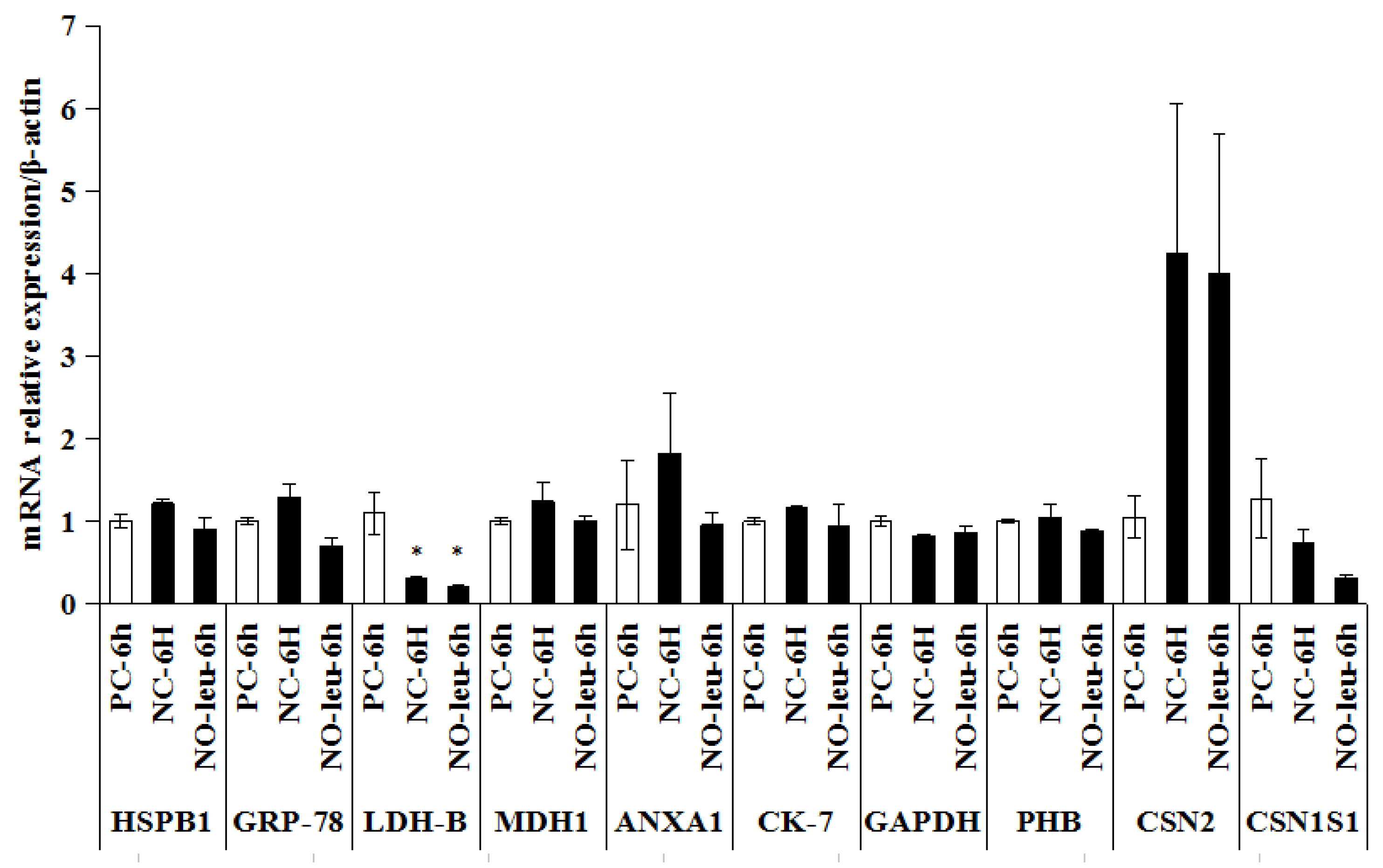
2.4. The mRNA Expression of Candidate Proteins in MAC-T Cells

Total RNA was extracted from MAC-T cells using TRIZOL reagent according to the manufacturer’s instructions (Thermo Fisher Scientific, Waltham, MA, USA), and the RNA quality was assessed by Agilent 2100 Bioanalyzer (Agilent Technologies Inc, Santa Clara, CA, USA). The real-time PCR was performed with a total reaction volume of 20 µL in 96-well plates using the CFX96 Real-Time PCR Detection System (Bio-Rad Laboratories, Hercules, CA, USA). β-actin was used as housekeeping gene. The primer sequences are shown in Table 1. Detailed procedure of real-time PCR was reported by Wang et al. (2014) [11].

2.5. Statistical Analysis

All data were presented as mean ± standard error (S.E.). The data of milk protein contents of dairy cows were analyzed with t-test while the data of total proteins content and mRNA expression in MAC-T Cells were analyzed with one-way analysis of variance (one-way ANOVA, SPSS Inc., Chicago, IL, USA). Differences were considered significant when p < 0.05.

3. Results

3.1. Milk Protein Contents for Individual Animals

In this study, milk protein contents from individual primiparous Holstein cows in the same lactation stage were quite different, ranging from a minimum of about 2.69% to a maximum of about 3.86% (Figure 1). Subsequently, the milk samples were divided into two groups as group High (3.34 ± 0.10%) and group Low (2.86 ± 0.05%) (p < 0.01).

3.2. Differentially Expressed Proteins in Isolated MECs

In the isolated MECs, there were more than 450 detectable spots in each of the 2-DE images and ten differently expressed proteins were identified (Figure 2). Seven of them were down-regulated (heat shock protein beta-1 (HSPB1), 78 kDa glucose-regulated protein (GRP-78), l-lactate dehydrogenase B chain (LDH-B), malate dehydrogenase, cytoplasmic (MDH1), annexin I (ANXA1), cytokeratin-7 (CK-7), and glyceraldehyde-3-phosphate dehydrogenase (GAPDH)), whereas three were up-regulated in MECs of group High (prohibitin (PHB), beta casein (CSN2) (Spot 418 and 422), and alpha S1 casein (CSN1S1)) (Spot 425 and 208887)) (Table 2).

3.3. Effect of l-Leucine Depletion on Total Proteins Content in MAC-T Cells

After both 1 h and 6 h incubation, the total proteins synthesis in MAC-T cells of group NC were significantly lower than those of the groups of PC (p < 0.05; p < 0.01) and NO-leu (p < 0.05; p < 0.05) (Figure 3).

3.4. Differentially Expressed Proteins in Isolated MECs

The mRNA expression of LDH-B was found to be significantly down-regulated in both groups of NC (p < 0.05) and NO-leu (p < 0.05). No significant difference was found regarding the other genes (Figure 4).

4. Discussion

The present data indicated significant differences existed in the milk protein synthesis ability of individual cows, even when fed the same diet. Similar to a previous report, this study showed that the milk protein levels of 25 multi-lactation Holstein cows (6–12 weeks lactation) were ranging from 2.55% to 3.30% [2]. The milk samples were divided into two groups according to their protein contents and ten differently expressed proteins were identified in the isolated MECs using a 2-DE system. HSPB1 was suggested to play a role in testosterone-related myogenesis in beef cattle [12], and a negative association of its expression with meat tenderness was observed in Nellore cattle [13]. ANXA1 was previously reported to be expressed in mammary glands of lactating cows [14] and have a negative connection with milk cis-9, trans-11 CLA level in dairy cows [11]. LDH-B, an enzyme involved in cellular carbohydrate metabolic process and glycolytic process, can catalyze reversible conversion of pyruvate to lactate as it can convert NADH to NAD1+ and back. In the present study, the protein expression of LDH-B was less in MECs from cows with high protein level than that from cows with low protein level. In addition, LDH-B is down-regulated in adipose tissue from Korean bulls relative to steers [12], and LDH-B was reported as a tenderness marker protein for goat muscle [15]. Both CSN2 and CSN1S1 are specific milk proteins secreted by mammary gland cells. CSN2 might be related to mammary protein synthesis [16], and allele CSN2 B had the effect of increasing β-casein content and decreasing content of αS1-casein [17]. A previous study has also indicated that genotype of CSN3, another milk protein, can significantly affect milk yield and composition in dairy cows [4]. However, whether CSN2 and CSN1S1 have similar functions remains unknown. For the other five proteins, little information was found on their roles involved in milk protein synthesis. GRP-78 is involved in protein folding and assembly [18]. MDH1 is an oxidoreductase that plays a part in tricarboxylic acid cycle [19]. CK-7 can stimulate DNA synthesis in cells [20]. GAPDH is an energy metabolism-related enzyme in the glycolytic pathway [21], whereas PHB has a role in regulating proliferation [22].
l-leucine is recognized as a major regulator of milk protein synthesis [8,16,23]. In this study, our data proved that l-leucine depletion could decrease milk protein content in MAC-T cells; in accordance with some previous reports showing that depletion of leucine could reduce protein synthesis [7,24]. In order to improve the understanding of leucine-specific signaling in milk protein synthesis, the association of these genes with l-leucine was checked in MAC-T cells but not in MECs due to limitations in sample availability. Interestingly, mRNA expression of LDH-B was significantly down-regulated when l-leucine was depleted. Moreover, it has been reported that l-leucine can inhibit oxidation of pyruvate [25]. These data suggest that there is a positive association between l-leucine and LDH-B. It is well known that l-leucine can regulate protein synthesis through mTOR pathway [16,24]. Whether LDH-B regulates milk protein synthesis via the l-leucine-mTOR pathway remains unclear and needs further study. In addition, no significant difference was found regarding the other genes, which may be due to the possible random errors in the experiment.

5. Conclusions

Milk protein content and global protein expression were found to be significantly different in animals fed the same diet. LDH-B is negatively related to the milk protein content of dairy cows and has a positive relationship with l-leucine. Our findings may provide some theoretical basis to study individual difference in milk protein synthesis ability of dairy cows.

Author Contributions

T.W. and H.G.L. conceived and designed the experiments. T.W. and S.W.J. executed the experiment and analyzed the samples. T.W. and H.G.L. revised the manuscript. M.J.K. and U.S.J. gave help during the animal experiment.

Funding

This work was supported by Korea Institute of Planning and Evaluation for Technology in Food, Agriculture, Forestry (IPET) through Agri-Bio Industry Technology Development Program (117030-03-2-HD020), Preferential Funding of Science and Technology Innovation and Entrepreneurship Projects for Returned Overseas Scientific Research Personnel in Jilin Province (G11), and Youth Top-notch Talent Support Program of Jilin Agricultural University (2016005).

Acknowledgments

The authors thank Yu Guo Zhen for helps during the submission of this manuscript.

Conflicts of Interest

The authors declare no conflicts of interest.

References

  1. Murphy, J.J.; O’Mara, F. Nutritional manipulation of milk protein concentration and its impact on the dairy industry. Livest. Prod. Sci. 1993, 35, 117–134. [Google Scholar] [CrossRef]
  2. Robinson, P.H. Manipulating milk protein production and level in lactating dairy cows. Part1-Difficult to manipulate factors. Calif. Dairy 1999, 8, 6–7. [Google Scholar]
  3. Robinson, P.H. Manipulating milk protein percentage and production in lactating dairy cows. Part 2-Factors that can be manipulated. Calif. Dairy 1999, 8, 4–5, 12–13. [Google Scholar]
  4. Gustavsson, F.; Buitenhuis, A.J.; Johansson, M.; Bertelsen, H.P.; Glantz, M.; Poulsen, N.A.; Lindmark, M.H.; Stålhammar, H.; Larsen, L.B.; Bendixen, C.; et al. Effects of breed and casein genetic variants on protein profile in milk from Swedish Red, Danish Holstein, and Danish Jersey cows. J. Dairy Sci. 2014, 97, 3866–3877. [Google Scholar] [CrossRef] [PubMed] [Green Version]
  5. Heck, J.M.L. Milk Genomics: Opportunities to Improve the Protein and Fatty Acid Composition in Raw Milk. Ph.D. Thesis, Wageningen University, Wageningen, The Netherlands, 2009. [Google Scholar]
  6. Kim, E. Mechanisms of amino acid sensing in mTOR signaling pathway. Nutr. Res. Pract. 2009, 3, 64–71. [Google Scholar] [CrossRef] [PubMed] [Green Version]
  7. Appuhamy, J.A.D.R.N.; Knoebel, N.A.; Nayananjalie, W.A.; Escobar, J.; Hanigan, M.D. Isoleucine and leucine independently regulate mTOR signaling and protein synthesis in MAC-T cells and bovine mammary tissue slices. J. Nutr. 2012, 142, 484–491. [Google Scholar] [CrossRef] [PubMed]
  8. Appuhamy, J.A.D.R.N. Regulatory Roles of Essential Amino Acids, Energy, and Insulin in Mammary Cell Protein Synthesis. Ph.D. Thesis, Virginia Polytechnic Institute and State University, Blacksburg, VA, USA, 2010. [Google Scholar]
  9. Wang, T.; Oh, J.J.; Lim, J.N.; Hong, J.E.; Kim, J.H.; Kim, J.H.; Kang, H.S.; Choi, Y.J.; Lee, H.G. Effects of lactation stage and individual performance on milk cis-9, trans-11 conjugated linoleic acids content in dairy cows. Asian Austral J. Anim. 2013, 26, 189–194. [Google Scholar] [CrossRef]
  10. Wang, T.; Lee, H.G.; Hwang, J.H.; Oh, J.J.; Lim, J.N.; Kang, H.S.; Kang, H.S.; Joo, J.K.; Lee, K.S. Myoglobin: An Exogenous Reference Marker for Proteomics Analysis. Food Sci. Biotechnol. 2013, 22, 393–408. [Google Scholar] [CrossRef]
  11. Wang, T.; Lim, J.N.; Bok, J.D.; Kim, J.H.; Kang, S.K.; Lee, S.B.; Hwang, J.H.; Lee, K.H.; Kang, H.S.; Choi, Y.J.; et al. Association of protein expression in isolated milk epithelial cells and cis-9, trans-11 CLA concentrations in milk from dairy cows. J. Sci. Food Agric. 2014, 94, 1835–1843. [Google Scholar] [CrossRef]
  12. Zhang, Q.K.; Lee, H.G.; Han, J.A.; Kang, S.K.; Lee, N.K.; Baik, M.G.; Choi, Y.J. Differentially expressed proteins associated with myogenesis and adipogenesis in skeletal muscle and adipose tissue between bulls and steers. Mol. Biol. Rep. 2012, 39, 953–960. [Google Scholar] [CrossRef]
  13. Malheiros, J.M.; Enríquez-Valencia, C.E.; da Silva Duran, B.O.; de Paula, T.G.; Curi, R.A.; de Vasconcelos Silva, J.A., II; Dal-Pai-Silva, M.; de Oliveira, H.N.; Chardulo, L.A.L. Association of CAST2, HSP90AA1, DNAJA1 and HSPB1 genes with meat tenderness in Nellore cattle. Meat Sci. 2018, 138, 49–52. [Google Scholar] [CrossRef] [PubMed]
  14. Norio, K.; Takafumi, S.; Akira, Y.; Toru, M. Distribution of Annexins I, II, and IV in Bovine Mammary Gland. J. Dairy Sci. 1995, 78, 2382–2387. [Google Scholar]
  15. Wei, Y.C.; Li, X.; Zhang, D.Q.; Liu, Y.F. Comparison of protein differences between high- and low-quality goat and bovine parts based on iTRAQ technology. Food Chem. 2019, 289, 240–249. [Google Scholar] [CrossRef] [PubMed]
  16. Bionaz, M.; Loor, J.J. Gene Networks Driving Bovine Mammary Protein Synthesis during the Lactation Cycle. Bioinform. Biol. Insights 2011, 5, 83–98. [Google Scholar] [CrossRef] [PubMed]
  17. Bonfatti, V.; Martino, G.D.; Cecchinato, A.; Vicario, D.; Carnier, P. Effects of β-κ-casein (csn2-csn3) haplotypes and β-lactoglobulin (blg) genotypes on milk production traits and detailed protein composition of individual milk of simmental cows. J. Dairy Sci. 2010, 93, 3797–3808. [Google Scholar] [CrossRef] [PubMed]
  18. Ti, Z.W.; Li, Z.Y. Glucose regulated protein 78: A critical link between tumor microenvironment and cancer hallmarks. Biochim. Biophys. Acta 2012, 1826, 13–22. [Google Scholar]
  19. Han, X.; Tong, Y.; Tian, M.; Zhang, Y.; Sun, X.; Wang, S.; Qiu, X.; Ding, C.; Yu, S. Enzymatic Activity Analysis and Catalytic Essential Residues Identification of Brucella abortus Malate Dehydrogenase. Sci. World J. 2014, 8, 973751. [Google Scholar]
  20. Smith, F.J.; Porter, R.M.; Corden, L.D.; Lunny, D.P.; Lane, E.B.; McLean, W.H. Cloning of human, murine, and marsupial keratin 7 and a survey of K7 expression in the mouse. Biochem. Biophys. Res. Commun. 2002, 297, 818–827. [Google Scholar] [CrossRef]
  21. Takaoka, Y.; Goto, S.; Nakano, T.; Tseng, H.P.; Yang, S.M.; Kawamoto, S.; Ono, K.; Chen, C.L. Glyceraldehyde-3-phosphate dehydrogenase (GAPDH) prevents lipopolysaccharide (LPS)-induced, sepsis-related severe acute lung injury in mice. Sci. Rep. 2014, 4, 5204. [Google Scholar] [CrossRef] [Green Version]
  22. Wang, Y.J.; Guo, X.L.; Li, S.A.; Zhao, Y.Q.; Liu, Z.C.; Lee, W.H.; Xiang, Y.; Zhang, Y. Prohibitin is involved in the activated internalization and degradation of protease-activated receptor 1. Biochim. Biophys. Acta 2014, 1843, 1393–1401. [Google Scholar] [CrossRef] [Green Version]
  23. Moshel, Y.; Rhoads, R.E.; Barash, I. Role of amino acids in translational mechanisms governing milk protein synthesis in murine and ruminant mammary epithelial cells. J. Cell Biochem. 2006, 98, 685–700. [Google Scholar] [CrossRef] [PubMed]
  24. Gao, H.N.; Hu, H.; Zheng, N.; Wang, J.Q. Leucine and histidine independently regulate milk protein synthesis in bovine mammary epithelial cells via mTOR signaling pathway. J. Zhejiang Univ. Sci. B 2015, 16, 560–572. [Google Scholar] [CrossRef] [PubMed]
  25. Chang, T.W.; Goldberg, A.L. Leucine inhibits oxidation of glucose and pyruvate in skeletal muscles during fasting. J. Biol. Chem. 1978, 253, 3696–3701. [Google Scholar] [PubMed]
Figure 1. Milk protein contents of primiparous dairy cows (93.8 ± 11.56 milking days). ** p < 0.01.
Animals 09 00442 g001
Figure 2. Representative two-dimensional gel electrophoresis (2-DE) images of isolated milk epithelial cells (MECs) (A) and differently expressed proteins (B).
Animals 09 00442 g002
Figure 3. Effects of l-leucine limitations (1 h, 6 h) on total proteins content in mammary alveolar cells (MAC-T cells). NC: Negative control, Earle’s Balanced Salts Solution + insulin (5 μg/mL) + hydrocortisone (1 μg/mL) + prolactin (5 μg/mL); PC: Positive control, NC medium supplemented with 0.70 l-arginine, 0.15 l-histidine, 0.42 l-isoleucine, 0.45 l-leucine, 0.5 l-lysine, 0.12 l-methionine, 0.22 l-phenylalanine, 0.45 l-threonine, 0.04 l-tryptophan, and 0.45 l-valine (all in mmol/L); NO-leu: PC media devoid of l-leucine. * p < 0.05, ** p < 0.01.
Animals 09 00442 g003
Figure 4. Verification test of candidate proteins obtained from isolated milk epithelial cells (MECs) in MAC-T cells after l-leucine limitations for 6 h by real time PCR. NC: Negative control, Earle’s Balanced Salts Solution + insulin (5 μg/mL) + hydrocortisone (1 μg/mL) + prolactin (5 μg/mL); PC: Positive control, NC medium supplemented with 0.70 l-arginine, 0.15 l-histidine, 0.42 l-isoleucine, 0.45 l-leucine, 0.5 l-lysine, 0.12 l-methionine, 0.22 l-phenylalanine, 0.45 l-threonine, 0.04 l-tryptophan, and 0.45 l-valine (all in mmol/L); NO-leu: PC media devoid of l-leucine. * p < 0.05. There were 3 biological samples in each group and the trial was performed in triplicate.
Animals 09 00442 g004
Table 1. Primer sequences for candidate genes used in this study.
Gene 1Accession NumberPrimers
ACTBNM_173979.3forwardGCGTGGCTACAGCTTCACC
reverseTTGATGTCACGGACGATTTC
HSPB1NM_001025569.1forwardGCCGGAAACAAGTAAAGACC
reverseGGTGAGGATGTCCAGTGATG
GRP-78NM_001075148.1forwardCTTCTCGGAGACCCTGACTC
reverseCACTTTCTGGACAGGCTTCA
LDH-BNM_174100.1forwardGTGGAGTGGAGTGAATGTGG
reverseTGTCTGTTCCCATTTCTGGA
MDH1NM_001034628.2forwardCAACCATGCCAAAGTGAAAC
reverseGCCAGCTGTCATCTTTCAGA
ANXA1NM_175784.3forwardTTCTTTGCTGAGAAGCTCCA
reverseCAGAGCGGGAAACCATAATC
CK-7NM_001046411.1forwardCATGAACAAGGTGGAGTTGG
reverseAGCTCTTTCAGCTCCTGCTC
GAPDHNM_001034034.2forwardCGTTCGACAGATAGCCGTAA
reverseTCACCATCTTGTCTCAGGGA
PHBNM_001034572.2forwardGAGATCCTCAAGTCCGTGGT
reverseACCAGCTCTCTCTGGGTGAT
CSN2NM_181008.2forwardGTGAGGAACAGCAGCAAACA
reverseTTTTGTGGGAGGCTGTTAGG
CSN1S1NM_181029.2forwardGCTGAGGAACGACTTCACAG
reverseAGGCCAGTTCCTGATTCACT
1 ACTB β-Actin, HSPB1 Heat shock protein beta-1, GRP-78 78 kDa glucose-regulated protein, LDH-B l-lactate dehydrogenase B chain, MDH1 Malate dehydrogenase cytoplasmic, ANXA1 Annexin I, CK-7 Cytokeratin-7, GAPDH Glyceraldehyde-3-phosphate dehydrogenase, PHB Prohibitin, CSN2beta casein, CSN1S1 alpha S1 casein.
Table 2. List of differently expressed proteins (≥2-fold or ≤0.5-fold) in isolated MECs.
Spot No.UniProtKB/Swiss-Prot EntryProtein Name 1Theory; Calculation Mol. Mass (kDa)/PIMASCOT ScorePeptides MatchedSequence Coverage (%)Molecular FunctionsProtein Expression (Area)
HLH/L
396Q3T149HSPB122.4/5.98;750.4523360.70Stress resistance and actin organization0.04230.20460.207
22.4/6.40
404Q0VCX2GRP-7872.4/5.07;131.033810.53Facilitating the assembly of multimeric protein complexes inside the ER0.02420.22170.109
72.4/5.16
408Q5E9B1LDH-B36.7/6.02;150.545617.37Oxidoreductase in cellular carbohydrate metabolic process and glycolysis0.09340.38520.242
36.7/6.44
409Q3T145MDH136.4/6.16;20.4493.59Oxidoreductase in cellular carbohydrate metabolic process and malate metabolic process0.12910.51640.250
36.4/6.58
411P46193ANXA139.0/6.37;2103.2452246.24Plays important roles in the innate immune response and has anti-inflammatory activity0.27050.64480.420
39.0/6.81
412Q29S21CK-751.5/5.79;59.14172.58Blocks interferon-dependent interphase and stimulates DNA synthesis in cells0.07420.19840.374
51.5/5.97
413P10096GAPDH35.8/8.51;17.5877.93Oxidoreductase and transferase in apoptosis glycolysis, translation regulation0.13430.32340.415
24.2/8.53
414Q3T165PHB29.8/5.57;703.8422552.57Has a role in regulating proliferation0.19090.07772.457
29.8/5.76
418P02666CSN225.1/5.26;224.156119.14Antioxidant activity; negative regulation of catalytic activity; transporter activity0.84300.34492.444
23.6/5.34
422P02666CSN225.1/5.26;106.403019.140.63920.28822.218
23.6/5.34
425P02662CSN1S124.5/4.98;94.574119.83Antioxidant activity; transporter activity; important role in the capacity of milk to transport calcium phosphate0.20550.10242.008
13.8/5.55
208887P02662CSN1S124.5/4.98;79.253219.830.20660.08202.518
13.8/5.55
1 Heat shock protein beta-1, GRP-78 78 kDa glucose-regulated protein, LDH-B l-lactate dehydrogenase B chain, MDH1 Malate dehydrogenase cytoplasmic, ANXA1 Annexin I, CK-7 Cytokeratin-7, GAPDH Glyceraldehyde-3-phosphate dehydrogenase, PHB Prohibitin, CSN2 beta casein, CSN1S1 alpha S1 casein.

