Article

Detection and Genetic Characterization of Enterocytozoon hepatopenaei in Giant Freshwater Prawn (Macrobrachium rosenbergii) Imported into South Korea

1
Laboratory of Aquatic Biomedicine, College of Veterinary Medicine, Kyungpook National University, Daegu 41566, Republic of Korea
2
Institute for Veterinary Biomedical Science, Kyungpook National University, Daegu 41566, Republic of Korea
3
Quarantine and Inspection Division, National Fishery Products Quality Management Service, 337, Haeyang-ro, Yeongdo-gu, Busan 49111, Republic of Korea
4
Department of Veterinary Medicine, Faculty of Veterinary Science, Chulalongkorn University, Bangkok 10330, Thailand
5
Department of Food Science and Biotechnology, College of Bionano Technology, Gachon University, Seongnam 13120, Republic of Korea
*
Authors to whom correspondence should be addressed.
Animals 2025, 15(22), 3286; https://doi.org/10.3390/ani15223286
Submission received: 1 September 2025 / Revised: 31 October 2025 / Accepted: 10 November 2025 / Published: 13 November 2025
(This article belongs to the Section Aquatic Animals)

Simple Summary

Enterocytozoon hepatopenaei (EHP) is an intracellular parasite that causes substantial economic losses to the global shrimp industry. Although penaeid shrimps are particularly vulnerable to EHP infection and have been extensively studied, Macrobrachium rosenbergii (giant freshwater prawn), another important species widely farmed in warm-climate regions, is also affected by this pathogen. Despite the increasing global incidence of EHP infection in M. rosenbergii, studies on this species remain limited compared to those on penaeid shrimps. Here, we monitored the EHP infection in M. rosenbergii imported from India and Vietnam to South Korea and conducted genetic analyses to better characterize its genetic diversity.

Abstract

This study investigated Macrobrachium rosenbergii imported from India (15 batches, N = 180) and Vietnam (7 batches, N = 84) between 2023 and 2024, for Enterocytozoon hepatopenaei (EHP) monitoring and genetic analysis. Polymerase chain reaction assays detected EHP in 13.3% (2/15) and 71.4% (5/7) samples from India and Vietnam, respectively. The sequence of the small subunit ribosomal ribonucleic acid region of the EHPs isolated from M. rosenbergii showed no significant differences from those available in GenBank. Interestingly, spore wall protein (SWP) 1 region analysis revealed that M. rosenbergii EHPs could be divided into three groups, some of which were closely related to Penaeus vannamei EHPs. Similarly, the internal transcribed spacer-1 (ITS-1) region analysis divided M. rosenbergii EHPs into two groups, with some showing close relationships with P. vannamei EHPs. Phylogenetic analyses based on the SWP 1 and ITS-1 regions suggested that EHPs infecting M. rosenbergii exhibited greater genetic diversity than those infecting P. vannamei. This study provides the first report of EHP detection in M. rosenbergii imported from India and Vietnam to South Korea. Further genome-based analyses are necessary for a comprehensive genetic characterization of EHPs infecting M. rosenbergii from various geographical regions.

1. Introduction

Microsporidia are intracellular parasites that infect various hosts and produce environmentally resistant spores [1]. Their spores have a thick-layered wall, which enables them to survive for extended periods under harsh environmental conditions, such as high temperatures [2]. The spore wall is composed of an outer exospore and an inner endospore made of chitin and protein [2]. When a spore is exposed to environmental stimuli, it can increase the internal osmotic pressure, cause swelling and eventually injecting the polar filament into a host cell [3]. To date, over 50 genera of microsporidia have been identified in aquatic arthropods, including 20 genera that infect crustaceans belonging to the class Malacostraca, including shrimp, crabs, and lobsters [4].
Among these, Enterocytozoon hepatopenaei (EHP) causes substantial economic losses to shrimp farming [5]. EHP infection is mostly asymptomatic, making early detection difficult and leading to its spread across farms. In shrimp, this microsporidium proliferates in the cytoplasm of hepatopancreatic tubular epithelial cells, leading to growth retardation and size variation among individuals [5,6,7,8]. Notably, although EHP infection alone rarely causes high mortality, co-infection with other pathogens such as bacteria and viruses can significantly increase mortality rates and exacerbate symptoms [9,10]. EHP infection was initially reported in penaeid shrimps, including Penaeus monodon [9], Penaeus vannamei [11], and Penaeus stylirostris [12]; however, it has more recently been detected in M. rosenbergii in China [13], South Korea [14], and Japan (2023, available in the GenBank database).
Macrobrachium rosenbergii (commonly known as giant freshwater prawn) is the largest freshwater prawn species worldwide and widely farmed in various regions across the globe, including Northwest India, Bangladesh, China, Indonesia, Thailand, Vietnam, the Philippines, Northern Australia, and South Korea [15,16,17]. Since 1980, global production has increased significantly, exceeding 220,000 tons in 2020 [18]. In South Korea, over 47 tons of M. rosenbergii was imported from major prawn-producing countries between 2019 and 2023 [19].
The genetic diversity of EHP and its potential for horizontal transmission across various shrimp species highlight the need for molecular characterization to develop effective management strategies for EHP. This study aimed to highlight the potential risk of introducing EHPs into South Korea via imported shrimp, for which EHP monitoring is currently not conducted. In this study, we investigated the prevalence and genetic diversity of EHP in imported M. rosenbergii by analyzing the small subunit ribosomal RNA (SSU rRNA) and spore wall protein (SWP) 1 regions of EHP. Phylogenetic analyses were performed using these sequences for comparison with EHP strains available in the GenBank database.

2. Materials and Methods

2.1. Sample Collection

A total of 22 batches (12 prawns per batch) of frozen M. rosenbergii imported from India (N = 15 batches) and Vietnam (N = 7 batches) were collected from local retail markets in South Korea. All samples were transported to the laboratory on ice and stored at −80 °C until further analysis. Detailed information on the M. rosenbergii samples is presented in Table 1.

2.2. DNA Extraction and EHP Monitoring

From each batch, M. rosenbergii (N = 10) was randomly selected, and hepatopancreatic tissues were pooled as one sample. DNA was extracted from 30 mg hepatopancreatic tissue using the DNeasy Blood & Tissue Kit (Qiagen, Hilden, Germany) according to the manufacturer’s instructions. EHP was detected by PCR using the 510-F/510-R primer pair, targeting the SSU rRNA [12] (Table 2).

2.3. Sequence Analysis of SSU rRNA, SWP 1, and Internal Transcribed Spacer-1 Regions of EHP

EHP-positive samples (identified using the 510-F/510-R primer pair) were further analyzed by sequencing the SSU rRNA [11], SWP 1 region [13], and internal transcribed spacer-1 (ITS-1) regions [21] (Table 2). The resulting amplicons were sequenced at Bioneer (Daejeon, Republic of Korea). The SSU rRNA, SWP 1, and ITS-1 sequences obtained in this study were deposited in the GenBank database (Supplementary Table S1). The obtained sequences were compared to other sequences listed in GenBank using BLASTn (ver. 2.17.0) to confirm the identity of the EHP sequences. Pairwise distances between the obtained sequences and reference EHP sequences derived from the GenBank database were compared using Geneious Prime (ver. 2024; http://www.geneious.com).

2.4. Phylogenetic Tree Analysis

Phylogenetic analyses were conducted using SSU rRNA, SWP 1, and ITS-1 sequences from EHP strains available in the GenBank database. (1) SSU rRNA: P. vannamei EHP (China, KX981865; India, KY643648; Republic of Korea, MZ819965; Vietnam, KP759285) and M. rosenbergii EHP (Republic of Korea, OP363710); (2) SWP 1: P. vannamei EHP (India, MH365434; Indonesia, KY593133; Republic of Korea, MZ541056; Thailand, MG015710), M. rosenbergii EHP (China, MW269619); and (3) ITS-1: P. vannamei EHP (China, OR162445 and OR168076; South Korea, ON015652; Thailand, MNPJ00000000) (all sequences were retrieved from the GenBank database: http://www.ncbi.nlm.nih.gov/genbank, accessed on 25 July 2024). The sequences were initially aligned using ClustalW (ver. 2.1), and non-overlapping genomic regions at the fragment ends were trimmed. The trimmed sequences were used for phylogenetic analyses. Phylogenetic trees based on the SSU rRNA, SWP 1, and ITS-1 sequences of EHP strains were constructed using the maximum-likelihood method with 1000 bootstrap replications implemented in MEGA X (version 11.0.13) [22].

3. Results

3.1. EHP Monitoring in Imported M. rosenbergii

PCR analysis identified EHP in two M. rosenbergii samples from India (23-026C6-2-INDIA and 23-026C7-2-INDIA; 2/15, 13.3%) and five M. rosenbergii samples from Vietnam (23-026C6-1-VIE, 23-026C7-1-VIE, 23-026C7-3-VIE, 23-026C9-2-VIE, and 23-026C11-1-VIE; 5/7, 71.4%) (Table 1).

3.2. Sequence Analysis of SSU rRNA, SWP 1, and ITS-1 Regions of EHP

SSU rRNA sequences were successfully obtained from six EHP-positive M. rosenbergii samples, except 23-026C11-1-VIE, which exhibited weak PCR amplification result. BLASTn searches revealed that the six sequences shared over 99.0% identity (query cover = 100%) with P. vannamei EHPs (originating from China, India, Republic of Korea, and Vietnam) and M. rosenbergii EHP (originating from Republic of Korea). The results of the pairwise distance analysis between the obtained six M. rosenbergii EHPs in this study and reference P. vannamei and M. rosenbergii EHPs available in the GenBank database are shown in Supplementary Table S2.
SWP 1 region sequences were successfully obtained from all EHP-positive M. rosenbergii samples. BLASTn searches revealed that two samples (23-026C7-2-INDIA and 23-026C7-3-VIE) shared 99.44% identity (query cover = 100%) with P. vannamei EHPs (originating from India and Thailand) and 92.05% identity (query cover = 98%) with M. rosenbergii EHP (originating from China). The remaining five samples (23-026C6-1-VIE, 23-026C6-2-INDIA, 23-026C7-1-VIE, 23-026C9-2-VIE, and 23-026C11-1-VIE) showed 97.78% identity (query cover = 100%) with P. vannamei EHPs (originating from India and Thailand) and 92.61% identity (query cover = 98%) with M. rosenbergii EHP (originating from China). The results of pairwise distance analysis between the seven M. rosenbergii EHPs obtained in this study and reference P. vannamei and M. rosenbergii EHPs available in GenBank are presented in Supplementary Table S3.
The ITS-1 region sequences were successfully obtained from seven EHP-positive M. rosenbergii samples. BLASTn searches revealed that two samples (23-026C7-2-INDIA and 23-026C7-3-VIE) had 93.51% identity (query cover = 90.0%), and the other five samples (23-026C6-1-VIE, 23-026C6-2-INDIA, 23-026C7-1-VIE, 23-026C9-2-VIE, and 23-026C11-1-VIE) had 80.76% identity (query cover = 100%) with P. vannamei EHPs (originating from China and South Korea). Pairwise distance analysis results between the seven M. rosenbergii EHPs obtained in this study and the reference P. vannamei EHPs available in the GenBank database are shown in Supplementary Table S4.

3.3. Phylogenetic Analysis

Phylogenetic analysis based on SSU rRNA sequences was performed using the six M. rosenbergii EHP sequences obtained in this study; previously reported P. vannamei EHP sequences (N = 4) from China, India, South Korea, and Vietnam; and previously reported M. rosenbergii EHP sequence (N = 1) from South Korea available in the GenBank database. The resulting phylogenetic tree based on SSU rRNA sequences revealed that the M. rosenbergii EHPs from this study grouped closely with previously reported P. vannamei and M. rosenbergii EHPs (Figure 1).
Phylogenetic analysis of the SWP 1 region was conducted using seven M. rosenbergii EHP sequences from this study; P. vannamei EHP sequences (N = 4) from India, Indonesia, South Korea, and Thailand; and an M. rosenbergii EHP sequence (N = 1) from China, available in the GenBank database. The tree based on the SWP 1 region was divided into three groups (Groups 1–3; Figure 2). Group 1 included two M. rosenbergii EHPs identified in this study (23-026C7-2-INDIA and 23-026C7-3-VIE), which were grouped with previously reported P. vannamei EHPs from Asian countries. Group 2 comprised the remaining five M. rosenbergii EHPs from this study (23-026C6-1-VIE, 23-026C6-2-INDIA, 23-026C7-1-VIE, 23-026C9-2-VIE, and 23-026C11-1-VIE), whereas Group 3 contained the previously reported M. rosenbergii EHP from China [13] (Figure 2 and Supplementary Figure S1).
Phylogenetic analysis of the ITS-1 region was performed using the seven M. rosenbergii EHP sequences from this study, along with P. vannamei EHP sequences (N = 4) from China, South Korea, and Thailand, available in the GenBank database. The phylogenetic tree based on the ITS-1 region is divided into two groups (Groups 1 and 2), with identical compositions in the SWP 1 region (Figure 3). Group 1 included two M. rosenbergii EHPs identified in this study (23-026C7-2-INDIA and 23-026C7-3-VIE), which were grouped with previously reported P. vannamei EHPs from Asian countries. Group 2 comprised the remaining five M. rosenbergii EHPs (23-026C6-1-VIE, 23-026C6-2-INDIA, 23-026C7-1-VIE, 23-026C9-2-VIE, and 23-026C11-1-VIE) (Figure 3 and Supplementary Figure S2). This grouping pattern of M. rosenbergii EHPs in this study was similar to that observed in the SWP 1 region analysis, suggesting consistent genetic grouping.

4. Discussion

EHP infection in M. rosenbergii has been increasingly reported worldwide, including China [13], Republic of Korea [14], and Japan (OQ860232, retrieved from GenBank). EHP infections in M. rosenbergii are similar to those in P. vannamei, typically showing no noticeable clinical signs except growth retardation [14,23,24]. Histopathological changes such as inflammation, hemocytic infiltration, and tubular epithelial cell sloughing in the hepatopancreas are common [25], making EHP infections easy to overlook, often resulting in significant yield loss [26]. A previous study [27] has emphasized the importance of quarantining EHP in crustaceans imported to Republic of Korea. However, currently, no regulatory guidelines for EHP in the crustacean trade are available, and EHP is not listed as a quarantined infectious disease for imported shrimp either in Republic of Korea or by the World Organisation for Animal Health [28]. Furthermore, despite the recent global increase in EHP infections in M. rosenbergii, research on this issue has been limited compared to studies on EHP in penaeid shrimp.
In the present study, we confirmed the presence of EHP in frozen M. rosenbergii imported from India and Vietnam to Republic of Korea. Interestingly, EHP was not detected in samples collected in 2024, which may reflect the increased global awareness of the risk of EHP. Strict controls implemented by producing countries after the WOAH classified EHP as an emerging disease are likely to contribute to the declining prevalence. However, to the best of our knowledge, this is the first report of EHP detection in M. rosenbergii imported from these countries to Republic of Korea. Additionally, we analyzed the nucleotide sequence and phylogenetic tree of three regions (SSU rRNA, SWP 1, and ITS-1) to understand the genetic characteristics of M. rosenbergii EHPs. The SSU rRNA region is highly conserved and has been used to accurately assess the evolutionary relationships among EHP isolates, as the SWP 1 and ITS-1 regions have high sequence variability and have been used for species-level identification and analysis of genetic diversity within species. Although the SSU rRNA sequences of the EHP-positive M. rosenbergii showed high nucleotide identities (≥99.4%) with previously reported EHP sequences, more variable regions, SWP 1, and ITS-1 revealed phylogenetic differentiation.
Phylogenetic analysis of the SWP 1 region divided into three groups (Groups 1, 2, and 3) and seven M. rosenbergii EHPs formed two distinct groups (Groups 1 and 2). Notably, among the seven M. rosenbergii EHPs, two (23-026C7-2-INDIA: PP238913 and 23-026C7-3-VIE: PP238914) were closely related to P. vannamei EHPs from Asian countries (Group 1), whereas the remaining five were grouped into another group (Figure 2). This conflicts with the findings by Wang et al. (2023), who reported that M. rosenbergii EHPs are genetically distinct from P. vannamei EHPs in Asian countries [13]. In addition, the seven M. rosenbergii EHPs were distinct from the previously reported Chinese M. rosenbergii EHP (Group 3, Figure 1). The ITS-1 region, known for its genetic diversity, is commonly used for genotyping and diagnosing microsporidia [21,29,30]. In this study, we obtained seven ITS-1 sequences from M. rosenbergii EHPs. Similar to the SWP 1 phylogeny, two EHPs (23-026C7-2-INDIA: PP265525 and 23-026C7-3-VIE: PP265526) were grouped with P. vannamei EHPs from Asian countries (Group 1), whereas the remaining five were distinct from Group 1 and formed Group 2 (Figure 3). The heterogeneity within M. rosenbergii observed in the SWP 1 and ITS-1 analyses in this study suggests that M. rosenbergii EHPs can be classified into two groups: those closely related to P. vannamei EHPs and those that are genetically distinct. Interestingly, the samples from India and Vietnam were grouped in the same branch. Therefore, genetic variation was not clearly related to origin. Moreover, shrimp size had no clear relationship with genetic variation, as shrimp of different sizes were included in the same group. Whether the phylogenetic similarities shown in the SWP 1 and ITS-1 regions also affect the virulence factors of EHP remains unclear. Nevertheless, two factors (genetic similarity between M. rosenbergii EHP and P. vannamei EHP and genetic diversity within M. rosenbergii EHP) suggest genomic divergence within EHP species and a possible host range expansion, encompassing both marine and freshwater environments. The adaptation of the pathogen to various environments and many other species raises concerns about disease quarantine and management.
The global trade of crustaceans has created opportunities for spreading pathogens such as EHP across borders. EHP can adapt to various crustacean hosts and potentially be transmitted for up to 14 days under freezing conditions [31]. Therefore, effective EHP monitoring is essential for both global crustacean trade and domestic aquaculture. Although conventional PCR is commonly used for EHP detection, strict surveillance using quantitative methods, such as qPCR or digital PCR, is necessary for accurate management, which can significantly improve the aquaculture industry. While this study focused primarily on the genetic analysis of M. rosenbergii EHPs, further research is needed to examine the transferability between crustacean species and pathogen activity under varying conditions, such as temperature, salinity, and pH, to clearly understand its infectivity and pathogenicity. In addition, genetic diversity may reflect differences in origin or sample variation, suggesting that further investigation with broad sampling is required. Our findings contribute to a comprehensive understanding of M. rosenbergii EHP and provide a foundation for further epidemiological studies, transmission route investigation, and improved EHP infection management in M. rosenbergii.

5. Conclusions

This study monitored EHP occurrence in 22 frozen M. rosenbergii samples (10 shrimp from each batch were pooled and treated as one sample) imported from India (N = 15) and Vietnam (N = 7) to Republic of Korea, via PCR assays. The genes in SSU rRNA, SWP 1, and ITS-1 regions of EHP-positive samples were analyzed. The genetic analysis of the SSU rRNA region revealed that the M. rosenbergii EHPs identified in this study were nearly identical (≥99.4%) to those available in the GenBank database. However, the sequence of SWP 1 and ITS-1 regions varied, where phylogenetic analysis separated M. rosenbergii EHPs into two distinct groups: one closely related to P. vannamei EHPs and the other genetically distinct. These findings, along with the genetic similarity between M. rosenbergii EHP and P. vannamei EHP and genetic diversity within M. rosenbergii EHP, indicate genomic divergence within EHP species and a possible expansion of the host range, encompassing both marine and freshwater environments. This is the first report of EHP detection in M. rosenbergii imported from India and Vietnam to Republic of Korea. These findings highlight the need for stringent monitoring of EHP in M. rosenbergii before global trade as well as the establishment of guidelines for controlling EHP infection.

Supplementary Materials

The following supporting information can be downloaded at: https://www.mdpi.com/article/10.3390/ani15223286/s1, Table S1: Detailed information on the SSU rRNA, SWP 1, and ITS-1 sequences of Enterocytozoon hepatopenaei (EHP) obtained from this study; Table S2: Nucleotide sequence identities of the SSU rRNA region among M. rosenbergii EHPs obtained in this study and other EHPs available in the GenBank database; Table S3: Nucleotide sequence identities of the SWP 1 region among M. rosenbergii EHPs obtained in this study and other EHPs available in the GenBank database; Table S4: Nucleotide sequence identities of the ITS-1 region among M. rosenbergii EHPs obtained in this study and other EHPs available in the GenBank database; Figure S1: Alignment of nucleotide sequence of the SWP region among M. rosenbergii EHPs obtained in this study and other EHPs available in the GenBank database; Figure S2: Alignment of nucleotide sequence of the ITS-1 region among M. rosenbergii EHPs obtained in this study and other EHPs available in the GenBank database.

Author Contributions

Writing—original draft preparation, H.J.J.; formal analysis, B.K., S.Y.B. and Y.K.; writing—review and editing, P.P., J.Y.H., J.H.K. and J.E.H.; supervision, J.H.K. and J.E.H. All authors have read and agreed to the published version of the manuscript.

Funding

This research was supported by a grant from the National Fisheries Products Quality Management Service and the Basic Science Research Programs through the National Research Foundation of Korea (NRF) (NRF2022R1I1A3066435, RS-2024-00336046, RS-2025-16066853, and RS-2025-25400247).

Institutional Review Board Statement

Not applicable.

Informed Consent Statement

Not applicable.

Data Availability Statement

The original contributions presented in this study are included in the article/Supplementary Materials. Further inquiries can be directed to the corresponding authors.

Conflicts of Interest

The authors declare no conflicts of interest.

References

  1. Texier, C.; Vidau, C.; Viguès, B.; El Alaoui, H.; Delbac, F. Microsporidia: A model for minimal parasite–host interactions. Curr. Opin. Microbiol. 2010, 13, 443–449. [Google Scholar] [CrossRef]
  2. Dunn, A.M.; Smith, J.E. Microsporidian life cycles and diversity: The relationship between virulence and transmission. Microbes Infect. 2001, 3, 381–388. [Google Scholar] [CrossRef]
  3. Huang, Q.; Chen, J.; Lv, Q.; Long, M.; Pan, G.; Zhou, Z. Germination of microsporidian spores: The known and unknown. J. Fungi 2023, 9, 774. [Google Scholar] [CrossRef]
  4. Stentiford, G.D.; Dunn, A.M. Microsporidia in aquatic invertebrates. In Microsporidia: Pathogens of Opportunity, 1st ed.; Louis, M.W., James, J.B., Eds.; Wiley-Blackwell: Hoboken, NJ, USA, 2014; Chapter 23; pp. 579–604. [Google Scholar] [CrossRef]
  5. Geetha, R.; Avunje, S.; Solanki, H.G.; Priyadharshini, R.; Vinoth, S.; Anand, P.R.; Ravisankar, T.; Patil, P.K. Farm-level economic cost of Enterocytozoon hepatopenaei (EHP) to Indian Penaeus vannamei shrimp farming. Aquaculture 2022, 548, 737685. [Google Scholar] [CrossRef]
  6. Newman, S.G. Microsporidian Impacts Shrimp Production—Industry Efforts Address Control, Not Eradication; Global Aquaculture Advocate; The Global Seafood Alliance: Portsmouth, NH, USA, 2015; pp. 16–17, (March/April). [Google Scholar]
  7. Shen, H.; Fan, X.; Qiao, Y.; Jiang, G.; Wan, X.; Cheng, J.; Li, H.; Dou, Y.; Li, H.; Wang, L.; et al. The links among Enterocytozoon hepatopenaei infection, growth retardation and intestinal microbiota in different sized shrimp Penaeus vannamei. Aquac. Rep. 2021, 21, 100888. [Google Scholar] [CrossRef]
  8. Wang, Y.; Zhou, J.; Yin, M.; Ying, N.; Xiang, Y.; Liu, W.; Ye, J.; Li, X.; Fang, W.; Tan, H. A modification of nested PCR method for detection of Enterocytozoon hepatopenaei (EHP) in giant freshwater prawn Macrobrachium rosenbergii. Front. Cell Infect. Microbiol. 2022, 12, 1013016. [Google Scholar] [CrossRef]
  9. Chayaburakul, K.; Nash, G.; Pratanpipat, P.; Sriurairatana, S.; Withyachumnarnkul, B. Multiple pathogens found in growth-retarded black tiger shrimp Penaeus monodon cultivated in Thailand. Dis. Aquat. Organ. 2004, 60, 89–96. [Google Scholar] [CrossRef] [PubMed]
  10. Aranguren, L.F.; Han, J.E.; Tang, K.F. Enterocytozoon hepatopenaei (EHP) is a risk factor for acute hepatopancreatic necrosis disease (AHPND) and septic hepatopancreatic necrosis (SHPN) in the Pacific white shrimp Penaeus vannamei. Aquaculture 2017, 471, 37–42. [Google Scholar] [CrossRef]
  11. Tangprasittipap, A.; Srisala, J.; Chouwdee, S.; Somboon, M.; Chuchird, N.; Limsuwan, C.; Srisuvan, T.; Flegel, T.W.; Sritunyalucksana, K. The microsporidian Enterocytozoon hepatopenaei is not the cause of white feces syndrome in whiteleg shrimp Penaeus (Litopenaeus) vannamei. BMC Vet. Res. 2013, 9, 139. [Google Scholar] [CrossRef]
  12. Tang, K.F.; Pantoja, C.R.; Redman, R.M.; Han, J.E.; Tran, L.H.; Lightner, D.V. Development of in situ hybridization and PCR assays for the detection of Enterocytozoon hepatopenaei (EHP), a microsporidian parasite infecting penaeid shrimp. J. Invertebr. Pathol. 2015, 130, 37–41. [Google Scholar] [CrossRef]
  13. Wang, Y.; Chen, J.; Na, Y.; Li, X.C.; Zhou, J.F.; Fang, W.H.; Tan, H.X. Ecytonucleospora hepatopenaei n. gen. et comb. (Microsporidia: Enterocytozoonidae): A redescription of the Enterocytozoon hepatopenaei (Tourtip et al., 2009), a microsporidian infecting the widely cultivated shrimp Penaeus vannamei. J. Invertebr. Pathol. 2023, 201, 107988. [Google Scholar] [CrossRef]
  14. Jang, G.I.; Kim, S.M.; Oh, Y.K.; Lee, S.J.; Hong, S.Y.; Lee, H.E.; Kwon, M.G.; Kim, B.S. First Report of Enterocytozoon hepatopenaei Infection in Giant Freshwater Prawn (Macrobrachium rosenbergii de Man) Cultured in the Republic of Korea. Animals 2022, 12, 3149. [Google Scholar] [CrossRef]
  15. New, M.B. History and Global Status of Freshwater Prawn Farming. In Freshwater Prawn Farming: The Farming of Macrobrachium rosenbergii.; New, M.B., Valenti, W.C., Eds.; Blackwell Science Ltd.: Oxford, UK, 2000; pp. 1–11. [Google Scholar]
  16. New, M.B.; Nair, C.M. Global scale of freshwater prawn farming. Aquac. Res. 2012, 43, 960–969. [Google Scholar] [CrossRef]
  17. Pillai, B.R.; Ponzoni, R.W.; Das Mahapatra, K.; Panda, D. Genetic improvement of giant freshwater prawn Macrobrachium rosenbergii: A review of global status. Rev. Aquac. 2022, 14, 1285–1299. [Google Scholar] [CrossRef]
  18. Food and Agriculture Organization (FAO). Global Production by Production Source Quantity (1950–2020). 2022. Available online: https://www.fao.org/fishery/statistics-query/en/global_production/global_production_quantity (accessed on 12 November 2022).
  19. National Fishery Products Quality Management Service (NFQS). 2024. Available online: https://www.nfqs.go.kr/hpmg/data/actionExportQuarantineStatisticsForm.do?menuId=M0000225 (accessed on 29 February 2024).
  20. Jaroenlak, P.; Sanguanrut, P.; Williams, B.A.P.; Stentiford, G.D.; Flegel, T.W.; Sritunyalucksana, K.; Itsathitphaisarn, O. A nested PCR assay to avoid false positive detection of the microsporidian Enterocytozoon hepatopenaei (EHP) in environmental samples in shrimp farms. PLoS ONE 2016, 11, e0166320. [Google Scholar] [CrossRef] [PubMed]
  21. Lee, J.H.; Jeon, H.J.; Seo, S.; Lee, C.; Kim, B.; Kwak, D.M.; Rhee, M.H.; Piamsomboon, P.; Nuraini, Y.L.; Je, C.U.; et al. The use of the internal transcribed spacer region for phylogenetic analysis of the microsporidian parasite Enterocytozoon hepatopenaei infecting whiteleg shrimp (Penaeus vannamei) and for the development of a nested PCR as its diagnostic tool. J. Microbiol. Biotechnol. 2024, 34, 1146. [Google Scholar] [CrossRef] [PubMed]
  22. Tamura, K.; Stecher, G.; Kumar, S. Molecular Evolutionary Genetics Analysis Version 11. Mol. Biol. Evol. 2021, 38, 3022–3027. [Google Scholar] [CrossRef]
  23. Sritunyalucksana, K.; Sanguanrut, P.; Salachan, P.V.; Thitamadee, S.; Flegel, T.W. Urgent Appeal to Control Spread of the Shrimp Microsporidian Parasite Enterocytozoon hepatopenaei (EHP); Network of Aquaculture Centres in Asia-Pacific (NACA): Bangkok, Thailand, 2014; pp. 4–6. Available online: https://enaca.org/?id=101 (accessed on 24 November 2014).
  24. Zhou, J.; Jiang, F.; Ding, F. The preliminary analysis of the reasons for the poor growth of Macrobrachium rosenbergii in pond. J. Shanghai Ocean Univ. 2017, 26, 853–861. [Google Scholar]
  25. Sonthi, M.; Kasikidpongpan, N. Histopathological changes in the hepatopancreas (Macrobrachium rosenbergii) infected with microsporidian (Enterocytozoon hepatopenaei). J. Agric. Ext. 2018, 35, 1015–1020. [Google Scholar]
  26. Chaijarasphong, T.; Munkongwongsiri, N.; Stentiford, G.D.; Aldama-Cano, D.J.; Thansa, K.; Flegel, T.W.; Sritunyalucksana, K.; Itsathitphaisarn, O. The shrimp microsporidian Enterocytozoon hepatopenaei (EHP): Biology, pathology, diagnostics and control. J. Invertebr. Pathol. 2021, 186, 107458. [Google Scholar] [CrossRef]
  27. Han, J.E.; Lee, S.C.; Park, S.C.; Jeon, H.J.; Kim, K.Y.; Lee, Y.S.; Park, S.; Han, S.H.; Kim, J.H.; Choi, S.K. Molecular detection of Enterocytozoon hepatopenaei and Vibrio parahaemolyticus-associated acute hepatopancreatic necrosis disease in Southeast Asian Penaeus vannamei shrimp imported into Korea. Aquaculture 2020, 517, 734812. [Google Scholar] [CrossRef]
  28. National Fishery Products Quality Management Service (NFQS). Quarantine of Imported and Exported Aquatic Organisms. 2024. Available online: https://www.nfqs.go.kr/hpmg/quar/actionImportExportQuarantineForm.do?menuId=M0000192 (accessed on 7 March 2024).
  29. Gresoviac, S.J.; Khattra, J.S.; Nadler, S.A.; Kent, M.L.; Devlin, R.H.; Vivares, C.P.; De La Fuente, E.; Hedrick, R.P. Comparison of small subunit ribosomal RNA gene and internal transcribed spacer sequences among isolates of the intranuclear microsporidian Nucleospora salmonis. J. Eukaryot. Microbiol. 2000, 47, 379–387. [Google Scholar] [CrossRef] [PubMed]
  30. Haro, M.; Del Aguila, C.; Fenoy, S.; Henriques-Gil, N. Intraspecies Genotype Variability of the Microsporidian Parasite Encephalitozoon hellem. J. Clin. Microbiol. 2003, 41, 4166–4171. [Google Scholar] [CrossRef] [PubMed]
  31. Cendales, Y.G.; Schofield, P.J.; Morahan, B.; McQuinn, T.; Starkey, C.; Gross, K.; Dhar, A.K. Impact of commercial freezing on transmission of Enterocytozoon hepatopenaei. J. Invertebr. Pathol. 2025, 211, 108293. [Google Scholar] [CrossRef] [PubMed]
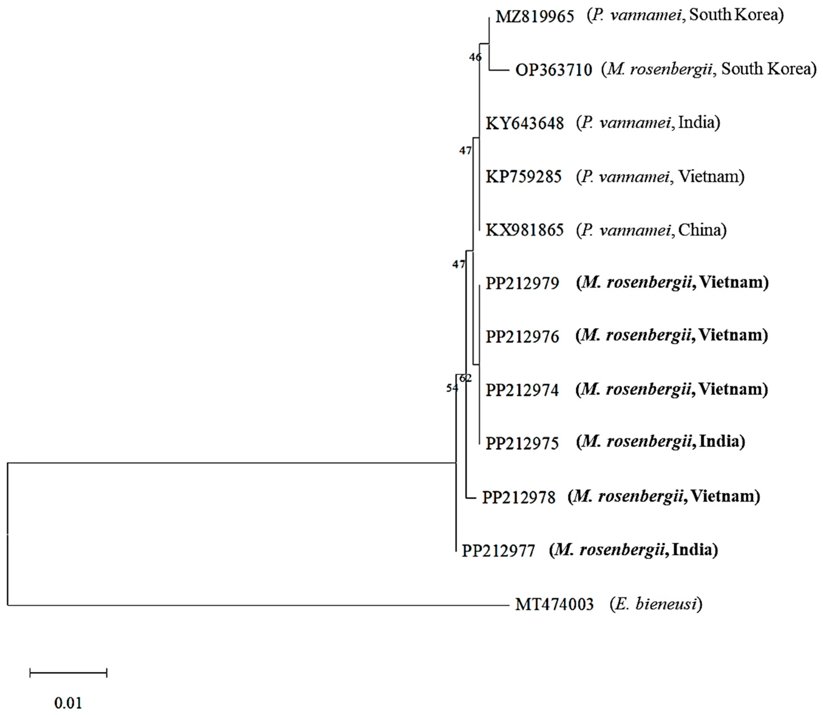
Figure 1. Maximum-likelihood phylogenetic tree based on nucleotide sequences of the small subunit ribosomal RNA (SSU rRNA) region of Enterocytozoon hepatopenaei (EHP), including Macrobrachium rosenbergii EHPs obtained in this study (in bold; GenBank accession Nos. PP212974, PP212975, PP212976, PP212977, PP212978, and PP212979). Enterocytozoon bieneusi (GenBank accession No. MT474003; family Enterocytozoonidae; order Enterocytozoonida) was used as an outgroup. Numbers at the branches indicate bootstrap values obtained using 1000 replicates. The scale bars represent 0.01 nucleotide substitutions per site. The trimmed alignment length was 778 bp.
Animals 15 03286 g001
Figure 2. Maximum-likelihood phylogenetic tree based on nucleotide sequences of the spore wall protein (SWP) 1 region of Enterocytozoon hepatopenaei (EHP), including Macrobrachium rosenbergii EHPs obtained in this study (in bold; GenBank accession Nos. PP908959, PP238911, PP238912, PP238913, PP238914, PP238915, and PP238916). Enterocytozoon bieneusi (GenBank accession No. MT490633) was used as an outgroup. Numbers at the branches indicate bootstrap values obtained using 1000 replicates. The scale bars represent 0.05 nucleotide substitutions per site. The trimmed alignment length was 151 bp.
Animals 15 03286 g002
Figure 3. Maximum-likelihood phylogenetic tree based on the nucleotide sequences of the internal transcribed spacer-1 (ITS-1) region of Enterocytozoon hepatopenaei (EHP), including Macrobrachium rosenbergii EHPs obtained in this study (in bold; GenBank accession Nos. PP265522, PP265523, PP265524, PP265525, PP265526, PP265527, and PP265528). Enterocytozoon bieneusi (GenBank accession No. AF023245) was used as an outgroup. Numbers at the branches indicate bootstrap values obtained using 1000 replicates. The scale bars represent 0.10 nucleotide substitutions per site. The trimmed alignment length was 305 bp.
Animals 15 03286 g003
Table 1. Sample information and PCR results.
Sample IDCountryCollection Month/YearLength (cm)Weight (g)EHP Detection 1
23-026C5-1-VIEVietnamMay 202320.0–23.0121.1–157.7
23-026C6-1-VIEVietnamJune 202314.0–18.333.0–60.0+
23-026C6-2-INDIAIndiaJune 202320.5–23.093.6–153.4+
23-026C7-1-VIEVietnamJuly 202315.0–18.649.5–89.4+
23-026C7-2-INDIAIndiaJuly 202319.0–22.5105.5–183.6+
23-026C7-3-VIEVietnamJuly 202314.4–18.031.4–75.8+
23-026C8-1-INDIAIndiaAugust 202320.9–26.195.1–199.4
23-026C8-2-VIEVietnamAugust 202314.7–16.351.8–80.5
23-026C9-1-INDIAIndiaSeptember 202319.0–22.892.6–130.7
23-026C9-2-VIEVietnamSeptember 202315.0–16.445.7–77.8+
23-026C10-1-INDIAIndiaOctober 202319.5–22.890.6–140.2
23-026C11-1-VIEVietnamOctober 202315.8–18.543.4–79.0+
24-004C2-1-INDIAIndiaFebruary 202418.9–21.496.1–161.6
24-004C2-2-INDIAIndiaFebruary 202419.0–21.497.4–159.0
24-004C3-1-INDIAIndiaMarch 202420.2–22.7110.6–159.4
24-004C3-2-INDIAIndiaMarch 202419.2–22.9101.3–137.6
24-004C4-1-INDIAIndiaApril 202418.4–22.176.9–139.6
24-004C4-2-INDIAIndiaApril 202418.9–21.987.5–143.9
24-004C4-3-INDIAIndiaApril 202423.3–26.6185.0–289.9
24-004C5-1-INDIAIndiaMay 202422.5–25.595.5–143.0
24-004C5-2-INDIAIndiaMay 202419.4–21.986.8–134.5
24-004C6-1-INDIAIndiaJune 202422.2–25.2109.56–148.15
1 For EHP detection, the 510-F/R primer set was used [6]. +: positive, −: negative.
Table 2. Primer information used in this study.
TargetPrimerSequence (5′ to 3′)Amplicon Size (bp)Reference
SSU rRNA 1510-FGCCTGAGAGATGGCTCCCACGT510[12]
510-RGCGTACTATCCCCAGAGCCCGA
SSU rRNA 218S-FCACCAGGTTGATTCTGCCTGA1146[12]
18S-RTCTGAAATAGTGACGGGCGG
SWP 11F (1st)TTGCAGAGTGTTGTTAAGGGTTT514[20]
1R (1st)CACGATGTGTCTTTGCAATTTTC
2F’ (2nd)GCAGAGTGTTGTTAAGGGTTTAAG182[13]
2R’ (2nd)GCTGTTTGTCWCCAACTGTATT
ITS-1ITS1-1F (1st)CGCCCGTCACTATTTCAGAT603[21]
ITS1-1R (1st)TACGTTCGTCATCGCTGCTA
ITS1-2F (2nd)GAACCTGCTGTGGGATCATT400
ITS1-2R (2nd)AATTTTTGCTTGGCTCATTCT
1 For EHP monitoring. 2 For sequence analysis.
Disclaimer/Publisher’s Note: The statements, opinions and data contained in all publications are solely those of the individual author(s) and contributor(s) and not of MDPI and/or the editor(s). MDPI and/or the editor(s) disclaim responsibility for any injury to people or property resulting from any ideas, methods, instructions or products referred to in the content.