Review

State-of-the-Art Age Determination Methods for Amphibians and Reptiles

by
Fabio Maria Guarino
1,* and
Marcello Mezzasalma
2
1
Department of Biology, University of Naples Federico II, Via Cinthia 26, 80126 Naples, Italy
2
Department of Biology, Ecology and Earth Science, University of Calabria, Via P. Bucci 4/B, 87036 Rende, Italy
*
Author to whom correspondence should be addressed.
Animals 2025, 15(18), 2722; https://doi.org/10.3390/ani15182722
Submission received: 14 July 2025 / Revised: 11 September 2025 / Accepted: 16 September 2025 / Published: 17 September 2025
(This article belongs to the Special Issue Age Determination, Growth and Longevity of Amphibians and Reptiles)

Simple Summary

Accurately assessing animals’ age parameters (age at maturity, mean age, and longevity) and growth-related parameters is fundamental in many fields of biology. Numerous methods with varying degrees of accuracy and precision have been developed to estimate the age and growth rates of amphibians and reptiles in the wild. The capture–mark–recapture technique potentially provides the most reliable data on age–size relationships, but requires a large amount of work, is time-consuming, and is often impractical for some wild populations. Skeletochronology has proven to be the most reliable among indirect age determination methods and has been successfully applied in numerous species of amphibians and reptiles. Skeletochronology is based on the interpretation of the growth marks that form within mineralized tissues, such as bones, during animals’ lives. Other indirect methods of age estimation based on the same principle (counting of growth marks), but applied to hard non-skeletal structures (epidermal scutes, claws, osteoderms), result in less reliable assessments and/or cannot be applied to all taxa. Recently, the use of molecular and biochemical methods such as measurements of telomere length and DNA methylation has provided new promising tools for the estimation of age, but these methods still need further refinement and testing on a larger number of species.

Abstract

In this work, we provide an overview of the age determination methods of amphibians and reptiles, critically evaluating their methodological principles, utility (including range of applicability), as well as experimental and biological limitations. Potentially, the most reliable age estimation method is the capture–mark–recapture (CMR) technique, allowing a precise chronological age estimation of wild individuals. However, this is a time-consuming method and is not always applicable. Among the indirect methods of age estimation, skeletochronology is one of the most reliable techniques and has been successfully applied in numerous species of amphibians and reptiles. Skeletochronology is based on the interpretation of the growth marks (especially lines of arrested growth or LAGs) that form in hard-mineralized tissues. Other indirect methods such as the counting of growth marks on epidermal scutes, dermal scales, and claw sections have shown to be less reliable and/or have a more limited range of applicability. Recently, the estimation of chronological age using molecular methods has acquired new perspectives from different approaches. Among them, measurements of telomere length and DNA methylation represent promising and minimally invasive methods, but their effective use requires further refinement and testing on a larger number of species.

1. Introduction

Understanding the timing and evolution of life histories is a key theme in evolutionary biology as well as crucial for studies in ecology, zoology, and conservation biology [1,2,3,4,5,6]. Age at maturity, mean age, longevity, and growth-related parameters are among the most important organismal traits and allow predictions about species’ life histories. Amphibians and reptiles represent two particularly interesting groups to better understand the proximate and evolutionary causes of aging and longevity in vertebrates. While they constitute two very different groups of vertebrates in terms of evolutionary history and biological characteristics, they can be considered together in terms of addressing age estimation methods because many of these methods can be applied to either group.
Amphibians and reptiles include many species with a maximum longevity of higher than 100 years (e.g., Proteus salamanders, Galapagos tortoise) [1,7] as well as the current record holder for the shortest life span in tetrapods (about five months, in the Labord’s chameleon, Furcifer labordi) [8]. Wild amphibians and reptiles show a greater variability in growth rates and longevity than birds and mammals [5]. Furthermore, in amphibians and reptiles the age structure and other demographic traits can highly differ among populations of the same species living at different altitudes and/or latitudes (i.e., [9,10,11,12,13,14]). Like endothermic vertebrates, in amphibians and reptiles both growth and longevity rates are correlated to the age of sexual maturity [5].
Multiple factors can exert different selective pressures that affect the longevity of amphibian and reptile species. Species exposed to reduced extrinsic and intrinsic mortality pressures (less predation, lower metabolic rates, and shorter activity periods) live longer [15,16]. However, it should be taken into account that in longevity studies, the chances of finding older individuals increase with a larger study sample.
In general, for animals, we can distinguish life span from life expectancy [17]. The first (also referred to as “maximum”, “potential”, or “specific longevity”) represents the maximum age that the species can attain in optimal conditions. The life span is generally fixed, although in various animal species can be variable to some extent and, in some cases, it can be extended by the assumption of some particular substances (i.e., lithium, see [18,19]). In turn, life expectancy (also termed “ecological longevity”) represents the average length of the life of individuals living in the wild. It is highly dependent on environmental conditions (such as temperature, trophic resources, predation, and diseases) and it may vary among different populations. In amphibians and reptiles, the difference between life span and life expectancy can be significant, and it is usually greater than that of similar-sized endotherms. In addition, longevity in captive individuals is usually much higher than longevity in the wild for a variety of factors, including the lack of natural predators. For example, the common toad Bufo bufo can attain 36 years in captivity [20], whereas its longevity in the wild varies considerably among populations, but has been never documented to exceed 15 years [21]. Similarly, the longevity of the Mediterranean gecko Hemidactylus turcicus is 8 years in captivity while it is 4 years in the wild [22].
In general, age estimates can be defined as “absolute age” (termed also “chronological age”) when the age is estimated according to standard time measurements (usually years for long-lived species) or “relative age” (termed also “categorical age”) when the animal is assigned to an age class [23]. Relative age is also synonymous of “biological age” since it expresses the aging phase of an individual (e.g., newborn, subadult, adult) and can be defined on the basis of morpho-functional characteristics associated with individual development (e.g., skeletal changes, ontogenetic color and/or pattern changes, sexual maturity). In some field of studies, such as demography (i.e., [24,25,26,27,28]) it is necessary to infer the absolute age, while for others, including, for example, reproductive biology (i.e., [29]) or ethology (i.e., [30]), an assessment of the relative age can be appropriate for most studies.
Numerous methods with varying degrees of accuracy and precision have been developed to estimate the individual age and growth rates of amphibians and reptiles. In this contribution, we summarize and critically evaluate the methodological principle, the utility (including accuracy, efficiency, and range of applicability) as well as the experimental and biological limitations of these methods. Finally, we consider the still unresolved critical issues presented by the available methods and topics insufficiently explored, proposing some future research avenues. This review is intended as an informative introduction to age estimation for non-specialists as well as a methodological summary and a reference guide for more experienced researchers.

2. Materials and Methods

Peer-reviewed scientific papers were identified and selected by electronic literature searches in several engines including Web of Science®, PubMed®, Google Scholar® and JSTOR® from April 2025 to June 2025. We specifically searched for terms: “age estimation” or “life expectancy” or “longevity” or “lifespan” or “capture-mark-recapture” or “sclerochronology” or “skeletochronology” or “size frequency” or “growth marks” or “telomere length” or “DNA methylation” or “DNAm” or “amino acid racemization” or “bomb radiocarbon” AND “amphibians” or “reptiles” or “frogs” or “caecilians” or “salamanders” or “newts” or “amphisbaenians” or “lizards” or “snakes” or “squamates” or “turtles” or “crocodiles” or “Sphenodon” or “tuatara”. A further search was performed checking the literature cited by the papers retrieved during the initial search and the authors’ personal databases.
Bone terminology and relative abbreviations used in the following sections generally follow refs. [17,20].

3. Methods of Age Estimation

3.1. Capture–Mark–Recapture Method

Potentially, the most reliable age estimation method is the capture–mark–recapture (CMR) technique, which virtually allows a precise chronological age estimation of wild animals [31]. This method requires the capture of animals of a known age (preferably hatchlings and neonates) during an initial sampling period, their marking, release and recapture at periodic intervals. This technique is generally used in combination with body length measurements to infer age-specific growth rates [31,32]. Furthermore, the CMR technique can be useful for validating other methods of age estimation such as those based on the interpretation of incremental growth layers [32,33,34,35]. Besides being one of the first methods of age determination, the CMR method is also one of the most frequently used (i.e., [36,37]); for other references see [31]). Furthermore, CMR can be applied to certain taxa, such as snakes (i.e., [38,39]) for which other methods of age determination cannot be applied without sacrificing the individual or causing significant impairment (as in the case of skeletochronology, see below). The main limitations of the CMR method include requirements of long-term observations, which can hardly be achieved on some wild populations.

3.2. Size Frequency Method

The extrapolation from size frequency data is an indirect method for estimating age, and, in the past, it was frequently applied to amphibians [40,41] and reptiles (i.e., [42,43,44]). The main weakness of this method is that it assumes that body length and age are positively correlated, while this has been shown to be false for many species (in one or both sexes) or between different populations of the same species [24,31,45,46]. As a result, individuals that fall into the same size class can often have very different ages as shown using other methods (e.g., skeletochronology, see below). Furthermore, several studies provided evidence that linear growth may stop well before senescence in reptiles including those taxa until recently considered indeterminate growers, such as crocodilians [47,48,49,50], and/or very long-living species, such as the Blanding’s turtle Emydoidea blandingii [51].

3.3. Sclerocronology

Most of the indirect methods of age estimation are based on the correlation between the growth of individual and the progressive layering of its hard tissues such as otoliths (only for bony fishes, see [33]); epidermal scutes (i.e., [52]), claws (i.e., [53,54]), bones (i.e., [55,56,57,58,59,60,61,62,63,64,65,66,67,68,69]), teeth (only for mammals), osteoderms (i.e., [70]), and dermal scales (i.e., [71]). These methods are overall called sclerochronology, and more specifically skeletochronology when they involve only mineralized hard tissues [17,33,72] (Table 1).
While the counting of growth marks on otoliths (also termed otolithometry) has been widely applied in bony fishes (teleosts) (see [73]), it has never been used in amphibians and reptiles. In fact, unlike the otoliths of most bony fish, which are stony masses, the inner ear labyrinth of amphibians and reptiles contains much smaller crystals of calcium carbonate (otoconia or statoconia), which are unsuitable for otolithometric applications [74].
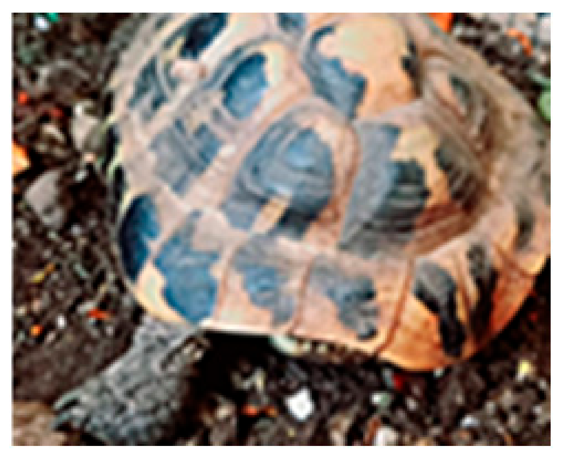
The counting of growth marks or rings on scutes (Figure 1A) has been tested only in several species of tortoises and terrapins [75,76,77,78,79,80,81]. Indeed, it finds its main constraint in the fact that there are few species of amphibians and reptiles with keratinized epidermal scutes. Moreover, according to several researchers, the use of scute rings may produce age estimates in chelonians that are, at best, approximations, except in the youngest specimens [78], in individuals near the age of maturity [82], or up to a certain age (for example 15 years in the Gopherus polyfemus) [79]. Furthermore, this method might be practicable for some populations, but only after calibrating the relationship between ring counts and age for each circumstance (including species and geographical location) [83]. Nevertheless, a recent long-term CMR study on Testudo graeca showed that, when correctly applied, individual age calculated by counting shell growth rings can be more precise than other techniques [81], underlining that while age estimation in turtles is challenging, a combination of methods could improve its accuracy.
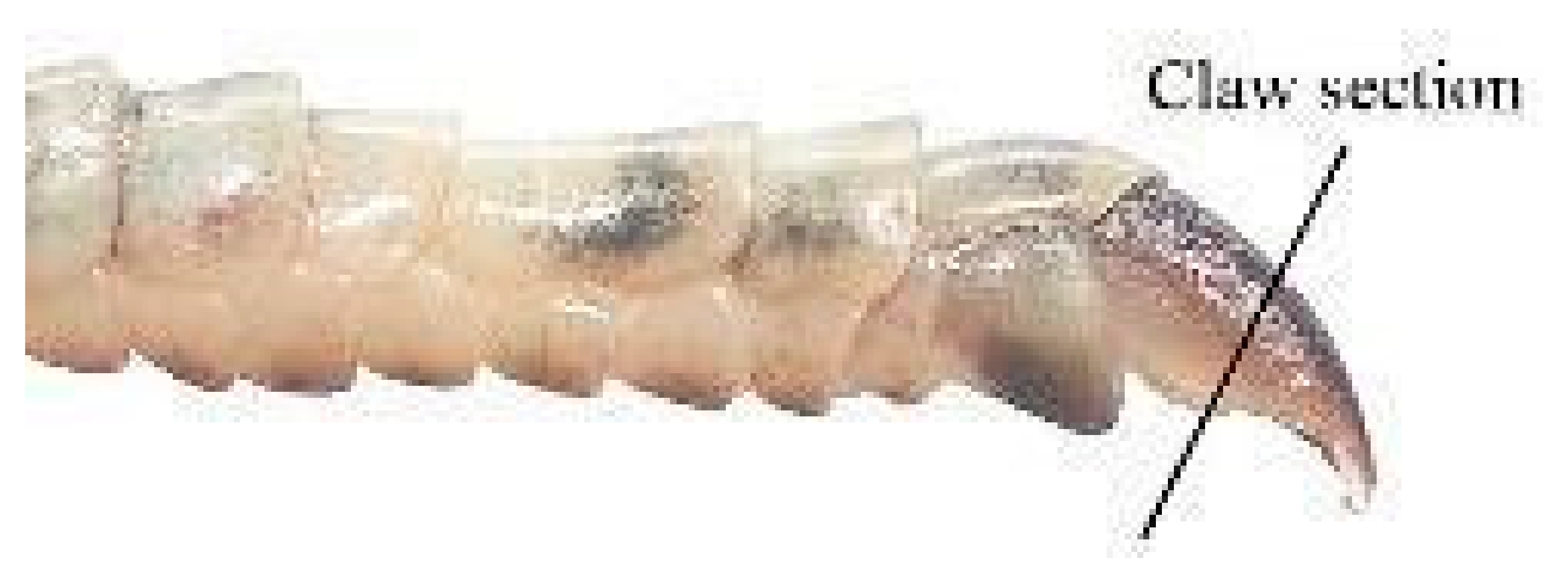
The analysis of claw sections (Figure 1B) for the purpose of age determination could potentially be applied to a large number of reptiles but, so far, it was only performed on a few species, including the terrapins Chrysemys picta and Deirochelis reticularia [53] and some lizard species of the genus Darevskia [54]. In terrapins, this method was applied to individuals under 30 years of age, so there may be upper age limits on the usefulness of claw sections as a precise tool for age determination [53].
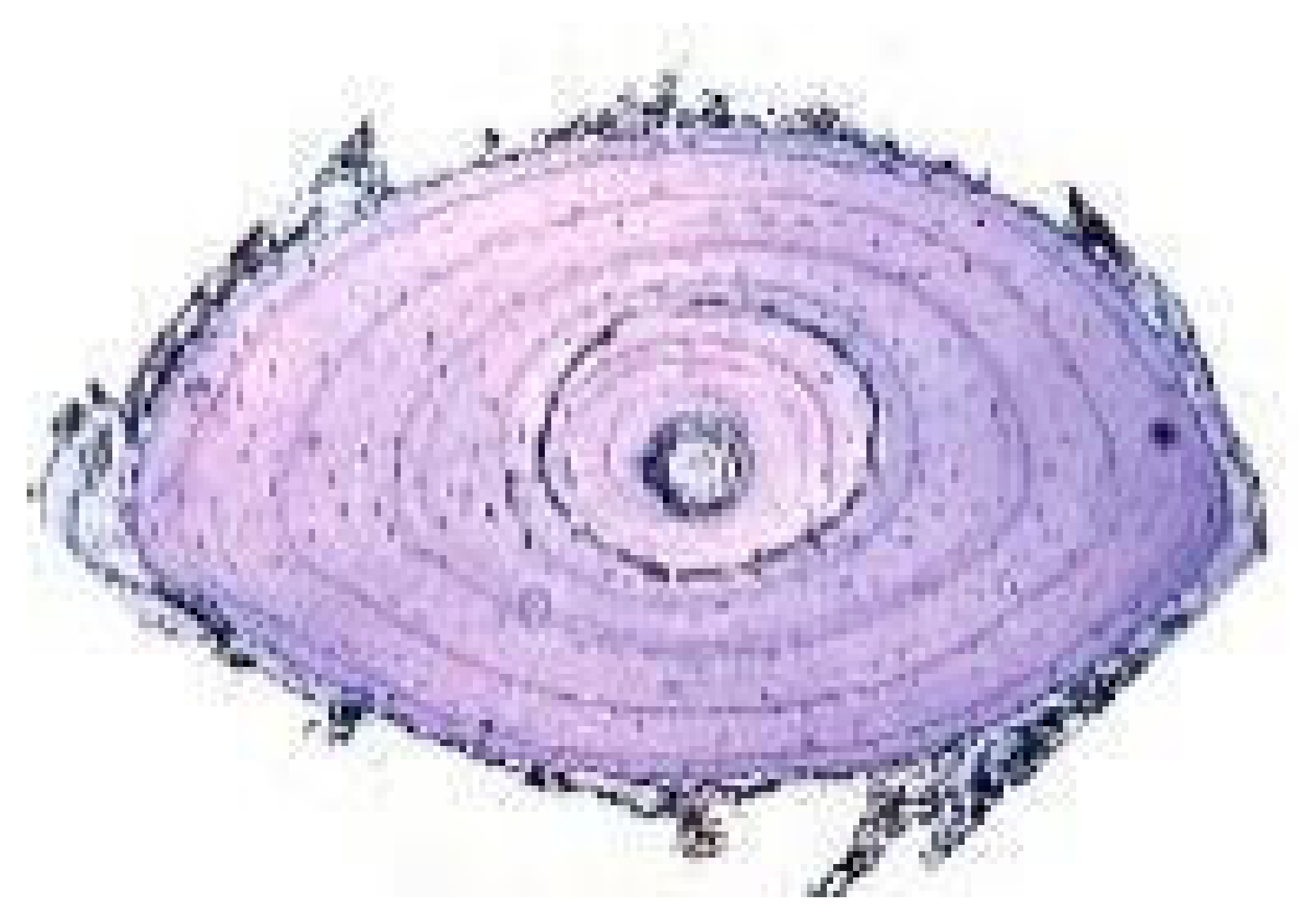
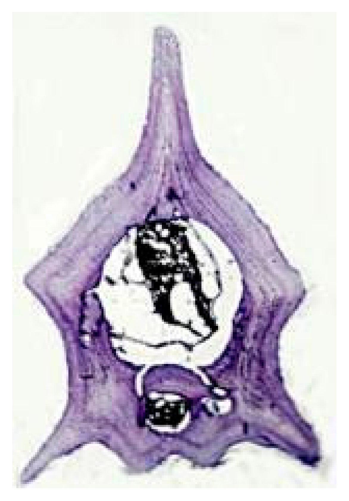
Unlike all the previously mentioned indirect methods, skeletochronological studies have been successfully applied to a large number of species of amphibians and reptiles and include several hundreds of published papers (at least 369 for amphibians, see [84], and at least 468 papers covering 236 reptile species, see [85]), representing the most used age estimation method for these vertebrates. Skeletochronology is a histological method based on the interpretation of the growth marks occurring in the mineralized tissues of an individual throughout its life cycle, corresponding to the slowing or stopping of the somatic growth, and reflecting endogenous biological rhythms, which are generally synchronized with environmental conditions [33]. In general, three categories of growth marks can be recognized in the bone: lines of arrested growth (LAGs), representing a temporary arrest of the bone growth; annuli, which corresponds to periods of slow bone deposition; zones or opaque layers, which are the widest marks and correspond to periods of active osteogenesis [17,33]. Also, one LAG can be present within an annulus. Once LAGs with annual periodicity have been identified, the age estimation of the individual can be assessed from their count (including any totally reabsorbed LAG) while growth rates can be estimated from the distance between the LAGs. In amphibians and reptiles, annual growth marks can be observed in several bones, including skull bones (i.e., [55,58,65]), vertebrae (i.e., [66,67,69]), and long, tubular bones, such as the femur, humerus, and phalanx (i.e., [60,86,87,88,89,90]). Growth marks can be detectable also in osteoderms of some reptile groups, such as crocodilians and Anguidae, as well as in the dermal scales of amphibians of the order Gymnophiona. However, within these taxa, a limited number of species (including the Crocodylus niloticus, [91]; C. johnstoni [70], A. veronensis [68], and Dermophis mexicanus [71]) have been studied in order to determine individual age through the growth mark-counting of mineralized dermal structures, with contrasting results in terms of efficiency and accuracy. In Diploglossidae, it has been shown that regenerated and non-regenerated tail osteoderms probably do not follow the same growth rates and that skeletochronology should be applied only to non-regenerated osteoderms [92]. Therefore, estimating age by counting growth marks that form in this mineralized tissue should be viewed with caution in the case of some species, i.e., crocodiles [70], and it is probably unreliable in other species [66,68,71]. The teeth are also unsuitable for skeletochronological analysis because majority of amphibians and reptiles are polyphyodonts [93,94]. In a few exceptions, such as the agamids where post-canine acrodont teeth are not replaced during their lifetime, the growth layers formed in the teeth are indistinct when they are examined in section and cannot be used for age determination [95]. On the other hand, the use of so-called tide marks on tooth roots, which are visible, for example, in the Crocodylus niloticus, is currently anecdotal [72]. However, available data show that, while growth marks can be visualized in very thin flat bones by backlighting them (Figure 1C), they have better optical sharpness using stained decalcified bone sections [72] (Figure 1D,E). In general, long bones are the most useful and reliable skeletal elements with regard to growth mark visualization and ease in sample preparation [14,33,96]. Furthermore, long bones such as the femur, humerus, and phalanges usually provide corresponding age estimations in amphibians and reptiles [20,63,97]. A few exceptions are represented by some very long-living species (e.g., Sphenodon punctatus, Bombina variegata, and Homonota darwini) in which age assessments based on the phalanges are slightly underestimated compared with those based on the femur [64,98,99]. Furthermore, since different bones can differ in growth patterns and in bone remodeling rates [20,100,101], it can be useful to perform preliminary comparisons between the different types of long bones in understudied taxa, before undertaking a skeletochronological study. Obviously, limbless taxa, such as Gymnophiona, snakes, and many lizard species (i.e., Dibamidae, Anguidae) or taxa with much reduced limbs (i.e., several species of skinks) represent a special challenge. In these taxa, skeletochronology can be applied to skull bones, such as the ectopterygoid (as in the snake Trimesurus flavoviridis) [102], the supra-angular of the mandibula (as in the snakes Vipera aspis and Trimesurus flavoviridis) [58,102,103] or the caudal vertebrae (as in the cecilian Geotrypetes seraphini, in the limbless lizards of the genus Anguis, and in the snakes Thamnophis and Trimesurus) [66,68,69,104,105]. However, excluding studies on samples from museum collections (i.e., [106,107,108], skeletochronology performed on cranial bones, vertebrae, femur or humerus usually requires the sacrifice of the animal. On the contrary, skeletochronology performed on phalanges obtained by toe-clipping allows both the marking of individuals and the collection of bone samples for skeletochronological analysis without the sacrifice of any animal, potentially also enabling demographic studies over long intervals of time [14,28,109]. Technical accounts of skeletochronology including its validation and possible solutions to the main difficulties (e.g., endosteal resorption of LAGs, double LAGs, supernumerary LAGs) encountered in skeletochronological studies are given by [14,33,110,111]. It is worth mentioning that the study of Schucht et al. [112] questioned the ambiguity of histology-based skeletochronology because halved bones (at level of the area with the smallest medullary cavity) provided some discrepant age estimates when comparing petrographic ground sections and conventional microtomized sections. Taking into account the fundamental differences between these two histological techniques, more research is needed to better standardize some methodological aspects, with particular regard to the unambiguous recognition of the different types of growth marks.

3.4. Other Morphological Methods

Among the traditional morphological methods of age estimation (other than skeletochronology) is the measurement of the eyeball lens, which grows continually, though not continuously, throughout the animal’s life. Although this method has been widely tested for mammals, very limited data are available for amphibians and reptiles (for a list of species see [73,77,113]. Furthermore, as pointed out by Zug [77], small differences in the lens mass can lead to disproportionate differences in age estimation; consequently, the protocol (including the type of fixative and duration of lens fixation) should be standardized as much as possible.
Some other morphological methods of age determination have been proposed in the past but later proven ineffective. One of the best-known examples is the count of testicular lobes in salamanders, which were thought to form progressively over successive breeding seasons and could therefore be used to estimate individual age (i.e., [114,115]). However, in a study on the Lissotriton vulgaris and T. cristatus, a considerable variation in lobe numbers within age classes (determined by skeletochronology) was found and no male showed more than three or four lobes [116]. The alleged correlation between lobe number and individual age was later refuted [117], and the method has since fallen out of use.

3.5. Molecular Methods

In recent years, the estimation of chronological age using molecular methods has acquired new perspectives from different approaches. In general, different molecular methods represent indirect methods of age determination, similarly to sclerochronology. After opportune calibrations (e.g., for the taxon, tissue, or cell type) they can provide either relative or absolute age estimates with a margin of error (of, e.g., 3–10 years). Telomere length (TL) shortening is a well-known hallmark of both cellular senescence and organismal aging [118]. TL shortening with body length and individual age has been tested on different taxa of amphibians and reptiles, using different molecular techniques such as Southern blot and qPCR [119,120,121,122].
The first experimental study on a natural amphibian population (of Epidalea calamita) reported a clear correlation between telomere shortening and age, especially during the first 1–2 years [122]. However, besides providing some interesting results, methods based on TL also showed several limitations, and there are still too few studies to conclude its full reliability in amphibians and reptiles. In particular, age determination using TL has been applied in several studies on different lizard taxa (including, e.g., the Psammodromus algirus, Lacerta agilis, Chlamydosaurus kingii, and Ctenophorus pictus), showing contrasting results [123,124,125]. Overall, these studies showed a correlation of telomere shortening with age in some taxa but also a lack of universality. In fact, TL patterns appear to vary among species, sexes, tissues, and cell types and are prone to variations linked to other factors [123,124,125]. For example, in the sand lizard (L. agilis), differences in telomere length were correlated to sex and stress patterns; in females, an increase in the TL was correlated with age, but in males, no distinct age pattern was recorded, and TL was negatively impacted by tail autotomy and larger body size [125].
Studies on the epigenetic process of DNA methylation (DNAm) are another recent promising experimental approach for age determination. The methodology employed is based on the quali–quantitative detection and measurement of changes in DNAm occurring in an individual during the natural course of aging. In general, the process of DNAm refers to the addition to the 5′ cytosine of cytosine–guanine pairs (CpGs) of a methyl group (CH3) by the DNA methyltransferase enzyme class as a part of mechanisms of DNA packaging and gene regulation [126].
In vertebrates, DNAm is necessary for several processes including regular development, inactivation of genes and whole chromosomes (e.g., X chromosomes in mammals), suppression of transposable elements, and genomic imprinting [127].
To date, DNAm techniques for age estimation have been used several times for mammals and tested in birds [128] and, to a limited extent, amphibians [129,130] but not in reptiles. In particular, two species of the genus Xenopus (X. laevis and X. tropicalis) were used as model organisms to develop age-related epigenetic clocks to estimate relative and chronological age [129,130]. Interestingly, a combination of the human and clawed frog epigenetic clock showed evolutionarily conserved methylation patterns, enabling age estimation in both species using a single formula [130].
However, similarly to TL, DNAm also presents several issues as a reliable method for age determination. In fact, the general methylation status is not fixed within any single organisms, species, or cell type. In particular, different organisms usually exhibit differential levels of DNAm in different tissues and/or age and developmental stage (i.e., [131,132], and different cell types can regulate DNAm in response to physiological and environmental changes [126]. Overall, despite presenting a high diagnostic potential, techniques of age determination-based DNAm are more expensive when compared with some alternatives and might prove challenging to standardize.

3.6. Biochemical Methods

Amino acid racemization (AAR) has been used in age determination in fish and mammals [73], but its reliability in amphibians and reptiles is currently unexplored and would likely require some methodological adaptations. The method is based on the evaluation of the temporal rates of conversion of L-amino acids to their D-forms over time, and it is potentially able to provide aging estimates of preserved museum samples or even fossils [133]. The efficiency of this method is linked to the identification of tissues with low-turnover rates in order to accurately measure aging [134]. In vertebrates, AAR has provided age estimates with a lower accuracy (of about +/−3–10 years) compared with other methods, and its limitations also include a potentially destructive sampling and the absence of calibration in most taxa [73,135]. Nevertheless, the potential use of AAR on preserved samples offers promising applications of the method on biological samples from public and private collections, avoiding invasive practices on endangered natural populations.
Radiocarbon methods (particularly bomb-14C) have been used in the age assessment of turtles [136] but not in amphibians. In particular, they have been successfully applied to living specimens and conserved hard tissues of Hawksbill sea turtles (Eretmochelys imbricata), helping to provide individual age estimates and date keratinized growth bands on the scute [136]. Using bomb-14C, Van Houtan et al. [136] found that about eight scute rings were deposited on the scute each year, aligning chronological data with biological age [136]. In the end, while radiocarbon dating had very limited empirical applications in reptiles, its use appears promising and potentially extendable to amphibian taxa.
Furthermore, techniques using stable isotopes of carbon and nitrogen have been applied to reconstruct seasonal feeding patterns and timeframes of tissue turnover in turtles and lizards (i.e., [137,138]), but they do not provide direct aging estimates. However, when isotope profiling is used in combination with skeletochronology (applied on growth lines) it may provide some useful correlations between age (counted via LAGs) and isotopic changes, helping to better understand life-stage events, feeding patterns, and habitat transitions [139].

4. Future Research Perspectives

Since the end of the last century, numerous studies conducted on the growth and longevity of different species of amphibians and reptiles have greatly improved our understanding of their demographic life-history traits, mainly using skeletochronology (i.e., [11,27,34,109,140,141,142]). However, some aspects remain insufficiently explored, including whether variations in age structure or in other demographic traits can be attributed to natural geographical variations, climate change, or other human-induced effects (i.e., chemical pollution, land cover change). For example, a recent study by Zamaletdinov et al. [143] on three green frog species (genus Pelophylax) showed specific characteristics of their age structure in relation to the degree of habitat transformation. Considering both their main limitations and exciting future perspectives, studies on age at maturity, mean age, longevity, and growth-related parameters remain fundamental windows on the natural and evolutionary history of vertebrates, which are potentially able to determine novel scenarios on the macroevolution of aging and life itself.
Although phalangeal skeletochronology can be considered one of the most efficient non-lethal methods for estimating the age and growth in most amphibians and reptiles, it presents some critical issues that can prevent an accurate LAG counting. For example, bone remodeling can totally destroy some LAGs [17,20,110,111], possible aperiodic (false) or supernumerary LAGs [33,72] can be confused with true LAGs, and very closely adjacent LAGs might be hard to correctly count in the outer margin of the bone sections of the longest-lived individuals [60]. To date, while it is possible to approximate the number of completely resorbed LAGs using the back calculation method [33,144,145,146], the false or supernumerary LAGs remain difficult to address and are often the cause of approximations in absolute age determination [112]. Therefore, the development of non-lethal age estimation methods which can be alternative to or synergic with phalangeal skeletochronology is desirable for challenging species (e.g., snakes, lizards with largely reduced limbs, or limbless taxa) and can be useful also to overcome the aforementioned general difficulties of the skeletochronological method. In particular, promising molecular and biochemical methods (with particular regard to AAR and DNAm) should be further developed and tested on a wider variety of taxa of amphibians and reptiles as they may either provide non-destructive alternatives to skeletochronology or refine the age estimation based on LAG count.

5. Conclusions

Studies on the age determination of individuals belonging to natural populations are fundamental to better understanding population dynamics and structure, life-history strategies, and ecological interactions. Their application ultimately informs conservation actions and policies of wildlife management. This importance is reflected in the ongoing research of accurate, efficient, and non-invasive universal methodologies, being able to cover distinct organisms with marked differences in their natural history, physiology, and morphology (e.g., limbless squamates). In recent years, several methodological innovations have been proposed for age assessments of amphibians and reptiles, but traditional methods based on the identification of morphological growth marks still represent the more efficient alternative. This is also due to the lack of testing of the new methods, which leaves their efficiency widely unexplored and does not allow for an accurate assessment of their potential on most taxa of amphibians and reptiles. Nevertheless, the future of age estimation of natural populations is probably linked to either the development of new methods or to the multidisciplinary, synergic application of the existing ones.

Author Contributions

Conceptualization, F.M.G. and M.M.; methodology, F.M.G. and M.M.; software, F.M.G. and M.M.; validation, F.M.G. and M.M.; formal analysis, F.M.G. and M.M.; investigation, F.M.G. and M.M.; resources, F.M.G. and M.M.; data curation, F.M.G. and M.M.; writing—original draft preparation, F.M.G. and M.M.; writing—review and editing, F.M.G. and M.M.; visualization, F.M.G. and M.M.; supervision, F.M.G. and M.M. All authors have read and agreed to the published version of the manuscript.

Funding

This research received no external funding.

Institutional Review Board Statement

Not applicable.

Informed Consent Statement

Not applicable.

Data Availability Statement

Not applicable.

Acknowledgments

We thank Emanuela Fiorella Guarino for Figure 1A. We thank the anonymous reviewers for their valuable comments.

Conflicts of Interest

The authors declare no conflicts of interest.

References

  1. de Magalhães, J.P.; Costa, J.A. database of vertebrate longevity records and their relation to other life-history traits. J. Evol. Biol. 2009, 22, 1770–1774. [Google Scholar]
  2. Mezzasalma, M.; Andreone, F.; Glaw, F.; Guarino, F.M.; Odierna, G.; Petraccioli, A.; Picariello, O. Changes in heterochromatin content and ancient chromosome fusion in the endemic Malagasy boid snakes Sanzinia and Acrantophis (Squamata: Serpentes). Salamandra 2019, 55, 140–144. [Google Scholar]
  3. Mezzasalma, M.; Guarino, F.M.; Odierna, G. Lizards as model organisms of sex chromosome evolution: What we really know from a systematic distribution of available data? Genes 2021, 12, 1341. [Google Scholar] [CrossRef] [PubMed]
  4. Petraccioli, A.; Guarino, F.M.; Kupriyanova, L.; Mezzasalma, M.; Odierna, G.; Picariello, O.; Capriglione, T. Isolation and characterization of interspersed repeated sequences in the common lizard, Zootoca vivipara, and their conservation in Squamata. Cytog. Genome Res. 2019, 157, 65–76. [Google Scholar]
  5. Reinke, B.A.; Cayuela, H.; Janzen, F.J.; Lemaître, J.F.; Gaillard, J.M.; Lawing, A.M.; Iverson, J.B.; Christiansen, D.J.; Martínez-Solano, I.; Sánchez-Montes, G.; et al. Diverse aging rates in ectothermic tetrapods provide insights for the evolution of aging and longevity. Science 2022, 376, 1459–1466. [Google Scholar] [CrossRef]
  6. Stott, I.; Salguero-Gómez, R.; Jones, O.R.; Ezard, T.H.G.; Gamelon, M.; Lachish, S.; Lebreton, J.D.; Simmonds, E.G.; Gaillard, J.M. Life histories are not just fast or slow. Trends Ecol. Evol. 2024, 39, 830–840. [Google Scholar] [CrossRef]
  7. Voituron, J.; de Fraipont, M.; Issartel, J.; Guillaume, O.; Clobert, J. Extreme lifespan of the human fish (Proteus anguinus): A challenge for ageing mechanisms. Biol. Lett. 2010, 7, 105–107. [Google Scholar]
  8. Eckart, F.; Kappeler, P.M.; Krauss, C. Highly variable lifespan in an annual reptile, Labord’s chameleon (Furcifer labordi). Sci. Rep. 2017, 7, 11397. [Google Scholar] [CrossRef]
  9. Hemelaar, A. Age, growth and other population characteristics of Bufo bufo from different latitudes and altitudes. J. Herpetol. 1988, 22, 369–388. [Google Scholar] [CrossRef]
  10. Wapstra, E.; Swan, R.; O’Reilly, J.M. Geographic variation in age and size at maturity in a small Australian viviparous skink. Copeia 2001, 3, 646–655. [Google Scholar] [CrossRef]
  11. Morrison, C.; Hero, J.M. Geographic variation in life history characteristics of amphibians: A review. J. Anim. Ecol. 2003, 72, 270–279. [Google Scholar] [CrossRef]
  12. Guarino, F.M.; Di Già, I.; Sindaco, R. Age structure by skeletochronology in a declining population of Rana temporaria from northern Italy. Acta Zool. Acad. Sci. Hung. 2008, 54, 99–112. [Google Scholar]
  13. Zhang, L.; Lu, X. Amphibians live longer at higher altitudes but not at higher latitudes. Biol. J. Linn. Soc. 2012, 106, 623–632. [Google Scholar] [CrossRef]
  14. Sinsch, U. Review: Skeletochronological assessment of demographic life-history traits in amphibians. Herpetol. J. 2015, 25, 5–13. [Google Scholar]
  15. Stark, G.; Meiri, S. Cold and dark captivity: Drivers of amphibian longevity. Glob. Ecol. Biogeogr. 2018, 27, 1384–1397. [Google Scholar] [CrossRef]
  16. Stark, G.; Tamar, K.; Itescu, Y.; Feldman, A.; Meiri, S. Cold and isolated ectotherms: Drivers of reptilian longevity. Biol. J. Linn. Soc. 2018, 125, 730–740. [Google Scholar] [CrossRef]
  17. Castanet, J. Age estimation and Longevity in reptiles. Gerontology 1994, 40, 174–192. [Google Scholar] [CrossRef]
  18. Zarse, K.; Terao, T.; Tian, J.; Iwata, N.; Ishii, N.; Ristow, M. Low-dose lithium uptake promotes longevity in humans and metazoans. Eur. J. Nutr. 2011, 50, 387–389. [Google Scholar] [CrossRef]
  19. Coutts, F.; Palmos, A.B.; Duarte, R.R.R.; De Jong, S.; Lewis, C.M.; Dima, D.; Powell, T.R. The polygenic nature of telomere length and the anti-ageing properties of lithium. Neuropsychopharmacology 2019, 44, 757–765. [Google Scholar] [PubMed]
  20. Smirina, E. Age determination and longevity in amphibians. Gerontology 1994, 40, 133–146. [Google Scholar] [CrossRef] [PubMed]
  21. Cvetković, D.; Tomašević, N.; Ficetola, G.F.; Crnobrnja-Isailović, J.; Miaud, C. Bergmann’s rule in amphibians: Combining demographic and ecological parameters to explain body size variation among populations in the common toad Bufo bufo. J. Zool. Syst. Evol. Res. 2009, 47, 171–180. [Google Scholar] [CrossRef]
  22. Werner, Y.; Frankenberg, E.; Volokita, M.; Harari, R. Longevity of geckos (Reptilia: Lacertilia: Gekkonoidea) in captivity: An analytical review incorporating new data. Isr. J. Zool. 1993, 39, 105–124. [Google Scholar]
  23. Read, F.L.; Hohn, A.; Lockyer, C.H. A review of age estimation methods in marine mammals with special reference to monodontid. NAMMCO Sci. Publ. 2018, 10. [Google Scholar] [CrossRef]
  24. Denton, J.S.; Beebee, T.J.C. Density related features of natterjack toad (Bufo calamita) populations in Britain. J. Zool. 1993, 229, 105–119. [Google Scholar] [CrossRef]
  25. Driscoll, D.A. Skeletochronological assessment of age structure and population stability for two threatened frog species. Austr. J. Ecol. 1999, 24, 182–189. [Google Scholar] [CrossRef]
  26. Guarino, F.M.; De Pous, P.; Crottini, A.; Mezzasalma, M.; Andreone., F. Age structure and growth in a population of Pelobates varaldii (Anura, Pelobatidae) from northwestern Morocco. Amphib. Reptil. 2011, 32, 550–556. [Google Scholar]
  27. Sinsch, U.; Dehling, J.M. Tropical anurans mature early and die young: Evidence from eight Afromontane Hyperolius species and a meta-analysis. PLoS ONE 2017, 12, e0171666. [Google Scholar] [CrossRef] [PubMed]
  28. Atkins, Z.S.; Clemann, N.; Chapple, D.G.; Edwards, A.M.; Sinsch, U.; Hantzschmann, A.M.; Schroder, M.; Scroggie, M.P.; Robert, K.A. Demographic and life history variation in two sky-island populations of an endangered alpine lizard. J. Zool. 2020, 310, 34–44. [Google Scholar] [CrossRef]
  29. Ngo, B.V.; Ngo, C.D. Reproductive activity and advertisement calls of the Asian common toad Duttaphrynus melanostictus (Amphibia, Anura, Bufonidae) from Bach Ma National Park, Vietnam. Zool. Stud. 2012, 52, 12. [Google Scholar] [CrossRef]
  30. Jansen, F.T.; Paukstis, G.L.; Brodie, E.D. Observations on basking behavior of hatchling turtles in the wild. J. Herpetol. 1992, 26, 217–219. [Google Scholar] [CrossRef]
  31. Halliday, T.; Verrell, P. Body Size and Age in Amphibians and Reptiles. J. Herpetol. 1988, 22, 253–265. [Google Scholar] [CrossRef]
  32. Avens, L.; Snover, M.L. Age and age estimation in sea turtles. In The Biology of Sea Turtles; Wyneken, J., Lohmann, K.J., Musick, J.A., Eds.; CRC Press: Boca Raton, FL, USA, 2011; Volume III, pp. 97–134. [Google Scholar]
  33. Castanet, J.; Francillon-Vieillot, H.; Meunier, F.J.; de Ricqles, A. Bone and individual aging. In Bone Growth; Hall, B.K., Ed.; CRC Press: Boca Raton, FL, USA, 1993; pp. 245–283. [Google Scholar]
  34. El Mouden, E.H.; Znari, M.; Brown, R.P. Skeletochronology and mark ± recapture assessments of growth in the North African agamid lizard (Agama impalearis). J. Zool. 1999, 249, 455–461. [Google Scholar] [CrossRef]
  35. Kumbar, S.M.; Pancharatna, K. Annual Growth Layers in the Phalanges of the Indian Skipper Frog Rana cyanophlyctis (Schn.). Copeia 2002, 3, 870–872. [Google Scholar] [CrossRef]
  36. Barbault, R.; Mou, Y.P. A population analysis of the common wall lizard Podarcis muralis in Southwestern France. In Studies in Herpetology; Roček, Z., Ed.; Charles University Press: Prague, Czech Republic, 1986; pp. 513–515. [Google Scholar]
  37. James, C.G. Growth rates and age at maturity of simpatric scincid lizards (Ctenotus) in Central Australia. J. Herpetol. 1991, 15, 284–295. [Google Scholar] [CrossRef]
  38. Brown, W.S. Lifetime Reproduction in a Northern Metapopulation of Timber Rattlesnakes (Crotalus horridus). Herpetologica 2016, 72, 331–342. [Google Scholar] [CrossRef]
  39. Brown, W.S.; Kery, M.; Hines, J.E. Survival of timber rattlesnakes (Crotalus horridus) estimated by capture-recapture models in relation to age, sex, color morph, time, and birthplace. Copeia 2007, 3, 656–671. [Google Scholar] [CrossRef]
  40. Bell, G. The life of the smooth newt (Triturus vulgaris) after metamorphosis. Ecol. Monogr. 1977, 47, 279–299. [Google Scholar] [CrossRef]
  41. Daugherty, C.H.; Sheldon, A.C. Age-determination, growth and life history of a Montana population of the tailed frog (Ascaphus truei). Herpetologica 1982, 38, 461–468. [Google Scholar] [CrossRef]
  42. Tinkle, D.W. The life and demography of the side-blotched lizard, Uta stansburiana. Misc. Publ. Mus. Zool. Univ. Mich. 1967, 132, 1–182. [Google Scholar]
  43. Galan, P. Demography and population dynamics of the lacertid lizard Podarcis bocagei in north-west Spain. J. Zool. Lond. 1999, 249, 203–218. [Google Scholar]
  44. Bjorndal, K.A.; Bolten, A.B.; Koike, B.; Schroeder, B.A.; Shaver, D.J.; Teas, W.G.; Witzel, W.N. Somatic growth function for immature loggerhead sea turtles, Caretta caretta, in southeastern U.S. waters. Fish. Bull. 2001, 99, 240–246. [Google Scholar]
  45. Platz, J.E.; Lahtrop, A.; Hofbauer, L.; Vradenburg, M. Age distribution and longevity in the Ramsey Canyon leopard frog, Rana subaquavocalis. J. Herpetol. 1997, 31, 552–557. [Google Scholar] [CrossRef]
  46. Erismis, U.C.; Arikan, H.; Konuk, M.; Guarino, F.M. Age structure and growth in Caucasian parsley frogs Pelodytes caucasicus (Boulenger, 1896) from Turkey. Russ. J. Herpetol. 2009, 16, 19–26. [Google Scholar]
  47. Woodward, H.N.; Horner, J.R.; Farlow, J.O. Osteohistological evidence for determinate growth in the American alligator. J. Herpetol. 2011, 45, 339–342. [Google Scholar] [CrossRef]
  48. Wilkinson, P.M.; Rainwater, T.R.; Woodward, A.R.; Leone, E.H.; Carter, C. Determinate growth and reproductive lifespan in the American alligator (Alligator mississippiensis): Evidence from long-term recaptures. Copeia 2016, 104, 843–852. [Google Scholar] [CrossRef]
  49. Rainwater, T.R.; Woodward, H.N.; Woodward, A.R. Evidence of determinate growth in an American alligator (Alligator mississippiensis) based on long-term recapture and osteohistological confirmation. Anat. Rec. 2022, 305, 3101–3108. [Google Scholar]
  50. Hoffman, D.K.; Goldsmith, E.R.; Houssaye, A.; Maidment, S.C.R.; Felice, R.N.; Mannion, P.D. Evolution of growth strategy in alligators and caimans informed by osteohistology of the late Eocene early-diverging alligatoroid crocodylian Diplocynodon hantoniensis. J. Anat. 2025, 247, 165–178. [Google Scholar]
  51. Congdon, J.D.; Nagle, R.D.; Kinney, O.M.; van Loben Sels, R.C. Hypotheses of aging in a long-lived vertebrate, Blanding’s turtle (Emydoidea blandingii). Exp. Gerontol. 2001, 36, 813–827. [Google Scholar] [CrossRef]
  52. Tracy, C.R.; Tracy, R.C. Estimating age of desert tortoises (Gopherus agassizi) from scute rings. Copeia 1995, 4, 974–979. [Google Scholar]
  53. Thomas, R.B.; Beckman, D.W.; Thompson, K.; Buhlmann, K.A.; Gibbons, J.W.; Moll, D.L. Estimation of age for Trachemys scripta and Deirochelis reticularia by counting annual growth layers in claws. Copeia 1997, 4, 964–966. [Google Scholar]
  54. Galoyan, E.; Sopilko, N.; Kovalyeva, A.; Chamkina, A. Double-check in lizard age estimation: Use of phalanx bone and keratin claw sheath lamellas. Asian Herp. Res. 2024, 15, 59–61. [Google Scholar]
  55. Senning, W.C. A study of age determination and growth of Necturus maculosus based on parasphenoid bone. Am. J. Anat. 1940, 66, 483–495. [Google Scholar] [CrossRef]
  56. Peabody, F.E. Annual growth zones in living and fossil Vertebrates. J. Morphol. 1961, 108, 11–62. [Google Scholar] [CrossRef]
  57. Schroeder, E.E.; Baskett, T.E. Age estimation, growth rates and population structure in Missouri bullfrogs. Copeia 1968, 1968, 538–592. [Google Scholar] [CrossRef]
  58. Castanet, J.; Naulleau, J. La squelettochronologie chez les Reptiles. II. Résultats expérimentaux sur la signification des marques de croissance squelettiques chez les Serpents. Remarques sur la croissance et la longévités de la vipère aspic. Ann. Sci. Nat. Zool. 1985, 7, 41–62. [Google Scholar]
  59. Ryser, J. Determination of growth and maturation in the common frog, Rana temporaria, by skeletochronology. J. Zool. 1988, 216, 673–685. [Google Scholar] [CrossRef]
  60. Francillon-Vieillot, H.; Arntzen, W.J.; Geraudie, J. Age, growth and longevity of sympatric Triturus cristatus, T. marmoratus and their hybrids (Amphibia, Urodela): A skeletochronological comparison. J. Herpetol. 1990, 24, 13–22. [Google Scholar] [CrossRef]
  61. Goshe, L.R.; Snover, M.L.; Hohn, A.A.; Balazs, G.H. Validation of back-calculated body lengths and timing of growth mark deposition in Hawaiian green sea turtles. Ecol. Evol. 2016, 10, 3208–3215. [Google Scholar] [CrossRef]
  62. Pereira, M.E.; Bona, P.; Siroski, P.; Chinsamy, A. Analyzing the life history of Caimans: The growth dynamics of Caiman latirostris from an osteohistological approach. J. Morphol. 2025, 286, e70010. [Google Scholar]
  63. Comas, M.; Reguera, S.; Zamora-Camacho, F.J.; Salvadó, H.; Moreno-Rueda, G. Comparison of the effectiveness of phalanges vs. humeri and femurs to estimate age with skeletochronology. Anim. Biodiv. Conserv. 2016, 39, 237–240. [Google Scholar] [CrossRef]
  64. Castanet, J.; Newman, D.J.; Saint Girons, H. Skeletochronological data on the growth, age, and population structure of the tuatara, Sphenodon punctatus, on Stephens and Lady Alice islands, New Zealand. Herpetologica 1988, 44, 25–37. [Google Scholar]
  65. Avens, L.; Taylor, J.C.; Goshe, L.R.; Jones, T.T.; Hastings, M. Use of skeletochronological analysis to estimate the age of leatherback sea turtles Dermochelys coriacea in the western North Atlantic. Endanger. Species Res. 2009, 8, 165–177. [Google Scholar] [CrossRef]
  66. Scholtz, S.; Orliz, M.; Gonwuouo, L.N.; Kupfer, A. Demography and life history of a viviparous Central African caecilian amphibian. J. Zool. 2010, 280, 17–24. [Google Scholar] [CrossRef]
  67. Guarino, F.M. Structure of the femora and autotomous (postpygal) caudal vertebrae in the three-toed skink Chalcides chalcides (Reptilia: Squamata:Scincidae) and its applicability for age and growth rate determination. Zool. Anz. 2010, 248, 273–283. [Google Scholar] [CrossRef]
  68. Guarino, F.M.; Mezzasalma, M.; Odierna, G. Usefulness of postpygal caudal vertebrae and osteoderms for skeletochronology in the limbless lizard Anguis veronensis Pollini, 1818 (Squamata: Sauria: Anguidae). Herpetozoa 2016, 29, 69–75. [Google Scholar]
  69. Waye, H.L.; Gregory, P.T. Determining the age of garter snakes (Thamnophis spp.) by means of skeletochronology. Can. J. Zool. 1998, 76, 288–294. [Google Scholar] [CrossRef]
  70. Tucker, A. Validation of skeletochronology to determine age of freshwater crocodiles (Crocodylus johnstoni). Mar. Fresh Res. 1997, 48, 343–351. [Google Scholar] [CrossRef]
  71. Wake, M.H.; Nygren, K.M. Variation in scales in Dermophis mexicanus (Amphibia: Gymnophiona): Are scales of systematic utility? Fieldiana Zool. 1987, 36, 1–8. [Google Scholar]
  72. de Buffrénil, V.; Quilhac, A.; Castanet, J. Cyclical growth and skeletochronology. In Vertebrate Skeletal Histology and Palaeohistology; de Buffrénil, V., de Ricqlès, A., Zylberberg, L., Padian, K., Eds.; CRC Press: Boca Raton, FL, USA, 2021; pp. 626–644. [Google Scholar]
  73. Zhang, Y.; Bi, J.; Ning, Y.; Feng, J. Methodology Advances in Vertebrate Age Estimation. Animals 2024, 14, 343. [Google Scholar] [CrossRef]
  74. Rosauer, E.A.; Redmond, J.R. Comparative crystallography of vertebrate otoconia. J. Laryngol. Otol. 1985, 9, 21–28. [Google Scholar] [CrossRef]
  75. Castanet, J.; Cheylan, M. Les marques de croissance des os el des ecailles comme indicaleur de rage chez Testudo hermanni et Testudo graeca (Reptilia, Chelonia, Testudinidae). Can. J. Zool. 1979, 57, 1649–1665. [Google Scholar] [CrossRef]
  76. Galbraith, D.A.; Brooks, R.J. Age Estimates for Snapping Turtles. J. Wildl. Manag. 1989, 53, 502–508. [Google Scholar] [CrossRef]
  77. Zug, G. Age determination in turtles. Herp. Circ. 1991, 20, 1–28. [Google Scholar]
  78. Brooks, R.J.; Krawchuk, M.A.; Stevens, C.; Koper, N. Testing the precision and accuracy of age estimation using lines in scutes of Chelydra serpentina and Chrysemys picta. J. Herpetol. 1997, 31, 521–529. [Google Scholar] [CrossRef]
  79. Aresco, M.J.; Guyer, C. Efficacy of using scute annuli to determine growth histories and age of Gopherus polyphemus in Southern Alabama. Copeia 1998, 4, 1094–1100. [Google Scholar] [CrossRef]
  80. Litzgus, J.D.; Brooks, R.J. Testing the validity of counts of plastral scute rings in spotted turtles, Clemmys guttata. Copeia 1998, 1, 222–225. [Google Scholar] [CrossRef]
  81. Vlad, S.E.; Stănescu, F.; Cogălniceanu, D. Age estimation in tortoises: An evaluation of methods for the spur-thighed tortoise (Testudo graeca). Amphibia-Reptilia 2024, 45, 265–277. [Google Scholar] [CrossRef]
  82. Germano, D.J.; Bury, R.B. Age determination in turtles: Evidence of annual deposition of scute rings. Chelonian Conserv. Biol. 1998, 3, 123–132. [Google Scholar]
  83. Wilson, D.S.; Tracy, C.R.; Tracy, R.C. Estimating age of turtles from growth rings: A critical evaluation of the technique. Herpetologica 2003, 59, 178–194. [Google Scholar] [CrossRef]
  84. Peng, Z.; Zhang, L.; Lu, X. Global gaps in age data based on skeletochronology for amphibians. Integr. Zool. 2022, 17, 752–763. [Google Scholar] [CrossRef]
  85. Szekely, D.; Stănescu, F.; Szekely, P.; Telea, A.E.; Cogălniceanu, D. A review of age estimation methods in non-avian reptiles by growth marks in hard tissues. Integr. Zool. 2025, 20, 15–32. [Google Scholar] [CrossRef]
  86. Miaud, C.; Guyétant, R.; Elmberg, J. Variations in life-history traits in the common frog Rana temporaria (Amphibia: Anura): A literature review and new data from the French Alps. J. Zool. 1999, 249, 61–73. [Google Scholar] [CrossRef]
  87. Castanet, J.; Baez, M. Adaptation and evolution in Gallotia lizards from the Canary Islands: Age, growth maturity and longevity. Amphibia-Reptilia 1991, 12, 81–112. [Google Scholar] [CrossRef]
  88. Guarino, F.M.; Crottini, A.; Mezzasalma, M.; Randrianirina, J.E.; Andreone, F. A skeletochronological estimate of age and growth in a large riparian frog from Madagascar (Anura, Mantellidae, Mantidactylus). Herpetozoa 2019, 32, 39–44. [Google Scholar] [CrossRef]
  89. Ma, M.; Luo, S.; Tang, X.; Chen, Q. Age structure and growth pattern of a high-altitude lizard population based on age determination by skeletochronology. J. Exp. Zool. A Ecol. Integr. Physiol. 2022, 337, 491–500. [Google Scholar] [CrossRef] [PubMed]
  90. Seren, N.; Megía-Palma, R.; Simčič, T.; Krofel, M.; Guarino, F.M.; Pinho, C.; Žagar, A.; Carretero, M.A. Functional responses in a lizard along a 3.5-km altitudinal gradient. J. Biogeogr. 2023, 50, 2042–2056. [Google Scholar] [CrossRef]
  91. Hutton, J.M. Dermination of Living Nile Crocodiles from the age determination of living nile crocodiles from the cortical stratification of bone. Copeia 1986, 2, 332–341. [Google Scholar] [CrossRef]
  92. Bochaton, C.; De Buffrenil, V.; Lemoine, M.; Bailon, S.; Ineich, I. Body Location and Tail Regeneration Effectson Osteoderms Morphology—Are They Useful Tools for Systematic, Paleontology, and Skeletochronology in Diploglossine Lizards (Squamata, Anguidae)? J. Morphol. 2015, 276, 1333–1344. [Google Scholar] [CrossRef]
  93. David-Beal, T.; Chisaka, H.; Delgado, S.; Sire, J.Y. Amphibian teeth: Current knowledge, unanswered questions, and some directions for future research. Biol. Rev. 2007, 82, 49–81. [Google Scholar] [CrossRef] [PubMed]
  94. Razmadze, D.; Salomies, L.; Di Poi, N. Squamates as a model to understand key dental features of vertebrates. Dev. Biol. 2024, 516, 1–19. [Google Scholar] [CrossRef]
  95. Smirina, E.; Ananjeva, N. Growth layers in bones and acrodont teeth of the agamid lizard Laudakia stoliczkana (Blanford, 1875) (Agamidae, Sauria). Amphibia-Reptilia 2007, 28, 193–204. [Google Scholar] [CrossRef]
  96. Guarino, F.M.; Di Nocera, F.; Pollaro, F.; Galiero, G.; Iaccarino, D.; Iovino, D.; Mezzasalma, M.; Petraccioli, A.; Odierna, G.; Maio, N. Skeletochronology, age at maturity and cause of mortality of loggerhead sea turtles Caretta caretta stranded along the beaches of Campania (south-western Italy, western Mediterranean Sea). Herpetozoa 2020, 33, 39–51. [Google Scholar] [CrossRef]
  97. Guarino, F.M.; Andreone, F.; Mezzasalma, M.; Licata, F.; Puoti, S.; Santos, B.; Cocca, W.; Solofoniaina Fidy, J.; Ndriantsoa, S.N.; Noel, J.; et al. Life History Traits and Longevity of the Invasive Asian Common Toad Duttaphrynus melanostictus (Schneider, 1799) in Madagascar. Animals 2023, 13, 2099. [Google Scholar] [CrossRef]
  98. Guarino, F.M.; Cammarota, M.; Angelini, F. Skeletochronology on femurs and phalanxes from some species of Italian Amphibians. Riv. Idrobiol. 1999, 38, 55–59. [Google Scholar]
  99. Piantoni, C.; Ibargüengoitía, N.R.; Cussac, V.E. Growth and age of the southernmost distributed gecko of the world (Homonota darwini) studied by skeletochronology. Amphibia-Reptilia 2006, 27, 393–400. [Google Scholar] [CrossRef]
  100. Castanet, J.; Baez, M. Data on age and longevity in Gallotia galloti (Sauria, Lacertidae) assessed by skeletochronology. Herp. J. 1988, 1, 218–222. [Google Scholar]
  101. Woodward, H.N.; Padian, K.; Lee, A.H. Skeletochronology. In Bone Histology of Fossil Tetrapods: Advancing Methods, Analysis, and Interpretation; Padian, K., Lamm, E.-T., Eds.; University of California Press: Berkeley and Los Angeles, CA, USA, 2013; pp. 195–215. [Google Scholar]
  102. Hayashi, Y.; Tanaka, H. Age determination in the venomous snake, habu Trimesurus flavoviridis. Jpn. J. Exp. Med. 1981, 51, 209–213. [Google Scholar]
  103. Nishimura, M. Age estimation of habu, Trimesurus flavoviridis (Viperidae) using growth bands in angulares. Amphibia-Reptilia 1990, 11, 411–418. [Google Scholar]
  104. Minakami, K. An estimation of age and life-span of the genus Trimesurus (Reptilia, Serpentes, Viperidae) on Amani Oshima Island. Jpn. J. Herpetol. 1979, 13, 147–152. [Google Scholar] [CrossRef]
  105. Kidov, A.A.; Ivanov, A.A.; Ivolga, R.A.; Kondratova, T.E.; Kidova, E.A. Age Structure of the Population of Anguis colchica orientalis (Reptilia, Anguidae) in the Talysh Mountains. Biol. Bull. 2023, 40, 2857–2860. [Google Scholar] [CrossRef]
  106. Andreone, F.; Guarino, F.M. Giant and long-lived: Age structure in Macroscincus coctei, an extinct skink from Cape Verde. Amphibia Reptilia 2003, 4, 459–470. [Google Scholar]
  107. Chinsamy, A.; Valenzuela, N. Skeletochronology of the endangered side-neck turtle, Podocnemis expansa. S. Afr. J. Sci. 2008, 104, 311–314. [Google Scholar]
  108. Fornasiero, S.; Bonnet, X.; Dendi, F.; Zuffi, M.A.L. Growth, longevity and age at maturity in the European whip snakes, Hierophis viridiflavus and H. carbonarius. Acta Herpetol. 2016, 11, 135–149. [Google Scholar]
  109. Sinsch, U.; Leskovar, C.; Drobig, A.; König, A.; Rüdiger Gross, W. Life-history traits in green toad (Bufo viridis) populations: Indicators of habitat quality. Can. J. Zool. 2007, 65, 665–673. [Google Scholar] [CrossRef]
  110. Lyapkov, S.M. Skeletochronology of Amphibians and Reptiles: Fundamentals of Methodology, Variety of Problems, and Prospects. Biol. Bull. 2025, 52, 109. [Google Scholar] [CrossRef]
  111. Skutschas, P.P.; Kolchanov, V.V. Determination of individual age and ontogenetic stages of fossil tetrapods using paleohistological methods. Biol. Bull. 2025, 52, 112. [Google Scholar] [CrossRef]
  112. Schucht, P.J.; Klein, N.; Lambertz, M. What’s my age again? On the ambiguity of histology-based skeletochronology. Proc. R. Soc. B 2021, 288, 20211166. [Google Scholar] [CrossRef]
  113. Augusteyn, R.C. Growth of the eye lens: I. Weight accumulation in multiple species. Mol. Vis. 2014, 20, 410. [Google Scholar]
  114. Humphrey, R.R. The multiple testis in urodeles. Biol. Bull. 1922, 43, 45–67. [Google Scholar] [CrossRef]
  115. Tilley, S.G. Structure and dynamics of populations of the salamander Desmognathus ochrophaeus Cope in different habitats. Ecology 1974, 55, 808–817. [Google Scholar] [CrossRef]
  116. Dolmen, D. Skeletal growth marks and testis lobulation as criteria for age in Triturus species (Amphibia) in central Norway. Acta Zool. 1982, 63, 73–80. [Google Scholar] [CrossRef]
  117. Bruce, R.C.; Castanet, J.; Francillon-Vieillot, H. Skeletochronological analysis of variation in age structure, body size, and life-history in three species of Demognathine salamanders. Herpetologica 2002, 58, 181–193. [Google Scholar] [CrossRef]
  118. Vaiserman, A.; Krasnienkov, D. Telomere Length as a Marker of Biological Age: State-of-the-Art, Open Issues, and Future Perspectives. Front. Gene 2021, 11, 630186. [Google Scholar] [CrossRef] [PubMed]
  119. Scott, N.M.; Haussmann, M.F.; Elsey, R.M.; Trosclai, P.L.; Vleck, C.M. Telomere Length Shortens with Body Length in Alligator mississippiensis. Southeast. Nat. 2006, 5, 685–692. [Google Scholar] [CrossRef]
  120. Bronikowski, A.M. The evolution of aging phenotypes in snakes: A review and synthesis with new data. Age 2008, 30, 169–176. [Google Scholar] [CrossRef]
  121. Xu, M.; Wu, B.; Yan, P.; Zhu, H. Telomere Length Shortens with Age in Chinese Alligators (Alligator sinensis). J. Appl. Anim. Res. 2009, 36, 109–112. [Google Scholar] [CrossRef]
  122. Sánchez-Montes, G.; Solano, I.M.; Díaz-Paniagua, C.; Vilches, A.; Ariño, A.H.; Gomez-Mestre, I. Telomere attrition with age in a wild amphibian population. Biol. Lett. 2020, 16, 20200168. [Google Scholar] [CrossRef]
  123. Ujvari, B.; Biro, P.A.; Charters, J.E.; Brown, G.; Heasman, K.; Beckmann, C.; Madsen, T. Curvilinear telomere length dynamics in a squamate reptile. Funct. Ecol. 2017, 31, 753–759. [Google Scholar] [CrossRef]
  124. Rollings, N.; Friesen, C.R.; Whittington, C.M.; Johansson, R.; Shine, R.; Olsson, M. Sex- and tissue-specific differences in telomere length in a reptile. Ecol. Evol. 2019, 9, 6211–6219. [Google Scholar] [CrossRef]
  125. Burraco, P.; Comas, M.; Reguera, S.; Zamora-Camacho, F.J.; Moreno-Rueda, G. Telomere length mirrors age structure along a 2200-m altitudinal gradient in a Mediterranean lizard. Comp. Biochem. Phys. Part A Mol. Integr. Physiol. 2020, 247, 110741. [Google Scholar] [CrossRef]
  126. Moore, L.; Le, T.; Fan, G. DNA Methylation and Its Basic Function. Neuropsychopharmacology 2013, 38, 23–38. [Google Scholar] [CrossRef]
  127. Mattei, A.L.; Bailly, N.; Meissner, A. DNA methylation: A historical perspective. Trends Genet. 2022, 38, 676–707. [Google Scholar] [CrossRef] [PubMed]
  128. De Paoli-Iseppi, R.; Deagle, B.E.; Polanowski, A.M.; McMahon, C.R.; Dickinson, J.L.; Hindell, M.A.; Jarman, S.N. Age estimation in a long-lived seabird (Ardenna tenuirostris) using DNA methylation-based biomarkers. Mol. Ecol. Resour. 2019, 19, 411–425. [Google Scholar] [CrossRef] [PubMed]
  129. Morselli, M.; Bennett, R.; Shaidani, N.I.; Horb, M.; Peshkin, L.; Pellegrini, M. Age-associated DNA methylation changes in Xenopus frogs. Epigenetics 2023, 18, 2201517. [Google Scholar] [CrossRef] [PubMed]
  130. Zoller, J.A.; Parasyraki, E.; Lu, A.T.; Haghani, A.; Niehrs, C.; Horvath, S. DNA methylation clocks for clawed frogs reveal evolutionary conservation of epigenetic aging. Geroscience 2024, 46, 945–960. [Google Scholar] [CrossRef]
  131. Day, K.; Waite, L.L.; Thalacker-Mercer, A.; West, A.; Bamman, M.M.; Brooks, J.D.; Myers, R.M.; Absher, D. Differential DNA methylation with age displays both common and dynamic features across human tissues that are influenced by CpG landscape. Genome Biol. 2013, 14, R102. [Google Scholar] [CrossRef]
  132. Freire-Aradas, A.; Phillips, C.; Girón-Santamaría, L.; Mosquera-Miguel, A.; Gómez-Tato, A.; de Cal, M.Á.C.; Álvarez-Dios, J.; Lareu, M.V. Tracking age-correlated DNA methylation markers in the young. Forensic Sci. Int. Genet. 2018, 36, 50–59. [Google Scholar] [CrossRef]
  133. Csapó, J.; Csapó-Kiss, Z.; Némethy, S.; Folestad, S.; Tivesten, A.; Martin, T.G. Age determination based on amino acid racemization: A new possibility. Amino Acids 1994, 7, 317–325. [Google Scholar] [CrossRef]
  134. Prastyo, E.; Auerkari, E.I.; Suhartono, A.W.; Pasaribu, R.S.; Putra, A.G.A.; Auerkari, P. Performance of Forensic Age Estimation by Aspartic Acid Racemization and DNA Methylation: A Systematic Review. F1000Research 2024, 13, 1368. [Google Scholar] [CrossRef]
  135. Monum, T.; Jaikang, C.; Sinthubua, A.; Prasitwattanaseree, S.; Mahakkanukrauh, P. Age estimation using aspartic amino acid racemization from a femur. Aust. J. Forensic Sci. 2017, 51, 417–425. [Google Scholar] [CrossRef]
  136. Van Houtan, K.S.; Andrews, A.H.; Jones, T.T.; Murakawa, S.K.; Hagemann, M.E. Time in tortoiseshell: A bomb radiocarbon-validated chronology in sea turtle scutes. Proc. Biol. Sci. 2016, 283, 20152220. [Google Scholar] [CrossRef]
  137. Murray, I.W.; Wolf, B.O. Tissue carbon incorporation rates and diet-to-tissue discrimination in ectotherms: Tortoises are really slow. Physiol. Biochem. Zool. 2012, 85, 96–105. [Google Scholar]
  138. Grundler, M.R.; Pianka, E.R.; Pelegrin, N.; Cowan, M.A.; Rabosky, D.L. Stable isotope ecology of a hyper-diverse community of scincid lizards from arid Australia. PLoS ONE 2017, 12, e0172879. [Google Scholar] [CrossRef] [PubMed]
  139. Warne, R.W.; Wolf, B.O. Nitrogen stable isotope turnover and discrimination in lizards. Rapid Commun. Mass Spectr. 2021, 35, e9030. [Google Scholar] [CrossRef] [PubMed]
  140. Guarino, F.M.; Tessa, G.; Mercurio, V.; Andreone, F. Rapid sexual maturity and short life span in the blue-legged frog and the rainbow frog from the arid Isalo Massif, southern-central Madagascar. Zoology 2010, 113, 378–384. [Google Scholar] [CrossRef] [PubMed]
  141. Cabezas-Cartes, F.; Boretto, J.M.; Ibarguengoytía, N.R. Effects of climate and latitude on age at maturity and longevity of lizards studied by skeletochronology. Integr. Comp. Biol. 2018, 58, 1086–1097. [Google Scholar] [CrossRef]
  142. Hantzschmann, A.M.; Gollmann, B.; Gollmann, G.; Sinsch, U. The fast slow continuum of longevity among yellow-bellied toad populations (Bombina variegata): Intrinsic and extrinsic drivers of variation. Peer J. 2019, 7, e8233. [Google Scholar] [CrossRef]
  143. Zamaletdinov, R.I.; Svinin, A.O.; Fayzulin, A.I.; Ermakov, O.A.; Mikhaylova, R.I.; Litvinchuk, S.N. Age structure of water frogs of the genus Pelophylax in the Middle Volga River region (European Russia). Animals 2025, 15, 1273. [Google Scholar] [CrossRef]
  144. Hemelaar, A. An improved method to estimate the number of year rings resorbed in phalanges of Bufo bufo (L.) and its application to populations from different latitudes and altitudes. Amphibia-Reptilia 1985, 6, 323–343. [Google Scholar] [CrossRef]
  145. Zug, G.; Wynn, A.H.; Ruckdeschel, C. Age determination of loggerhead sea turtles, Caretta caretta, by incremental growth marks in the skeleton. Smithson. Contrib. Zool. 1986, 427, 1–34. [Google Scholar] [CrossRef]
  146. Guarino, F.M.; Di Maio, A.; Caputo, V. Age estimation by phalangeal skeletochronology of Caretta caretta from Mediterranean Sea. Ital. J. Zool. 2004, 71 (Suppl. S2), 175–180. [Google Scholar] [CrossRef]
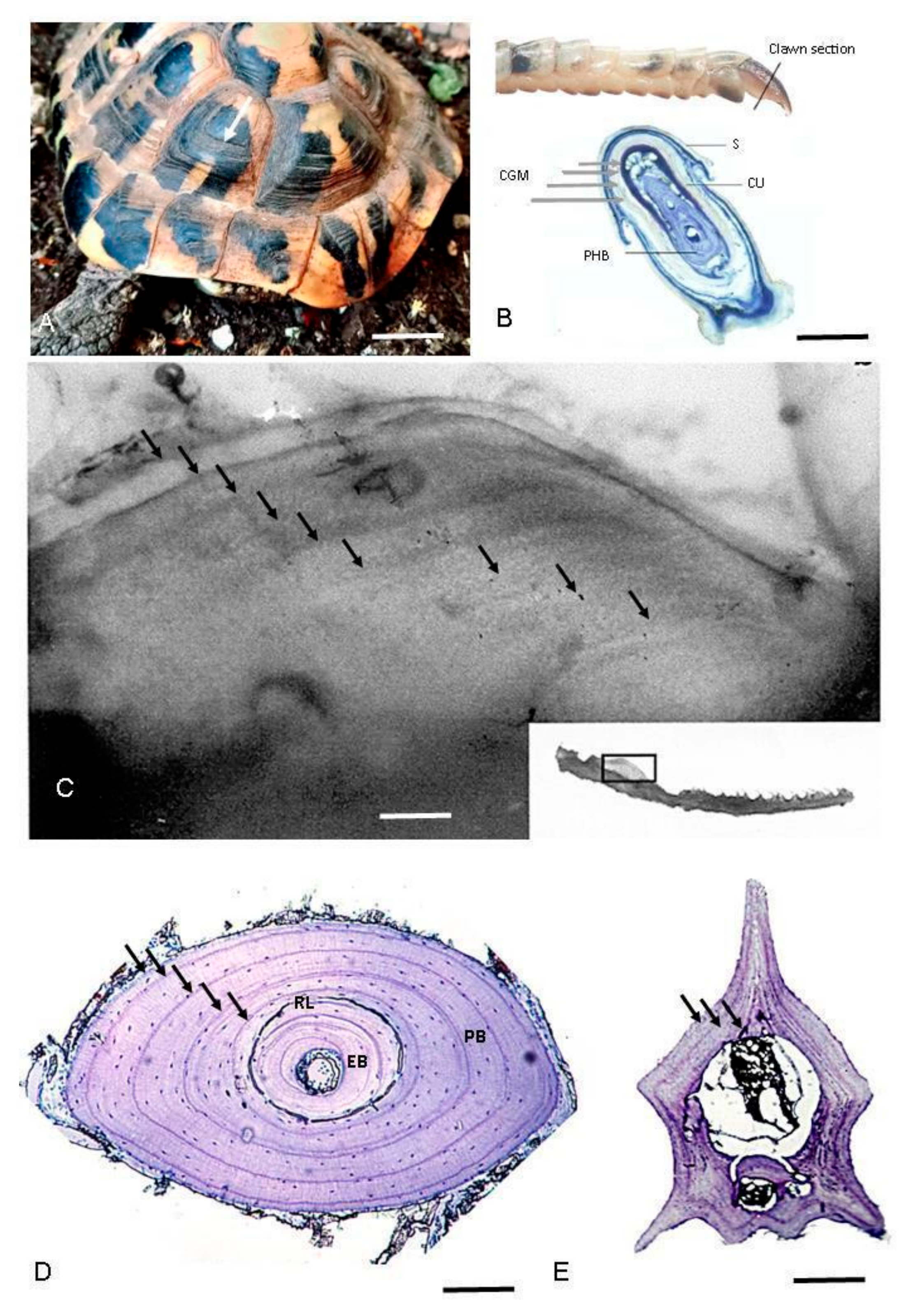
Figure 1. Representative growth marks in hard tissues of amphibians and reptiles. (A) Scute rings in Testudo hermanni; (B) Claw section of Darevskia portschinskii (Squamata, Lacertidae) (modified from [51]); (C) Growth marks seen by transparency (backlighting of the bone) in the mandibular plate from Hierophis viridiflavus (Squamata, Colubridae). (D) Phalanx cross-section of Boehmantis microtympanum (Anura, Mantellidae), decalcified with 5% acid nitric and stained with Mayer’s Hematoxylin; (E) Caudal vertebra cross-section of Hierophis viridiflavus (Squamata, Colubridae), decalcified with 5% acid nitric and stained with Mayer’s Hematoxylin. White arrow points to scute ring (only the first one is indicated); gray arrows point to claw growth marks; black arrows point to LAGs in the bone section. CGM: claw growth marks; EB: endosteal bone; PB, periosteal bone; PHB: phalanx bone; RL: reversal line; S: scale; and U: unguis. Scale bar corresponds to 18 mm (A), 200 µm (B), 380 µm (C), 130 µm (D), and 600 µm (E).
Animals 15 02722 g001
Table 1. Methods for age determination using growth marks applied to the different groups of amphibians and reptiles as classified by traditional (Linnaean) taxonomy. The number in superscript shows a study example in which the technique has been successfully applied for a certain group. Further studies and taxon-specific range of applicability of different bones/mineralized tissue are reported in the main text. T: tested NA: not applicable; NT: not tested although potentially applicable.
Scutes
Growth Marks
Animals 15 02722 i001
Claws
Growth Marks
Animals 15 02722 i002
Skeletochronology
Cranial Bones
Animals 15 02722 i003
Limb Long Bones
Animals 15 02722 i004
Vertebrae
Animals 15 02722 i005
FrogsNANAT 4T 9NT
SalamandersNANAT 5T 10NT
CaeciliansNA NANTNAT 15
TurtlesT 1T 2T 6T 11NT
CrocodilesNTNTT 7T 12NT
LizardsNT T 3T 7T 13T 16
AmphisbeniansNTNANTNANT
SnakesNTNAT 8NAT 17
TuatarasNTNTNTT 14NT
1 [52]; 2 [53]; 3 [54]; 4 [57]; 5 [55]; 6 [65]; 7 [56]; 8 [58]; 9 [59]; 10 [60]; 11 [61]; 12 [62]; 13 [63]; 14 [64]; 15 [66]; 16 Only for short-limbed [67] or limbless lizards [68]; 17 [69].
Disclaimer/Publisher’s Note: The statements, opinions and data contained in all publications are solely those of the individual author(s) and contributor(s) and not of MDPI and/or the editor(s). MDPI and/or the editor(s) disclaim responsibility for any injury to people or property resulting from any ideas, methods, instructions or products referred to in the content.