Article

Natural Infection by Fasciola hepatica in Red Deer (Cervus elaphus) from NW Spain: The Usefulness of Necropsy, Coprology, and Three Enzyme-Linked Immunosorbent Assays for the Diagnosis

by
Sara González Hidalgo
1,
Natividad Diez Baños
1,
María del Rosario Hidalgo Argüello
1,* and
Angelica Martínez-Delgado
2
1
Department of Animal Health, Parasitology and Parasitic Diseases, Faculty of Veterinary Science, University of León, 24071 León, Spain
2
Department of Biotechnology and Food Science, Faculty of Sciences, University of Burgos, 09001 Burgos, Spain
*
Author to whom correspondence should be addressed.
Animals 2025, 15(18), 2649; https://doi.org/10.3390/ani15182649
Submission received: 16 July 2025 / Revised: 26 August 2025 / Accepted: 3 September 2025 / Published: 10 September 2025

Simple Summary

The objective of this study was to investigate the epidemiology of Fasciola hepatica in Cervus elaphus kept in natural conditions in the Riaño Regional Hunting Reserve, north-western of Spain. One hundred red deer were examined and classified according to age, sex, and sampling season. After the necropsy of the animals, the liver was removed and inspected. Faecal samples were collected and processed using the coprological sedimentation technique. The prevalence of this trematode by necropsy was 12%, with a low number of specimens per animal ( x ¯ = 2.7 ± 1.5; range 1–6). The young animals and the males had a higher prevalence with statistically significant differences only according to the host age. Significant variations were also observed when considering the seasons of the year, with the highest number of infected animals in spring. The histopathological study revealed the presence of lesions compatible with chronic fasciolosis. The shedding of F. hepatica eggs was quite low in terms of prevalence (6%) and mean intensity of infection ( x ¯ = 27.3 ± 20.6, range 4–60). The samples were also analysed using excretory/secretory antigens (FhES) and a 2.9 kDa recombinant protein (FhrAPS) used for diagnosis of early and current fasciolosis in livestock. A commercial kit for serodiagnosis of F. hepatica in sheep and cattle, based on a monoclonal antibody (BIO K 211), was also evaluated in red deer. The prevalence of seropositivity of F. hepatica by FhES-ELISA was 32%, by FhrAPS-ELISA 13%, and by BIO K 211, 9%, being higher in adult animals, in males, and in spring.

Abstract

The objective of this study was to investigate the epidemiology of Fasciola hepatica in Cervus elaphus kept in natural conditions in the Riaño Regional Hunting Reserve, north-west of Spain, where several species of domestic and wild animals coexist. One hundred red deer were examined and classified according to age, sex, and sampling season. After the necropsy of the animals, the liver was removed and inspected to obtain the adult parasites of F. hepatica. Faecal samples were collected and processed using the coprological sedimentation technique. The prevalence of this trematode by necropsy was 12%, with a low number of specimens per animal ( x ¯ = 2.7 ± 1.5; range 1–6). The young animals and the males had a higher prevalence than the adults and the females, finding statistically significant differences only according to the host age. Significant variations were also observed when considering the seasons of the year, with the highest number of infected animals in spring. The histopathological study revealed the presence of lesions compatible with a chronic fasciolosis similar to that found in domestic animals. The shedding of F. hepatica eggs was quite low in terms of prevalence (6%) and mean intensity of infection ( x ¯ = 27.3 ± 20.6, range 4–60 epg), being in young animals, in males, and in spring, where the greatest excretion of eggs was observed. These results suggest that the deer are suitable definitive hosts for F. hepatica in the northwest of the Iberian Peninsula, but they are unusual hosts. The serum samples were analysed using a native excretory/secretory antigen (FhES) and a 2.9 kDa recombinant protein (FhrAPS) used for diagnosis of early and current fasciolosis in livestock. A commercial kit for serodiagnosis of F. hepatica in sheep and cattle, based on a monoclonal antibody (BIO K 211), was also evaluated in red deer. The seroprevalence of seropositivity of F. hepatica by FhES-ELISA was 32%, by FhrAPS-ELISA 13%, and by BIO K 211, 9%. In the three serological tests, the seroprevalence obtained was higher in adult animals, in males, and in spring. In the three serological tests used to understand the epidemiology of F. hepatica in red deer, we have observed that the sensitivity was low, perhaps due to the use of an anti-bovine IgG1 as a conjugate, so in future immunodiagnostic tests, it would be more desirable to obtain an anti-deer IgG, probably achieving better results. Due to these results, it is essential to investigate other serological or molecular tests that allow us to know the real importance of F. hepatica in deer and other wild animals. The role of deer as a reservoir of this trematode does not appear to be very important.

1. Introduction

Fasciolosis is a parasitic disease caused by digenean trematodes belonging to the family Fasciolidae and the genus Fasciola. There are three described species, one of them only recorded in the hippopotamus in Zimbabwe, F. nyanzae [1]. The other two species, F. hepatica and F. gigantica, infect livestock and humans but have a different geographical distribution. F. hepatica is present in Europe, Asia, Africa, the Americas, and Oceania in areas where Galba/Fossaria lymnaeids are distributed, whereas F. gigantica appears restricted to Africa except the Maghreb and Asia except the high-altitude areas of the Himalayas. Its absence in other continents is being related to the lack of Radix species [2]. Both species cause a foodborne zoonotic disease with wild aquatic plants contaminated with metacercariae as a source of infection, and it was considered a secondary disease until the end of the 19th century [3]. Since then the situation has changed drastically, becoming an endemic or semi-endemic disease in different parts of the world. Different studies suggest that the number of human cases is increasing [4].
In our country, F. hepatica is the most important species affecting a wide range of hosts, including domestic ruminants (sheep, goats, cows), wild animals (deer, fallow deer, roe deer, Iberian ibex, chamois, mouflon, wild boar), donkeys, horses, rabbits, hares, and humans [5,6]. Its existence is linked to the presence of the semi-aquatic snail Galba truncatula as the main intermediate host [7,8]. The development of its life cycle in the environment includes a metacercarial stage that encysts on pasture and other vegetation and depends on geoclimatic, ecological, and anthropogenic factors such as altitude [9], temperature [10], precipitation, humidity, evapotranspiration [11], vegetation, and soil type [12]. Due to this, its spatial distribution is very uneven, and therefore there are large variations in prevalence [13].
The parasite causes liver damage, locating itself in the bile ducts and gallbladder of the animals. It occurs in three clinical forms—acute, subacute, and chronic—depending on the metacercariae ingested in a given time and the substantial liver damage caused by the migration of large numbers of juvenile fluke, with significant consequences for the health and welfare of the animal, causing great socio-economic impact. Acute infections can be fatal due to loss of liver function, with chronic infections that manifest themselves in anaemia, weight loss, and ill-thrift being more common [14,15,16].
Traditionally, F. hepatica infections are diagnosed by detecting eggs in faeces through coprological sedimentation or flotation techniques, but those techniques have a low sensitivity and can only detect patent infection [17]. In addition, it is difficult to establish a correlation between parasite burden and egg count in faeces. Even so, it remains a valuable diagnostic tool [18]. Infections can also be confirmed at necropsy. It is the safest and most effective method to be able to observe adult parasites and liver lesions.
Other methods for diagnosing include ELISAs testing for coproantigens and antibodies in serum and milk samples. These ELISAs are typically based on native fluke excretory/secretory (ES) products due to their strong immunogenic properties [19]. More recent testing methods have used several antigenic fractions of Fasciola spp., purified antigens and recombinant antigens, for the serodiagnosis of fasciolosis in human and animal species. Cysteine proteases, such as cathepsins, have proved to be good biomarkers of infection, and several tests have been developed to detect antibodies against this family of proteins, cathepsins L being the most used antigens [20,21,22]. Alternatively, animal fasciolosis can be diagnosed by immunological tests that detect coproantigens released by adult flukes living in bile ducts, as the ultrasensitive capture ELISA (MM3 COPRO) using the monoclonal antibody (mAb) MM3, and which is commercialised by Bio-X Diagnostics (La Jemelle, Belgium) [23,24,25]. Other diagnostic methods include molecular genetic techniques such as polymerase chain reaction (PCR) [26,27,28] or the loop-mediated isothermal amplification (LAMP) technique [29,30].
Although fasciolosis has been extensively studied in domestic animals and humans, the application of these methods in wild populations is necessary to elucidate the importance of F. hepatica in these hosts. However, it should be approached with caution because the course of the disease and previous health status with respect to other diseases are unknown [31].
The objectives of this study were (1) to investigate the epidemiology of Fasciola hepatica infection in red deer kept in natural conditions in a mountain area in the north of the province of León (Spain), (2) to know the effect of intrinsic factors, such as age, sex, or sampling seasons, on the F. hepatica infection, and (3) to evaluate and compare necropsy, coprology, and serological tests (excretory/secretory antigens, a recombinant F. hepatica surface antigen, and a commercial kit (BIO K 211) based on a monoclonal antibody) for fasciolosis in red deer under field conditions.

2. Materials and Methods

2.1. Study Area

The present study was carried out in the Riaño Regional Hunting Reserve (43°03′14″ N, 4°57′33.85″ W), Province of León, north-western Spain, and belonging to the “Picos de Europa” National Park in the Cantabrian Mountains (Figure 1). The reserve is a protected area of 78,995 ha, and the greater part of its territory is in the Atlantic region, where the landscape is considerably mountainous, with altitudes between 1200 and 1500 m above sea level. The average monthly temperature is 10.2 °C (−12 °C/36 °C). Winter is long and cold, and summer is short and temperate with an average temperature of 16–17 °C. Rainfall occurs throughout the year, with an average annual precipitation of 1088.6 mm (maximum value in October, minimum in July), and snowfall occurs mainly between November and March [32]. The region has a wide and complex fluvial network, as well as numerous tributaries in the form of mountain streams.
The progressive abandonment of agricultural lands in this area has led to wild animals in the area (red deer, roe deer, chamois, wild boar, and Spanish ibex) sharing pastures and forests with domestic animals, mainly sheep and goats “farming in notable declive”, and cattle and horses raised for meat.

2.2. Animal Sampling

A total of 100 red deer were culled by the technical staff of the Reserve in selective hunting. For management reasons, it was not possible to obtain samples during the summer months. The animals sampled were classified according to their sex, age groups: young ≤ 2 years old and adults > 2 years old and sampling seasons (winter, spring and autumn), (Table 1).

2.3. Parasitological Examination

Once the animals were culled, and upon arrival at the faculty, they were identified and transferred to an enable room where a systematic and complete necropsy was performed. After accessing the abdominal cavity through the linea alba, the liver was extracted, first performing a macroscopic inspection of the entire surface of the organ. Next, the bile ducts and portal vein were opened, and the parenchyma was filleted into pieces about 1–3 cm thick. The pieces were pressed to facilitate the release of the trematodes, which were collected, washed with physiological solution, enumerated, and preserved in 70% ethanol at −20 °C for subsequent studies. The areas of the liver with representative lesions were preserved in 10% buffered formalin. After 48 h of fixation, they were paraffin-embedded, and tissue sections of 3 µm were stained with hematoxylin-eosin for histopathological study.
Blood samples were taken by cardiac puncture or from the thoracic cavity. Sera were obtained by centrifugation, placed in vials, labelled, and preserved at −20 °C until used. Also, faecal samples were collected directly from the rectum of the red deer and, after being labelled and identified, were immediately processed using the coprological sedimentation technique [33]. The samples were examined under a light microscope to detect and quantify F. hepatica eggs, which were expressed as the number of eggs per gram of faeces (epg).

2.4. Serological Procedures

Adult F. hepatica parasites collected from cattle livers at the slaughterhouse were washed several times in phosphate-buffered saline (0.1 mol/L PBS, pH 7.5) and finally incubated in RPMI culture medium [34,35]. Subsequently, the antigens obtained were dialyzed, lyophilized, and maintained as FhES (F. hepatica).
The presence of specific antibodies (IgG) against F. hepatica was investigated by enzyme-linked immunoassays (ELISA) using different antigens that had previously been tested in domestic ruminants experimentally and naturally infected [36,37]. The protocol was adapted to deer to detect the exposure against F. hepatica. The optimal concentrations of antigen, serum, and conjugate were previously determined by experiments using serum samples from deer that were positive and negative, by sedimentation, and by necropsy methods.
ELISA using excretory/secretory antigens of F. hepatica (FhES) was used at 1 μg ml−1 concentration, sera diluted (1/100) in PBS containing 0.05% Tween and 1% skimmed milk, and then anti-deer IgG peroxidase-labelled (Sigma Aldrich, St. Loius, MO, USA) was added and tested at 1/1000 [19].
Detection of IgG antibodies against the 2.9 kDa recombinant surface protein (APS) was performed in U-bottom microtiter plates coated with 3 μg ml−1 FhrAPS, sera diluted at 1/100, and anti-deer IgG peroxidase-labelled tested at 1/1000 [38,39].
The optical densities (OD) were read at 492 nm in an ELISA reader (Bio-Rad, Inc., Hercules, CA, USA, model 680 XR). Negative control sera were obtained from 5 young red deer that did not have flukes at necropsy and were negative for sedimentation. In order to establish the cut-off point, positive values were the mean optical density (OD) of negative sera plus 2 standard deviations (SD) [40,41]. Positive absorbance values were established in ≥0.1483 (FhES) and ≥0.1689 (FhrAPS).
Also, circulating IgG was investigated with a commercial test, “ELISA Kit for serodiagnosis of F. hepatica in bovine and ovine samples” (BIO K 211, Bio-X Diagnostics, La Jemelle, Belgium). This test uses as a conjugate a peroxidase-labelled anti-bovine IgG1 manufacturer and includes the interpretation of the results.

2.5. Statistical Analysis

The prevalence and mean intensity of the infection were determined considering host age, sex, and seasons of sampling [42]. The chi-square non-parametric test (χ2) was used to determine significant differences in prevalence, and the Mann–Whitney and Kruskal–Wallis tests were used to compare the intensity, taking different variables (host sex, age, and seasons) into account. The 95% confidence intervals (CI) for prevalence and mean intensity were calculated [43]. The sensitivity and specificity of the indirect ELISA were calculated according to the following formulas: sensitivity = true positive/true positive + false negative and specificity = true negative/false positive + true negative. The level of agreement among the ELISA methods, the necropsy, and/or coprology was assessed by the Kappa index (κ). Correlations between OD values and the number of flukes were assessed using Spearman’s rank correlation coefficient (ρ). Statistical significance was determined at the p ≤ 0.05 level. The statistical analysis was performed using the SPSS 18.0 software package (SPSS, Inc., Chicago, IL, USA).

3. Results

3.1. Necropsy

The prevalence of adult trematodes of F. hepatica obtained by necropsy was 12% (95% CI: 5.6–18.4%) with a mean intensity of 2.7 ± 1.6, 95% CI: 2.4–3, range 1–6 flukes/infected animal. The young animals had a higher prevalence (20%, 95% CI: 2.6–37.4%) than the adults (10%, 95% CI: 3.3–16.7), and the males (15.4%, 95% CI: 1.5–29.3%) had a higher prevalence than the females (10.8%, 95% CI: 3.8–17.8%), finding statistically significant differences only according to the host age (χ2 = 4.082, p = 0.043).
Significant variations were observed when considering the seasons of the year (χ2 = 6.270, p = 0.044), with the highest number of infected animals in spring (22.8%; 95% CI: 9–36) decreasing sharply in winter (7.9%, 95% CI: 1–16%) and autumn (3.7%, 95% CI: 1–11%). Regarding the mean intensity of infection, very low values were observed in all the parameters analysed, with no statistically significant differences found between them (Table 1).
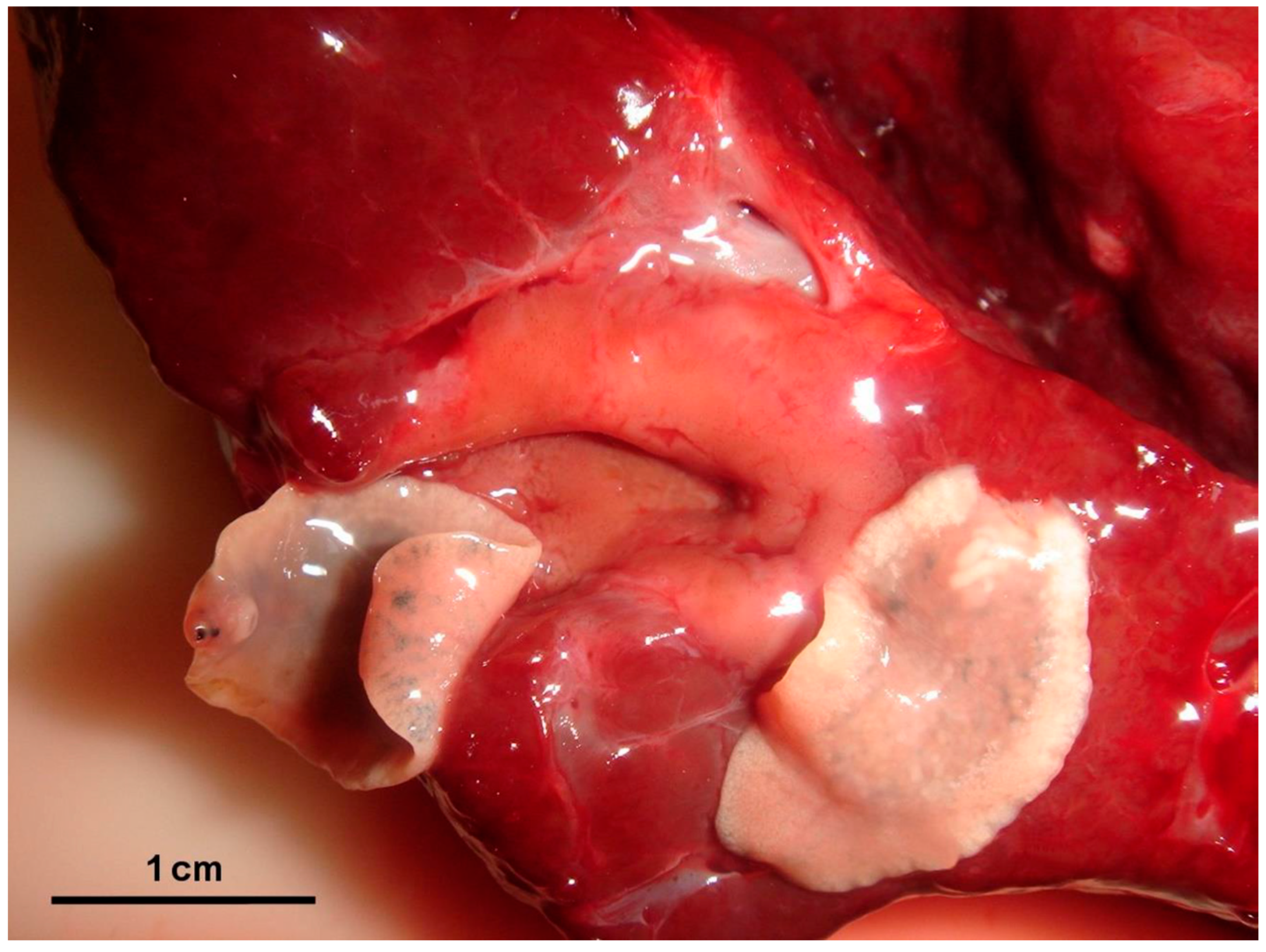
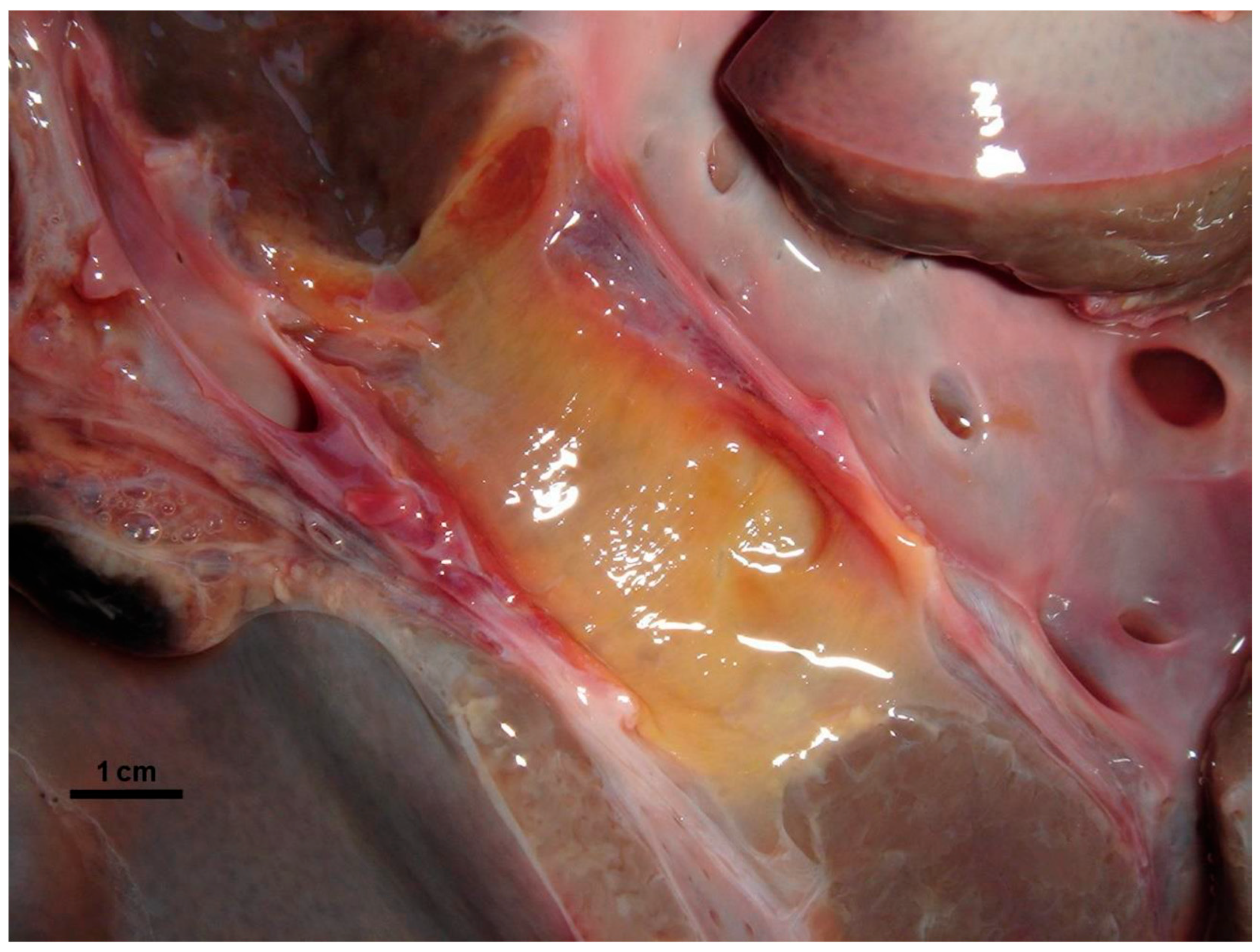
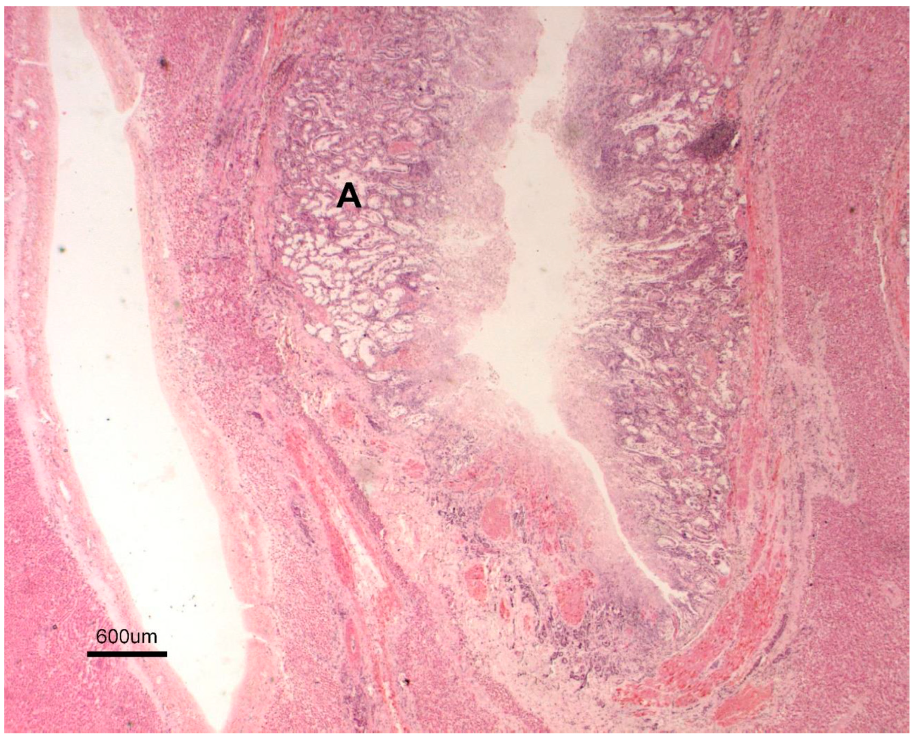
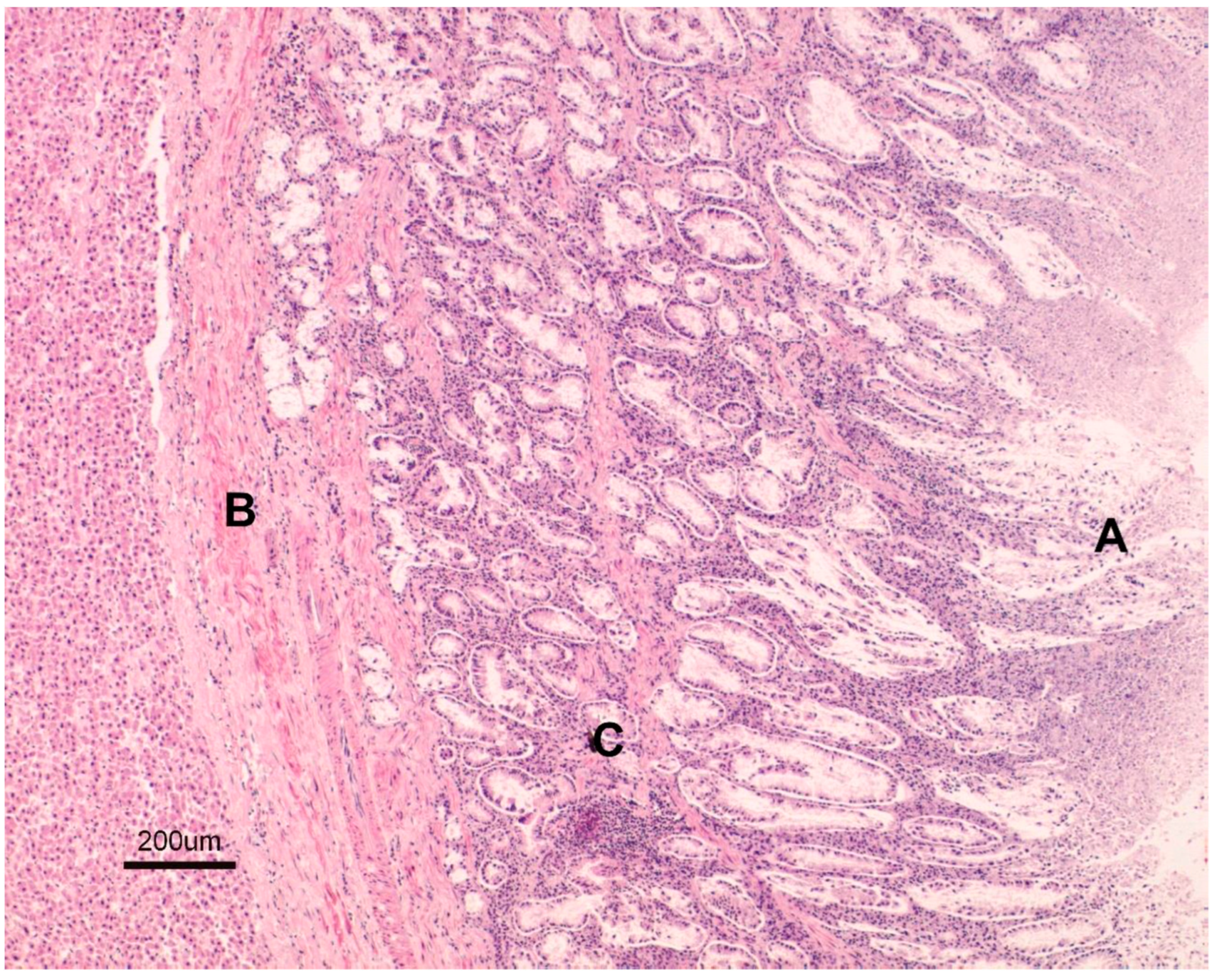
Gross lesions were characterised by changes produced by the adult trematodes in the intrahepatic bile ducts, which had thickened walls and dilated lumens as they contained some parasites (Figure 2). Large areas of fibrosis were also observed in the liver parenchyma, calcification, and encapsulation of flukes. Furthermore, in the inner part of the hepatic lymph nodes, a blackish colour, associated with the presence of iron-porphyrin pigments, was observed (Figure 3). The main histopathological lesions were observed in the septal bile ducts and were characterised by chronic cholangitis and cholangiectasis (Figure 4). Affected bile ducts showed a moderate-severe epithelial hyperplasia with diffuse lymphocytic infiltration (Figure 5).

3.2. Coprology

The shedding of F. hepatica eggs was quite low in terms of prevalence (6%, 95% CI: 1.4–10.6) and mean intensity of infection ( x ¯ = 27.3 ± 20.6, 95% CI: 23.2–31.4, range 4–60). Males (7.7%, 95% CI: 1–17.9%) and young animals (10%, 95% CI: 1–23.1%) had the highest prevalence, while females ( x ¯ = 28.5 ± 23.6, 95% CI: 23.1–33.9) and adult animals ( x ¯ = 32.5 ± 22.2; 95% CI: 27.6–37.4) had the highest mean intensity of infection. Regarding the seasons of the year, the highest prevalence and mean intensity of infection were observed in spring, with significant differences when comparing with the autumn (χ2 = 1.27, p = 0.053), but not with the winter values (p ≥ 0.05) (Table 1).
We must point out that only three deer harboured adult worms and excreted eggs in their faeces.

3.3. Serology

The prevalence of seropositivity of F. hepatica by FhES-ELISA was 32% (95% CI: 23–41%), by FhrAPS-ELISA 13% (95% CI: 6–20%), and by BIO K 211, 9% (95% CI: 3–15%). In the three serological tests, the seroprevalence obtained was higher in adult animals (11.3–36.3%) than in young (0–15%) and in males (15.4–42.3%) than in females (6.9–28.4%). Only significant differences were observed between adult and young animals tested with the FhES-ELISA (p ≤ 0.05). If we consider seasonal sampling, with the three tests analysed, the seroprevalence was higher in spring than in the rest of the seasons, but no statistically significant differences were found (Table 1).
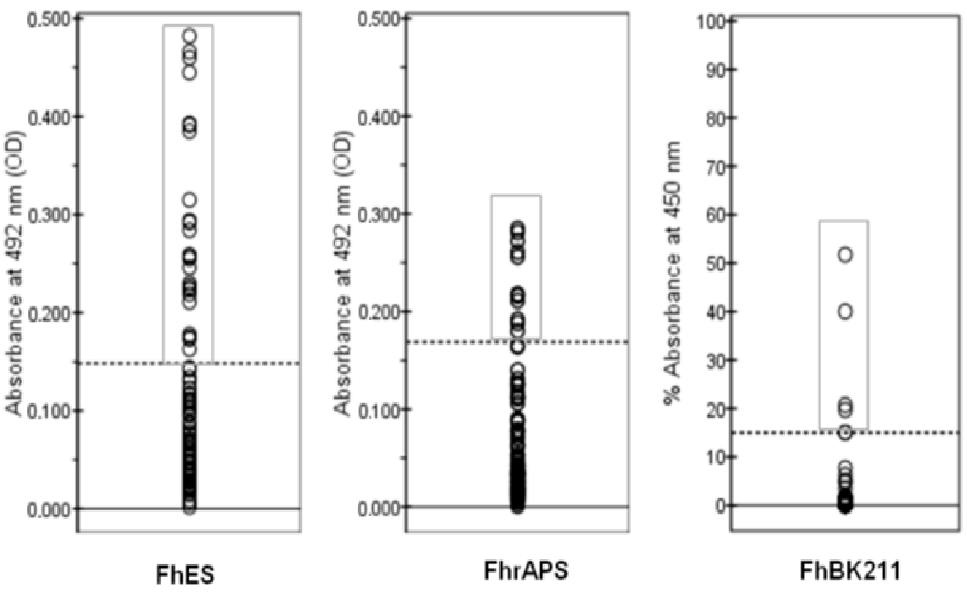
When comparing the results between the three methods evaluated and the necropsy, it was observed that the highest number of false positives was detected with the FhES-ELISA (n = 25), and the highest number of false negatives was observed by both FhrAPS-ELISA (n = 10) and BIO K 211 (n = 9) methods (Figure 6). Five animals presented flukes in the liver and were negative by all three ELISA methods. The number of flukes correlated significantly with OD values only in the case of BIO K 211-ELISA (ρ = 0.675; p = 0.016).
Regarding the results obtained for sensitivity (Table 2), it was observed that its highest value was obtained for the FhES-ELISA (58.33%), followed by the BIO K 211-ELISA (25%) and the FhrAPS-ELISA (16.67%). The highest specificity and positive likelihood ratio (PLR) were obtained with the BIO K 211-ELISA. The concordance among the necropsy and the three ELISA methods evaluated was low: κ = 0.040 for FhrAPS-ELISA, κ = 0.174 for FhES-ELISA, and κ = 0.184 for BIO K 211-ELISA. The highest level of agreement was obtained between the FhES-ELISA and the BIO K 211-ELISA (κ = 0.326, p < 0.001).
The results obtained when comparing the three ELISAs studied and the coprology were also analysed. In the results obtained for sensitivity, it was observed that its highest value was obtained for the FhES-ELISA (66.67%), followed by the BIO K 211-ELISA (33.33%) and the FhrAPS-ELISA (16.67%); however, the specificity was much higher with the BIO K 211-ELISA than with the rest of the ELISAs analysed. The concordance between the three ELISAs and the coprology was very low, as can be seen from the results of the Kappa index (κ) (Table 3).

4. Discussion

The present study has detected the presence of F. hepatica in deer in a mountainous area in northern Spain where wild animals share their habitat with domestic livestock in a traditional free-range grazing system. Its knowledge is a fact of general biological interest from the veterinary and public health point of view.
The prevalence and intensity of infection found in this work has been low, both that obtained through necropsy (12%, x ¯ = 2.7 ± 1.5 flukes/infected animal) and that of the eggs faecal counts (6%, x ¯ = 27.3 ± 20.6 epg), although considerably higher than that observed in previous studies on this host: 7.27% flukes by necropsy, 3.64% eggs by coprology in this same study area [44], 1.64% adult flukes, 1.19% egg count in the central area of Spain [45], or 7% adult flukes in the south [19]. They were also very different from those found in other European deer populations, ranging between 0% in Germany [46], 7% in Ireland [47], 27.7% (flukes), 12.3% (faeces) in Scotland [31], or 31.3% in Belarus [48]. These large variations observed in the prevalence of infection are mainly due to environmental factors (topography, geochemistry, climate, and land use) that directly intervene in the development of the biological cycle of F. hepatica and the risk of infection, so they will be different in each of the study areas [49]. In addition to these factors, we must take into account susceptibility to infection in the different species of wildlife hosts. Thus, in our country, a low prevalence, 1.87% flukes and 0.53% eggs, was found in Iberian ibex in the Sierra Nevada mountains [5]; in roe deer, 1.87% flukes and 0.25% eggs; and in chamois, 0% flukes 0.39% eggs in our study area [50]; in 2% of fallow deer sampled in the south [19]; and in 6.1% of chamois in the Pyrenees [51]. These data contrast with the significant increase in prevalence obtained in domestic livestock since the last decade of the 19th century [52,53,54,55].
With both techniques we observed that the prevalence was higher in young animals and in males, while adult animals and females presented the highest intensity of infection. This same pattern has been observed in other parasitisms studied in wild animals in the same sampling area, where age would be related to the development of the hosts’ immune system, sex with the hormonal differences between them, different feeding habitats, land use, or physiological variations [56,57]. Regarding the seasons, the highest prevalence and intensity of infection were observed in spring, which correlates with the chronobiological pattern of F. hepatica in temperate areas [58,59].
The presence of trematodes in the inspection of the liver is 100% specific but does not always present the same sensitivity. This may be due to the pathogenesis caused by the parasite, with the formation of large areas of fibrosis in the liver parenchyma and bile duct walls. This results in significant calcification, leading to encapsulation of the flukes, preventing their migration through the bile ducts. This would result in fewer flukes reaching the bile ducts, preventing their sexual maturity; fewer adults detected at necropsy; and fewer eggs excreted in the faeces. This fact has been reported by various authors in deer and fallow deer [31,60]. The lesions found in the liver of this host are consistent with chronic fasciolosis described in domestic ruminants and other species [61,62,63]. Like necropsy, the coprological sedimentation technique for egg counting is highly specific but has low sensitivity. In addition to the above mentioned, this technique depends on a wide variety of factors, such as the faecal production of the animal, the volume of the sample analysed, the number of adult parasites present in the host, or the migration of immature stages and therefore the non-elimination of eggs. Furthermore, it must be taken into account that there are hourly, daily, and weekly variations in the elimination of eggs, especially in mild infections such as in deer [64,65,66].
When analysing both methods together, we found great disparity in results, and there is no correlation between the number of adults found and the egg count in the faeces. Nor is it known the number of eggs that adult parasites can eliminate or the longevity of F. hepatica in its hosts. However, from an epidemiological point of view, the use, especially of the coprological technique, is effective in knowing the parasitological status of certain hosts in a given area and is the technique used initially in the diagnosis of any parasitic process.
The data obtained in this study confirm that red deer, like other wild animals, are suitable definitive hosts for F. hepatica (they can complete their biological cycle), but they are unusual hosts (low rates of infection and partially inhibited development) [56,67]. For this reason, some authors state that their role as reservoirs can be reduced and mediated by ecological barriers that limit exposure and/or inherent resistance to infection [51,68], while others maintain that red deer could have an important role in the spreading of F. hepatica in areas shared with domestic ruminants [69].
Immunological techniques have been used successfully in the diagnosis of fasciolosis due to their high sensitivity, but studies of these techniques and their comparison with necropsy or the shedding of eggs in faeces are scarce. This study evaluates three ELISA methods for the detection of IgG antibodies in serum samples for the diagnosis of F. hepatica infection in red deer using excretory/secretory antigens, a recombinant F. hepatica surface antigen, and a commercial kit (BIO K 211) based on a monoclonal antibody. Many of these techniques have been tested in experimental infections in domestic animals, but it has been proven that the sensitivity and specificity of several of these methods decrease considerably under field conditions [36,38,65], and the values observed in the present study agree with these observations.
Thus, the 2.9 kDa recombinant protein had a sensitivity of 16.67% in the present work, while its values in sheep under experimental conditions were considerably higher (91%) [70], but these values decrease considerably (67.7%) in sheep naturally exposed to F. hepatica [36]. However, the sensitivity and specificity of the ELISA based on excretory/secretory antigens, 58% and 72% in the present study, respectively, showed higher values than those from sheep under field conditions (52.5%, 47.8%) but lower than those from sheep experimentally infected (86%, 97.5%) [36]. The monoclonal antibody is 100% sensitive and 100% specific for detecting early and long-term fasciolosis in sheep [24,71], values much higher than those found in this study.
Considering wild ruminants, the only comparable study was performed in Spain [17,39], analysing for the first time the presence of antibodies against excretory/secretory antigens of F. hepatica in these hosts. Our results were similar to those found in the red deer (34%), lower than those found in the fallow deer (58%), but higher than those observed in mouflon (12%), Iberian ibex (15%) from Southern Spain, and roe deer (25%) from Galicia. This is probably because red deer and fallow deer have been found to be more suitable hosts to F. hepatica infections than other wild ruminants due to their ecology and behaviour, despite controversy about their role in the epidemiology of this parasite.
Regarding factors such as age or sex, unlike what was observed in the necropsy and in the shedding of eggs in faeces, the seroprevalence was higher in adult animals and in males, which would be related mainly to ecological and physiological differences between them [57,72].
Several false positive results have been observed with the FhES-ELISA, and this seems to indicate the possibility of cross-reactions with antigens present in faeces common to other trematodes such as Dicrocoelium dendriticum and Paramphistomidae parasites [73,74,75,76]. On the other hand, some of these false positive results could correspond to primo-infected animals with juvenile flukes migrating through the intestinal wall and peritoneal cavity or animals that were previously exposed to the parasite but in which the infection has not been successfully established since the presence of antibodies do not always correlate with the existence of active infection but only with exposure to the parasite [77,78].
Also, the number of false negatives was higher with the recombinant protein and the monoclonal antibody than with the excretory/secretory antigens, despite the fact that the first two antigens are highly sensitive to detecting current and long-term sheep fasciolosis, even in animals infected with low numbers of flukes [70,71], or the discovery of five animals that harboured flukes and that were negative to the three assays, suggesting that other causes may be impairing the normal functioning of the immune system in these animals.

5. Conclusions

The present study has detected the presence of F. hepatica in deer in a mountainous area in northern Spain where wild animals share their habitat with domestic livestock in the area in a traditional free-range grazing system.
Regarding the diagnosis, we have observed that there was no correlation between the coprological technique and the necropsy findings, while the serological tests showed great variability of results both in prevalence and in sensitivity and specificity, so it would be essential to investigate other serological or molecular tests that allow us to know the real importance of F. hepatica in deer and other wild animals.
The ecological conditions of this area favour the natural development of the F. hepatica cycle, and it is confirmed that the deer are suitable definitive hosts for this trematode. However, the low results obtained in terms of prevalence and intensity of infection, number of eggs excreted in faeces and seroprevalence seem to indicate that the pastures shared by domestic and wild ruminants are little contaminated with the infective phase of Fasciola hepatica from deer, and their role as reservoir and transmission to domestic livestock does not appear to be very important. It is not known whether other wild animals could act as more suitable hosts for maintaining the disease.

Author Contributions

S.G.H.: Investigation; methodology; resources; writing—original draft; writing—review and editing. N.D.B.: Investigation; methodology; supervision; validation; writing—original draft; writing—review and editing. M.d.R.H.A.: Funding acquisition; investigation; methodology; project administration; resources; supervision; writing—review and editing. A.M.-D.: Investigation; methodology; supervision; validation; visualisation; writing—review and editing. All authors have read and agreed to the published version of the manuscript.

Funding

This study was supported by a Research Project 2004-02580-C03-03/BOS from the Spanish “Ministerio de Ciencia y Tecnología”.

Institutional Review Board Statement

Ethical review and approval was waived for this study because it did not involve live animals. The animals were culled by the technical staff of the Riaño Regional Reserve in selective hunting.

Informed Consent Statement

Not applicable.

Data Availability Statement

The data that support the findings of this study are included in the article.

Acknowledgments

We are sincerely grateful to the Junta de Castilla y León for providing animals and to all the forestry staff of the “Riaño Regional hunting Reserve” by its cooperation, especially to J.J. Martínez, of Wildlife Service in León province. (English revision by M.C.U. and P.C.U. in acknowledgement).

Conflicts of Interest

The authors declare no conflicts of interest.

References

  1. Schols, R.; Carolus, H.; Hammoud, C.; Muzarabani, K.C.; Barson, M.; Huyse, T. Invasive snails, parasite spillback, and potential parasite spillover drive parasitic diseases of Hippopotamus amphibius in artificial lakes of Zimbabwe. BMC Biol. 2021, 19, 160–181. [Google Scholar] [CrossRef]
  2. Mas-Coma, S.; Valero, M.A.; Bargues, M.D. Fasciola, Lymnaeids and Human Fascioliasis, with a Global Overview on Disease Transmission, Epidemiology, Evolutionary Genetics, Molecular Epidemiology and Control. Adv. Parasitol. 2009, 69, 141–146. [Google Scholar]
  3. Chen, M.G.; Mott, K.E. Progress in assessment of morbidity due to Fasciola hepatica infection, a review of recent literature. Trop. Dis. Bull. 1990, 87, 1–38. [Google Scholar]
  4. WHO. Foodborne Trematode Infections, World Health Organization. Available online: https://www.who.int/news-room/fact-sheets/detail/foodborne-trematode-infections (accessed on 23 February 2024).
  5. Alasaad, S.; Li, Q.Y.; Lin, R.Q.; Martín-Atance, P.; Granados, J.E.; Díez-Baños, P.; Pérez, J.M.; Zhu, X.Q. Genetic variability among Fasciola hepatica samples from different host species and geographical localities in Spain revealed by the novel SRAP marker. Parasitol. Res. 2008, 103, 181–186. [Google Scholar] [CrossRef]
  6. Barges, M.D.; Artiga, P.; Khoubbane, M.; Ortiz, P.; Naquira, C.; Mas-Coma, S. Molecular characterisation of Galba truncatula, Lymnae neotrofica and L. schirazensis from Cajamarca, Peru and their potential role in transmission of human and animal fascioliasis. Parasi. Vectors 2012, 5, 174. [Google Scholar] [CrossRef] [PubMed]
  7. Manga-González, M.Y.; González-Lanza, C.; González-Warleta, M.; Miñambres, B.; Carro-Corral, C.; Iglesias, J.; Castillejo, J.; Mezo, M. Infection kinetic of Galba truncatula (Mollusca, Basommatophora) by Fasciola hepatica and Calicophoron daubneyi (Trematoda, Digenea) in Galicia (Spain). Acta Parasitol. Portuguesa. 2009, 16, 118–119. [Google Scholar]
  8. Correa, A.C.; Escobar, J.S.; Durand, P.; Renaud, F.; David, P.; Jarne, P.; Pointier, J.P.; Hurtrez-Boussés, S. Bridging gaps in the molecular phylogeny of the Lymnaeidae (Gastropoda: Pulmonata), vectors of Fascioliasis. BMC Evol. Biol. 2010, 10, 381. [Google Scholar] [CrossRef]
  9. Alves, D.P.; Carneiro, M.B.; Martins, I.V.F.; Bernardo, C.C.; Donatele, D.M.; Pereira Junior, O.S.; Almeida, B.R.; Avelar, B.R.; Leao, A.G.G. Distribution and factors associated with Fasciola hepatica infection in cattle in the south of Espirito Santo State, Brasil. J. Venom. Anim. Toxins Incl. Trop. Dis. 2011, 17, 271–276. [Google Scholar]
  10. Howell, A.K.; Williams, D.J.L. The epidemiology and control of liver flukes in cattle and sheep. Vet. Clin. N. Am. Food Anim. Pract. 2020, 36, 109–123. [Google Scholar] [CrossRef] [PubMed]
  11. Stuen, S.; Ersdal, C. Fasciolosis-An increasing challenge in the sheep industry. Animals 2022, 12, 1491. [Google Scholar] [CrossRef]
  12. Urquart, G.M.; Armour, J.; Duncan, J.L.; Dunn, A.M.; Jennings, F.W. Veterinary Parasitology, 2nd ed.; Blackwell Science Ltd.: Oxford, UK, 1996. [Google Scholar]
  13. Beesley, N.J.; Caminade, C.; Charlier, J.; Flynn, R.J.; Hodkinson, J.E.; Martinez-Moreno, A.M.; Martinez-Valladares, M.; Perez, J.; Rinaldi, L.; Williams, D.J.L. Fasciola and fasciolosis in ruminants in Europe: Identifying research needs. Emerg. Dis. 2018, 65, 199–216. [Google Scholar] [CrossRef]
  14. Howell, A.; Baylis, M.; Smith, R.; Pinchbeck, G.; Williams, D. Epidemiology and impact of Fasciola hepatica exposure in high-yielding dairy herds. Prev. Vet. Med. 2015, 121, 41–48. [Google Scholar] [CrossRef] [PubMed]
  15. Mazeri, S.; Rydevik, G.; Handel, I.; Bronsvoort, B.M.d.C.; Sargison, N. Estimation of the impact of Fasciola hepatica infection on time taken for UK beef cattle to reach slaughter weight. Sci. Rep. 2017, 7, 7319. [Google Scholar] [CrossRef] [PubMed]
  16. Charlier, J.; Rinaldi, L.; Musella, V.; Ploeger, H.W.; Chartier, C.; Vineer, H.R.; Hinney, B.; Von Samson-Himmelstjerna, G.; Băcescu, B.; Mickiewicz, M.; et al. Initial assessment of the economic burden of major parasitic helminth infections to the ruminant livestock industry in Europe. Prev. Vet. Med. 2020, 182, 105103. [Google Scholar] [CrossRef]
  17. Charlier, J.; Vercruysse, J.; Morgan, E.; Van Dijk, J.; Williams, D.J.L. Recent advances in the diagnosis, impact on production and prediction of Fasciola hepatica in cattle. Parasitology 2014, 141, 326–335. [Google Scholar] [CrossRef]
  18. Mazeri, S.; Sargison, N.; Kelly, R.F.; Bronsvoort, B.M.d.C.; Handel, I. Evaluation of the performance of five diagnostic tests for Fasciola hepatica infection in naturally infected cattle using a bayesian no gold standard approach. PLoS ONE 2016, 11, e0161621. [Google Scholar] [CrossRef]
  19. Arias, M.S.; Martínez-Carrasco, C.; León-Vizcaíno, L.; Paz-Silva, A.; Díez-Baños, P.; Morrondo, P.; Alonso, F. Detection of antibodies in wild ruminants to evaluate exposure to liver trematodes. J. Parasitol. 2012, 98, 754–759. [Google Scholar] [CrossRef] [PubMed]
  20. Gonzales Santana, B.; Dalton, J.P.; Vasquez Camargo, F.; Parkinson, M.; Ndao, M. The diagnosis of human fascioliasis by enzyme-linked immunosorbent assay (ELISA) using recombinant Cathepsin L protease. PLoS Negl. Trop. Dis. 2013, 7, e2414. [Google Scholar] [CrossRef]
  21. Martínez-Sernández, V.; Perteguer, M.J.; Hernández-González, A.; Mezo, M.; González-Warleta, M.; Orbegozo-Medina, R.A.; Romarís, F.; Paniagua, E.; Gárate, T.; Ubeira, F.M. Comparison of recombinant cathepsins L1, L2, and L5 as ELISA targets for serodiagnosis of bovine and ovine fascioliasis. Parasitol. Res. 2018, 117, 1521–1534. [Google Scholar] [CrossRef]
  22. Siles-Lucas, M.; Becerro-Recio, D.; Serrat, J.; González-Miguel, J. Fascioliasis and fasciolopsiasis: Current knowledge and future trends. Res. Vet. Sci. 2021, 124, 27–35. [Google Scholar] [CrossRef]
  23. Mezo, M.; González-Warleta, M.; Carro, C.; Ubeira, F.M. An ultrasensitive capture ELISA for detection of Fasciola hepatica coproantigens in sheep and cattle using a new monoclonal antibody (MM3). J. Parasitol. 2004, 90, 845–852. [Google Scholar] [CrossRef] [PubMed]
  24. Mezo, M.; Gonzalez-Warleta, M.; Ubeira, F.M. The use of MM3 monoclonal antibodies for the early immunodiagnosis of ovine fascioliasis. J. Parasitol. 2007, 93, 65–72. [Google Scholar] [CrossRef]
  25. Mezo, M.; González-Warleta, M.; Castro-Hermida, J.A.; Martínez-Sernández, V.; Ubeira, F.M. Field evaluation of the enhanced MM3-COPRO ELISA test for the diagnosis of Fasciola hepatica infection in sheep. PLoS ONE 2022, 17, e0265569. [Google Scholar] [CrossRef]
  26. Marcilla, A.; Bargues, M.D.; Mas-Coma, S. A PCR–RFLP assay for the distinction between Fasciola hepatica and Fasciola gigantica. Mol. Cell. Probes 2002, 16, 327–333. [Google Scholar] [CrossRef]
  27. Martínez-Pérez, J.M.; Robles-Pérez, D.; Rojo-Vázquez, F.A.; Martínez-Valladares, M. Comparation of three different techniques to diagnose Fasciola hepatica infection in experimentally and naturally infected sheep. Vet. Parasitol. 2012, 190, 80–86. [Google Scholar] [CrossRef]
  28. Ai, L.; Dong, S.J.; Zhang, W.Y.; Elsheikha, H.M.; Mahmmod, Y.S.; Lin, R.Q.; Yuan, Z.G.; Shi, Y.L.; Huang, W.Y.; Zhu, X.Q. Specific PCR-based assays for the identification of Fasciola species: Their development, evaluation and potential usefulness in prevalence surveys. Ann. Trop. Med. Parasitol. 2013, 104, 65–72. [Google Scholar] [CrossRef]
  29. Notomi, T.; Okayama, H.; Masubuchi, H. Loop-mediated isothermal amplification of DNA. Nucleic Acids Res. 2000, 28, E63. [Google Scholar] [CrossRef]
  30. Nagamine, K.; Hase, T.; Notomi, T. Accelerated reaction by loop-mediated isothermal amplification using loop primers. Mol. Cell. Probes 2002, 16, 223–229. [Google Scholar] [CrossRef] [PubMed]
  31. French, A.S.; Zadoks, R.N.; Skuce, P.J.; Mitchell, G.; Gordon-Gibbs, D.K.; Craine, A.; Shaw, D.; Gibb, S.W.; Taggart, M.A. Prevalence of liver fluke (Fasciola hepatica) in wild red deer (Cervus elaphus): Coproantigen ELISA is a practicable alternative to faecal egg counting for surveillance in remote populations. PLoS ONE 2016, 11, e0162420. [Google Scholar] [CrossRef] [PubMed]
  32. AEMET, State Meteorology Station. Ministry of the Environment and Rural and Marine Management. 2024. Available online: https://www.aemet.es/es/portada (accessed on 20 February 2025).
  33. MAFF, Ministry of Agriculture, Fisheries and Food. Manual of Veterinary Parasitological Laboratory Techniques; H. M. Stationery Office: London, UK, 1986; p. 160.
  34. Sánchez-Andrade, R.; Paz-Silva, A.; Suárez, J.L.; Panadero, R.; Díez-Baños, P.; Morrondo, P. Use of a sandwich-enzyme-linked immunosorbent assay (SEA) for the diagnosis of natural Fasciola hepatica infection in cattle from Galicia (NW Spain). Vet. Parasitol. 2000, 93, 39–46. [Google Scholar] [CrossRef]
  35. Sánchez-Andrade, R.; Paz-Silva, A.; Suárez, J.L.; Panadero, R.; Pedreira, J.; López, C.; Díez-Baños, P.; Morrondo, P. Influence of age and breed on natural bovine fasciolosis in an endemic area (Galicia, NW Spain). Vet. Res. Commun. 2002, 26, 361–370. [Google Scholar] [CrossRef]
  36. Arias, M.; Morrondo, P.; Hillyer, G.V.; Sánchez-Andrade, R.; Suárez, J.L.; Lomba, C.; Pedreira, J.; Díaz, P.; Díez-Baños, P.; Paz-Silva, A. Immunodiagnosis of current fasciolosis in sheep naturally exposed to Fasciola hepatica by using a 2.9 kDa recombinant protein. Vet. Parasitol. 2007, 146, 46–49. [Google Scholar] [CrossRef] [PubMed]
  37. Arias, M.S.; Piñeiro, P.; Hillyer, G.V.; Suárez, J.L.; Francisco, I.; Cortiñas, F.J.; Díez-Baños, P.; Morrondo, P.; Sánchez-Andrade, R.; Paz-Silva, A. An approach of the laboratory to the field: Assessment of the influence of cattle management on the seroprevalence of fascioliasis by using polyclonal- and recombinant-based ELISAs. J. Parasitol. 2010, 96, 626–631. [Google Scholar] [CrossRef]
  38. Paz-Silva, A.; Hillyer, G.V.; Sánchez-Andrade, R.; Rodríguez Medina, J.R.; Arias, M.S.; Morrondo, P.; Díez-Baños, P. Isolation, identification and expression of a Fasciola hepatica cDNA encoding a 2.9-kDa recombinant protein for the diagnosis of ovine fasciolosis. Parasitol. Res. 2005, 95, 129–135. [Google Scholar] [CrossRef]
  39. Arias, M.S.; Piñeiro, P.; Sanchez-Andrade, R.; Suárez, J.L.; Hillyer, G.V.; Díez-Baños, P.; Paz-Silva, A.; Morrondo, P. Relationship between exposure to Fasciola hepatica in roe deer (Capreolous capreolous) and cattle extensively reared in an endemic area. Res. Vet. Sci. 2013, 95, 1031–1035. [Google Scholar] [CrossRef]
  40. Paz-Silva, A.; Sánchez-Andrade, R.; Suárez, J.L.; Pedreira, J.; Arias, M.S.; López, C.; Panadero, R.; Díaz, P.; Díez-Baños, P.; Morrondo, P. Prevalence of natural ovine fasciolosis shown by demonstrating the presence of serum circulating antigens. Parasitol. Res. 2003, 91, 328–331. [Google Scholar] [CrossRef]
  41. Thrusfield, M. Diagnosting Testing in Veterinary Epidemiology; Blackwell Science: Oxford, UK, 2007; pp. 305–330. [Google Scholar]
  42. Bush, A.O.; Lafferty, K.D.; Lotz, J.M.; Shostak, A.W. Parasitology meets ecology on its own terms: Margolis et al. revisited. J. Parasitol. 1997, 83, 575–583. [Google Scholar] [CrossRef]
  43. Martin, S.W.; Meek, A.H.; Willeberg, P. Veterinary Epidemiology: Principles and Methods; Iowa State University Pres: Ames, IA, USA, 1988; p. 343. [Google Scholar]
  44. Hidalgo-Argüello, M.R.; Díez-Baños, M.N.; Martínez-Delgado, A. Parasitic infections in red deer and their repercussions on animal and human health: A study in the north of the province of León. In Parasitic Diseases of Wild Animals and Sustainable Environment: The Wild/Domestic Animal Interface; Hernández-Rodríguez, S., Hidalgo-Argüello, M.R., De la Fuente-López, C., Eds.; Universidad Complutense de Madrid: Madrid, Spain, 2010; pp. 63–95. [Google Scholar]
  45. Valcárcel, F. Hepatic helminths in red deer in two climatic regions in Spain. Res. Rev. Parasitol. 2003, 63, 1–4. [Google Scholar]
  46. Rehbein, S.; Lutz, W.; Visser, M.; Winter, R. Investigation of the parasite fauna of wildlife in North Rhine-Westfalia. 2. Endoparasites of red deer. Z. Jagdwiss. 2002, 48, 69–93. [Google Scholar]
  47. Sleeman, D.P. Parasites of deer in Ireland. J. Life Sci. 1983, 4, 203–210. [Google Scholar]
  48. Shimalov, V.V.; Shimalov, V.T. Findings of Fasciola hepatica Linnaeus 1758, in wild animals in Belorussia Polesye. Parasitol. Res. 2000, 86, 342. [Google Scholar] [CrossRef] [PubMed]
  49. Bennema, S.C.; Ducheyne, E.; Vercruysse, J.; Claerebout, E.; Hendrickx, G.; Charlier, J. Relative importance of management, meteorological and environmental factors in the spatial distribution of Fasciola hepatica in dairy cattle in a temperate climate zone. Int. J. Parasitol. 2011, 41, 225–233. [Google Scholar] [CrossRef]
  50. Díez-Baños, N.; Martínez-Delgado, A.; Hidalgo-Argüello, M.R. Comparative survey of hepatic parasites in wild ruminants from three regional hunting reserves in the north of León province (Spain). Acta Parasitol. Portuguesa. 2009, 16, 138–139. [Google Scholar]
  51. Roldán, C.; Begovoeva, M.; López-Olvera, J.R.; Velarde, R.; Cabezón, O.; Molinar Min, A.R.; Pizzato, F.; Pasquetti, M.; Fernández Aguilar, X.; Mentaberre, G.; et al. Endemic occurrence of Fasciola hepatica in an alpine ecosystem, Pyrenees, Northeastern Spain. Transbound. Emerg. Dis. 2021, 68, 2589–2594. [Google Scholar] [CrossRef]
  52. Manga-González, Y.; González-Lanza, C.; Lavín, P. Encuesta parasitológica y ensayos con netobimim frente a Dicrocoelium dendriticum y Fasciola hepatica en ganado vacuno mantenido en pastoreo. Acta Parasitol. Portuguesa. 2001, 8, 348. [Google Scholar]
  53. Rapsch, C.; Schweizer, G.; Grimm, F.; Kohler, I.; Bauer, C.; Deplazes, P.; Braun, U.; Torgerson, P.R. Estimating the true prevalence of Fasciola hepatica in cattle slaughtered in Switzerland in the absence of an absolute diagnostic test. Int. J. Parasitol. 2006, 36, 1153–1158. [Google Scholar] [CrossRef]
  54. Arias, M.; Lomba, C.; Dacal, V.; Vázquez, L.; Pedreira, J.; Francisco, I.; Piñeiro, P.; Cazapal-Monteiro, C.; Suárez, J.L.; Díez-Baños, P.; et al. Prevalence of mixed trematode infections in an abattoir receiving cattle from northern Portugal and north-west Spain. Vet. Rec. 2011, 168, 408. [Google Scholar] [CrossRef]
  55. Mehmood, K.; Zhang, H.; Sabir, A.J.; Abbas, R.Z.; Ijaz, M.; Durrani, A.Z.; Saleem, M.H.; Ur Rehman, M.; Iqbal, M.K.; Wang, Y.; et al. A review on epidemiology, global prevalence and economical losses of fasciolosis in ruminants. Microb. Pathog. 2017, 109, 253–262. [Google Scholar] [CrossRef]
  56. Martínez Delgado, A. Contribución al Conocimiento del Estado Sanitario del Ciervo (Cervus elaphus) en el Norte de España: Procesos Parasitarios. Ph.D. Thesis, Universidad de León, León, Spain, 2016. [Google Scholar]
  57. González, S.; Del Rio, M.L.; Díez-Baños, N.; Martínez, A.; Hidalgo, M.R. Contribution to the knowledge of gastrointestinal nematodes in roe deer (Capreolus capreolus) from the Province of León, Spain: An epidemiological and molecular study. Animals 2023, 13, 3117. [Google Scholar] [CrossRef]
  58. Cordero, M.; Rojo, F.A.; Martínez, A.R.; Sánchez, M.C.; Hernández, S.; Navarrete, I.; Díez, P.; Quiroz, H.; Carvahlo, M. Parasitología Veterinaria; Mc Graw Hill Interamericana de España, S.A.U.: Madrid, Spain, 1999; p. 968. [Google Scholar]
  59. Taylor, M.A. Emerging parasitic diseases of sheep. Vet. Parasitol. 2012, 189, 2–7. [Google Scholar] [CrossRef] [PubMed]
  60. Lamb, J.; Doyle, E.; Barwick, J.; Chambers, M.; Kahn, L. Prevalence and pathology of liver fluke (Fasciola hepatica) in fallow deer (Dama dama). Vet. Parasitol. 2021, 293, 109427. [Google Scholar] [CrossRef]
  61. Lalor, R.; Cwiklinski, K.; Eliza, N.; Calvani, D.; Dorey, A.; Hamon, S.; López Corrales, J.; Dalton, J.P.; De Marco Verissimo, C. Pathogenicity and virulence of the liver flukes Fasciola hepatica and Fasciola gigantica that cause the zoonosis Fasciolosis. Virulence 2021, 12, 2839–2867. [Google Scholar] [CrossRef]
  62. Mezo, M.; González-Warleta, M.; Castro-Hermida, J.A.; Muino, L.; Ubeira, F.M. Association between anti-F. hepatica antibody levels in milk and production losses in dairy cows. Vet. Parasitol. 2011, 180, 237–242. [Google Scholar] [CrossRef]
  63. Kostenberger, K.; Tichy, A.; Bauer, K.; Pless, P.; Wittek, T. Associations between fasciolosis and milk production, and the impact of anthelmintic treatment in dairy herds. Parasitol. Res. 2017, 116, 1981–1987. [Google Scholar] [CrossRef] [PubMed]
  64. Dorchies, P. Comparison of methods for the veterinary diagnosis of liver flukes (Fasciola hepatica) in cattle. Bull. Univ. Agric. Sci. Vet. Med. Cluj-Napoca. Vet. Med. 2007, 64, 14–19. [Google Scholar]
  65. Charlier, J.; De Meulemeester, L.; Claerebout, E.; Williams, D.; Vercruysse, J. Qualitative and quantitative evaluation of coprological and serological techniques for the diagnosis of fasciolosis in cattle. Vet. Parasitol. 2008, 153, 44–51. [Google Scholar] [CrossRef]
  66. Brockwell, Y.; Spithill, T.; Anderson, G.; Grillo, V.; Sangster, N. Comparative kinetics of serological and coproantigen ELISA and faecal egg count in cattle experimentally infected with Fasciola hepatica and following treatment with triclabendazole. Vet. Parasitol. 2013, 196, 417–426. [Google Scholar] [CrossRef]
  67. Barth, D.; Schaich, K. Untersuchungen zur experimentellen Fasciolose bei Reh-(Capreolus capreolus) und Rotwild (Cervus elaphus). Z. Jagdwiss. 1973, 19, 183–197. [Google Scholar]
  68. Pybus, M.J. Liver flukes. In Parasitic Diseases of Wild Mammals, 2nd ed.; Samuel, W.M., Pybus, M.J., Kocan, A.A., Eds.; Iowa State University Press: Ames, IA, USA, 2001; pp. 121–149. [Google Scholar]
  69. Pritchard, G.C.; Forbes, A.B.; Williams, D.J.L.; Salimi-Bejestani, M.R.; Daniel, R.G. Emergence of fasciolosis in cattle in East Anglia. Vet. Rec. 2005, 157, 578–582. [Google Scholar] [CrossRef]
  70. Arias, M.; Hillyer, G.V.; Sánchez-Andrade, R.; Suárez, J.L.; Pedreira, J.; Lomba, C.; Díaz, P.; Morrondo, P.; Díez-Baños, P.; Paz-Silva, A. A 2.9-kDa Fasciola hepatica-recombinant protein based ELISA test for the detection of current-ovine fasciolosis trickle infected. Vet. Parasitol. 2006, 137, 67–73. [Google Scholar] [CrossRef]
  71. Valero, M.A.; Ubeira, F.M.; Khoubbane, M.; Artigas, P.; Muiño, L.; Mezo, M.; Pérez-Crespo, I.; Periago, V.; Mas-Coma, S. MM3-ELISA evaluation of coproantigen release and serum antibody production in sheep experimentally infected with Fasciola hepatica and F. gigantica. Vet. Parasitol. 2009, 159, 77–81. [Google Scholar] [CrossRef] [PubMed]
  72. Wilson, K.; Bjørnstad, O.N.; Dobson, A.P.; Merler, S.; Poglayen, G.; Randolph, S.E.; Read, A.F.; Skorping, A. Heterogeneities in macroparasite infections: Patterns and processes. In The Ecology of Wildlife Diseases; Hudson, P.J., Rizzoli, A., Grenfell, B.T., Heesterbeek, H., Dobson, A.P., Eds.; Oxford University Press: Oxford, UK, 2002; pp. 6–44. [Google Scholar]
  73. Revilla-Nuín, B.; Manga-González, M.Y.; Miñambres, B.; González-Lanza, C. Partial characterization and isolation of 130 kDa antigenic protein of Dicrocoelium dendriticum adults. Vet. Parasitol. 2005, 134, 229–240. [Google Scholar] [CrossRef]
  74. Gordon, D.K.; Roberts, L.C.P.; Lean, N.; Zadoks, R.N.; Sargison, N.D.; Skuce, P.J. Identification of the rumen fluke, Calicophoron daubneyi, in GB livestock: Possible implications for liver fluke diagnosis. Vet. Parasitol. 2013, 195, 65–71. [Google Scholar] [CrossRef]
  75. Sanchís, J.; Sánchez-Andrade, R.; Macchi, M.I.; Piñeiro, P.; Suárez, J.L.; Cazapal-Monteiro, C.; Maldini, G.; Venzal, J.M.; Paz-Silva, A.; Arias, M.S. Infection by Paramphistomidae trematodes in cattle from two agricultural regions in NW Uruguay and NW Spain. Vet. Parasitol. 2013, 191, 165–171. [Google Scholar] [CrossRef]
  76. Kelley, J.M.; Stevenson, M.A.; Rathinasamy, V.; Rawlin, G.; Beddoe, T.; Spithill, T.W. Analysis of daily variation in the release of faecal eggs and coproantigen of Fasciola hepatica in naturally infected dairy cattle and the impact on diagnostic test sensitivity. Vet. Parasitol. 2021, 298, 109504. [Google Scholar] [CrossRef] [PubMed]
  77. Espino, A.M.; Díaz, A.; Pérez, A.; Finlay, C.M. Dynamics of antigenemia and coproantigens during a human Fasciola hepatica outbreak. J. Clin. Microbiol. 1998, 36, 2723–2726. [Google Scholar] [CrossRef] [PubMed]
  78. Sánchez-Andrade, R.; Paz-Silva, A.; Suárez, J.L.; Panadero, R.; Pedreira, J.; Díez-Baños, P.; Morrondo, P. Effect of fasciolicides on the antigenaemia in sheep naturally infected with Fasciola hepatica. Parasitol. Res. 2001, 87, 609–614. [Google Scholar] [CrossRef]
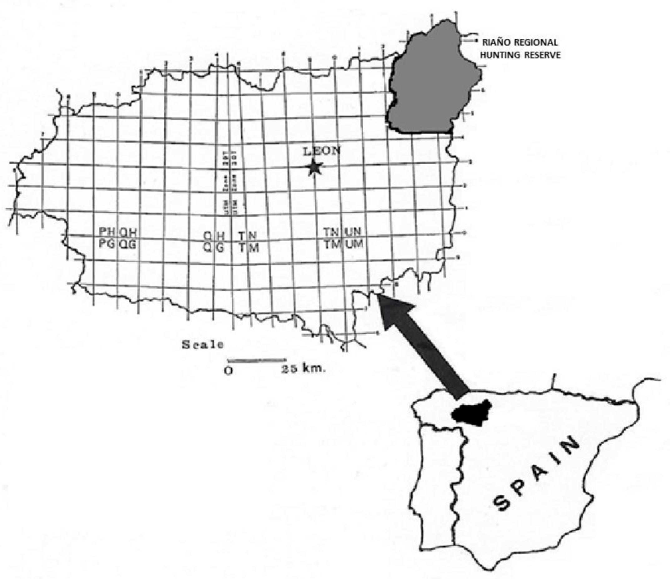
Figure 1. Riaño Regional hunting Reserve.
Animals 15 02649 g001
Figure 2. Large bile duct contained adults of F. hepatica. Note the thickened wall of the ecstatic bile duct.
Animals 15 02649 g002
Figure 3. Severe epithelial hyperplasia and a blackish colour in the inner part of the hepatic lymph nodes.
Animals 15 02649 g003
Figure 4. Large bile duct (H-E. 10×). A. Septal bile duct showing chronic cholangitis.
Animals 15 02649 g004
Figure 5. Detail of a large bile duct (H-E. 31×). A. Severe epithelial hyperplasia. B. Portal fibrosis. C. Severe inflammatory infiltrates.
Animals 15 02649 g005
Figure 6. Distribution of OD values obtained with the FhES-ELISA (excretory-secretory products), FhrAPS-ELISA (2.9 kDa recombinant protein), and the FhBK211 (commercial version, BIO K 211, of the monoclonal antibody, MM3) in 88 serum samples from red deer negative to the “gold standard” (necropsy). Each circle represents the OD value for each animal tested with the 3 methods. The dashed lines represent cut-off values of the iELISA (≥0.1689 for FhrAPS, ≥0.1483 for FhES, and >15% absorbance for FhBK211). Circles inside the square represent false positive results.
Animals 15 02649 g006
Table 1. Prevalence (%) and mean intensity (±SD) of infection by F. hepatica and percentage of animals by iELISA using FhES, FhrAPS and BIO K 211 relative to host age, sex, and seasons of sampling in red deer from NW Spain.
Host AgeSexSeasonsTotal
n = 100
≤2 Years
n = 20
>2 Years
n = 80
Males
n = 26
Females
n = 74
Autum
n = 27
Winter
n = 38
Spring
n = 35
Necropsy %201015.410.83.77.922.812
x ¯ ± SD2.7 ± 1.72.6 ± 1.61.7 ± 0.93.1 ± 1.611.7 ± 1.53.2 ± 1.52.7 ± 1.5
Range1–51–61–31–611–32–61–6
Egg output (epg) %1057.75.47.42.68.66
x ¯ ± SD17 ± 18.432.5 ± 22.225 ± 21.228.5 ± 23.620 ± 14.14028 ± 28.827.3 ± 20.6
Range4–3010–6010–404–6010–30404–604–60
FhES-ELISA %1536.342.328.43723.737.132
FhrAPS-ELISA %51523.19.53.713.22013
BIO K 211-ELISA %011.315.46.911.15.311.49
Table 2. Statistical analysis of the results obtained between the ELISAs studied and the necropsy.
FhES-ELISAFhrAPS-ELISABIO K 211-ELISA
Infected animals (n = 12)723
Uninfected animals (n = 88)637782
Sensitivity %58.3316.6725
Specificity %71.6087.5093.18
PPV %21.8815.3833.33
NPV %92.6588.5190.11
PLR2.0531.3333.700
NLR0.5820.9520.805
κ-value0.1740.0400.184
p-value0.0370.6870.065
Seroprevalence (95% CI)32 (23–41)13 (6–20)9 (3–15)
PPV = positive predictive value; NPV= negative predictive value; PLR = positive likelihood ratio; NLR = negative likelihood ratio.
Table 3. Statistical analysis of the results obtained between the ELISAs studied and the 40 coprology.
FhES-ELISAFhrAPS-ELISABIO K 211-ELISA
Infected animals (n = 6)412
Uninfected animals (n = 94)668287
Specificity %66.6716.6733.33
Specificity %70.2187.2392.55
PPV %12.503.126.25
NPV %97.0692.6494.12
PLR2.2381.3054.474
NLR0.4750.9550.720
κ-value0.122−0.0540.005
Seroprevalence (95% CI)32 (23–41)13 (6–20)9 (3–15)
PPV = positive predictive value; NPV= negative predictive value; PLR = positive likelihood ratio; NLR = negative likelihood ratio.
Disclaimer/Publisher’s Note: The statements, opinions and data contained in all publications are solely those of the individual author(s) and contributor(s) and not of MDPI and/or the editor(s). MDPI and/or the editor(s) disclaim responsibility for any injury to people or property resulting from any ideas, methods, instructions or products referred to in the content.