

Recurrent vs. Nonrecurrent Superficial Non-Healing Corneal Ulcers in Cats: A Multifactorial Retrospective Analysis

 , and
, and
Simple Summary
Abstract
1. Introduction
2. Materials and Methods
2.1. Study Design and Ethical Approval
2.2. Case Selection and Data Collection
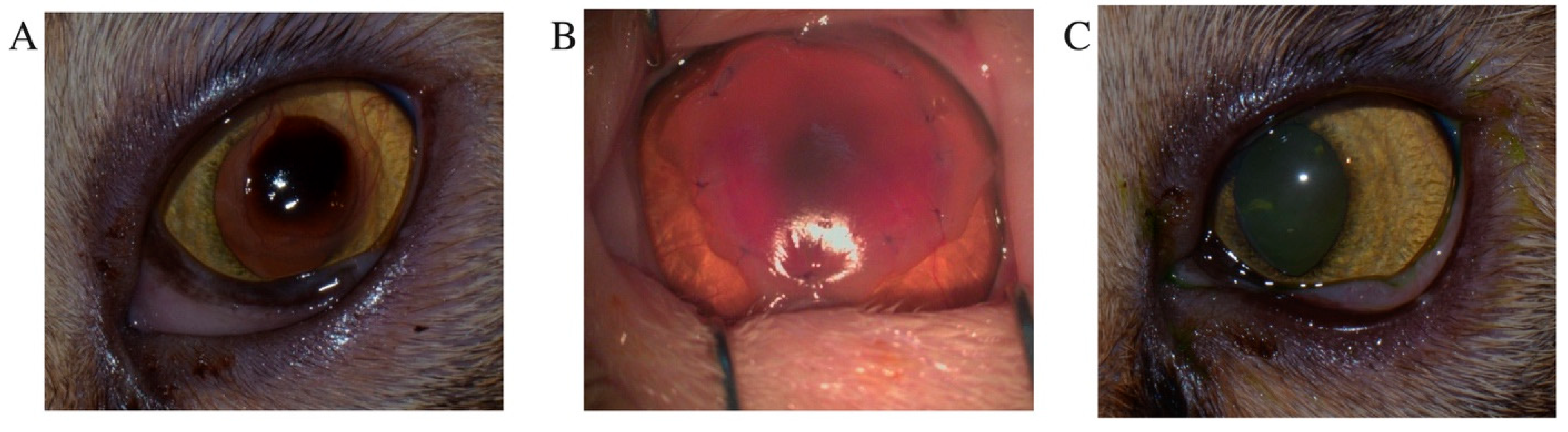
2.3. Classification of Cases
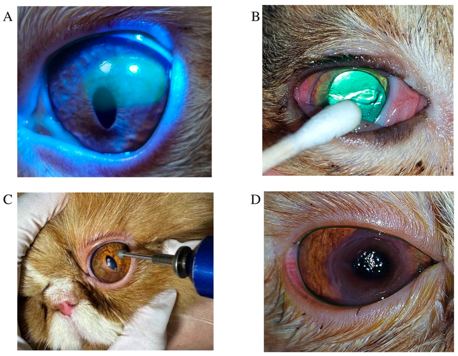
2.4. Ophthalmic Examination and Treatment Procedure
2.5. Statistical Analysis
3. Results
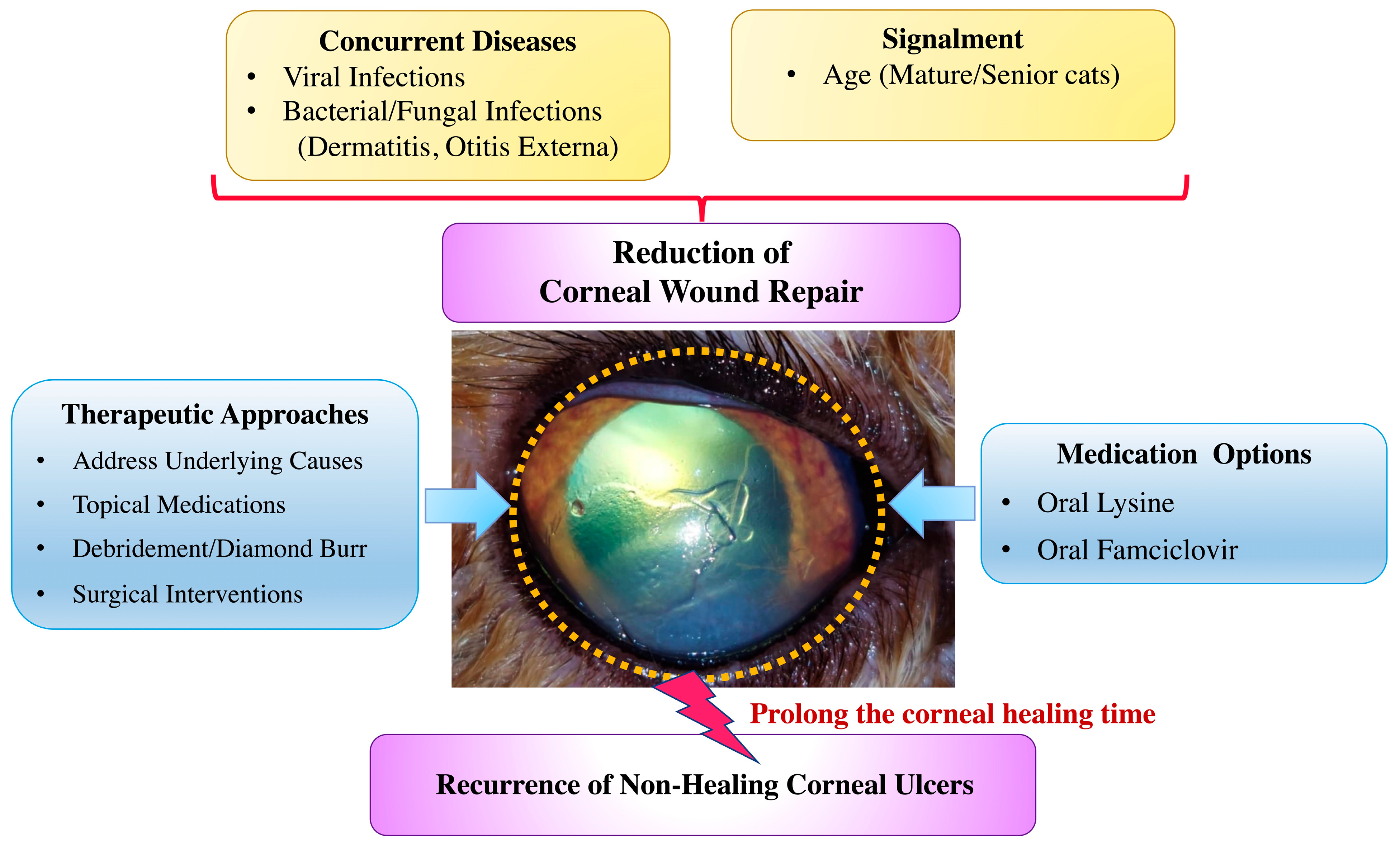
4. Discussion
5. Conclusions
Author Contributions
Funding
Institutional Review Board Statement
Informed Consent Statement
Data Availability Statement
Acknowledgments
Conflicts of Interest
Abbreviations
| FHV-1 | feline herpesvirus type 1 |
| NSAIDs | nonsteroidal anti-inflammatory drugs |
| SD | standard deviation |
References
- Maggs, D.J. Slatter’s Fundamentals of Veterinary Ophthalmology, 5th ed.; Maggs, D.J., Miller, P., Dacvo, D., Ofri, R., Eds.; Elsevier Health Sciences: St. Louis, MO, USA, 2013; pp. 184–219. [Google Scholar]
- Moore, P.A. Diagnosis and management of chronic corneal epithelial defects (indolent corneal ulcerations). Clin. Tech. Small Anim. Pract. 2003, 18, 168–177. [Google Scholar] [CrossRef] [PubMed]
- Morgan, R.; Abrams, K. A comparison of six different therapies for persistent corneal erosions in dogs and cats. Vet. Comp. Ophthalmol. 1994, 4, 38–43. [Google Scholar]
- Bentley, E.; Abrams, G.A.; Covitz, D.; Cook, C.S.; Fischer, C.A.; Hacker, D.; Stuhr, C.M.; Reid, T.W.; Murphy, C.J. Morphology and immunohistochemistry of spontaneous chronic corneal epithelial defects (SCCED) in dogs. Investig. Ophthalmol. Vis. Sci. 2001, 42, 2262–2269. [Google Scholar]
- Jégou, J.P.; Tromeur, F. Superficial keratectomy for chronic corneal ulcers refractory to medical treatment in 36 cats. Vet. Ophthalmol. 2015, 18, 335–340. [Google Scholar] [CrossRef]
- La Croix, N.C.; van der Woerdt, A.; Olivero, D.K. Non-healing corneal ulcers in cats: 29 cases (1991–1999). J. Am. Vet. Med. Assoc. 2001, 218, 733–735. [Google Scholar] [CrossRef]
- Featherstone, H.J.; Sansom, J. Feline corneal sequestra: A review of 64 cases (80 eyes) from 1993 to 2000. Vet. Ophthalmol. 2004, 7, 213–227. [Google Scholar] [CrossRef]
- Gosling, A.A.; Labelle, A.L.; Breaux, C.B. Management of spontaneous chronic corneal epithelial defects (SCCEDs) in dogs with diamond burr debridement and placement of a bandage contact lens. Vet. Ophthalmol. 2013, 16, 83–88. [Google Scholar] [CrossRef]
- Dees, D.D.; Fritz, K.J.; Wagner, L.; Paglia, D.; Knollinger, A.M.; Madsen, R. Effect of bandage contact lens wear and postoperative medical therapies on corneal healing rate after diamond burr debridement in dogs. Vet. Ophthalmol. 2017, 20, 382–389. [Google Scholar] [CrossRef]
- Anastassiadis, Z.; Bayley, K.D.; Read, R.A. Corneal diamond burr debridement for superficial non-healing corneal ulcers in cats. Vet. Ophthalmol. 2022, 25, 476–482. [Google Scholar] [CrossRef]
- Chandler, H.L.; Gemensky-Metzler, A.J.; Bras, I.D.; Robbin-Webb, T.E.; Saville, W.J.; Colitz, C.M. In vivo effects of adjunctive tetracycline treatment on refractory corneal ulcers in dogs. J. Am. Vet. Med. Assoc. 2010, 237, 378–386. [Google Scholar] [CrossRef]
- Kim, S.E.; Lee, M.-K.; Seo, K. Clinical Application of Serum Eye Drops for Herpetic Keratitis in Cats: A Pilot Study. Int. J. Appl. Res. Vet. Med. 2018, 16, 221–225. [Google Scholar]
- Rankin, A.J.; Sebbag, L.; Bello, N.M.; Crumley, W.R.; Allbaugh, R.A. Effects of oral administration of anti-inflammatory medications on inhibition of paracentesis-induced blood-aqueous barrier breakdown in clinically normal cats. Am. J. Vet. Res. 2013, 74, 262–267. [Google Scholar] [CrossRef]
- Bossuyt, S. The use of therapeutic soft contact bandage lenses in the dog and the cat: A series of 41 cases. Vlaams Diergeneeskd. Tijdschr. 2016, 85, 343–348. [Google Scholar]
- Grinninger, P.; Verbruggen, A.; Kraijer-Huver, I.; Djajadiningrat-Laanen, S.; Teske, E.; Boevé, M. Use of bandage contact lenses for treatment of spontaneous chronic corneal epithelial defects in dogs. J. Small Anim. Pract. 2015, 56, 446–449. [Google Scholar] [CrossRef] [PubMed]
- Prabhasawat, P.; Kosrirukvongs, P.; Booranapong, W.; Vajaradul, Y. Application of preserved human amniotic membrane for corneal surface reconstruction. Cell Tissue Bank. 2000, 1, 213–222. [Google Scholar] [CrossRef] [PubMed]
- Barachetti, L.; Giudice, C.; Mortellaro, C.M. Amniotic membrane transplantation for the treatment of feline corneal sequestrum: Pilot study. Vet. Ophthalmol. 2010, 13, 326–330. [Google Scholar] [CrossRef] [PubMed]
- Moore, P.A. Feline corneal disease. Clin. Tech. Small Anim. Pract. 2005, 20, 83–93. [Google Scholar] [CrossRef]
- Ali, K.; Mostafa, A.; Soliman, S. Complicated corneal ulceration in cats: Diagnosis and treatment outcomes of 80 cases (2014–2018). J. Hell. Vet. Med. Soc. 2021, 72, 3041–3050. [Google Scholar] [CrossRef]
- Blocker, T.; Van Der Woerdt, A. A comparison of corneal sensitivity between brachycephalic and domestic short-haired cats. Vet. Ophthalmol. 2001, 4, 127–130. [Google Scholar] [CrossRef]
- Gelatt, K.; Peiffer, L.; Stevens, J. Chronic ulcerative keratitis and sequestrum in the domestic cat. J. Am. Anim. Hosp. Assoc. 1973, 9, 204–213. [Google Scholar]
- Rujirekasuwan, N.; Sattasathuchana, P.; Theerapan, W.; Thengchaisri, N. Comparative analysis of ocular biometry, ocular protrusion, and palpebral fissure dimensions in brachycephalic and nonbrachycephalic dog breeds. Vet. Radiol. Ultrasound 2024, 65, 437–446. [Google Scholar] [CrossRef]
- Barrett, P.M.; Scagliotti, R.H.; Merideth, R.E.; Jackson, P.A.; Alarcon, F.L. Absolute corneal sensitivity and corneal trigeminal nerve anatomy in normal dogs. Prog. Vet Comp. Ophthalmol. 1991, 1, 245–254. [Google Scholar]
- Packer, R.M.; Hendricks, A.; Burn, C.C. Impact of facial conformation on canine health: Corneal ulceration. PLoS ONE 2015, 10, e0123827. [Google Scholar] [CrossRef]
- Uhl, L.K.; Saito, A.; Iwashita, H.; Maggs, D.J.; Mochel, J.P.; Sebbag, L. Clinical features of cats with aqueous tear deficiency: A retrospective case series of 10 patients (17 eyes). J. Feline Med. Surg. 2019, 21, 944–950. [Google Scholar] [CrossRef]
- Sebbag, L.; Uhl, L.K.; Schneider, B.; Hayes, B.; Olds, J.; Mochel, J.P. Investigation of Schirmer tear test-1 for measurement of tear production in cats in various environmental settings and with different test durations. J. Am. Vet. Med. Assoc. 2020, 256, 681–686. [Google Scholar] [CrossRef]
- Lia, I.; Ionascu, I.; Cerrada, I.; Birtoiu, A.; Huguet, E. Human amniotic membrane transplantation in the treatment of feline corneal sequestrum: Preliminary results. AgroLife Sci. J. 2016, 5, 81–88. [Google Scholar]
- Startup, F. Corneal necrosis and sequestration in the cat: A review and record of 100 cases. J. Small Anim. Pract. 1988, 29, 476–486. [Google Scholar] [CrossRef]
- Graham, K.L.; White, J.D.; Billson, F.M. Feline corneal sequestra: Outcome of corneoconjunctival transposition in 97 cats (109 eyes). J. Feline Med. Surg. 2017, 19, 710–716. [Google Scholar] [CrossRef] [PubMed]
- Vawer, G. Corneal mummification in colourpoint cats. Vet. Rec. 1981, 109, 413. [Google Scholar] [CrossRef]
- Goulle, F. Use of porcine small intestinal submucosa for corneal reconstruction in dogs and cats: 106 cases. J. Small Anim. Pract. 2012, 53, 34–43. [Google Scholar] [CrossRef]
- Dulaurent, T.; Azoulay, T.; Goulle, F.; Dulaurent, A.; Mentek, M.; Peiffer, R.L.; Isard, P.F. Use of bovine pericardium (Tutopatch®) graft for surgical repair of deep melting corneal ulcers in dogs and corneal sequestra in cats. Vet. Ophthalmol. 2014, 17, 91–99. [Google Scholar] [CrossRef]
- Gaskell, R.; Povey, R. Experimental induction of feline viral rhinotracheitis virus re-excretion in FVR-recovered cats. Vet. Rec. 1977, 100, 128–133. [Google Scholar] [CrossRef] [PubMed]
- Nasisse, M.; Guy, J.; Davidson, M.; Sussman, W.; Fairley, N. Experimental ocular herpesvirus infection in the cat. Sites of virus replication, clinical features and effects of corticosteroid administration. Investig. Ophthalmol. Vis. Sci. 1989, 30, 1758–1768. [Google Scholar]
- Maggs, D.J.; Collins, B.K.; Thorne, J.G.; Nasisse, M.P. Effects of L-lysine and L-arginine on in vitro replication of feline herpesvirus type-1. Am. J. Vet. Res. 2000, 61, 1474–1478. [Google Scholar] [CrossRef] [PubMed]
- Cooper, A.E.; Thomasy, S.M.; Drazenovich, T.L.; Kass, P.H.; Potnis, S.S.; Leutenegger, C.M.; Maggs, D.J. Prophylactic and therapeutic effects of twice-daily famciclovir administration on infectious upper respiratory disease in shelter-housed cats. J. Feline Med. Surg. 2019, 21, 544–552. [Google Scholar] [CrossRef]
- Malik, R.; Lessels, N.S.; Webb, S.; Meek, M.; Graham, P.G.; Vitale, C.; Norris, J.M.; Power, H. Treatment of feline herpesvirus-1 associated disease in cats with famciclovir and related drugs. J. Feline Med. Surg. 2009, 11, 40–48. [Google Scholar] [CrossRef]
- Thomasy, S.M.; Shull, O.; Outerbridge, C.A.; Lim, C.C.; Freeman, K.S.; Strom, A.R.; Kass, P.H.; Maggs, D.J. Oral administration of famciclovir for treatment of spontaneous ocular, respiratory, or dermatologic disease attributed to feline herpesvirus type 1: 59 cases (2006–2013). J. Am. Vet. Med. Assoc. 2016, 249, 526–538. [Google Scholar] [CrossRef]
- La Croix, N.C. Ocular manifestations of systemic disease in cats. Clin. Tech. Small Anim. Pract. 2005, 20, 121–128. [Google Scholar] [CrossRef]
- Hollingsworth, S.R.; Holmberg, B.J. Ocular disease in the intensive care unit. In Small Animal Critical Care Medicine; Elsevier (Saunders): St. Louis, MO, USA, 2009; pp. 739–743. [Google Scholar]
- Lim, C.C.; Reilly, C.M.; Thomasy, S.M.; Kass, P.H.; Maggs, D.J. Effects of feline herpesvirus type 1 on tear film break-up time, Schirmer tear test results, and conjunctival goblet cell density in experimentally infected cats. Am. J. Vet. Res. 2009, 70, 394–403. [Google Scholar] [CrossRef]
- Gould, D. Feline herpesvirus-1: Ocular manifestations, diagnosis and treatment options. J. Feline Med. Surg. 2011, 13, 333–346. [Google Scholar] [CrossRef]

| Parameters | Nonrecurrent Non-Healing Corneal Ulcers | Recurrent Non-Healing Corneal Ulcers |
|---|---|---|
| No. of patient | 101 (83.5%) | 20 (16.5%) |
| No. of eyes | 113 (83.1%) | 23 (16.9%) |
| Lesion in 1 eye | 89 (88.1%) | 17 (85.0%) |
| Lesion in 2 eyes | 12 (11.8%) | 3 (15.0%) |
| Age (years) | 5.1 ± 4.6 | 7.2 ± 4.3 * |
| Gender | ||
| Male | 57 (56.4%) | 8 (40.0%) |
| Female | 44 (43.5%) | 12 (60.0%) |
| Non-brachycephalic cat | 55 (54.5%) | 12 (60.0%) |
| Brachycephalic cat | 46 (45.5%) | 8 (40.0%) |
| Breed | ||
| Domestic Shorthair | 45 (44.6%) | 10 (50.0%) |
| Persian | 25 (24.8%) | 5 (25.0%) |
| British Shorthair | 10 (9.9%) | 1 (5.0%) |
| Exotic Shorthair | 9 (8.9%) | 1(5.0%) |
| American Wirehair | 4 (4.0%) | - |
| Maine Coon | 4 (4.0%) | - |
| Scottish Fold | 2 (2.0%) | 1 (5.0%) |
| Himalayan | 1 (1.0%) | - |
| Bengal | 1 (1.0%) | - |
| Ragdoll | - | 2 (10.0%) |
| Body weight (kg) | 4.20 ± 1.54 | 4.77 ± 1.50 |
| Parameters | Nonrecurrent Non-Healing Corneal Ulcers | Recurrent Non-Healing Corneal Ulcers | p-Value |
|---|---|---|---|
| Healing time (days) | |||
| Debridement | 25.5 ± 23.1 (N = 88) | 32.3 ± 34.4 (N = 20) | 0.272 |
| Diamond burr | 47.5 ± 27.6 (N = 12) | 14 (N = 1) | - |
| Debridement + Surgery | 50.5 ± 27.6 (N = 13) | 54.5 ± 8.5 (N = 2) | 0.694 |
| Healing time after surgery (days) | 19.13 ± 13.13 (N = 13) | 29.5 ± 19.09 (N = 2) | 0.580 |
| Number of cats with corneal sequestrum (95%CI) | 12 (5.94–16.30%) | 3 (−0.72–26.81%) | 0.735 |
| Exotic Shorthair | 5 | - | - |
| Persian | 3 | - | |
| Domestic Shorthair | 2 | 2 | - |
| American Wirehair | 1 (2 eyes) | - | - |
| Ragdoll | - | 1 | - |
| Day found sequestrum after debridement | 17.6 ± 14.8 (N = 10, 8.8%) | 19.3 ± 15.0 (N = 3, 13.04%) | 0.871 |
| Day found sequestrum after diamond burr | 13.0 ± 7.0 (N = 2, 1.77%) | - | - |
| Treatment Methods | Nonrecurrent Non-Healing Corneal Ulcers N (95% CI) | Recurrent Non-Healing Corneal Ulcers N (95% CI) | p-Value |
|---|---|---|---|
| Debridement | 88 (70.2–85.5%) | 20 (73.2–100.0%) | 0.326 |
| Diamond burr | 12 (4.9–16.3%) | 1 (−4.0–12.7%) | 0.351 |
| Debridement + Surgery | 13 (5.6–17.4%) | 2 (−2.8–20.2%) | 0.695 |
| Medications | Nonrecurrent Non-Healing Corneal Ulcers, N (%) | Recurrent Non-Healing Corneal Ulcers, N (%) | p-Value |
|---|---|---|---|
| Artificial tear drops | |||
| Yes | 106 (93.8%) | 22 (95.6%) | 1.000 |
| No | 7 (6.2%) | 1 (4.4%) | |
| Autologous serum eye drops | |||
| Yes | 2 (1.8%) | 0 (0%) | 1.000 |
| No | 111 (98.2%) | 23 (100.0%) | |
| Oral NSAIDs | |||
| Yes | 46 (40.7%) | 7 (30.4%) | 0.351 |
| No | 67 (59.3%) | 16 (69.6%) | |
| Oral lysine | |||
| Yes | 30 (26.5%) | 11 (47.8%) | 0.049 |
| No | 83 (73.5%) | 12 (52.2%) | |
| Oral famciclovir | |||
| Yes | 3 (2.6%) | 4 (17.4%) | 0.016 |
| No | 110 (97.4%) | 19 (82.6%) | |
| Oral antibiotics | |||
| Amoxicillin-clavulanate | 6 (5.3%) | 0 (0%) | 0.285 |
| Doxycycline | 43 (38.1%) | 8 (34.8%) | |
| No antibiotics | 64(56.6%) | 15 (65.2%) |
| Concurrent Disease | Nonrecurrent Non-Healing Corneal Ulcers N (95% CI) | Recurrent Non-Healing Corneal Ulcers N (95% CI) | p-Value |
|---|---|---|---|
| Viral infection | 7 (1.8–10.6%) | 7 (11.6–49.2%) ** | <0.001 |
| Renal disease | 5 (0.6–8.2%) | 3 (0.7–26.8%) | 0.109 |
| Endocrine/Metabolic disorders | 4 (0.1–6.9%) | 1 (−4.0–12.7%) | 0.851 |
| Neoplasia/Mass | 4 (0.1–6.9%) | 2 (−2.8–20.2%) | 0.272 |
| Bacterial/Fungal infections | 3 (−0.3–5.6%) | 3 (−0.7–26.8%) * | 0.027 |
| Neurological disease | 3 (−0.3–5.6%) | 1 (−4.0–12.7%) | 0.661 |
| Gastrointestinal/Hepatic diseases | 2 (−0.7–4.2%) | 0 | 0.520 |
| Trauma | 2 (−0.7–4.2%) | 0 | 0.520 |
| Total (with concurrent diseases) | 30 (18.4–34.7%) | 17 (11.6–49.2%) | 0.703 |
| No concurrent disease | 83 (65.3–81.6%) | 6 (8.1–44.0%) ** | <0.001 |
Disclaimer/Publisher’s Note: The statements, opinions and data contained in all publications are solely those of the individual author(s) and contributor(s) and not of MDPI and/or the editor(s). MDPI and/or the editor(s) disclaim responsibility for any injury to people or property resulting from any ideas, methods, instructions or products referred to in the content. |
© 2025 by the authors. Licensee MDPI, Basel, Switzerland. This article is an open access article distributed under the terms and conditions of the Creative Commons Attribution (CC BY) license (https://creativecommons.org/licenses/by/4.0/).
Share and Cite
Rujirekasuwan, N.; Sattasathuchana, P.; Sritrakoon, N.; Thengchaisri, N. Recurrent vs. Nonrecurrent Superficial Non-Healing Corneal Ulcers in Cats: A Multifactorial Retrospective Analysis. Animals 2025, 15, 2104. https://doi.org/10.3390/ani15142104
Rujirekasuwan N, Sattasathuchana P, Sritrakoon N, Thengchaisri N. Recurrent vs. Nonrecurrent Superficial Non-Healing Corneal Ulcers in Cats: A Multifactorial Retrospective Analysis. Animals. 2025; 15(14):2104. https://doi.org/10.3390/ani15142104
Chicago/Turabian StyleRujirekasuwan, Nuanwan, Panpicha Sattasathuchana, Natthanet Sritrakoon, and Naris Thengchaisri. 2025. "Recurrent vs. Nonrecurrent Superficial Non-Healing Corneal Ulcers in Cats: A Multifactorial Retrospective Analysis" Animals 15, no. 14: 2104. https://doi.org/10.3390/ani15142104
APA StyleRujirekasuwan, N., Sattasathuchana, P., Sritrakoon, N., & Thengchaisri, N. (2025). Recurrent vs. Nonrecurrent Superficial Non-Healing Corneal Ulcers in Cats: A Multifactorial Retrospective Analysis. Animals, 15(14), 2104. https://doi.org/10.3390/ani15142104