Article

Risk Factors for the Occurrence of Cutaneous Neoplasms in Dogs: A Retrospective Study by Cytology Reports, 2019–2021

by
Issa Carolina García-Reynoso
1,
Cesar Augusto Flores-Dueñas
1,
Nohemí Castro-del Campo
2,
Mariana Jácome-Ibarra
1,
José Carlomán Herrera-Ramírez
1,
Sergio Daniel Gómez-Gómez
1,
Miguel Ángel Rodríguez-Gaxiola
2 and
Soila Maribel Gaxiola-Camacho
2,*
1
Institute for Research in Veterinary Sciences, Autonomous University of Baja California, Mexicali 21387, Mexico
2
Faculty of Veterinary Medicine and Zootechnics, Autonomous University of Sinaloa, Culiacán 80260, Mexico
*
Author to whom correspondence should be addressed.
Animals 2025, 15(14), 2069; https://doi.org/10.3390/ani15142069
Submission received: 9 May 2025 / Revised: 20 June 2025 / Accepted: 9 July 2025 / Published: 14 July 2025
(This article belongs to the Special Issue Advances in Animal Clinical Pathology)

Simple Summary

Cutaneous neoplasms represent a significant health concern in dogs worldwide, yet epidemiological data from arid regions remain limited. This retrospective study analyzed 698 canine cutaneous neoplasms diagnosed via fine needle aspiration cytology in Mexicali, Mexico, where extreme desert conditions create unique environmental challenges. We found a notably high malignancy rate (56.59%), exceeding reports from temperate regions. Transmissible venereal tumor (TVT) and mast cell tumors comprised 28.35% and 27.84% of malignant cases, respectively. A paradoxical age distribution emerged, where young dogs (0–4 years) showed the highest malignancy rates, driven by endemic TVT affecting intact animals. Conversely, Schnauzers demonstrated protective effects against malignancy compared to mixed breeds. These findings reveal how regional factors (intense UV radiation, limited reproductive control, and local canine population management practices) create distinct cancer patterns in arid environments. Understanding these unique patterns is crucial for developing targeted prevention strategies, improving diagnostic approaches, and optimizing treatment protocols in settings where cytology frequently guides clinical decision-making.

Abstract

Studies worldwide report cutaneous neoplasms in dogs; however, data in the arid regions of Mexico remain scarce. Here we report the main malignant cutaneous neoplasms diagnosed by fine needle aspiration cytology (FNAC), and describe the associations with age, sex and breed in Mexicali. Neoplastic lesions accounted for 25.52% (698/2735) of the cases, of which 56.59% (395/698) were malignant. The highest prevalence was observed in dogs aged 9–12 years (n = 193), intact males (n = 162), and mixed-breed dogs (n = 247). Round cell neoplasms (n = 309), including lymphoma, transmissible venereal tumors (TVT), and mast cell tumors (MCT), were the most common cell lineage. Using dogs aged 0–4 years as the reference group, dogs aged 9–12 years had 0.241 times the odds of developing malignant neoplasms (95% CI: 0.141–0.415, p = 0.0025). Using neutered males as the reference group, intact females showed 2.499 times the odds of developing malignant neoplasms (95% CI: 1.462–4.271, p = 0.0042). Compared to mixed-breed dogs, Schnauzers (OR = 0.161) showed significantly lower odds of malignancy (95% CI: 0.082–0.317, p = 0.0004), while Pitbull Terriers had 1.748 times more chance of present malignant neoplasia (95% CI: 1.014–3.013, p < 0.0001). This study provides significant epidemiological evidence on canine cutaneous neoplasms in an arid region of Mexico, identifying key risk factors and distribution patterns that can guide preventive, diagnostic, and therapeutic strategies tailored to regional characteristics.

1. Introduction

Cutaneous lesions of various origins are easily noticeable and therefore represent a common reason for veterinary consultation. Since many of these lesions share similar macroscopic characteristics and clinical signs [1], establishing a reliable diagnosis is crucial for providing accurate treatment and prognosis. Cutaneous neoplasms are the most frequently diagnosed type of cancer in dogs, making up approximately 30–40% of all canine neoplasms [2,3]. Their high prevalence, along with the diversity of clinical presentations and biological behaviors, poses a significant diagnostic and therapeutic challenge in veterinary practice [4].
Two primary diagnostic methods are frequently utilized: fine needle aspiration cytology (FNAC), a rapid, safe, cost-effective, and accessible technique with high sensitivity and specificity [5]; and histopathology, which, while regarded as the gold standard technique, tends to be more expensive, invasive, and requires a longer processing time. Recent studies have shown that FNAC has a sensitivity of 89.3–90.5% and specificity of 97.2–97.9% for diagnosing canine cutaneous neoplasms when performed by experienced pathologists [6,7]. It is important to note that worldwide, various factors, such as economic constraints and perceptions of invasiveness, limit the use of histopathological studies, with confirmation rates ranging from 15 to 25% in Latin America to 65–70% in developed countries [8,9]. In this context, FNAC has been established as a crucial diagnostic tool that provides valuable information to guide therapeutic decisions [10]. Furthermore, despite a lower number of cytological studies compared to histopathological papers, many publications exist on specific tumor types [11,12,13,14,15,16,17].
Among neoplasms in dogs, cutaneous neoplasms are the most common, with prevalence rates ranging from 33% [18] to 50% [19], although most of these neoplasms are benign [20]. The most frequently reported cutaneous neoplasms in dogs include mast cell tumors (MCTs), histiocytomas, cutaneous adnexal neoplasms, and lipomas [21]. The reported incidence varies considerably between geographic regions, suggesting that environmental, genetic, and population management factors may influence their development [22,23].
The epidemiology of canine cutaneous neoplasms has been extensively studied in developed countries. In the United States, MCTs account for 16–21% of all cutaneous neoplasms, followed by lipomas (8–10%), sebaceous gland adenomas (7–9%), and histiocytomas (4–8%) [24,25]. In Europe, similar patterns have been reported, although with significant regional variations [26,27]. Well-established risk factors include advanced age, certain breed predispositions (particularly in Boxers, Boston Terriers, and Retrievers for MCTs), and reproductive status [28,29].
The impact of environmental factors on the development of cutaneous neoplasms has gained considerable attention. Ultraviolet (UV) radiation exposure has been associated with an increased risk of squamous cell carcinomas, particularly in dogs with light-colored coats and in anatomical regions with lower hair density [30,31]. In arid and semi-arid areas, where UV radiation intensity is elevated throughout the year, this factor could have particular relevance [32]. Additionally, environmental pollutants, pesticides, and herbicides have been implicated in canine cutaneous carcinogenesis [33,34].
Although several studies have reported the frequency of cutaneous neoplasms in dogs worldwide, only a few have retrospectively analyzed large populations and identified risk factors associated with malignant cutaneous neoplasms [24,35,36,37,38,39,40]. In Latin America, epidemiological studies on canine cutaneous neoplasms are limited. Research in Brazil has reported a prevalence of malignant neoplasms of 40–45%, with transmissible venereal tumor (TVT), being particularly common due to high stray dog populations [41,42]. In Mexico, the few available studies have focused on central regions of the country, reporting similar patterns but with limited data on specific risk factors [43,44].
Identification of specific risk factors for malignant cutaneous neoplasms is crucial for developing preventive and early detection strategies. Recent molecular studies have identified mutations in genes such as c-KIT, BRAF, and p53 in MCTs and other cutaneous neoplasms, suggesting that certain breeds may have specific genetic predispositions [45,46]. Furthermore, the role of chronic inflammation, recurrent dermatitis, and non-healing wounds as predisposing factors for malignant transformation has been increasingly recognized [47,48].
However, data on the canine population in arid cities of Mexico remain scarce. The city of Mexicali, in the state of Baja California in Northwestern Mexico, presents unique environmental characteristics, including extreme temperatures (exceeding 45 °C in summer), low relative humidity, high UV radiation intensity throughout the year, and frequent dust storms [49]. These conditions could influence the epidemiology of cutaneous neoplasms differently from what has been reported in other regions. Additionally, local socioeconomic factors affecting access to preventive veterinary services and reproductive control could impact the prevalence of certain neoplasms such as TVT [50].
Therefore, the objective of this study was to report the main benign and malignant cutaneous neoplasms diagnosed through FNAC, in the domestic dog population of Mexicali, and to describe their frequency and associations with age, breed, and sex. This regional epidemiological knowledge is essential for guiding preventive, diagnostic, and therapeutic strategies adapted to the specific characteristics of the canine population in arid regions of Mexico.

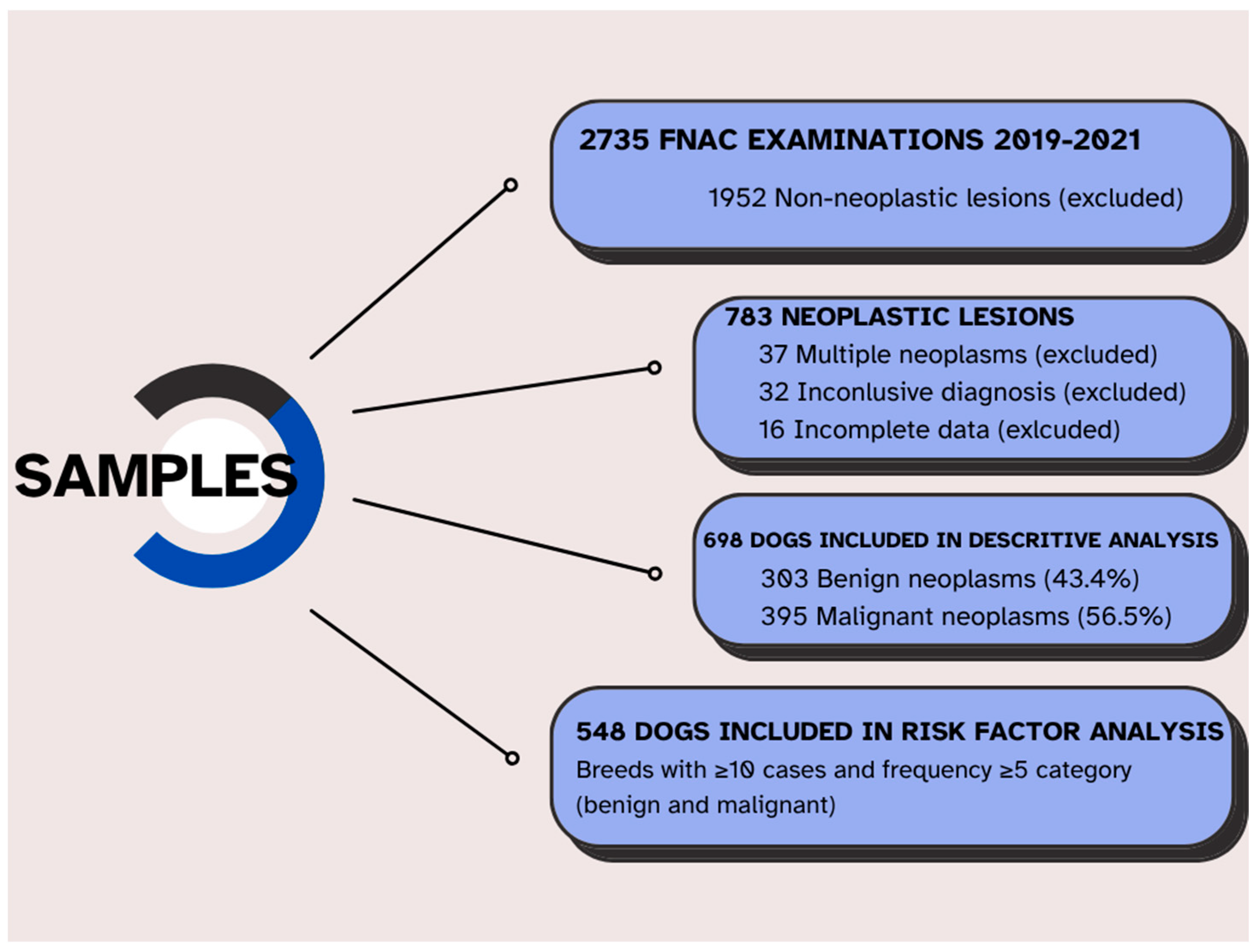
2. Materials and Methods

This retrospective, cross-sectional study analyzed cytological records from 1 January 2019 to 31 December 2021. Data were obtained from two sources: the Veterinary Teaching Hospital of the Autonomous University of Baja California and a private reference clinical pathology laboratory, both located in Mexicali, Baja California, Mexico. Ethical approval was waived due to the retrospective nature of this study and the use of existing clinical records. All procedures complied with institutional guidelines.
A total of 2735 fine-needle aspiration cytology (FNAC) studies were performed on dogs presenting cutaneous or subcutaneous nodules during the study period (817 in 2019, 757 in 2020, and 1161 in 2021). Dogs were eligible for inclusion if they had a single cutaneous or subcutaneous nodule evaluated by FNAC, a complete medical record including age, sex, reproductive status, breed, and a definitive cytological diagnosis. Cases were excluded if they had non-neoplastic lesions (n = 1952), inconclusive cytological results (n = 32), multiple distinct neoplasms (n = 37), or incomplete demographic data (n = 16). After applying these criteria, a total of 698 dogs with a single neoplastic lesion and complete records were included in the descriptive analysis. Subsequently, to perform the risk analysis study, we had to exclude 150 patients more, resulting in 548 individuals (Figure 1).
FNAC was performed using 22–23 gauge needles attached to 5–10 mL syringes. Multiple samples were collected from different areas of each lesion using non-aspiration techniques. Smears were air-dried and stained with modified Wright-Giemsa stain (Diff-Quik®, Siemens, Pomona, CA, USA), following the manufacturer’s instructions. All cytological samples were evaluated by a single board-certified veterinary clinical pathologist with over 10 years of experience. Neoplasms were classified and diagnosed cytologically according to the criteria described by Valenciano and Cowell [51]. Each lesion was categorized by biological behavior (benign or malignant), cell lineage (epithelial, mesenchymal, or round cell), and specific neoplasm type when cytological criteria were sufficient. To ensure diagnostic consistency, a random 10% of cases were re-evaluated, yielding an intra-observer agreement rate of 94%.
The following variables were recorded for each case: age (grouped into 0–4, 5–8, 9–12, and 13–18 years), sex and reproductive status (intact male, neutered male, intact female, spayed female), breed (as reported by owners and categorized per international breed standards), and neoplasm characteristics, including cytological diagnosis, and, when available, neoplasm location and size.
Descriptive statistics (frequencies and percentages) were calculated for all categorical variables. The association between malignancy and the variables of age, sex, and breed was assessed using the Wald chi-square test (χ2). A binary logistic regression was performed on breeds with more than 10 individuals and at least 5 individuals in every dependent variable (benign/malignant neoplasms), resulting in a dataset of 548 eligible dogs. Reference categories for multivariable analysis were obtained through stepwise backward selection, resulting in dogs aged 0–4 years, neutered males, and mixed-breed dogs. Odds ratios (OR) with 95% confidence intervals and p-values ≤ 0.05 were considered statistically significant. Statistical analyses were conducted using SAS (Statistical Analysis System) version 9.4.

3. Results

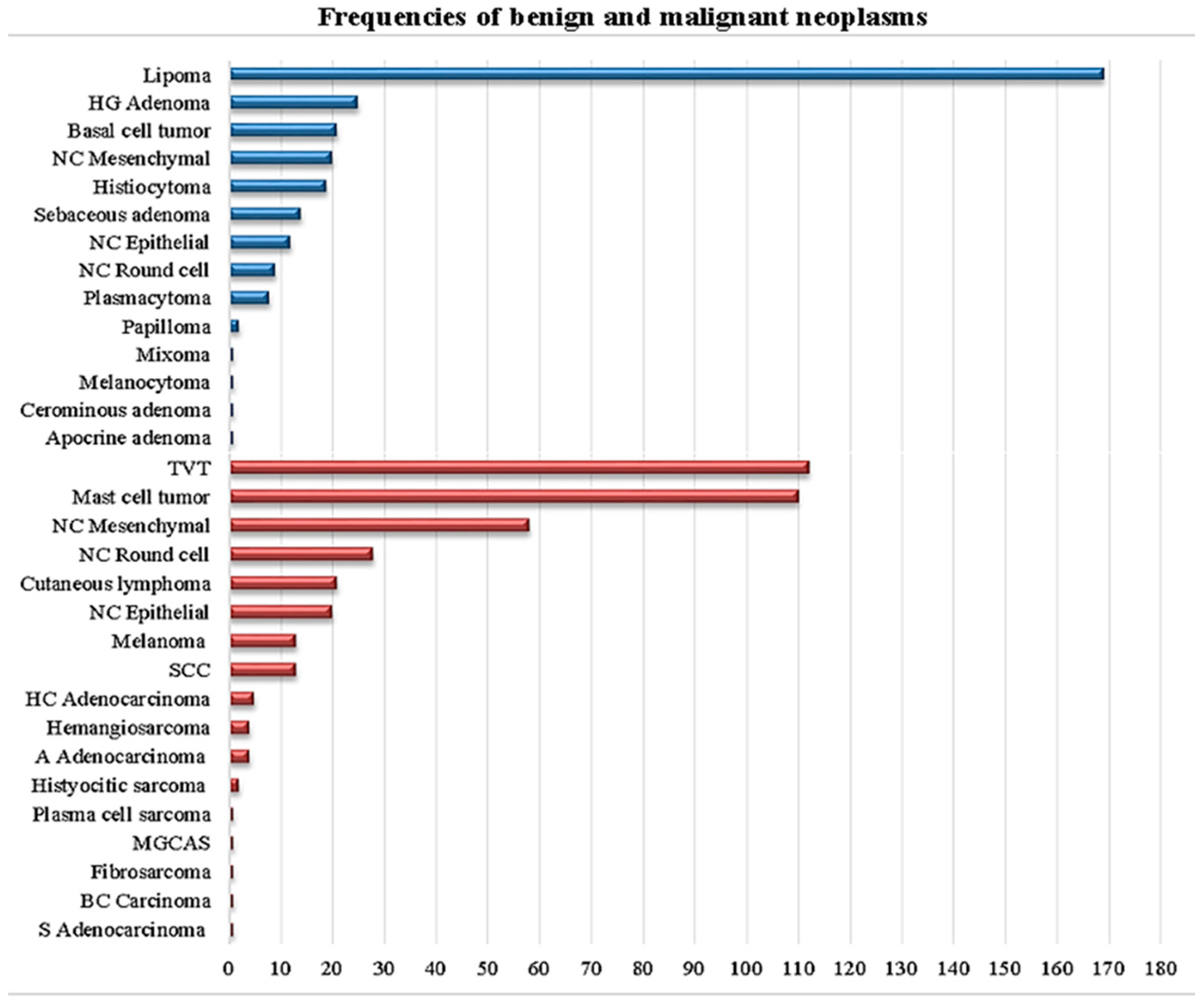
3.1. Descriptive Analysis

Over the three-year study period, 25.52% (698/2735) of cytological samples from cutaneous and subcutaneous masses were diagnosed as neoplastic lesions. Table 1 shows the frequency of breeds that presented at least one neoplasm. Of the 698 neoplastic cases, 303 (43.41%) were classified as benign, and 395 (56.59%) as malignant.
Dogs aged 9–12 years were most frequently affected (193/698, 27.65%), followed by those aged 5–8 years (187/698, 26.79%), 0–4 years (114/698, 16.33%), and 13–18 years (67/698, 9.59%). Regarding sex distribution, intact males represented the largest group (219/698, 31.37%), followed by intact females (187/698, 26.79%), spayed females (180/698, 25.78%), and neutered males (112/698, 16.04%). Mixed-breed dogs were most commonly affected (247/698, 35.38%), followed by Pitbull Terriers (94/698, 13.46%), Schnauzers (56/698, 8.02%), Chihuahuas (53/698, 7.59%), Poodles (38/698, 5.44%), and Labrador Retrievers (36, 5.15%). All remaining breeds represented 3.17% of the cases.
Classification by cell lineage revealed that round cell neoplasms were most prevalent (309/698, 44.26%), followed by mesenchymal (269/698, 38.53%) and epithelial neoplasms (120/698, 17.19%). Regarding the round cell neoplasms, 273/309 (88.34%) were malignant and 36/309 (11.65%) were benign. Mesenchymal neoplasms showed 78/269 (29.00%) malignant and 191/269 (71.00%) benign cases. Epithelial neoplasms comprised 44/120 (36.66%) malignant and 76/120 (63.33%) benign cases.
Figure 2 shows the frequency of the different types of neoplasms found, where the most frequently diagnosed were the following: lipoma (169/698, 24.21%), TVT (Figure 3) (112/698, 16.04%), MCT (110/698, 15.75%), unclassified malignant neoplasms (106/698, 15.18%), and unclassified benign neoplasms (41/698, 5.87%). Among the 303 benign neoplasms, the distribution was as follows: lipomas (169/303, 55.77%), hepatoid gland adenomas (25/303, 8.25%), basal cell neoplasms (21/303, 6.93%), unclassified mesenchymal neoplasms (20/303, 6.60%), histiocytomas (19/303, 6.27%), and nine additional neoplasm types representing the remaining 7.29%. Regarding the 395 malignant neoplasms, the distribution was: TVT (112/395, 28.35%), MCT (110/395, 27.84%), unclassified mesenchymal neoplasms (58/395, 14.68%), unclassified round cell neoplasms (28/395, 7.08%), cutaneous lymphoma (21/395, 5.31%), unclassified epithelial neoplasms (20/395, 5.06%), and 11 additional neoplasm types comprising the remaining 11.64%.
Age-specific analysis revealed distinct patterns for the most common neoplasms. Lipomas occurred most frequently in dogs aged 9–12 years (85/169, 50.29%) and 5–8 years (51/169, 30.17%). TVT showed the highest prevalence in dogs aged 0–4 years (67/112, 59.82%) and 5–8 years (33/112, 29.46%), with only 12/112 cases (10.71%) in dogs over 8 years old. MCT was most common in dogs aged 5–8 years (50/110, 45.45%) and 9–12 years (35/110, 31.81%).
Sex-specific patterns were evident for certain types of neoplasm. Hepatoid gland adenomas occurred predominantly in intact males (20/25, 80.00%), with only three cases in neutered males and two in intact females. TVT affected 58 intact males, 42 intact females, seven spayed females, and five neutered males, showing a clear predilection for intact animals (100/112, 89.29%). MCT showed a more even distribution across all sex categories, with no apparent predilection.

3.2. Risk Factors Analysis

Among breeds with at least ten individuals and more than four benign and malignant neoplasia, marked differences in malignancy rates were observed. Pitbull Terriers showed 72 malignant and 22 benign neoplasms (76.60% malignancy rate), as well as mixed breed dogs with 161 malignant and 86 benign neoplasms (65.18% malignancy rate). In contrast, Schnauzers demonstrated 13 malignant and 43 benign neoplasms (23.21% malignancy rate), while Poodles had 14 malignant and 24 benign neoplasms (36.84% malignancy rate) (Table 2). Although Dachshunds (n = 11) showed nine benign and only two malignant neoplasms, and Boxers (n = 21) presented exclusively with malignant neoplasms, including 18 MCT and three other malignant neoplasms, these breeds were selected as candidates for the risk factors study, given the statistical model constraints.
Multivariable logistic regression analysis of 548 dogs meeting the inclusion criteria identified significant predictors of malignancy (Table 3). Using dogs aged 0–4 years as the reference group, dogs aged 9–12 years demonstrated significantly lower odds (OR = 0.241, 95% CI = 0.141–0.415, p = 0.0025) of developing malignant neoplasia. Regarding sex and reproductive status, using neutered males as the reference group, intact females showed a 2.499-fold increase in the probability of developing malignant neoplasms (95% CI = 1.462–4.271, p = 0.0042). Breed analysis, compared to mixed-breed dogs, revealed significant protective effects for Schnauzers (OR = 0.161, 95% CI = 0.082–0.317, p = 0.0004) and a 1.748-fold increase in the probability of presenting malignant neoplasms in Pitbull Terriers (95% CI = 1.014–3.013, p < 0.0001).

4. Discussion

Cutaneous neoplasms are among the most common neoplasms in dogs, yet epidemiological studies in arid regions of Latin America remain scarce. This study offers key insights into the distribution of cutaneous neoplasms in the canine population of Northwestern Mexico, enabling comparisons with global data and contributing to the identification of specific risk factors that may guide preventive and diagnostic strategies in this region.
The higher proportion of malignant neoplasms (56.59%) observed in this study exceeds the prevalence reported in European (45.2%) [26] and North American (approximately 50%) studies [24], suggesting possible environmental and clinical management differences in the studied region. This elevated malignancy rate likely reflects the intense UV radiation exposure characteristic of Mexicali’s desert environment, where annual UV index values frequently exceed 11 (extreme category). Supporting this environmental hypothesis, studies from similarly high-UV regions demonstrate parallel patterns: Grenada reports hemangiosarcoma comprising 19.1% of cutaneous neoplasms [52], while Brazilian studies document that 27–80% of canine hemangiosarcomas are cutaneous in high-UV areas compared to only 14% in North America [53]. The molecular signature of UV-induced damage, TP53 mutations in 59.6% of hemangiosarcoma cases, and PIK3CA pathway alterations [54], provide mechanistic support for solar radiation as a primary driver of cutaneous malignancy in our region.
Statistical analysis revealed unexpected protective factors against malignant neoplasms. Using dogs aged 0–4 years as the reference group, the 9–12-year-old group demonstrated significantly lower odds of developing malignant neoplasms (OR = 0.241, 95% CI = 0.141–0.415, p < 0.0025). This finding appears paradoxical given the established principle of age-related cancer accumulation; however, the explanation lies, apparently, in the specific neoplastic distribution within our population. The exceptionally high prevalence of TVT in young dogs (59.82% of TVT cases occurred in dogs aged 0–4 years) creates an artificial peak in malignancy rates that obscures typical age-related cancer progression. When considering non-transmissible neoplasms alone, the expected pattern emerges, consistent with the Swiss Cancer Registry data, showing peak incidence rate ratios in 8–11-year-old dogs (IRR: 18.2) [55]. This finding underscores the importance of considering regional disease patterns when interpreting epidemiological data, as endemic diseases like TVT can significantly alter apparent risk factor associations.
Analysis by cell lineage revealed intrinsic biological patterns: round cell neoplasms were predominantly malignant (88.34%), whereas mesenchymal neoplasms were mostly benign (71.00%), consistent with molecular mechanisms documented in previous studies [40,41]. Round cell neoplasms, including lymphoma, mast cell tumors, and TVT, derive from mobile cells of the hematopoietic and immune systems whose inherent capacity for migration predisposes them to aggressive behavior. Recent genomic analyses reveal that these neoplasms frequently harbor mutations in genes associated with increased invasiveness and metastatic potential, including GNB1 mutations in 17.3% of mast cell tumors [56]. Conversely, mesenchymal neoplasms arising from connective tissues, predominantly lipomas in our study, typically maintain the differentiation and growth constraints of their cells of origin.
Among malignant neoplasms, TVT and MCT were the most frequent, representing 28.35% and 27.84% of cases, respectively. The high incidence of TVT in our study reinforces the well-established link between reproductive control and disease distribution. In countries like the United States and parts of Europe, where early spaying and neutering are common, TVT prevalence is significantly lower [42]. Similar TVT prevalence rates have been reported in studies from Brazil [9] and Mexico [43], confirming its status as a significant veterinary public health concern in regions with limited reproductive control measures. The predominance of TVT in animals younger than 9 years old (89.28%) emphasizes the importance of implementing comprehensive reproductive control programs as a primary prevention strategy.
MCT prevalence in our study aligns with global reports [24], confirming its status as one of the most common cutaneous neoplasms in dogs. In a large German epidemiological study analyzing 109,616 histopathological samples, MCT represented 9.7% of all canine neoplasms, ranking as the third most frequent neoplasm type [57]. Importantly, recent advances in cytological evaluation have demonstrated that FNAC can provide valuable prognostic information for MCT. Using specific cytological criteria, high-grade MCTs can be identified with 88% sensitivity and 94% specificity, with dogs having high-grade neoplasms being 25 times more likely to die within 2 years [13]. This prognostic capability positions cytology as a valuable decision-making tool that can guide initial clinical management, inform owners about prognosis, and help prioritize cases for surgical intervention or referral while treatment options are being evaluated.
Our findings regarding sex and reproductive status revealed that intact females had significantly higher odds (OR = 2.499) of developing malignant neoplasms compared to neutered males. This observation contrasts with numerous studies reporting increased cancer risk associated with neutering. For instance, the Veterinary Medical Database analysis indicated that spayed females had a 72% higher risk of developing hemangiosarcoma (OR = 1.72); meanwhile, neutered males exhibited a 14% increased risk (OR = 1.14) compared to intact animals [58]. Additionally, Vizslas demonstrated a 3.5-fold higher risk of MCTs following neutering [59]. Moreover, data from the Swiss Canine Cancer Registry (2008–2020) also indicated a higher proportion of skin neoplasms (30.01%) in neutered females [55].
However, the existing literature presents variable results depending on population characteristics. For example, a VetCompass study involving 168,636 dogs reported significantly reduced odds for MCT in neutered dogs (OR = 0.1, 95% CI: 0.1–0.2) [60], these findings are more consistent with ours. Such contradictory outcomes likely stem from regional differences: areas with high neutering prevalence, such as the USA (85%), display distinct cancer profiles compared to regions with predominantly intact populations, like Sweden (99%) [61].
The key distinction in our population is the notably high prevalence of transmissible venereal tumors (TVT), which represent 28.35% of malignant neoplasms, predominantly affecting young, intact animals (89.28%). In populations with low neutering rates, where TVT has a significant influence on neoplasm epidemiology, a protective effect may appear more pronounced in neutered animals. Moreover, in Mexico, neutering often correlates with improved veterinary care and reduced roaming behaviors, factors inherently linked to decreased TVT exposure [42,50].
Due to the lack of data on the age at neutering, our study was unable to assess the documented timing-related effects. For example, early neutering (before 12 months) in Golden Retrievers is associated with a higher incidence of lymphoma (10% vs. 3% in intact males), while late-neutered females show a higher incidence of MCT (5.7% vs. 0% in intact females) [62]. These breed- and timing-specific nuances were beyond the analytical scope of our study.
For hepatoid gland adenoma, we found that only 3.58% of the study population presented it, in contrast to studies that report frequencies of 7–25% [24,63]. Regarding sex, we found a male: female relationship of 23:2, which is very similar to Petterino et al., (2004) [64] where they report a 20:4 male:female relation, including five carcinomas among these 24 neoplasms, and adding the relevance of growing hormone-dependence in the presence of this adenoma, which is beyond the scope of this paper.
Breed analysis revealed significant protective effects for Schnauzers (OR = 0.161) compared to mixed-breed dogs, contradicting common assumptions about purebred cancer predisposition. This finding appears paradoxical given documented genetic risk factors in these breeds: Giant and Standard Schnauzers carry elevated KITLG gene copy number variations, which may be associated with digital squamous cell carcinoma [65].
For Pitbull Terriers, there was a 1.784 times greater probability of developing malignant neoplasms compared to mixed-breed dogs, corroborating previous observations on genetic susceptibility [28]. Mutations in the c-KIT proto-oncogene, identified in the Pitbull Terrier among other breeds, lead to constitutive activation of the KIT receptor, promoting uncontrolled cell proliferation and increasing tumor aggressiveness [45,47], which explains the higher MCT prevalence observed in this breed in this study. Additionally, regional breeding populations may harbor different genetic variants; regional legislations as well as regional culture may impact the number of dogs of this breed present in Mexico, Latin America, North America, or Europe. Furthermore, breed-specific characteristics such as coat density may confer protection against UV radiation in desert regions; thus, UV radiation in cutaneous carcinogenesis appears particularly relevant in Mexicali’s arid environment. Geographic epidemiology demonstrates clear associations between desert climates and specific neoplasm types, with Australian research showing 15% higher UV radiation exposure compared to equivalent northern latitudes [66]. UV-associated neoplasms show distinct patterns: cutaneous hemangiosarcoma correlates strongly with solar exposure, particularly affecting the ventral abdomen and sparsely haired areas in light-coated breeds [67]. The molecular evidence supporting UV-induced carcinogenesis, including characteristic alterations in TP53 mutations and PIK3CA pathway [54], suggests that environmental factors significantly contribute to the elevated malignancy rate observed in our population.
Although FNAC achieved complete classification in 94.13% of benign and 84.82% of malignant neoplasms, important limitations must be acknowledged. The technique cannot reliably differentiate morphologically similar round cell neoplasms without the use of ancillary methods, although in our high-TVT endemic region (Figure 3), the clinical context (young, intact animals, and genital/facial location) aids in diagnosis. Critical prognostic features, such as surgical margins, histologic grade (Patnaik/Kiupel), or lymphovascular invasion, cannot be assessed cytologically. Mesenchymal neoplasms, which comprise 38.53% of our cases, present particular diagnostic challenges as cytology cannot differentiate specific sarcoma subtypes. This limitation contributed to our overall incomplete classification rates. While histopathology remains the gold standard for definitive neoplasm diagnosis, economic constraints significantly limit its use in Latin American veterinary practice, with confirmation rates ranging from only 15 to 25% compared to 65–70% in developed countries [8,9]. This economic reality makes FNAC particularly relevant in our regional context, as it provides clinically actionable information at a fraction of the cost of histopathological examination. Despite its limitations, FNAC achieved complete classification in 78.93% of overall samples in our study, enabling appropriate clinical decision-making for the majority of patients. The high concordance between cytological and histopathological diagnoses, as reported in validation studies (89–90% sensitivity and 97% specificity) [6,7], supports FNAC as a valuable, though not infallible, first-line diagnostic tool when used by experienced pathologists. This is particularly crucial in settings where financial constraints preclude routine histopathological confirmation. This study thus reflects real-world veterinary practice in resource-limited regions, where treatment decisions must often rely on cytological findings, emphasizing the importance of understanding both the capabilities and limitations of FNAC when interpreting epidemiological patterns of cutaneous neoplasms.
This study has several limitations, including its retrospective nature and the use of convenience sampling, which may introduce bias in the prevalence estimates. Environmental factors such as specific UV exposure levels, coat color, lifestyle factors, and age at neutering were not systematically evaluated. The inability to confirm all cytological diagnoses histologically may affect accuracy; however, recent validation studies support the reliability of FNAC for most cutaneous neoplasms [68,69,70]. Despite these limitations, this study represents a significant contribution to the epidemiology of cutaneous neoplasms in arid regions of Latin America, revealing unexpected protective factors and risk patterns that challenge established paradigms and warrant further investigation.

5. Conclusions

This comprehensive analysis of 698 canine cutaneous neoplasms from an arid region of Mexico reveals that regional factors greatly influence cancer epidemiology. The high prevalence of malignant neoplasms likely reflects the intense UV radiation exposure typical of desert environments. The apparent increased risk in younger dogs is mainly attributable to endemic TVT, emphasizing the urgent need for reproductive control programs. Unexpected increased risk in intact females and Schnauzers suggests complex interactions between genetic, environmental, and socioeconomic factors that require further investigation. These findings underscore the importance of developing region-specific preventive strategies that consider local disease patterns, environmental conditions, and population characteristics rather than extrapolating from studies conducted in disparate geographic and socioeconomic contexts. Future prospective studies incorporating environmental monitoring, genetic profiling, and molecular characterization will further elucidate the complex interplay between genetic susceptibility and environmental carcinogens in canine cutaneous oncogenesis.

Author Contributions

Conceptualization, S.M.G.-C. and C.A.F.-D.; methodology, N.C.-d.C.; formal analysis, M.J.-I.; investigation, I.C.G.-R.; resources, I.C.G.-R.; data curation, M.J.-I.; writing—original draft preparation, I.C.G.-R., S.D.G.-G. and C.A.F.-D.; writing—review and editing, J.C.H.-R. and M.Á.R.-G.; visualization, S.D.G.-G. All authors have read and agreed to the published version of the manuscript.

Funding

This research received no external funding.

Institutional Review Board Statement

Not applicable.

Informed Consent Statement

Not applicable.

Data Availability Statement

All data are available upon request.

Conflicts of Interest

The authors declare no conflicts of interest.

Abbreviations

The following abbreviations are used in this manuscript:
FNACFine needle aspiration cytology
MCTMast cell tumors
TVTTransmissible venereal tumor
OROdds ratio
CIConfidence intervals

References

  1. Kukolj, V.; Nešić, S.; Marinković, D.; Aleksić-Kovačević, S. Prevalence and distribution of canine neoplastic and non-neoplastic cutaneous lesions in Serbia: A retrospective study of 2432 cases (2011–MID 2021). Acta Vet. Beogr. 2021, 71, 403–416. [Google Scholar] [CrossRef]
  2. Bronden, L.B.; Flagstad, A.; Kristensen, A.T. Veterinary cancer registries in companion animal cancer: A review. Vet. Comp. Oncol. 2007, 5, 133–144. [Google Scholar] [CrossRef]
  3. Merlo, D.F.; Rossi, L.; Pellegrino, C.; Ceppi, M.; Cardellino, U.; Capurro, C.; Ratto, A.; Sambucco, P.L.; Sestito, V.; Tanara, G.; et al. Cancer incidence in pet dogs: Findings of the Animal Tumor Registry of Genoa, Italy. J. Vet. Intern. Med. 2008, 2, 976–984. [Google Scholar] [CrossRef]
  4. Baioni, E.; Scanziani, E.; Vincenti, M.C.; Leschiera, M.; Bozzetta, E.; Pezzolato, M.; Desiato, R.; Bertolini, S.; Maurella, C.; Ru, G. Estimating canine cancer incidence: Findings from a population-based tumour registry in northwestern Italy. BMC Vet. Res. 2017, 13, 203. [Google Scholar] [CrossRef]
  5. Landgren, O.; MacDonald, A.P.; Tani, E.; Czader, M.; Grimfors, G.; Skoog, L.; Öst, Å.; Wedelin, C.; Axdorph, U.; Svedmyr, E.; et al. A prospective comparison of fine needle aspiration cytology and histopathology in the diagnosis and classification of lymphomas. Hematol. J. 2004, 5, 69–76. [Google Scholar] [CrossRef]
  6. Ghisleni, G.; Roccabianca, P.; Ceruti, R.; Stefanello, D.; Bonfanti, U.; Avallone, G.; Pezzolato, M.; Lombardo, R.; Finazzi, M.; Grieco, V. Correlation between Fine-Needle Aspiration Cytology and Histopathology in the Evaluation of Cutaneous and Subcutaneous Masses from Dogs and Cats. Vet. Clin. Pathol. 2006, 35, 228–233. [Google Scholar] [CrossRef] [PubMed]
  7. Simeonov, R.S. The Accuracy of Fine-Needle Aspiration Cytology in the Diagnosis of Canine Skin and Subcutaneous Masses. Comp. Clin. Path. 2012, 21, 1433–1438. [Google Scholar] [CrossRef]
  8. O’Neill, D.G.; Coulson, N.R.; Church, D.B.; Brodbelt, D.C. Demography and disorders of German Shepherd Dogs under primary veterinary care in the UK. Canine Genet. Epidemiol. 2017, 4, 7. [Google Scholar] [CrossRef]
  9. Pimentel, P.A.B.; Oliveira, C.S.F.; Horta, R.S. Epidemiological study of canine transmissible venereal tumor (CTVT) in Brazil, 2000–2020. Prev. Vet. Med. 2021, 197, 105526. [Google Scholar] [CrossRef]
  10. Sharkey, L.C.; Dial, S.M.; Matz, M.E. Maximizing the diagnostic value of cytology in small animal practice. Vet. Clin. N. Am. Small Anim. Pr. 2007, 37, 351–372. [Google Scholar] [CrossRef]
  11. Allen, S.W.; Prasse, K.W.; Mahaffey, E.A. Cytologic differentiation of benign from malignant canine mammary tumors. Vet. Pathol. 1986, 23, 649–655. [Google Scholar] [CrossRef]
  12. Weir, M.M.; Rosenberg, A.E.; Bell, D.A. Grading of spindle cell sarcomas in fine-needle aspiration biopsy specimens. Am. J. Clin. Pathol. 1999, 112, 784–790. [Google Scholar] [CrossRef] [PubMed]
  13. Camus, M.S.; Priest, H.L.; Koehler, J.W.; Driskell, E.A.; Rakich, P.M.; Ilha, M.R.; Krimer, P.M. Cytologic criteria for mast cell tumor grading in dogs with evaluation of clinical outcome. Vet. Pathol. 2016, 53, 1117–1123. [Google Scholar] [CrossRef]
  14. Sapierzyński, R.; Kliczkowska-Klarowicz, K.; Jankowska, U.; Jagielski, D. Cytodiagnostics of canine lymphomas—Possibilities and limitations. Pol. J. Vet. Sci. 2016, 19, 433–439. [Google Scholar] [CrossRef] [PubMed]
  15. Milovancev, M.; Townsend, K.L.; Gorman, E.; Bracha, S.; Curran, K.; Russell, D.S. Shaved margin histopathology and imprint cytology for assessment of excision in canine mast cell tumors and soft tissue sarcomas. Vet. Surg. 2017, 46, 879–885. [Google Scholar] [CrossRef]
  16. Dolka, I.; Czopowicz, M.; Gruk-Jurka, A.; Wojtkowska, A.; Sapierzyński, R.; Jurka, P. Diagnostic efficacy of smear cytology and Robinson’s cytological grading of canine mammary tumors with respect to histopathology, cytomorphometry, metastases and overall survival. PLoS ONE 2018, 13, e0191595. [Google Scholar] [CrossRef] [PubMed]
  17. Millanta, F.; Asproni, P.; Aquino, G.; Poli, A. Cytologic grading of canine and feline spindle-cell sarcomas of soft tissues and its correlation with histologic grading. Top. Companion Anim. Med. 2020, 41, 100458. [Google Scholar] [CrossRef]
  18. González-Chávez, M.T.; Pino, D.; Zamora, Y.; Matos, R.G. Consideraciones actuales sobre las neoplasias cutáneas en la especie canina. Rev. Salud Anim. 2020, 42, 1–19. [Google Scholar]
  19. Dobrin, A.A.; Militaru, M. Cytopathology and histopathology in diagnosis of malignant cutaneous and subcutaneous mesenchymal neoplasms in dogs—A review. Lucr. Ştiinţifice 2023, 56, 71–88. [Google Scholar]
  20. Martins, A.L.; Canadas-Sousa, A.; Mesquita, J.R.; Dias-Pereira, P.; Amorim, I.; Gärtner, F. Retrospective study of canine cutaneous tumors submitted to a diagnostic pathology laboratory in Northern Portugal (2014–2020). Canine Med. Genet. 2022, 9, 2. [Google Scholar] [CrossRef]
  21. Subapriya, S.; Pazhanivel, N.; Gokulakrishnan, M.; Nagarajan, B.; Kavitha, S.; Sumathi, D.; Vairamuthu, S. Incidence and pathology or skin tumours in dogs. PIJ 2021, 10, 620–629. [Google Scholar]
  22. Meirelles, A.E.; Oliveira, E.C.; Rodrigues, B.A.; Costa, G.R.; Sonne, L.; Tesser, E.S.; Driemeier, D. Prevalence of neoplasms in domestic animals diagnosed by cytological exam: 1001 cases (2008–2010). Pesqui. Vet. Bras. 2010, 30, 832–838. [Google Scholar] [CrossRef]
  23. Bonnett, B.N.; Egenvall, A.; Hedhammar, A.; Olson, P. Mortality in over 350,000 insured Swedish dogs from 1995–2000: I. Breed-, gender-, age- and cause-specific rates. Acta Vet. Scand. 2005, 46, 105–120. [Google Scholar] [CrossRef]
  24. Villamil, J.A.; Henry, C.J.; Bryan, J.N.; Ellersieck, M.; Schultz, L.; Tyler, J.W.; Hahn, A.W. Identification of the most common cutaneous neoplasms in dogs and evaluation of breed and age distributions for selected neoplasms. JAVMA 2011, 239, 960–965. [Google Scholar] [CrossRef] [PubMed]
  25. Blackwood, L.; Murphy, S.; Buracco, P.; De Vos, J.P.; De Fornel-Thibaud, P.; Hirschberger, J. European consensus document on mast cell tumours in dogs and cats. Vet. Comp. Oncol. 2012, 10, e1–e29. [Google Scholar] [CrossRef]
  26. Graf, R.; Pospischil, A.; Guscetti, F.; Meier, D.; Welle, M.; Dettwiler, M. Cutaneous tumors in Swiss dogs: Retrospective data from the Swiss Canine Cancer Registry, 2008–2013. Vet. Pathol. 2018, 55, 809–820. [Google Scholar] [CrossRef]
  27. Kiupel, M.; Webster, J.D.; Bailey, K.L.; Best, S.; DeLay, J.; Detrisac, C.J.; Fitzgerald, S.D.; Gamble, D.; Ginn, P.E.; Goldschmidt, M.H.; et al. Proposal of a 2-tier histologic grading system for canine cutaneous mast cell tumors to more accurately predict biological behavior. Vet. Pathol. 2011, 48, 147–155. [Google Scholar] [CrossRef]
  28. Warland, J.; Dobson, J. Breed predispositions in canine mast cell tumour: A single centre experience in the United Kingdom. Vet. J. 2013, 197, 496–498. [Google Scholar] [CrossRef]
  29. White, C.R.; Hohenhaus, A.E.; Kelsey, J.; Procter-Gray, E. Cutaneous MCTs: Associations with spay/neuter status, breed, body size, and phylogenetic cluster. J. Am. Anim. Hosp. Assoc. 2011, 47, 210–216. [Google Scholar] [CrossRef]
  30. Schmitt, J.; Seidler, A.; Diepgen, T.L.; Bauer, A. Occupational ultraviolet light exposure increases the risk for the development of cutaneous squamous cell carcinoma: A systematic review and meta-analysis. Br. J. Dermatol. 2011, 164, 291–307. [Google Scholar] [CrossRef]
  31. Hargis, A.M.; Thomassen, R.W. Animal model: Solar dermatosis (keratosis) and solar dermatosis with squamous cell carcinoma. Am. J. Pathol. 1979, 94, 193–196. [Google Scholar] [PubMed]
  32. Goldschmidt, M.H.; Shofer, F.S. Skin Tumors of the Dog and Cat; Pergamon Press: Oxford, UK, 1992. [Google Scholar]
  33. Hayes, H.M., Jr.; Hoover, R.; Tarone, R.E. Bladder cancer in pet dogs: A sentinel for environmental cancer? Am. J. Epidemiol. 1981, 114, 229–233. [Google Scholar] [CrossRef]
  34. Reif, J.S.; Lower, K.S.; Ogilvie, G.K. Residential exposure to magnetic fields and risk of canine lymphoma. Am. J. Epidemiol. 1995, 141, 352–359. [Google Scholar] [CrossRef]
  35. Rothwell, T.L.; Howlett, C.R.; Middleton, D.J.; Griffiths, D.A.; Duff, B.C. Skin neoplasms of dogs in Sidney. Aust. Vet. J. 1987, 64, 161–164. [Google Scholar] [CrossRef] [PubMed]
  36. Mukaratirwa, S.; Chipunza, J.; Chitanga, S.; Chimonyo, M.; Bhebhe, E. Canine cutaneous neoplasms: Prevalence and influence of age, sex and site on the presence and potential malignancy of cutaneous neoplasms in dogs from Zimbabwe. J. South Afr. Vet. Assoc. 2005, 76, 59–62. [Google Scholar] [CrossRef]
  37. Fajardo, R.; Alpízar, A.; Pérez, L.S.; Martínez, J.S.; Córdova, E. Prevalence of tumors in dogs from the municipality of Toluca, Mexico, from 2002 to 2008. Arch. Med. Vet. 2013, 45, 305–309. [Google Scholar] [CrossRef]
  38. García, E.; Alpízar, A.; Fajardo, R.; Córdova, D.; Pérez, L.; Martínez, S. Epidemiology of tumors in dogs in the capital of the state of Mexico from 2002–2016. Arq. Bras. Med. Vet. Zootec. 2019, 71, 1085–1092. [Google Scholar] [CrossRef]
  39. Hassan, B.B.; Al-Mokaddem, A.K.; Abdelrahman, H.A.; Samir, A.; Mousa, M.R. Cutaneous tumors in dogs: A retrospective epidemiological and histological study of 112 cases. Adv. Anim. Vet. Sci. 2022, 10, 170–182. [Google Scholar] [CrossRef]
  40. Pakhrin, B.; Kang, M.S.; Bae, I.H.; Park, M.S.; Jee, H.; You, M.H.; Kim, J.H.; Yoon, B.I.; Choi, Y.K.; Kim, D.Y. Retrospective study of canine cutaneous tumors in Korea. J. Vet. Sci. 2007, 8, 229–236. [Google Scholar] [CrossRef]
  41. Souza, T.M.; Fighera, R.A.; Irigoyen, L.F.; Barros, C.S.L. Estudo retrospectivo de 761 tumores cutâneos em cães. Ciênc. Rural. 2006, 36, 555–560. [Google Scholar] [CrossRef]
  42. Strakova, A.; Murchison, E.P. The changing global distribution and prevalence of canine transmissible venereal tumour. BMC Vet. Res. 2014, 10, 168. [Google Scholar] [CrossRef] [PubMed]
  43. Ortega-Pacheco, A.; Segura-Correa, J.C.; Jimenez-Coello, M.; Forsberg, C.L. Reproductive patterns and reproductive pathologies of stray bitches in the tropics. Theriogenology 2007, 67, 382–390. [Google Scholar] [CrossRef] [PubMed]
  44. Rivera-Calderón, L.G.; Fonseca-Alves, C.E.; Kobayashi, P.E.; Carvalho, M.; Drigo, S.A.; de Oliveira Vasconcelos, R.; Laufer-Amorim, R. Alterations in PTEN, MDM2, TP53 and AR protein and gene expression are associated with canine prostate carcinogenesis. Res. Vet. Sci. 2016, 106, 56–61. [Google Scholar] [CrossRef] [PubMed]
  45. London, C.A.; Malpas, P.B.; Wood-Follis, S.L.; Boucher, J.F.; Rusk, A.W.; Rosenberg, M.P.; Henry, C.J.; Mitchener, K.L.; Klein, M.K.; Hintermeister, J.G.; et al. Multi-center, placebo-controlled, double-blind, randomized study of oral toceranib phosphate (SU11654), a receptor tyrosine kinase inhibitor, for the treatment of dogs with recurrent (either local or distant) mast cell tumor following surgical excision. Clin. Cancer Res. 2009, 15, 3856–3865. [Google Scholar] [CrossRef]
  46. Mochizuki, H.; Kennedy, K.; Shapiro, S.G.; Breen, M. BRAF mutations in canine cancers. PLoS ONE 2015, 10, e0129534. [Google Scholar] [CrossRef]
  47. Sledge, D.G.; Webster, J.; Kiupel, M. Canine cutaneous mast cell tumors: A combined clinical and pathologic approach to diagnosis, prognosis, and treatment selection. Vet. J. 2016, 215, 43–54. [Google Scholar] [CrossRef]
  48. Pang, L.Y.; Argyle, D.J. The evolving cancer cell: Molecular characterization of canine cancer. In BSAVA Manual of Canine and Feline Oncology, 3rd ed.; Dobson, J.M., Lascelles, B.D.X., Eds.; British Small Animal Veterinary Association: Gloucester, UK, 2011; pp. 11–28. [Google Scholar]
  49. García-Cueto, O.R.; Santillán-Espinoza, M.T.; Martínez-López, M.; Tejeda-Martínez, A. Extreme temperature scenarios in Mexicali, Mexico under climate change conditions. Atmósfera 2013, 26, 509–520. [Google Scholar] [CrossRef]
  50. Ortega-Pacheco, A.; Jiménez-Coello, M. Debate for and against euthanasia in the control of dog populations. In Euthanasia—The "Good Death" Controversy in Humans and Animals; Kure, J., Ed.; InTech: Rijeka, Croatia, 2011; pp. 233–244. [Google Scholar] [CrossRef]
  51. Valenciano, A.C.; Cowell, R.L. Cowell and Tyler’s Diagnostic Cytology and Hematology of the Dog and Cat, 5th ed.; Elsevier: St. Louis, MO, USA, 2019. [Google Scholar]
  52. Chikweto, A.; McNeil, P.; Bhaiyat, M.I.; Stone, D.; Sharma, R.N. Neoplastic and nonneoplastic cutaneous tumors of dogs in Grenada, West Indies. ISRN Vet. Sci. 2011, 2011, 416435. [Google Scholar] [CrossRef]
  53. Nikula, K.J.; Benjamin, S.A.; Angleton, G.M.; Saunders, W.J.; Lee, A.C. Ultraviolet radiation, solar dermatosis, and cutaneous neoplasia in beagle dogs. Radiat. Res. 1992, 129, 11–18. [Google Scholar] [CrossRef]
  54. Megquier, K.; Turner-Maier, J.; Swofford, S.; Kim, J.H.; Sarver, A.L.; Wang, C.; Sakthikumar, S.; Johnson, J.; Koltookian, M.; Lewellen, M.; et al. Comparative genomics reveals shared mutational landscape in canine hemangiosarcoma and human angiosarcoma. Mol. Cancer Res. 2020, 18, 241–2421. [Google Scholar] [CrossRef]
  55. Dhein, E.S.; Heikklä, U.; Oevermann, A.; Blatter, S.; Meier, D.; Hartnack, S.; Guscetti, F. Incidence rates of the most common canine tumors based on data from the Swiss Canine Cancer Registry (2008 to 2020). PLoS ONE 2024, 19, e0302231. [Google Scholar] [CrossRef]
  56. Vozdova, M.; Kubickova, S.; Fictum, P.; Sliva, J.; Rubes, J. Recurrent gene mutations detected in canine mast cell tumours by next generation sequencing. Vet. Comp. Oncol. 2020, 18, 509–518. [Google Scholar] [CrossRef]
  57. Aupperle-Lellbach, H.; Grassinger, J.M.; Floren, A.; Törner, K.; Beitzinger, C.; Loesenbeck, G.; Müller, T. Tumour incidence in dogs in Germany: A retrospective analysis of 109,616 histopathological diagnoses (2014–2019). J. Comp. Pathol. 2022, 198, 33–55. [Google Scholar] [CrossRef] [PubMed]
  58. Robinson, K.L.; Bryan, M.E.; Atkinson, E.S.; Keeler, M.R.; Hahn, A.W.; Bryan, J.N. Neutering is associated with developing hemangiosarcoma in dogs in the Veterinary Medical Database: An age and time-period matched case-control study (1964–2003). Can. J. Vet. Res. 2020, 84, 106–114. [Google Scholar]
  59. Zink, M.C.; Farhoody, P.; Elser, S.E.; Ruffini, L.D.; Gibbons, T.A.; Rieger, R.H. Evaluation of the risk and age of onset of cancer and behavioral disorders in gonadectomized Vizslas. JAVMA 2014, 244, 309–319. [Google Scholar] [CrossRef] [PubMed]
  60. Shoop, S.J.W.; Marlow, S.; Church, D.B.; English, K.; McGreevy, P.D.; Stell Thomson, P.C.; O’Neill, D.G.; Brodbelt, D.C. Prevalence and risk factors for mast cell tumours in dogs in England. CAGH 2015, 2, 1. [Google Scholar] [CrossRef]
  61. Egenvall, A.; Bonnett, B.N.; Öhagen, P.; Olson, P.; Hedhammar, Å.; von Euler, H. Incidence of and survival after mammary tumors in a population of over 80,000 insured female dogs in Sweden from 1995 to 2002. Prev. Vet. Med. 2005, 69, 109–127. [Google Scholar] [CrossRef]
  62. Torres de la Riva, G.; Hart, B.L.; Farver, T.B.; Oberbauer, A.M.; Messam, L.L.M.; Willits, N.H.; Oberbauer, A.M.; Hart, L.A. Neutering dogs: Effects on joint disorders and cancers in golden retrievers. PLoS ONE 2013, 8, e55937. [Google Scholar] [CrossRef]
  63. Maniscalco, L.; Olimpo, M.; Parisi, L.; Buracco, P.; Mazzone, E.; Martinelli, G.; Martano, M.; Lussich, S.; Morello, E. A novel scoring system proposal to guide treatment of dogs with hepatoid gland tumors. Front. Vet. Sci. 2025, 12, 1451. [Google Scholar] [CrossRef]
  64. Petterino, C.; Martini, M.; Castagnaro, M. Immunohistochemical Detection of growth hormone (GH) in canine hepatoid gland tumors. J. Vet. Med. Sci. 2004, 66, 569–572. [Google Scholar] [CrossRef]
  65. Aupperle-Lellbach, H.; Heidrich, D.; Kehl, A.; Conrad, D.; Brockman, M.; Törner, K.; Beitzinger, C.; Müller, T. KITLG copy number germline variations in Schnauzer breeds and their relevance in digital squamous cell carcinoma in black giant Schnauzers. Vet. Sci. 2023, 10, 147. [Google Scholar] [CrossRef] [PubMed]
  66. Gies, P.; van Deventer, E.; Green, A.C.; Sinclair, C.; Tinker, R. Review of the global solar UV index 2015 workshop report. Health Phys. 2018, 114, 84–90. [Google Scholar] [CrossRef] [PubMed]
  67. Szivek, A.; Burns, R.E.; Gericota, B.; Affolter, V.K.; Verstraete, F.J.M.; Kass, P.H. Clinical outcome of dogs with cutaneous hemangiosarcoma treated with surgery alone: 104 cases (2008–2019). Vet. Comp. Oncol. 2011, 10, 65–73. [Google Scholar] [CrossRef] [PubMed]
  68. Mello, C.B.E.; Engelmann, A.M.; Kommers, G.D.; Flores, M.M.; Fighera, R.A.; Rodrigues, B.R.; Lamego, E.C.; da Silva, C.B.; Bueno, A.; de Andrade, C.M. Fine needle aspiration cytology: High accuracy in diagnosing cutaneous and subcutaneous neoplasms in dogs. Comp. Clin. Pathol. 2023, 32, 155–164. [Google Scholar] [CrossRef]
  69. Rinaldi, V.; Crisi, P.E.; Vignoli, M.; Pierini, A.; Terragni, R.; Cabibbo, E.; Boari, A.; Finotello, R. The Role of Fine Needle Aspiration of Liver and Spleen in the Staging of Low-Grade Canine Cutaneous Mast Cell Tumor. Vet. Sci. 2022, 9, 473. [Google Scholar] [CrossRef]
  70. Dank, G.; Buber, T.; Rice, A.; Kraicer, N.; Hanael, E.; Shasha, T.; Aviram, G.; Yehudayoff, A.; Kent, M.S. Training and validation of a novel non-invasive imaging system for ruling out malignancy in canine subcutaneous and cutaneous masses using machine learning in 664 masses. Front. Vet. Sci. 2023, 10, 1164438. [Google Scholar] [CrossRef]
Figure 1. Flow diagram illustrating the total number of patients evaluated, those diagnosed with neoplasms, those with sufficient data for inclusion in the descriptive analysis, and those ultimately included in the risk factor analysis.
Animals 15 02069 g001
Figure 2. Benign (blue) and malignant (red) neoplasms and their frequencies. From top: HG = Hepatoid gland, NC = Not classified, TVT = Transmissible venereal tumor, SSC = Squamous cell carcinoma, HC = Hepatoid cell, A = Apocrine, MGCAS = Multinucleated giant cell anaplastic sarcoma, BC = basal cell, S = Sebaceous.
Animals 15 02069 g002
Figure 3. Diff-Quik®-stained slides from FNAC. (A) Samples with TVT typically show moderate to high cellularity, accompanied by abundant degenerated neutrophils (arrows) and erythrocytes (arrowheads) (20×). (B) Common cytological features of TVT include well-defined cytoplasmic borders with mild basophilia and prominent vacuolation (arrow); a large, round nucleus with coarse, granular chromatin; and one to three distinct nucleoli (arrowhead); anisocytosis, anisokaryosis, macrokaryosis, and occasional atypical mitotic figures (asterisk) are also observed (40×).
Animals 15 02069 g003
Table 1. Dogs with complete data and one type of neoplasm.
BreedFreq.BreedFreq.BreedFreq.
Akita1Fila Brasileiro1Poodle38
Belgian Shepherd3French Bulldog 4Pug13
Boston Terrier4German Shepherd14Portuguese Water Dog1
Bull Terrier2Golden Retriever5Rat Terrier1
Basset Hound7Greyhound1Rottweiler6
Beagle7Great Dane4Saint Bernard2
Boxer21Jack Russell Terrier2Schnauzer56
Chihuahua53Labrador Retriever36Shar Pei3
Chow Chow2Malinois B. Shepherd1Shih Tzu10
Cocker Spaniel7Maltese6Siberian Husky9
Corgi1Miniature Pinscher 1Springer Spaniel2
Dachshund11Mixed-breed dogs247Swiss Shepherd1
Doberman4Old English Sheepdog1Weimaraner1
Dogo Argentino2Pekingese1Xoloitzcuintle1
Dogue of Bordeaux1Pitbull Terrier94Yorkshire Terrier4
English Bulldog 4Pointer2Total698
Table 2. Breeds with at least 10 individuals and a minimum of four benign and four malignant neoplasms were included in the chi-squared test. Boxers and Dachshunds were excluded from the analysis since they presented 3:18 and 9:2 benign: malignant neoplasms, respectively.
BreedBenignMalignantFrequency (%)
Chihuahua262753 (9.67)
German Shepherd7714 (2.55)
Labrador Retriever221436 (6.56)
Mixed-breed dogs86161247 (45.07)
Pitbull Terrier227294 (17.15)
Poodle241438 (6.93)
Schnauzer431356 (10.21)
Shih Tzu5510 (1.82)
Total235313548
Table 3. Multivariable logistic regression analysis of risk factors for malignant neoplasia in dogs (n = 548).
Variablen (%)Adjusted Odds Ratio95% CIp-Value
Age group (years)
0–4 years (reference)99 (18.06)1.00
5–8 years181 (33.02)0.6140.353–1.0670.1366
9–12 years 193 (35.21)0.2410.141–0.4150.0025 *
13–18 years75 (13.68)0.2950-155–05630.1256
Sex and Reproductive Status
Neutered male (reference)91 (16.60)1.00
Intact female 148 (27.00)2.4991.462–4.2710.0042 *
Spayed female147 (26.82)1.3650.807–2.3080.2492
Intact male162 (29.56)2.0041.190–3.3720.1387
Breed
Mixed breed (reference)247 (45.07)1.00
Chihuahua53 (9.67)0.5550.305–1.0100.7705
Labrador Retriever36 (6.56)0.3400.166–0.6980.2079
German Sheperd14 (2.55)0.5340.181–1.5730.9304
American Pit Bull Terrier94 (17.15)1.7481.014–3.013<0.0001 *
Poodle38 (6.93)0.3120.153–0.6330.1217
Schnauzer56 (10.21)0.1610.082–0.3170.0004 *
Shih Tzu10 (1.82)0.5340.150–1.8960.9405
CI = confidence interval. Reference categories are indicated in italics. Statistical significance with bold and *.
Disclaimer/Publisher’s Note: The statements, opinions and data contained in all publications are solely those of the individual author(s) and contributor(s) and not of MDPI and/or the editor(s). MDPI and/or the editor(s) disclaim responsibility for any injury to people or property resulting from any ideas, methods, instructions or products referred to in the content.

