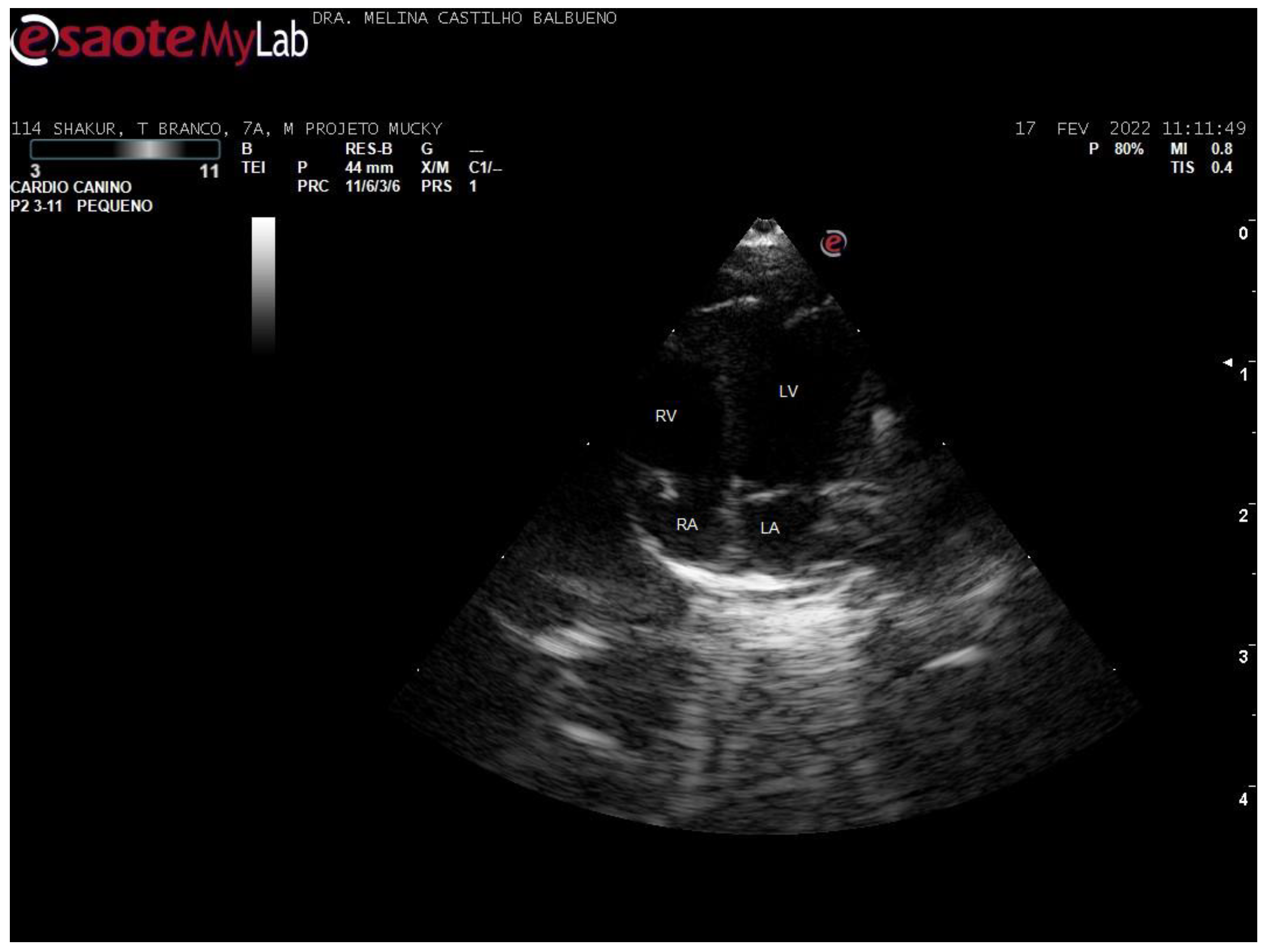
Echocardiographic Parameters of Callithrix spp. Under Human Care
Simple Summary
Abstract
1. Introduction
2. Materials and Methods
3. Results
4. Discussion
5. Conclusions
Author Contributions
Funding
Institutional Review Board Statement
Informed Consent Statement
Data Availability Statement
Acknowledgments
Conflicts of Interest
References
- Tang, H.L.; Wang, L.L.; Cheng, G.; Wang, L.; Li, S. Evaluation of the cardiovascular function of older adult Rhesus monkeys by ultrasonography. J. Med. Primatol. 2008, 37, 101–108. [Google Scholar] [PubMed]
- Pratt-Riccio, L.R.; Pratt-Riccio, E.K.; Bianco-Junior, C.; Alves, F.A.; Baptista, B.O.; Totino, P.R.R.; Muniz, J.A.P.C.; Castro, P.H.G.; Carvalho, L.J.M.; Daniel-Ribeiro, C.T. Using neotropical primate models for malaria research: A history of the 25 years of collaboration between the Malaria Research Laboratory (IOC, Fiocruz) and the National Primate Centre (IEC, SVS). Rev. Pan-Amaz. Saúde 2021, 12, 1–18. [Google Scholar]
- Burns, M.; Wachtman, L. Physical examination, diagnosis, and common clinical procedures. In The Common Marmoset in Captivity and Biomedical Research; Marini, R., Wachtman, L., Tardif, S., Mansfield, K., Fox, J., Eds.; Academic Press: Cambridge, MA, USA, 2019; pp. 145–175. [Google Scholar] [CrossRef]
- Lammey, M.L.; Lee, D.R.; Ely, J.J.; Sleeper, M.M. Sudden cardiac death in 13 captive chimpanzees (Pan troglodytes). J. Med. Primatol. 2008, 37, 39–43. [Google Scholar] [CrossRef] [PubMed]
- Chetboul, V. Advanced techniques in echocardiography in small animals. Vet. Clin. N. Am. 2010, 40, 529–543. [Google Scholar] [CrossRef] [PubMed]
- Brady, A.G.; Watford, J.W.; Massey, C.V.; Rodning, K.J.; Gibson, S.V.; Williams, L.E.; Abee, C.R. Studies of heart disease and failure in aged female squirrel monkeys (Saimiri sp.). Comp. Med. 2003, 53, 657–662. [Google Scholar]
- Rajendra, R.S.; Brady, A.G.; Parks, V.L.; Massey, C.V.; Gibson, S.V.; Abee, C.R. The normal and abnormal owl monkey (Aotus sp.) heart: Looking at cardiomyopathy changes with echocardiography and electrocardiography. J. Med. Primatol. 2010, 39, 143–150. [Google Scholar]
- Mitchell, C.; Rahko, P.S.; Blauwet, L.A.; Canaday, B.; Finstuen, J.A.; Foster, M.C.; Horton, K.; Ogunyankin, K.O.; Palma, R.A.; Velazquez, E.J. Guidelines for Performing a Comprehensive Transthoracic Echocardiographic Examination in Adults: Recommendations from the American Society of Echocardiography. J. Am. Soc. Echocardiogr. 2019, 32, 1–64. [Google Scholar]
- Yang, C.; Zhang, G. Callithrix jacchus (the common marmoset). Trends Genet. 2021, 37, 948–949. [Google Scholar] [CrossRef]
- Balthazar, D.A.; Vasconcelos, M.S.; Magalhães, B.S.N.; Kagohara, A.; Troccoli, F.; Galhões, A.; Santos Filho, M.; Paiva, J.P. Determination of echocardiographic parameters in spider monkey (Ateles spp.) collectives in captivity sedated with ketamine and midazolam. Braz. J. Vet. Med. 2020, 42, e107120. [Google Scholar]
- Moura, L.S.; Rodrigues, R.P.S.; Silva, A.B.S.; Pessoa, G.T.; Sousa, F.C.A.; Alves, J.J.R.P.; Bezerra Neto, L.; Macedo, K.V.; Vieira, M.C.; Alves, F.R. Standard, strain and strain rate echocardiography wih two-dimensional speckle tracking in a capuchin monkey (Cebus apella, Linnaeus, 1758). Arq. Bras. Cardiol. Imagem. Cardiovasc. 2018, 31, 57–66. [Google Scholar]
- Nakayama, S.; Koie, H.; Pai, C.; Ito-Fujishiro, Y.; Kanayama, K.; Sankai, T.; Yasutomi, Y.; Ageyama, N. Echocardiographic evaluation of cardiac function in cynomolgus monkeys over a wide age range. Exp. Anim. 2020, 69, 336–344. [Google Scholar] [CrossRef]
- Ueda, Y.; Duler, L.M.M.; Elliot, K.J.; Sosa, P.M.; Roberts, J.A.; Stern, J.A. Echocardiographic reference intervals with allometric scaling of 823 clinically healthy rhesus macaques (Macaca mulatta). BMC Vet. Res. 2020, 16, 1–12. [Google Scholar]
- Giannico, A.T.; Somma, A.T.; Lange, R.R.; Andrade, J.N.B.M.; Lima, L.; Souza, A.C.; Montiani-Ferreira, F. Electrocardiographic values in black tufted marmosets (Callithrix penicillata). Pesqui. Vet. Bras. 2013, 33, 937–941. [Google Scholar]
- Moussavi, A.; Mietsch, M.; Drummer, C.; Behr, R.; Mylius, J.; Boretius, S. Cardiac MRI in common marmosets revealing age-dependency of cardiac function. Sci. Rep. 2020, 10, 10221. [Google Scholar] [CrossRef] [PubMed]
- David, J.M.; Dick, J.R.E.J.; Hubbard, G.B. Spontaneous pathology of the common marmoset (Callithrix jacchus) and tamarins (Saguinus oedipus, Saguinus mystax). J. Med. Primatol. 2009, 38, 347–359. [Google Scholar] [CrossRef] [PubMed]
- Konoike, N.; Miwa, M.; Ishigami, A.; Nakamura, K. Hypoxemia after single-shot anesthesia in common marmosets. J. Med. Primatol. 2017, 46, 70–74. [Google Scholar] [PubMed]
- Goodroe, A.; Bakker, J.; Remarque, E.J.; Ross, C.N.; Scorpio, D. Evaluation of Anesthetic and Cardiorespiratory Effects after Intramuscular Administration of Three Different Doses of Telazol in Common Marmosets (Callithrix jacchus). Vet. Sci. 2023, 10, 116. [Google Scholar] [CrossRef] [PubMed]
- Guimarães, L.D.; Moraes, A.N. Anestesia em aves: Agentes anestésicos. Ciênc. Rural 2000, 30, 1073–1081. [Google Scholar]
- Balbueno, M.C.S.; Martins, J.A.; Malaga, S.K.; Forato, J.; Coelho, C.P. Dilated cardiomyopathy phenotype in Callithrix penicillata (E. Geoffroy, 1812): Case report. J. Med. Primatol. 2024, 53, e12678. [Google Scholar]


| Animals | ||||||
|---|---|---|---|---|---|---|
| Sex | Age Groups | |||||
| F | M | 0.4–2 y | 3–5 y | 6–8 y | 9–11 y | 12/15 y |
| 77 | 91 | 57 | 26 | 63 | 15 | 7 |
| Parameter | Normal Distribution | C. aurita | C. jacchus | C. penicillata | Callithrix sp. | Pspecies | Psex |
|---|---|---|---|---|---|---|---|
| Heart rate (bpm) | Yes | 242.2 ± 50.3 {212, 232.5, 283.3} [152–333] a | 289.7 ± 55.5 {248, 288, 326} [174–455] b | 270.1 ± 59.6 {233.8, 278, 313} [156–375] ab | 278.4 ± 54.8 {246.5, 273, 316} [172–385] ab | 0.009 * | 0.504 |
| Aortic root diameter (cm) | No | 0.39 ± 0.05 {0.35, 0.4, 0.43} [0.28–0.47] a | 0.36 ± 0.04 {0.33, 0.36, 0.4} [0.29–0.45] b | 0.39 ± 0.04 {0.35, 0.39, 0.42} [0.32–0.49] ab | 0.38 ± 0.04 {0.36, 0.39, 0.4} [0.26–0.48] a | 0.007 * | 0.989 |
| Left atrium diameter (cm) | No | 0.48 ± 0.06 {0.44, 0.49, 0.54} [0.37–0.59] a | 0.47 ± 0.06 {0.42, 0.48, 0.51} [0.33–0.61] a | 0.5 ± 0.05 {0.47, 0.49, 0.53} [0.41–0.61] a | 0.48 ± 0.06 {0.45, 0.48, 0.52} [0.35–0.62] a | 0.216 | 0.482 |
| Ratio of the left atrial dimension to the aortic annulus dimension | Yes | 1.24 ± 0.12 {1.15, 1.21, 1.32} [1.06–1.44] a | 1.3 ± 0.13 {1.22, 1.31, 1.37} [1.02–1.6] a | 1.3 ± 0.13 {1.21, 1.3, 1.37} [1.04–1.61] a | 1.26 ± 0.14 {1.15, 1.27, 1.38} [0.98–1.5] a | 0.154 | 0.472 |
| Interventricular septum thickness at end-diastole (cm) | No | 0.22 ± 0.02 {0.2, 0.22, 0.23} [0.16–0.26] a | 0.21 ± 0.03 {0.19, 0.21, 0.23} [0.14–0.29] a | 0.21 ± 0.03 {0.19, 0.21, 0.23} [0.16–0.28] a | 0.2 ± 0.03 {0.17, 0.2, 0.22} [0.15–0.28] a | 0.129 | 0.057 |
| Interventricular septum thickness at end-systole (cm) | No | 0.33 ± 0.05 {0.3, 0.32, 0.37} [0.23–0.44] a | 0.33 ± 0.04 {0.3, 0.32, 0.36} [0.24–0.45] a | 0.34 ± 0.05 {0.3, 0.34, 0.38} [0.26–0.46] a | 0.33 ± 0.05 {0.3, 0.33, 0.36} [0.22–0.47] a | 0.754 | 0.075 |
| Left ventricular internal dimension at end-diastole (cm) | No | 0.74 ± 0.12 {0.69, 0.76, 0.81} [0.39–0.87] a | 0.63 ± 0.09 {0.58, 0.62, 0.69} [0.45–0.83] b | 0.66 ± 0.11 {0.59, 0.68, 0.72} [0.46–0.91] ab | 0.64 ± 0.1 {0.57, 0.63, 0.72} [0.42–0.86] b | <0.001 * | 0.024 * |
| Left ventricular internal dimension at end-systole (cm) | No | 0.44 ± 0.08 {0.41, 0.46, 0.5} [0.23–0.59] a | 0.35 ± 0.06 {0.31, 0.33, 0.39} [0.21–0.54] b | 0.38 ± 0.09 {0.3, 0.37, 0.45} [0.23–0.61] b | 0.37 ± 0.08 {0.32, 0.36, 0.41} [0.23–0.59] b | <0.001 * | 0.077 |
| Left ventricular posterior wall thickness at end-diastole (cm) | No | 0.21 ± 0.02 {0.2, 0.21, 0.22} [0.15–0.24] a | 0.2 ± 0.02 {0.18, 0.2, 0.21} [0.15–0.25] a | 0.21 ± 0.04 {0.18, 0.2, 0.22} [0.15–0.33] a | 0.2 ± 0.04 {0.17, 0.21, 0.22} [0.11–0.3] a | 0.539 | 0.440 |
| Left ventricular posterior wall thickness at end-systole (cm) | No | 0.33 ± 0.05 {0.31, 0.34, 0.37} [0.21–0.4] a | 0.32 ± 0.05 {0.3, 0.32, 0.36} [0.21–0.45] a | 0.33 ± 0.05 {0.3, 0.34, 0.37} [0.22–0.47] a | 0.34 ± 0.04 {0.31, 0.33, 0.37} [0.23–0.45] a | 0.149 | 0.048 * |
| E-point-to-septal separation (cm) | No | 0.08 ± 0.02 {0.07, 0.09, 0.09} [0.05–0.11] a | 0.09 ± 0.02 {0.07, 0.08, 0.1} [0.06–0.16] a | 0.09 ± 0.01 {0.08, 0.09, 0.09} [0.07–0.11] a | 0.09 ± 0.02 {0.08, 0.08, 0.09} [0.06–0.15] a | 0.872 | 0.334 |
| Aortic flow (m/s) | No | 0.66 ± 0.19 {0.57, 0.69, 0.75} [0.33–1.02] a | 0.77 ± 0.23 {0.65, 0.74, 0.9} [0.33–1.85] a | 0.73 ± 0.19 {0.62, 0.72, 0.84} [0.4–1.26] a | 0.69 ± 0.2 {0.52, 0.7, 0.8} [0.36–1.13] a | 0.069 | 0.295 |
| Pulmonary flow (m/s) | No | 0.66 ± 0.19 {0.52, 0.6, 0.81} [0.41–1.06] a | 0.65 ± 0.2 {0.5, 0.61, 0.78} [0.29–1.19] a | 0.67 ± 0.23 {0.47, 0.59, 0.86} [0.37–1.17] a | 0.63 ± 0.23 {0.46, 0.63, 0.75} [0.29–1.25] a | 0.877 | 0.158 |
| Aortic flow gradient (mmHg) | No | 1.85 ± 1 {1.33, 1.9, 2.23} [0.4–4.1] a | 2.41 ± 1.14 {1.7, 2.2, 3.2} [0.4–5.3] a | 2.26 ± 1.21 {1.5, 1.9, 2.8} [0.7–6.4] a | 2.05 ± 1.11 {1.1, 1.9, 2.55} [0.5–5.1] a | 0.165 | 0.133 |
| Pulmonary flow gradient (mmHg) | No | 1.9 ± 1.11 {1.08, 1.4, 2.65} [0.7–4.5] a | 1.82 ± 1.11 {1, 1.5, 2.45} [0.3–5.7] a | 2 ± 1.38 {0.9, 1.35, 2.98} [0.5–5.5] a | 1.79 ± 1.29 {0.9, 1.6, 2.25} [0.3–6.2] a | 0.885 | 0.157 |
| Shortening fraction (%) | Yes | 39.7 ± 4.2 {37, 40.5, 43} [33–47] a | 43.6 ± 5.9 {40, 44, 48} [30–58] a | 42.6 ± 6.8 {36.3, 42.5, 48} [33–57] a | 42.4 ± 6.2 {38, 42, 47} [28–54] a | 0.086 | 0.812 |
| Ejection fraction (%) | No | 76.3 ± 5.6 {72.8, 76.5, 80} [67–88] a | 79.7 ± 6.4 {76.5, 81, 84.5} [62–92] a | 78.4 ± 6.9 {72, 79.5, 84} [67–91] a | 78.1 ± 6.5 {74, 78, 84} [60–89] a | 0.176 | 0.835 |
| E wave—Initial mitral diastolic flow (m/s) | Yes | 0.49 ± 0.21 {0.4, 0.52, 0.66} [0.05–0.8] a | 0.57 ± 0.15 {0.47, 0.6, 0.67} [0.3–0.88] a | 0.59 ± 0.17 {0.48, 0.54, 0.68} [0.34–0.99] a | 0.59 ± 0.18 {0.46, 0.56, 0.67} [0.3–1.24] a | 0.145 | 0.704 |
| A wave—Mitral late diastolic flow (m/s) | No | 0.42 ± 0.16 {0.32, 0.39, 0.49} [0.14–0.7] a | 0.41 ± 0.23 {0.28, 0.43, 0.54} [0–0.97] a | 0.47 ± 0.22 {0.32, 0.47, 0.58} [0–1.08] a | 0.39 ± 0.27 {0.23, 0.36, 0.6} [0–1.01] a | 0.535 | 0.604 |
| Ratio of E to A | No | 1.28 ± 0.66 {0.83, 1.22, 1.66} [0.14–2.85] a | 1.3 ± 0.43 {1.07, 1.24, 1.58} [0.56–2.67] a | 1.32 ± 0.41 {0.94, 1.28, 1.66} [0.49–2.09] a | 1.3 ± 0.49 {0.96, 1.23, 1.54} [0.63–2.84] a | 0.995 | 0.161 |
| Isovolumetric relaxation time (ms) | No | 41.8 ± 5.7 {36, 44, 45} [32–52] a | 35.3 ± 6.5 {32, 36, 40} [24–52] b | 36 ± 5.9 {32, 36, 40} [28–48] b | 38.3 ± 8.1 {32, 36, 44} [20–56] ab | 0.003 * | 0.424 |
Disclaimer/Publisher’s Note: The statements, opinions and data contained in all publications are solely those of the individual author(s) and contributor(s) and not of MDPI and/or the editor(s). MDPI and/or the editor(s) disclaim responsibility for any injury to people or property resulting from any ideas, methods, instructions or products referred to in the content. |
© 2025 by the authors. Licensee MDPI, Basel, Switzerland. This article is an open access article distributed under the terms and conditions of the Creative Commons Attribution (CC BY) license (https://creativecommons.org/licenses/by/4.0/).
Share and Cite
de Souza Balbueno, M.C.; Martins, J.A.; Malaga, S.K.; Vanstreels, R.E.T.; de Paula Coelho, C. Echocardiographic Parameters of Callithrix spp. Under Human Care. Animals 2025, 15, 1875. https://doi.org/10.3390/ani15131875
de Souza Balbueno MC, Martins JA, Malaga SK, Vanstreels RET, de Paula Coelho C. Echocardiographic Parameters of Callithrix spp. Under Human Care. Animals. 2025; 15(13):1875. https://doi.org/10.3390/ani15131875
Chicago/Turabian Stylede Souza Balbueno, Melina Castilho, Jessica Amancio Martins, Soraya Kezam Malaga, Ralph Eric Thijl Vanstreels, and Cideli de Paula Coelho. 2025. "Echocardiographic Parameters of Callithrix spp. Under Human Care" Animals 15, no. 13: 1875. https://doi.org/10.3390/ani15131875
APA Stylede Souza Balbueno, M. C., Martins, J. A., Malaga, S. K., Vanstreels, R. E. T., & de Paula Coelho, C. (2025). Echocardiographic Parameters of Callithrix spp. Under Human Care. Animals, 15(13), 1875. https://doi.org/10.3390/ani15131875


