Article

Diversity of Anaplasma phagocytophilum Strains from Roe Deer (Capreolus capreolus) and Red Deer (Cervus elaphus) in Poland

by
Anna W. Myczka
1,2,*,
Żaneta Steiner-Bogdaszewska
2,
Grzegorz Oloś
3,
Anna Bajer
1 and
Zdzisław Laskowski
2
1
Department of Eco-Epidemiology of Parasitic Diseases, Institute of Developmental Biology and Biomedical Sciences, Faculty of Biology, University of Warsaw, Ilji Miecznikowa 1, 02-096 Warsaw, Poland
2
Witold Stefański Institute of Parasitology, Polish Academy of Sciences, Twarda 51/55, 00-818 Warsaw, Poland
3
Institute of Environmental and Engineering and Biotechnology, University of Opole, Kardynała B. Kominka 6, 6a, 45-032 Opole, Poland
*
Author to whom correspondence should be addressed.
Animals 2024, 14(4), 637; https://doi.org/10.3390/ani14040637
Submission received: 11 January 2024 / Revised: 13 February 2024 / Accepted: 15 February 2024 / Published: 16 February 2024
(This article belongs to the Special Issue Eco-Epidemiology of Infectious Diseases in Wild Ungulates)

Abstract

:

Simple Summary

Bacteria continuously circulate in the natural environment and frequently cross into the human environment. While some of these bacteria can have a positive effect on human health, they are often accompanied by pathogenic forms. The aim of the present study was to more closely examine the zoonotic, tick-borne bacterium Anaplasma phagocytophilum, the causative agent of human and animal anaplasmosis. The study examined tissue samples from roe deer and red deer. Anaplasma phagocytophilum samples were genotyped based on DNA amplification, sequencing and phylogenetic analysis. Our results showed that detected bacteria from red deer belong to ecotype I and cluster I, which is why those bacteria are potentially pathogenic to humans. Detected bacteria from roe deer were mainly in ecotype II and clusters II and III, which means those bacteria are not pathogenic for humans.

Abstract

Background: The Gram-negative bacterium Anaplasma phagocytophilum is an intracellular pathogen and an etiological agent of human and animal anaplasmosis. Its natural reservoir comprises free-ranging ungulates, including roe deer (Capreolus capreolus) and red deer (Cervus elaphus). These two species of deer also constitute the largest group of game animals in Poland. The aim of the study was to genotype and perform a phylogenetic analysis of A. phagocytophilum strains from roe deer and red deer. Methods: Samples were subjected to PCR amplification, sequencing, and phylogenetic analysis of strain-specific genetic markers (groEL, ankA). Results: Five haplotypes of the groEL gene from A. phagocytophilum and seven haplotypes of ankA were obtained. The phylogenetic analysis classified the groEL into ecotypes I and II. Sequences of the ankA gene were classified into clusters I, II, and III. Conclusions: Strains of A. phagocytophilum from red deer were in the same ecotype and cluster as strains isolated from humans. Strains of A. phagocytophilum from roe deer represented ecotypes (I, II) and clusters (II, III) that were different from those isolated from red deer, and these strains did not show similarity to bacteria from humans. However, roe deer can harbor nonspecific strains of A. phagocytophilum more characteristic to red deer. It appears that the genetic variants from red deer can be pathogenic to humans, but the significance of the variants from roe deer requires more study.

1. Introduction

The Gram-negative bacterium Anaplasma phagocytophilum of the order Rickettsiales is a common intracellular parasite of both veterinary and medical interest [1,2]. A. phagocytophilum has been recorded globally [3,4,5,6,7,8,9,10,11,12,13,14,15,16]. The bacteria are typically transmitted by ticks and mainly by Ixodes ricinus in Europe [17,18]. There is currently no evidence of vertical transmission of A. phagocytophilum in the tick population; as such, the presence of A. phagocytophilum bacteria in the natural environment has most likely resulted from the overlap between the ecological niches of the vectors and animal reservoir hosts [19,20,21]. These bacteria are etiological agents of anaplasmosis in both humans (human granulocytic anaplasmosis, HGA) and animals [22,23]. The symptoms include fever, headache, chills, and gastric problems, with thrombocytopenia, leukopenia, and increased liver enzyme activity being noted in blood tests. However, all these symptoms are nonspecific, and diagnosing anaplasmosis can present a challenge without information about the tick bite [24,25]. HGA is common in North America, mainly in the USA, with over 5500 cases of anaplasmosis diagnosed in 2019; that same year in Europe, only about 300 cases were reported (Centers for Disease Control and Prevention—CDC) [25]. In animals, the occurrence of Anaplasma phagocytophilum was first discovered in sheep (Ovis aries) in Scotland as Cytoecetes phagocytophila [3]. In Europe, A. phagocytophilum strains appear to be more virulent among animals than humans [26]. Cattle infected with A. phagocytophilum present with high fever, anemia, leukopenia, and thrombocytopenia, as well as lower milk production, weight loss, and dullness. In wild ruminants, infection with A. phagocytophilum is asymptomatic and does not significantly affect the condition of the animals. However, in severe disease, the symptoms usually are weight loss, anorexia, and apathy [27,28]. Interestingly, while animal anaplasmosis has been observed in Asia [29,30], Africa [31,32], and Europe [33], no such record exists in North America [34,35]. A few fatal cases due to A. phagocytophilum infection have been reported in Norway, among farmed animals (sheep) and wild game animals (roe deer, Capreolus capreolus; moose, Alces alces) [36,37]. In many European countries, A.phagocytophilum was detected in wild and farm cervids [38]. In roe deer and red deer (Cervus elaphus), hosts were detected in Norway [37], Poland [39], France [40], Italy [41], Austria, Czech Republic [42], Germany [43], Hungary [44], Spain [45], and Slovakia [46] and additionally genotyped with various genetic markers such as 16S rDNA, mps2, msp4, groEL, and ankA.
In the research field of animals and humans anaplasmosis, five genetic markers are most common: 16S rDNA, groEL, ankA, msp2, and msp4 [1,2,20,26,38,39]. In this study, two genetic markers from this group, groEL and ankA, were used for the genotyping of A. phagocytophilum in two deer species. The groEL gene encodes heat-shock proteins, which enable the classification of A. phagocytophilum strains into four ecotypes [20,38,39]. The ecotypes of A. phagocytophilum clustered strains can occur in the similar or identical ecological niche in various hosts [39]. Variants I and II can infect a wide range of hosts: ecotype I is associated with goats (Capra hircus), hedgehogs (Erinaceus europaeus), roe deer, and humans, while ecotype II is mainly associated with wild ruminants—roe deer and moose. Ecotypes III and IV are more host-specific; i.e., ecotype III infects rodents, and ecotype IV infects birds [38,40]. All four ecotypes have been found in tick vectors [38,47,48]. The ankA gene encodes a cytoplasmic antigenic protein, whose sequence allows A. phagocytophilum strains to be divided into five clusters, each one associated with a certain host species [26,49,50]. Cluster I includes strains isolated from various hosts: humans and animals (domestic, wild, and farmed). Cluster II and III are formed by unique A. phagocytophilum genetic variants from roe deer and are probably not pathogenic to humans or other animals. Cluster IV is associated mainly with ruminants, e.g., sheep, European bison (Bison bonasus), and cows (Bos taurus taurus). Cluster V represents A. phagocytophilum variants circulating among species, namely rodents [26,40,49,50,51].
The aim of this study was to genotype A. phagocytophilum strains from two game species, viz. red and roe deer, with the use of groEL and ankA partial genetic markers. It focuses on the classification of A. phagocytophilum into ecotypes and clusters. The results of the phylogenetic analysis of the detected strains from red and roe deer can be used to determine which ones may, or may not, be potentially pathogenic to humans.

2. Materials and Methods

2.1. Materials

In total, 160 spleen and liver samples were collected from free-living and farmed deer. In 2017–2020, 145 samples were collected during hunting season in four areas: Pisz Forest (Warmian-Masurian Voivodeship), Bolimów Forest (Łódź Voivodeship), Kampinos National Park (Masovian Voivodeship), and Stobrawa-Turawa Forest (Opolskie Voivodeship). In addition, 15 spleen and liver samples were organized from farmed red deer from the Research Station of Witold Stefański Institute of Parasitology, Polish Academy of Sciences in Kosewo Górne (Warmian-Masurian Voivodeship). The samples are organized by host species, sex, and age in Table 1. All collected samples were previously examined for Anaplasma spp. With the use of the nested PCR amplification of 16S rDNA gene [52]. From the studied group of cervids, samples from 50 individuals of red deer and 39 individuals of roe deer were genotyped with the use of groEL and ankA genetic markers.

2.2. Methods

DNA was isolated from spleen and liver samples using a commercial DNA Mini Kit (Syngen, Poland) according to the manufacturer’s protocol. Positive samples for Anaplasma spp. were genotyped via the nested PCR amplification of two markers, groEL and ankA, according to Alberti et al. [53] and Massung et al. [54], respectively. Since the amplification of the ankA partial gene from roe deer A. phagocytophilum-positive samples was unsuccessful, new primers were developed at the Department of Ecology and Evolution of Parasitism of the Witold Stefański Institute of Parasitology, Polish Academy of Sciences (Table 2). The nested PCR amplification of the ankA gene with the new primers was performed according to Massung et al. [54]. For the new primers, a mix of two forward and one reverse primer was used in the first reaction. In the second reaction, a mix of two reverse and one forward were used for the amplification of a ~670-bp of the ankA gene. Amplification of the groEL partial gene was performed using the DNA Engine T100 Thermal Cycler (BioRad, Hercules, CA, USA).The first reaction was performed according to the following program: denaturation at 95 °C for two minutes, followed by 34 cycles of denaturation at 95 °C for 10 s, annealing at 50 °C for 15 s and extension at 72 °C for 30 s, with a final extension performed at 72 °C for five minutes. The second reaction was performed according to the following program: denaturation at 95 °C for one minute; followed by 34 cycles of denaturation at 95 °C for 10 s, annealing at 55 °C for 15 s, and extension at 72 °C for 30 s, with a final extension performed at 72 °C for three minutes. DNA amplification of the ankA partial gene was performed using the DNA Engine T100 Thermal Cycler (BioRad, Hercules, CA, USA). The first reaction was performed according to the following program: denaturation at 95 °C for three minutes, followed by 34 cycles of denaturation at 95 °C for 10 s, annealing at 50 °C for 10 s, and extension at 72 °C for 45 s, with a final extension performed at 72 °C for five minutes. The second reaction was performed according to the following program: denaturation at 95 °C for one minute, followed by 34 cycles of denaturation at 95 °C for 10 s, annealing at 55 °C for 20 s and extension at 72 °C for 30 s, with a final extension performed at 72 °C for three minutes. All the reactions were conducted in a 20 μL reaction mixture volume containing 2 μL of DNA, 0.1 μL (5U) of Gold Taq Polymerase (Syngen, Wrocław, Poland), 0,2 μL of dNTPs mix (25 mM) (Syngen, Poland), 1 μL of each primer (10 mM), 2 μL of polymerase buffer (Syngen, Poland), and 13,7 μL of nuclease-free water. The DNA of A. phagocytophilum isolated from Alces alces was used as a positive control (groEL—OM835684, ankA—OM835678) [16]. A negative control, consisting of nuclease-free water, was also added to the PCR mix instead of the tested DNA. The PCR products were visualized on a 1.2% agarose gel (Promega, Madison, WI, USA) stained with SimplySafe (EURx, Gdańsk, Poland) and a size-marked DNA Marker 100 bp LOAD DNA ladder (Syngen, Poland). Visualization was performed using ChemiDoc, MP Lab software (Imagine, BioRad, USA). The obtained PCR products were purified with the DNA clean-up Kit (Syngen, Poland), sequenced with the Sanger method by Genomed (Warsaw, Poland), and assembled using ContigExpress, Vector NTI Advance v.11.0 (Invitrogen Life Technologies, Carlsbad, CA, USA). The obtained sequences were compared with the GenBank database using BLAST (NCBI, Bethesda, MD USA) and then submitted to GenBank. Phylogenetic trees were constructed using Bayesian inference (BI), as implemented in the MrBayes version 3.2.0 software [55]. The HKY + I model was selected for the groEL sequences and the GTR + I + G model for ankA sequences as the best-fitting nucleotide substitution models, using JModelTest version 2.1.10 software [56,57]. The analysis was run for 2,000,000 generations, with 500,000 generations discarded as burn-in. The phylogenetic trees were visualized using the TreeView software (S&N Genealogy Supplies, Chilmark, Salisbury, Wiltshire, UK).

3. Results

3.1. GroEL Diversity

Five unique genetic variants (haplotypes) were identified among the twelve obtained sequences of the groEL partial gene fragment, including six from the red deer and six from the roe deer. The obtained sequences demonstrated 100% identity to the groEL sequences obtained from red deer in many European countries (listed in Table 3). The six sequences of the A. phagocytophilum groEL gene fragment obtained from red deer were identical (ON604852–ON604857) and classified as ecotype I. However, the six groEL sequences obtained from roe deer were classified into four genetic variants (haplotypes) (ON604846, ON604848, ON604849, and ON604850). The first haplotype group (G1) was represented by three sequences, namely ON604846, ON604847, and ON604851, while each remaining group included a single haplotype: ON604848 in GII, ON604849 in GIII, and ON604850 in GIV. These sequences demonstrated 100% identity to the groEL sequences obtained from roe deer in many European countries (Table 3). Haplotypes from group I and II belonged to ecotype II; haplotypes from group III and IV represented ecotype I (Figure 1).

3.2. AnkA Diversity

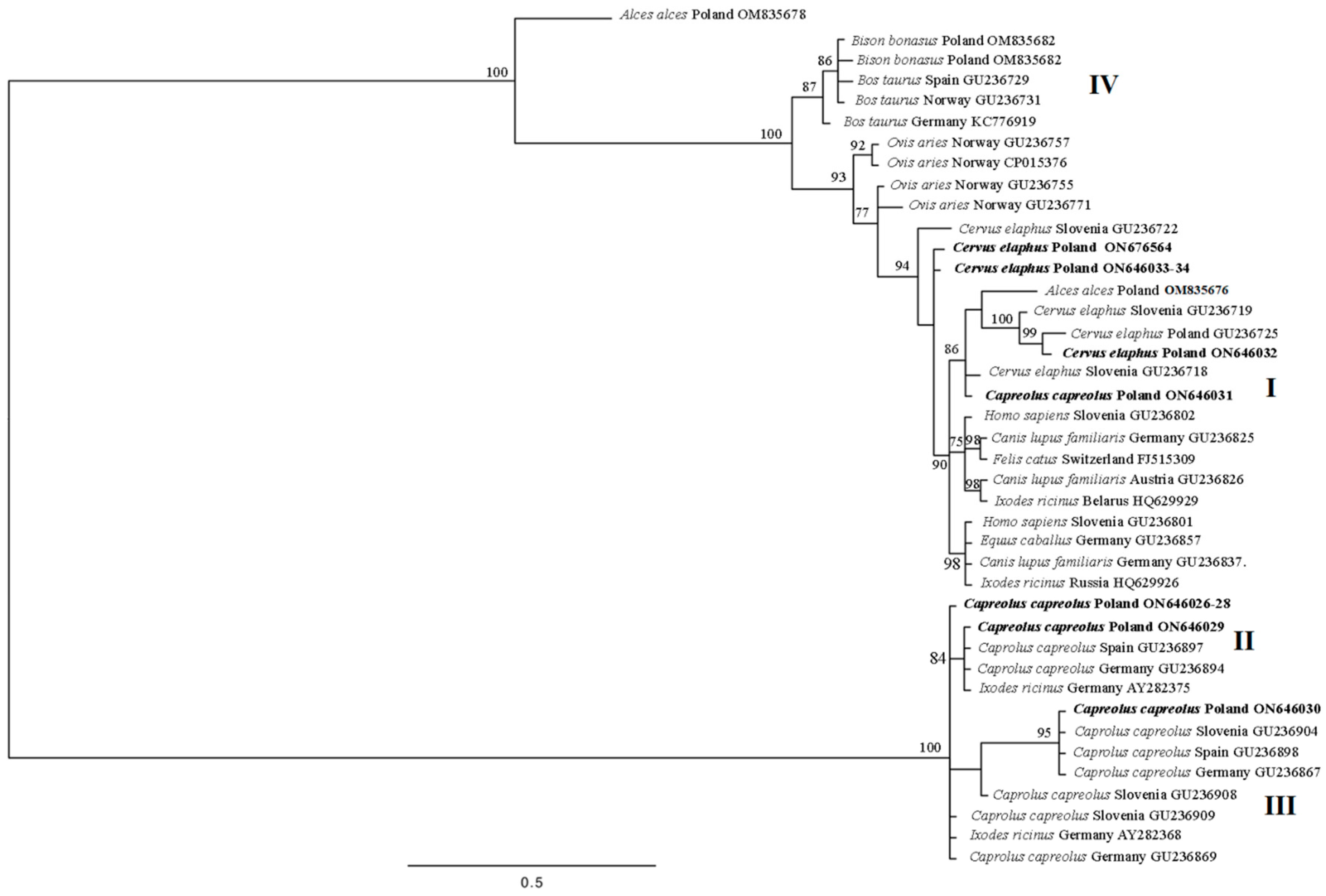
Seven haplotypes were identified among the ten obtained ankA sequences. Of these, four haplotypes (ON646026, ON646029, ON646030, and ON646031) were identified among six A. phagocytophilum sequences from roe deer. One haplotype encompassed three identical sequences (ON646026, ON646027, and ON646028). Three haplotypes of the A. phagocytophilum ankA gene fragment reported in this study (ON646026, ON646029, and ON646030) had already been reported in the GenBank database. The fourth haplotype, i.e., ON646031, had no 100% identity with any submission in GenBank (Table 4). Three haplotypes were identified among the four ankA sequences obtained from red deer. One of these haplotypes was represented by two identical sequences (ON646033 and ON646034) from two different individual red deer, while the other two demonstrated only one sequence each (ON646032 and ON676564). None of the red deer ankA gene variants displayed 100% similarity with GenBank records (Table 4). The phylogenetic analysis indicated that the sequences obtained from roe deer belonged to cluster I, II, and III, and all sequences from the red deer belonged to cluster I (Figure 2).

4. Discussion

The circulation of Anaplasma phagocytophilum in the natural environment among wild game animals is studied and monitored as part of the WHO’s “One Health” program. The balance referred to in the “One Health” program is threatened by the presence of a large and uncontrollable reservoir of Anaplasma genus bacteria, which in Poland consists of cervids. Therefore, our present study focused on the two cervid species, red deer and roe deer, which happen to be two of the largest populations of wild game animals in the country. Our findings based on the use of two genetic markers, groEL and ankA, provide a deeper insight into the diversity of A. phagocytophilum strains and allow us to better understand the nature of the parasite and its potential threat to human and animal health.
Our findings confirm the presence of a range of genetic haplotypes of Anaplasma phagocytophilum in both species of deer, including possible zoonotic strains from ecotype I and cluster I. They also confirm an association between certain haplotypes of A. phagocytophilum with roe deer (ecotypes II and clusters II and III) and red deer (ecotype I and cluster I). The roe deer were found to harbor one new haplotype of the ankA gene involved in cluster I; this phylogenetic finding is the first record of A. phagocytophilum from roe deer associated in cluster I [26].
Five haplotypes of the groEL sequence were identified among the deer. An analysis of the groEL sequences in BLAST showed that all these haplotypes can be classified as European ecotypes. The obtained haplotypes did not demonstrate any high similarity (90–95%), with any of the groEL sequences present in the GenBank database from Asia, MT010579.1, MN989864 [58], MH722253, MH722254 [59], KY379956; North America, AF383227 [60], AY219849 [61], JF494840 [62], DQ680012; and Middle America, MW699686, MW699687 [63]. Interestingly, in red deer, only one haplotype of groEL was recorded, which was found to belong to ecotype I. This genetic variant has quite a broad specificity and has previously been identified in a wide range of hosts, including red deer [48], horses (Equus caballus) [64], sheep [65], and ticks [48,66,67]. In samples from roe deer, four groups of groEL haplotypes were obtained (group I: ON604846, ON604847, ON604851, group II: ON604848, group III: ON604849, group IV: ON604850). The haplotypes from groups I and II belong to ecotype II, and those from groups III and IV belong to ecotype I (Figure 1). Ecotype I, as mentioned above, is associated with humans and domestic, farm, and wild game animals [38,48,64,65,66,67]. While sequences of A. phagocytophilum ecotype II are commonly reported throughout Europe [38,68,69], their occurrence seems restricted only to three host species: roe deer, moose, and the Ixodes genus ticks (Table 3). In Poland, among wild animals, previously analyzed strains of A. phagocytophilum have been found to occupy ecotypes I or II [16,39,68,69,70], which is in line with our findings. In addition, the fact that the obtained partial groEL gene haplotypes from red and roe deer are present in ecotype I and those from roe deer are in ecotype II is in agreement with previous reports on A phagocytophilum ecotype distribution [37].
Our findings confirm the presence of seven genetic variants of the Anaplasma phagocytophilum ankA sequence, belonging to clusters I, II, and III. Cluster I, which includes strains isolated from various hosts, encompassed samples from all red deer and one roe deer. The more host-specific clusters II and III included only the A. phagocytophilum ankA haplotypes from roe deer (Figure 2). All these findings are consistent with previous reports [26]. Three of the haplotypes isolated from roe deer (ON646026, ON646029, and ON646030) were recorded in three European countries, viz. Spain, Germany, and Slovenia, but only in roe deer (Table 4) [26] and in ticks of Ixodes genus (AY282386) [50]. Previous phylogenetic analyses of the A. phagocytophilum ankA sequences identified a strong association between clusters II and III and roe deer [26,40,49,50,51]. One haplotype of the ankA gene identified in the present study (ON646031—roe deer isolate S36) is new and did not share 100% identity with any sequence from the GenBank database. The closest match of this haplotype (99.83%) was observed with the ankA haplotype from red deer in Slovenia (GU236718) (Table 4) [26]. This haplotype was placed in cluster I (Figure 2), which also encompassed sequences obtained from red deer, humans, and domestic animals such as cats (Felis catus) and dogs (Canis lupus familiaris) [26]. A further analysis of the ankA and groEL gene fragments for the A. phagocytophilum isolate from roe deer S36 suggests that this individual was a carrier of an A. phagocytophilum strain not indicating an association with roe deer. Phylogenetic analysis of the ankA sequences from red deer classified them as cluster I; this result is in line with previous findings indicating it to be associated with various hosts, including humans, ticks, game animals like red deer and wild boar (Sus scrofa), domestic animals (cats, dogs), and horses [26,40,49,50,51,69]. The haplotype of A. phagocytophilum from red deer (ON646033) demonstrated the highest similarity (99.69%) with an ankA gene sequence from dogs [64], I. ricinus [71], horses [26], sheep [26], and humans [26,49]. The other two haplotypes, ON646032 and ON676564, were not identical but show close similarity with the same sequence. Haplotype ON646032 was 100% identical to GenBank sequence GU236718 obtained from red deer in Slovenia [26]; the second haplotype (ON676564) was found to share 99.68% similarity with the same sequence (Table 4). In Poland, studies that use ankA genetic marker are less frequent than those using groEL as a marker; however, in the few reports that are available, and in the present study, the A. phagocytophilum stains identified in wild animals were assigned to previously identified clusters: red deer and wild boars to cluster I, roe deer to clusters II and III, and bovids to cluster IV [16,26,68,69,72].
Our findings extend existing knowledge on the genetic diversity and host specificity of Anaplasma phagocytophilum strains in red and roe deer, the two most common species of wild game animals in Poland. Our phylogenetic and sequence analysis of the groEL and ankA gene fragments, as well as previous studies based on 16S rDNA [46], indicate that A. phagocytophilum sequences obtained from red deer are identical to A. phagocytophilum HGA variants identified in humans or are grouped within the same ecotypes and clusters. On the other hand, most of the haplotypes obtained from roe deer in this study seem host-specific and do not show any similarities to zoonotic strains according to our analysis based on groEL and ankA or on 16S rDNA [46]. However, our results indicate that roe deer can also be hosts for less typical A. phagocytophilum variants; this unusual haplotype was classified as cluster I, like haplotypes from red deer samples and humans. All these results show that a highly diverse range of A. phagocytophilum strains are present in cervids in Poland and are potentially zoonotic.

5. Conclusions

In Poland, roe deer are not likely to be natural reservoirs of HGA strains, but red deer can serve as reservoirs hosts of zoonotic strains. Also, we describe the possible occurrence of a nonspecific strain of A. phagocytophilum in roe deer: an A. phagocytophilum variant more characteristic of red deer. According to all of these findings, both wild hosts—Cervus elaphus and Capreolus capreolus—can likely serve as reservoirs of zoonotic Anaplasma phagocytophilum strains.

Author Contributions

A.W.M. and Z.L. designed the study; A.W.M. and Z.L. worked on methodology; Ż.S.-B., G.O. and A.W.M. collected biological samples; A.W.M. and Z.L. performed PCR and sequencing and analyzed sequence data; A.W.M., A.B. and Z.L. worked on the interpretation and discussion of the obtained results; A.W.M. wrote the first draft of the manuscript. All authors have read and agreed to the published version of the manuscript.

Funding

This research received no external funding.

Institutional Review Board Statement

Not applicable.

Informed Consent Statement

Not applicable.

Data Availability Statement

The data that support the findings of this study are available from the corresponding author upon reasonable request.

Acknowledgments

The authors would like to thank the scientists and staff of Witold Stefański Institute of Parasitology Polish Academy of Science, where most of the research included in this publication was performed. This study is part of A.W.M.’s Ph.D. thesis concerning the detection of Anaplasma phagocytophilum in ungulates in Poland.

Conflicts of Interest

The authors declare no conflicts of interest.

References

  1. Dumler, J.S.; Barbet, A.F.; Bekker, C.P.; Dasch, G.A.; Palmer, G.H.; Ray, S.C.; Rikihisa, Y.; Rurangirwa, F.R. Reorganization of genera in the families Rickettsiaceae and Anaplasmataceae in the order Rickettsiales: Unification of some species of Ehrlichia with Anaplasma, Cowdria with Ehrlichia and Ehrlichia with Neorickettsia, descriptions of six new species combinations and designation of Ehrlichia equi and ‘HGE agent’ as subjective synonyms of Ehrlichia phagocytophila. Int. J. Syst. Evol. Microbiol. 2001, 51 Pt. 6, 2145–2165. [Google Scholar] [CrossRef]
  2. Battilani, M.; De Arcangeli, S.; Balboni, A.; Dondi, F. Genetic diversity and molecular epidemiology of Anaplasma. Infect. Genet. Evol. 2017, 49, 195–211. [Google Scholar] [CrossRef]
  3. Gordon, W.S.; Brownlee, A.; Wilson, D.R.; MacLeod, J. Tick-borne fever (a hitherto undescribed disease of sheep). J. Comp. Pathol. 1932, 45, 301–307. [Google Scholar] [CrossRef]
  4. Pusterla, N.; Anderson, R.J.; House, J.K.; Pusterla, J.B.; DeRock, E.; Madigan, J.E. Susceptibility of cattle to infection with Ehrlichia equi and the agent of human granulocytic ehrlichiosis. J. Am. Vet. Med. Assoc. 2001, 218, 1160–1162. [Google Scholar] [CrossRef]
  5. Bown, K.J.; Begon, M.; Bennett, M.; Woldehiwet, Z.; Ogden, N.H. Seasonal dynamics of Anaplasma phagocytophila in a rodent-tick (Ixodes trianguliceps) system, United Kingdom. Emerg. Infect. Dis. 2003, 9, 63–70. [Google Scholar] [CrossRef]
  6. Aktas, M.; Altay, K.; Dumanli, N.; Kalkan, A. Molecular detection and identification of Ehrlichia and Anaplasma species in ixodid ticks. Parasitol. Res. 2009, 104, 1243–1248. [Google Scholar] [CrossRef]
  7. Santos, H.A.; Pires, M.S.; Vilela, J.A.; Santos, T.M.; Faccini, J.L.; Baldani, C.D.; Thomé, S.M.; Sanavria, A.; Massard, C.L. Detection of Anaplasma phagocytophilum in Brazilian dogs by real-time polymerase chain reaction. J. Vet. Diagn. Investig. 2011, 23, 770–774. [Google Scholar] [CrossRef]
  8. Barbet, A.F.; Al-Khedery, B.; Stuen, S.; Granquist, E.G.; Felsheim, R.F.; Munderloh, U.G. An emerging tick-borne disease of humans is caused by a subset of strains with conserved genome structure. Pathogens 2013, 2, 544–555. [Google Scholar] [CrossRef]
  9. Kim, K.H.; Yi, J.; Oh, W.S.; Kim, N.H.; Choi, S.J.; Choe, P.G.; Kim, N.J.; Lee, J.K.; Oh, M.D. Human granulocytic anaplasmosis, South Korea, 2013. Emerg. Infect. Dis. 2014, 20, 1708–1711. [Google Scholar] [CrossRef]
  10. Hofmann-Lehmann, R.; Wagmann, N.; Meli, M.L.; Riond, B.; Novacco, M.; Joekel, D.; Gentilini, F.; Marsilio, F.; Pennisi, M.G.; Lloret, A.; et al. Detection of ‘Candidatus Neoehrlichia mikurensis’ and other Anaplasmataceae and Rickettsiaceae in Canidae in Switzerland and Mediterranean countries. Schweiz. Arch. Tierheilkd. 2016, 158, 691–700. [Google Scholar] [CrossRef]
  11. Sosa-Gutierrez, C.G.; Vargas-Sandoval, M.; Torres, J.; Gordillo-Pérez, G. Tick-borne rickettsial pathogens in questing ticks, removed from humans and animals in Mexico. J. Vet. Sci. 2016, 17, 353–360. [Google Scholar] [CrossRef]
  12. Fukui, Y.; Ohkawa, S.; Inokuma, H. First Molecular Detection and Phylogenetic Analysis of Anaplasma phagocytophilum from a Clinical Case of Canine Granulocytic Anaplasmosis in Japan. Jpn. J. Infect. Dis. 2018, 71, 302–305. [Google Scholar] [CrossRef]
  13. Zaid, T.; Ereqat, S.; Nasereddin, A.; Al-Jawabreh, A.; Abdelkader, A.; Abdeen, Z. Molecular characterization of Anaplasma and Ehrlichia in ixodid ticks and reservoir hosts from Palestine: A pilot survey. Vet. Med. Sci. 2019, 5, 230–242. [Google Scholar] [CrossRef]
  14. Hurtado, C.; Torres, R.; Pérez-Macchi, S.; Sagredo, K.; Uberti, B.; de Souza Zanatto, D.C.; Machado, R.Z.; André, M.R.; Bittencourt, P.; Müller, A. Serological and molecular detection of Anaplasma phagocytophilum in Thoroughbred horses from Chilean racecourses. Ticks Tick. Borne Dis. 2020, 11, 101441. [Google Scholar] [CrossRef]
  15. Kolo, A.O.; Collins, N.E.; Brayton, K.A.; Chaisi, M.; Blumberg, L.; Frean, J.; Gall, C.A.; Wentzel, J.M.; Wills-Berriman, S.; Boni, L.; et al. Anaplasma phagocytophilum and Other Anaplasma spp. in Various Hosts in the Mnisi Community, Mpumalanga Province, South Africa. Microorganisms 2020, 8, 1812. [Google Scholar] [CrossRef]
  16. Myczka, A.W.; Kaczor, S.; Filip-Hutsch, K.; Czopowicz, M.; Plis-Kuprianowicz, E.; Laskowski, Z. Prevalence and Genotyping of Anaplasma phagocytophilum Strains from Wild Animals, European Bison (Bison bonasus) and Eurasian Moose (Alces alces) in Poland. Animals 2022, 12, 1222. [Google Scholar] [CrossRef]
  17. Karbowiak, G.; Biernat, B.; Stańczak, J.; Werszko, J.; Wróblewski, P.; Szewczyk, T.; Sytykiewicz, H. The role of particular ticks developmental stages in the circulation of tick-borne pathogens in Central Europe. 4. Anaplasmataceae. Ann. Parasitol. 2016, 62, 267–284. [Google Scholar] [CrossRef]
  18. Kowalec, M.; Szewczyk, T.; Welc-Falęciak, R.; Siński, E.; Karbowiak, G.; Bajer, A. Rickettsiales Occurrence and Co-occurrence in Ixodes ricinus Ticks in Natural and Urban Areas. Microb. Ecol. 2019, 77, 890–904. [Google Scholar] [CrossRef]
  19. Svitálková, Z.; Haruštiaková, D.; Mahríková, L.; Berthová, L.; Slovák, M.; Kocianová, E.; Kazimírová, M. Anaplasma phagocytophilum prevalence in ticks and rodents in an urban and natural habitat in South-Western Slovakia. Parasit. Vectors 2015, 8, 276. [Google Scholar] [CrossRef]
  20. Jaarsma, R.I.; Sprong, H.; Takumi, K.; Kazimirova, M.; Silaghi, C.; Mysterud, A.; Rudolf, I.; Beck, R.; Földvári, G.; Tomassone, L.; et al. Anaplasma phagocytophilum evolves in geographical and biotic niches of vertebrates and ticks. Parasit. Vectors 2019, 12, 328. [Google Scholar] [CrossRef]
  21. Lesiczka, P.M.; Hrazdilová, K.; Majerová, K.; Fonville, M.; Sprong, H.; Hönig, V.; Hofmannová, L.; Papežík, P.; Růžek, D.; Zurek, L.; et al. The Role of Peridomestic Animals in the Eco-Epidemiology of Anaplasma phagocytophilum. Microb. Ecol. 2021, 82, 602–612. [Google Scholar] [CrossRef]
  22. Rikihisa, Y. Mechanisms of obligatory intracellular infection with Anaplasma phagocytophilum. Clin. Microbiol. Rev. 2011, 24, 469–489. [Google Scholar] [CrossRef]
  23. Bakken, J.S.; Dumler, S. Human granulocytic anaplasmosis. Infect. Dis. Clin. N. Am. 2008, 22, 433–448. [Google Scholar] [CrossRef]
  24. Tylewska-Wierzbanowska, S.; Chmielewski, T. Zoonozy przenoszone przez kleszcze na terenie Polski. Post. Mikrobiol. 2010, 49, 191–197. (In Polish) [Google Scholar]
  25. Matei, I.A.; Estrada-Peña, A.; Cutler, S.J.; Vayssier-Taussat, M.; Varela-Castro, L.; Potkonjak, A.; Zeller, H.; Mihalca, A.D. A review on the eco-epidemiology and clinical management of human granulocytic anaplasmosis and its agent in Europe. Parasit. Vectors 2019, 12, 599. [Google Scholar] [CrossRef] [PubMed]
  26. Scharf, W.; Schauer, S.; Freyburger, F.; Petrovec, M.; Schaarschmidt-Kiener, D.; Liebisch, G.; Runge, M.; Ganter, M.; Kehl, A.; Dumler, J.S.; et al. Distinct host species correlate with Anaplasma phagocytophilum ankA gene clusters. J. Clin. Microbiol. 2011, 49, 790–796. [Google Scholar] [CrossRef] [PubMed]
  27. Stuen, S.; Granquist, E.G.; Silaghi, C. Anaplasma phagocytophilum—A widespread multi-host pathogen with highly adaptive strategies. Front. Cell Infect. Microbiol. 2013, 3, 31. [Google Scholar] [CrossRef] [PubMed]
  28. Razanske, I.; Rosef, O.; Radzijevskaja, J.; Krikstolaitis, R.; Paulauskas, A. Impact of tick-borne Anaplasma phagocytophilum infections in calves of moose (Alces alces) in southern Norway. Folia Parasitol. 2021, 68, 2021.023. [Google Scholar] [CrossRef] [PubMed]
  29. Zhan, L.; Cao, W.C.; Jiang, J.F.; Zhang, X.A.; Liu, Y.X.; Wu, X.M.; Zhang, W.Y.; Zhang, P.H.; Bian, C.L.; Dumler, J.S.; et al. Anaplasma phagocytophilum from Rodents and Sheep, China. Emerg. Infect. Dis. 2010, 16, 764–768. [Google Scholar] [CrossRef]
  30. Aktas, M.; Özübek, S. Bovine anaplasmosis in Turkey: First laboratory confirmed clinical cases caused by Anaplasma phagocytophilum. Vet. Microbiol. 2015, 178, 246–251. [Google Scholar] [CrossRef] [PubMed]
  31. Djiba, M.L.; Mediannikov, O.; Mbengue, M.; Thiongane, Y.; Molez, J.F.; Seck, M.T.; Fenollar, F.; Raoult, D.; Ndiaye, M. Survey of Anaplasmataceae bacteria in sheep from Senegal. Trop. Anim. Health Prod. 2013, 45, 1557–1561. [Google Scholar] [CrossRef]
  32. Ben Said, M.; Belkahia, H.; Messadi, L. Anaplasma spp. in North Africa: A review on molecular epidemiology, associated risk factors and genetic characteristics. Ticks Tick Borne Dis. 2018, 9, 543–555. [Google Scholar] [CrossRef]
  33. Woldehiwet, Z. The natural history of Anaplasma phagocytophilum. Vet. Parasitol. 2010, 167, 108–122. [Google Scholar] [CrossRef]
  34. Dugat, T.; Lagrée, A.C.; Maillard, R.; Boulouis, H.J.; Haddad, N. Opening the black box of Anaplasma phagocytophilum diversity: Current situation and future perspectives. Front. Cell. Infect. Microbiol. 2015, 5, 61. [Google Scholar] [CrossRef] [PubMed]
  35. Langenwalder, D.B.; Silaghi, C.; Nieder, M.; Pfeffer, M.; von Loewenich, F.D. Co-infection, reinfection and superinfection with Anaplasma phagocytophilum strains in a cattle herd based on ankA gene and multilocus sequence typing. Parasit. Vectors 2020, 13, 157. [Google Scholar] [CrossRef]
  36. Jenkins, A.; Handeland, K.; Stuen, S.; Schouls, L.; van de Pol, I.; Meen, R.T.; Kristiansen, B.E. Ehrlichiosis in a moose calf in Norway. J. Wildl. Dis. 2001, 37, 201–203. [Google Scholar] [CrossRef] [PubMed]
  37. Stuen, S.; Pettersen, K.S.; Granquist, E.G.; Bergström, K.; Bown, K.J.; Birtles, R.J. Anaplasma phagocytophilum variants in sympatric red deer (Cervus elaphus) and sheep in southern Norway. Ticks Tick Borne Dis. 2013, 4, 197–201. [Google Scholar] [CrossRef]
  38. Jahfari, S.; Coipan, E.C.; Fonville, M.; van Leeuwen, A.D.; Hengeveld, P.; Heylen, D.; Heyman, P.; van Maanen, C.; Butler, C.M.; Földvári, G.; et al. Circulation of four Anaplasma phagocytophilum ecotypes in Europe. Parasit. Vectors 2014, 7, 365. [Google Scholar] [CrossRef] [PubMed]
  39. Adamska, M. The role of different species of wild ungulates and Ixodes ricinus ticks in the circulation of genetic variants of Anaplasma phagocytophilum in a forest biotope in north-western Poland. Ticks Tick Borne Dis. 2020, 11, 101465. [Google Scholar] [CrossRef]
  40. Jouglin, M.; Chagneau, S.; Faille, F.; Verheyden, H.; Bastian, S.; Malandrin, L. Detecting and characterizing mixed infections with genetic variants of Anaplasma phagocytophilum in roe deer (Capreolus capreolus) by developing an ankA cluster-specific nested PCR. Parasit. Vectors 2017, 10, 377. [Google Scholar] [CrossRef]
  41. Grassi, L.; Franzo, G.; Martini, M.; Mondin, A.; Cassini, R.; Drigo, M.; Pasotto, D.; Vidorin, E.; Menandro, M.L. Ecotyping of Anaplasma phagocytophilum from Wild Ungulates and Ticks Shows Circulation of Zoonotic Strains in Northeastern Italy. Animals 2021, 11, 310. [Google Scholar] [CrossRef]
  42. Petrovec, M.; Sixl, W.; Schweiger, R.; Mikulasek, S.; Elke, L.; Wüst, G.; Marth, E.; Strasek, K.; Stünzner, D.; Avsic-Zupanc, T. Infections of wild animals with Anaplasma phagocytophila in Austria and the Czech Republic. Ann. N. Y. Acad. Sci. 2003, 990, 103–106. [Google Scholar] [CrossRef]
  43. Silaghi, C.; Fröhlich, J.; Reindl, H.; Hamel, D.; Rehbein, S. Anaplasma phagocytophilum and Babesia Species of Sympatric Roe Deer (Capreolus capreolus), Fallow Deer (Dama dama), Sika Deer (Cervus nippon) and Red Deer (Cervus elaphus) in Germany. Pathogens 2020, 9, 968. [Google Scholar] [CrossRef]
  44. Hornok, S.; Sugár, L.; Fernández de Mera, I.G.; de la Fuente, J.; Horváth, G.; Kovács, T.; Micsutka, A.; Gönczi, E.; Flaisz, B.; Takács, N.; et al. Tick- and fly-borne bacteria in ungulates: The prevalence of Anaplasma phagocytophilum, haemoplasmas and rickettsiae in water buffalo and deer species in Central Europe, Hungary. BMC Vet. Res. 2018, 14, 98. [Google Scholar] [CrossRef]
  45. Remesar, S.; Díaz, P.; Prieto, A.; García-Dios, D.; Fernández, G.; López, C.M.; Panadero, R.; Díez-Baños, P.; Morrondo, P. Prevalence and molecular characterization of Anaplasma phagocytophilum in roe deer (Capreolus capreolus) from Spain. Ticks Tick. Borne Dis. 2020, 11, 101351. [Google Scholar] [CrossRef]
  46. Stefanidesova, K.; Kocianova, E.; Boldis, V.; Kostanova, Z.; Kanka, P.; Nemethova, D.; Spitalska, E. Ecidence of Anaplasma phagocytophilum and Ricktettsia helvetica infection in free-ranging ungulates in central Slovakia. Eur. J. Wildl. Res. 2008, 54, 519–524. [Google Scholar] [CrossRef]
  47. Bown, K.J.; Lambin, X.; Ogden, N.H.; Begon, M.; Telford, G.; Woldehiwet, Z.; Birtles, R.J. Delineating Anaplasma phagocytophilum ecotypes in coexisting, discrete enzootic cycles. Emerg. Infect. Dis. 2009, 15, 1948–1954. [Google Scholar] [CrossRef] [PubMed]
  48. Stigum, V.M.; Jaarsma, R.I.; Sprong, H.; Rolandsen, C.M.; Mysterud, A. Infection prevalence and ecotypes of Anaplasma phagocytophilum in moose Alces alces, red deer Cervus elaphus, roe deer Capreolus capreolus and Ixodes ricinus ticks from Norway. Parasit. Vectors 2019, 12, 1. [Google Scholar] [CrossRef] [PubMed]
  49. Massung, R.F.; Owens, J.H.; Ross, D.; Reed, K.D.; Petrovec, M.; Bjoersdorff, A.; Coughlin, R.T.; Beltz, G.A.; Murphy, C.I. Sequence analysis of the ank gene of granulocytic ehrlichiae. J. Clin. Microbiol. 2000, 38, 2917–2922. [Google Scholar] [CrossRef] [PubMed]
  50. von Loewenich, F.D.; Baumgarten, B.U.; Schröppel, K.; Geissdörfer, W.; Röllinghoff, M.; Bogdan, C. High diversity of ankA sequences of Anaplasma phagocytophilum among Ixodes ricinus ticks in Germany. J. Clin. Microbiol. 2003, 41, 5033–5040. [Google Scholar] [CrossRef]
  51. Majazki, J.; Wüppenhorst, N.; Hartelt, K.; Birtles, R.; von Loewenich, F.D. Anaplasma phagocytophilum strains from voles and shrews exhibit specific ankA gene sequences. BMC Vet. Res. 2013, 9, 235. [Google Scholar] [CrossRef]
  52. Myczka, A.W.; Steiner-Bogdaszewska, Z.; Filip-Hutsch, K.; Olos´, G.; Czopowicz, M.; Laskowski, Z. Detection of Anaplasma phagocytophilum in Wild and Farmed Cervids in Poland. Pathogens 2021, 10, 1190. [Google Scholar] [CrossRef]
  53. Alberti, A.; Zobba, R.; Chessa, B.; Addis, M.F.; Sparagano, O.; Pinna Parpaglia, M.L.; Cubeddu, T.; Pintori, G.; Pittau, M. Equine and canine Anaplasma phagocytophilum strains isolated on the island of Sardinia (Italy) are phylogenetically related to pathogenic strains from the United States. Appl. Environ. Microbiol. 2005, 71, 6418–6422. [Google Scholar] [CrossRef]
  54. Massung, R.F.; Levin, M.L.; Munderloh, U.G.; Silverman, D.J.; Lynch, M.J.; Gaywee, J.K.; Kurtti, T.J. Isolation and propagation of the Ap-Variant 1 strain of Anaplasma phagocytophilum in a tick cell line. J. Clin. Microbiol. 2007, 45, 2138–2143. [Google Scholar] [CrossRef]
  55. Ronquist, F.; Huelsenbeck, J.P. MrBayes 3: Bayesian phylogenetic inference under mixed models. Bioinformatics 2003, 19, 1572–1574. [Google Scholar] [CrossRef] [PubMed]
  56. Guindon, S.; Gascuel, O. A simple, fast, and accurate algorithm to estimate large phylogenies by maximum likelihood. Syst. Biol. 2003, 52, 696–704. [Google Scholar] [CrossRef]
  57. Darriba, D.; Taboada, G.L.; Doallo, R.; Posada, D. jModelTest 2: More models, new heuristics and parallel computing. Nat. Methods 2012, 9, 772. [Google Scholar] [CrossRef]
  58. Lu, M.; Li, F.; Liao, Y.; Shen, J.J.; Xu, J.M.; Chen, Y.Z.; Li, J.H.; Holmes, E.C.; Zhang, Y.Z. Epidemiology and Diversity of Rickettsiales Bacteria in Humans and Animals in Jiangsu and Jiangxi provinces, China. Sci. Rep. 2019, 9, 13176. [Google Scholar] [CrossRef] [PubMed]
  59. Liz, J.S.; Sumner, J.W.; Pfister, K.; Brossard, M. PCR detection and serological evidence of granulocytic ehrlichial infection in roe deer (Capreolus capreolus) and chamois (Rupicapra rupicapra). J. Clin. Microbiol. 2002, 40, 892–897. [Google Scholar] [CrossRef]
  60. Liddell, A.M.; Stockham, S.L.; Scott, M.A.; Sumner, J.W.; Paddock, C.D.; Gaudreault-Keener, M.; Arens, M.Q.; Storch, G.A. Predominance of Ehrlichia ewingii in Missouri dogs. J. Clin. Microbiol. 2003, 41, 4617–4622. [Google Scholar] [CrossRef]
  61. Rejmanek, D.; Bradburd, G.; Foley, J. Molecular characterization reveals distinct genospecies of Anaplasma phagocytophilum from diverse North American hosts. J. Med. Microbiol. 2012, 61 Pt 2, 204–212. [Google Scholar] [CrossRef]
  62. Bermúdez, C.S.E.; Félix, M.L.; Domínguez, A.L.; Kadoch, N.; Muñoz-Leal, S.; Venzal, J.M. Molecular screening for tick-borne bacteria and hematozoa in Ixodes cf. boliviensis and Ixodes tapirus (Ixodida: Ixodidae) from western highlands of Panama. Curr. Res. Parasitol. Vector Borne Dis. 2021, 1, 100034. [Google Scholar] [CrossRef] [PubMed]
  63. Fröhlich, J.; Fischer, S.; Bauer, B.; Hamel, D.; Kohn, B.; Ahlers, M.; Obiegala, A.; Overzier, E.; Pfeffer, M.; Pfister, K.; et al. Host-pathogen associations revealed by genotyping of European strains of Anaplasma phagocytophilum to describe natural endemic cycles. Parasit. Vectors 2023, 16, 289. [Google Scholar] [CrossRef]
  64. Chastagner, A.; Dugat, T.; Vourc’h, G.; Verheyden, H.; Legrand, L.; Bachy, V.; Chabanne, L.; Joncour, G.; Maillard, R.; Boulouis, H.J.; et al. Multilocus sequence analysis of Anaplasma phagocytophilum reveals three distinct lineages with different host ranges in clinically ill French cattle. Vet. Res. 2014, 45, 114. [Google Scholar] [CrossRef]
  65. Bauer, B.U.; Răileanu, C.; Tauchmann, O.; Fischer, S.; Ambros, C.; Silaghi, C.; Ganter, M. Anaplasma phagocytophilum and Anaplasma ovis-Emerging Pathogens in the German Sheep Population. Pathogens 2021, 10, 1298. [Google Scholar] [CrossRef]
  66. Welc-Falęciak, R.; Kowalec, M.; Karbowiak, G.; Bajer, A.; Behnke, J.M.; Siński, E. Rickettsiaceae and Anaplasmataceae infections in Ixodes ricinus ticks from urban and natural forested areas of Poland. Parasit. Vectors 2014, 7, 121. [Google Scholar] [CrossRef]
  67. Luu, L.; Palomar, A.M.; Farrington, G.; Schilling, A.K.; Premchand-Branker, S.; McGarry, J.; Makepeace, B.L.; Meredith, A.; Bell-Sakyi, L. Bacterial Pathogens and Symbionts Harboured by Ixodes ricinus Ticks Parasitising Red Squirrels in the United Kingdom. Pathogens 2021, 10, 458. [Google Scholar] [CrossRef]
  68. Rymaszewska, A. Genotyping of Anaplasma phagocytophilum strains from Poland for selected genes. Folia Biol. 2014, 62, 37–48. [Google Scholar] [CrossRef] [PubMed]
  69. Myczka, A.W.; Szewczyk, T.; Laskowski, Z. The Occurrence of Zoonotic Anaplasma phagocytophilum Strains, in the Spleen and Liver of Wild Boars from North-West and Central Parts of Poland. Acta Parasitol. 2021, 66, 1082–1085. [Google Scholar] [CrossRef] [PubMed]
  70. Hildebrand, J.; Buńkowska-Gawlik, K.; Adamczyk, M.; Gajda, E.; Merta, D.; Popiołek, M.; Perec-Matysiak, A. The occurrence of Anaplasmataceae in European populations of invasive carnivores. Ticks Tick Borne Dis. 2018, 9, 934–937. [Google Scholar] [CrossRef]
  71. Katargina, O.; Geller, J.; Alekseev, A.; Dubinina, H.; Efremova, G.; Mishaeva, N.; Vasilenko, V.; Kuznetsova, T.; Järvekülg, L.; Vene, S.; et al. Identification of Anaplasma phagocytophilum in tick populations in Estonia, the European part of Russia and Belarus. Clin. Microbiol. Infect. 2012, 18, 40–46. [Google Scholar] [CrossRef] [PubMed]
  72. Michalik, J.; Stańczak, J.; Cieniuch, S.; Racewicz, M.; Sikora, B.; Dabert, M. Wild boars as hosts of human-pathogenic Anaplasma phagocytophilum variants. Emerg. Infect. Dis. 2012, 18, 998–1001. [Google Scholar] [CrossRef] [PubMed]
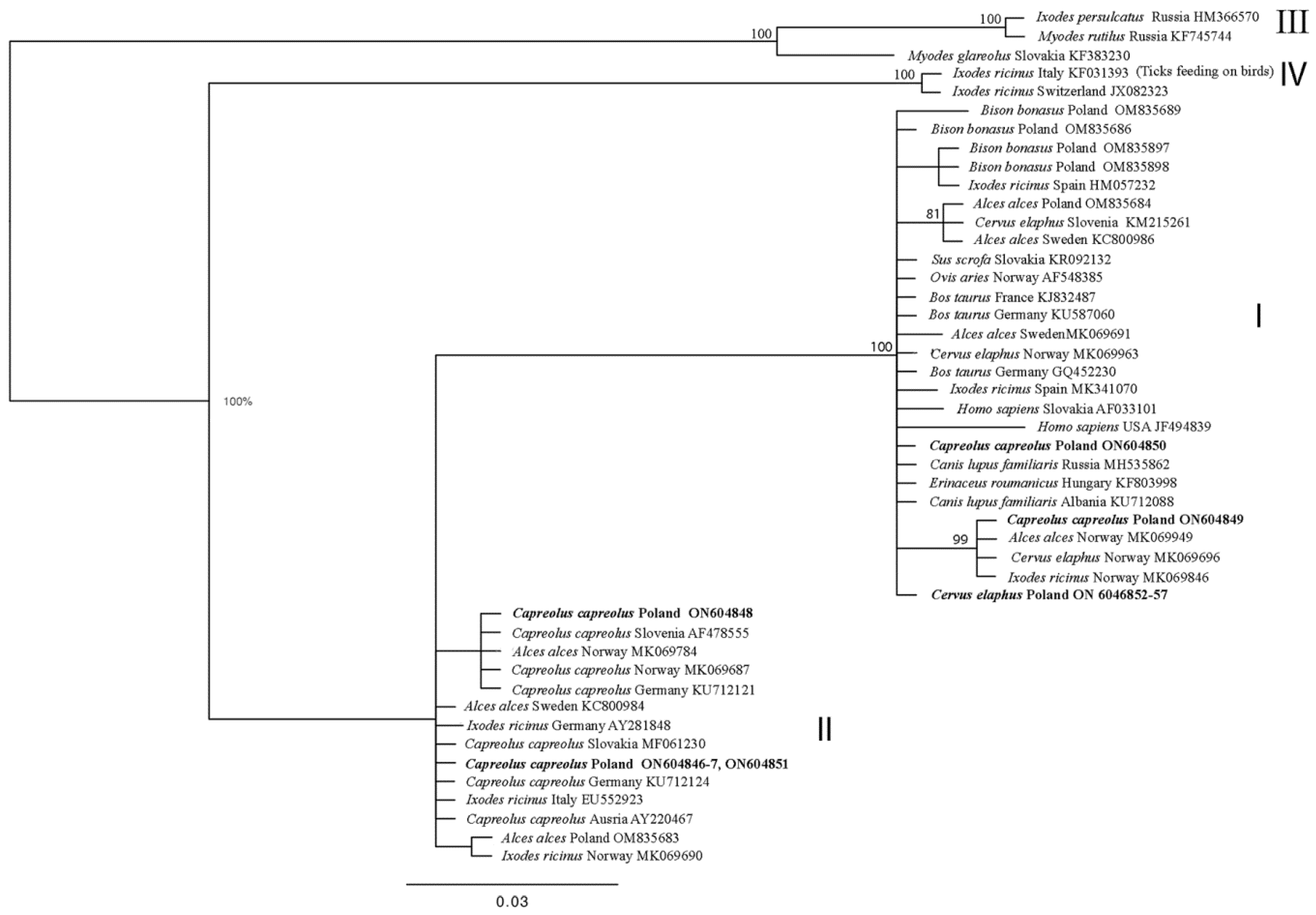
Figure 1. Phylogenetic tree of groEL partial gene (486 bp) of Anaplasma phagocytophilum haplotypes, constructed using Bayesian inference (BI) analysis with MrBayes version 3.2 [55]. The HKY + I model was chosen as the best-fitting nucleotide substitution model using JModelTest version 2.1.10 software [56,57]. The analysis was run for 2,000,000 generations, with 500,000 generations discarded as burn-in. In bold are sequences from this study. I–IV: ecotypes of A. phagocytophilum according to Jahfari et al. [38].
Animals 14 00637 g001
Figure 2. Phylogenetic tree of the ankA partial gene (633 bp) haplotypes from Anaplasma phagocytophilum, constructed using Bayesian inference (BI) analysis with MrBayes version 3.2 [55]. The GTR + I + G model was chosen as the best-fitting nucleotide substitution model using JModelTest version 2.1.10 software [56,57]. The analysis was run for 2,000,000 generations, with 500,000 generations discarded as burn-in. Sequences from this study are in bold. I–IV: clusters of A. phagocytophilum according to Scharf et al. [26].
Animals 14 00637 g002
Table 1. Demographic characteristics of the study population. The numbers of farmed cervids presented in parentheses.
SpeciesAdultsJuvenileTotal
MalesFemales
Red deer8 (0)49 (15)33 (0)90 (15)
Roe deer7491470
In total159847160
Table 2. Primer designed and used to amplify ankA gene fragment from roe deer samples.
ReactionPrimersTm (°C)Reference
PCRankAF1a 5′-TGCTGTAAATGAAGAAATTACAACTTC-3′
ankAF1b 5′-TGGTGTAAATGAAGAAATTACAACTC-3′
ankARC 5′-GCCTTTAGTAGTACTCTACATGC-3′
53 °C
52 °C
54 °C
this study
Nested—PCRankAF2a 5′-CTGACCGCTGAAGCACTAA-3′
ankAR1a 5′-GAAGCCAGATGCAGTAACGA-3′
ankAR1b 5′-GAAGCAAGATGCAGTAACGA-3′
51 °C
52 °C
50 °C
Table 3. Haplotypes of the A. phagocytophilum groEL gene fragment and reference sequences from the GenBank database, divided into ecotypes. Sequences from this study in bold.
EcotypeHostNo. GenBank Sequence (This Study)Sequences with 100% SimilarityHostCountry
Ired deer
(Cervus elaphus)
ON604852-87MK069963red deerNorway
KU712106Austria
KJ832471horseFrance
MZ348280sheepGermany
MK069889Ixodes ricinusNorway
MW732493UK
KF312358Poland
roe deer
(Capreolus capreolus)
ON604849MK069949mooseNorway
MK069696red deer
MK069797I. ricinus
ON604850AY281823I. ricinusGermany
KJ832474cowFrance
GQ452227goatSwitzerland
MW013536hedgehogCzech Republic
IIroe deer
(Capreolus capreolus)
ON604846-47
ON6048451
MN093177I. ricinusNederland
KU712112roe deerGermany
KC800984moose
AY220467roe deerAustria
ON604848KF031380I. ricinusItaly
HQ629905Estonia
MK069774mooseNorway
GQ988754roe deerAustria
KU712121Germany
Table 4. Haplotypes of A. phagocytophilum ankA gene fragment and reference sequences from the GenBank database, divided into clusters. Sequences from this study in bold.
ClusterHostNo. GenBank Sequence (This Study)Sequences with Highest IdentityHostCountry
Ired deer
(Cervus elaphus)
ON646032GU236718 (100%)red deerSlovenia
ON646033-34KJ832286 (99.69%)dogFrance
HQ629928
(99.69%)
Ixodes ricinusEstonia
ON676564GU236718
(99.68%)
red deerSlovenia
roe deer
(Capreolus capreolus)
ON646031GU236718 (99.83%)red deerSlovenia
KJ832286 (99.66%)dogFrance
HQ629928
(99.66%)
Ixodes ricinusEstonia
IIroe deer
(Capreolus capreolus)
ON646026-28GU236909
(100%)
roe deerSlovenia
GU236894
(100%)
Germany
GU236900
(100%)
Spain
AY282386
(100%)
Ixodes ricinusGermany
ON646029GU236897
(100%)
roe deerSpain
GU236874
(100%)
Germany
AY282375
(100%)
Ixodes ricinus
IIIroe deer
(Capreolus capreolus)
ON646030GU236904
(100%)
roe deerSlovenia
GU236898
(100%)
Spain
Disclaimer/Publisher’s Note: The statements, opinions and data contained in all publications are solely those of the individual author(s) and contributor(s) and not of MDPI and/or the editor(s). MDPI and/or the editor(s) disclaim responsibility for any injury to people or property resulting from any ideas, methods, instructions or products referred to in the content.