Article

Antibiotic Resistance of Bacteria Isolated from Clinical Samples and Organs of Rescued Loggerhead Sea Turtles (Caretta caretta) in Southern Italy

1
Istituto Zooprofilattico Sperimentale del Mezzogiorno, Via Salute 2, 80055 Portici, Italy
2
Department of Marine Animal Conservation and Public Engagement, Stazione Zoologica Anton Dohrn, Via Nuova Macello 16, 80055 Portici, Italy
*
Authors to whom correspondence should be addressed.
These authors have contributed equally to this work.
Animals 2024, 14(14), 2103; https://doi.org/10.3390/ani14142103
Submission received: 21 June 2024 / Revised: 13 July 2024 / Accepted: 17 July 2024 / Published: 18 July 2024

Abstract

:

Simple Summary

Antimicrobial resistance is threatening health and the economy worldwide, not only in clinical settings, but also in livestock and wildlife management. Among the numerous wild animals affected, sea turtles are particularly exposed to antibiotics, due to biological and habitat characteristics. This study analysed the hospitalisation records in a sea turtle rescue centre in southern Italy during the last decade, focusing on the antibiotic resistance of bacteria isolated from clinical samples and organs. Resistance patterns to 7 antibiotics were examined in 138 bacteria isolated from 60 loggerhead sea turtles, highlighting 6 main bacterial families with different resistance rates. Vibrionaceae represented the predominant family, although the resistance rates did not exceed 10.5%. Similar results were described for Shewanellaceae, whereas the less represented families were those with the highest resistance rates and numbers of multidrug-resistant strains, especially Enterobacteriaceae and Morganellaceae. Previous antibiotic therapies appeared to enhance resistance, emphasizing the need to improve antimicrobial stewardship. Isolated bacteria are considered opportunistic pathogens, but their antibiotic resistance might compromise treatment and rehabilitation, potentially affecting the population health of these endangered species. Given the possible transfer of resistance across species, these findings should be considered from a One Health perspective, including other animals, humans and the environment.

Abstract

Antimicrobial resistance affects all environments, endangering the health of numerous species, including wildlife. Increasing anthropic pressure promotes the acquisition and dissemination of antibiotic resistance by wild animals. Sea turtles, being particularly exposed, are considered sentinels and carriers of potential zoonotic pathogens and resistant strains. Therefore, this study examined the antibiotic resistance profiles of bacteria isolated from loggerhead sea turtles hospitalised in a rescue centre of Southern Italy over a 9-year period. Resistance to ceftazidime, doxycycline, enrofloxacin, flumequine, gentamicin, oxytetracycline and sulfamethoxazole-trimethoprim was evaluated for 138 strains isolated from the clinical samples or organs of 60 animals. Gram-negative families were the most isolated: Vibrionaceae were predominant, followed by Shewanellaceae, Pseudomonadaceae, Enterobacteriaceae and Morganellaceae. These last three families exhibited the highest proportion of resistance and multidrug-resistant strains. Among the three Gram-positive families isolated, Enterococcaceae were the most represented and resistant. The opportunistic behaviour of all the isolated species is particularly concerning for diseased sea turtles, especially considering their resistance to commonly utilised antibiotics. Actually, the multiple antibiotic resistance was higher when the sea turtles were previously treated. Taken together, these findings highlight the need to improve antimicrobial stewardship and monitor antibiotic resistance in wildlife, to preserve the health of endangered species, along with public and environmental health.

1. Introduction

Antimicrobial resistance (AMR) is one of the major problems challenging human and animal health in our century. The discovery and development of antibiotics was among the most important and successful cornerstones in the healthcare field. For many years, AMR has been contrasted by the introduction of new molecules on the market. However, in recent years, the development of new antibiotics has slowed down, and this, along with other aspects, has allowed an increase in antibiotic resistance [1,2]. Actually, antibiotic resistance is an ancient phenomenon, because it is the result of the adaptation of organisms to their environment; on the one hand, bacteria develop resistance mechanisms to defend themselves and survive; on the other, many antibiotics are naturally occurring molecules, and for this reason, intrinsic resistance may exist [3,4]. However, the current worldwide emergency mainly refers to acquired resistance, which is largely caused by the improper or massive use of antibiotics and which threatens global health [5,6].
Indeed, the appearance and dissemination of AMR has direct and indirect repercussions on different sectors involving humans and animals. In clinical settings, AMR compromises the ability to effectively treat infections, forces the use of last-resort molecules, increases the chances of complications in vulnerable populations, and prolongs treatment or hospitalisation periods, ultimately resulting in inflated healthcare costs [7,8]. In livestock farming, all of this translates into serious economic losses, since reductions in feed conversion, growth rates and reproductive performances contribute to decreased productivity, especially in low–middle income countries, thus widening the gap with other countries [5,9].
For wildlife conservation, the consequences of AMR have probably been overlooked and generally considered minor, compared to the particular interest raised by the potential role of wild animals as reservoirs of antibiotic-resistant bacteria (ARB), especially in relation to possible risks for public health [10,11]. Although wildlife is a recognised part of resistance dynamics, as suggested by several reports of ARB in different species and habitats [12], the origins, routes and implications of this dynamics are still not fully understood [10,13]. Wild animals are unlikely to be subjected to antibiotic treatment; therefore, research has been oriented towards their acquisition of ARB from the environment. In this regard, numerous studies have suggested that increasing interactions between humans, domestic animals and wild animals enhance the risk of transferring ARB to wildlife, and vice versa [11,12,14]. Specifically, the increase in human pressure (including the loss and fragmentation of habitats, the release of agricultural and urban wastewater, climate change, etc.) facilitates the dissemination of pathogens and antibiotic resistance in the environment. Aquatic ecosystems are considered particularly impacted, also, considering the role played by water in the dispersion of antibiotics and ARB [13,15,16]. Surface water, wastewater and recreational water have been previously monitored for the presence of ARB, and several environments (e.g., fish farms, animal manure, hospitals, spill events) have been identified as potential sources of antibiotic residues and ARB. Through seepage or sewage, resistance elements can reach and contaminate underground waters, lagoons or effluents, ending up in marine habitats and resulting in the appearance and transfer of resistance in marine organisms [15,16,17].
From a different point of view, some wild species, due to their characteristics and responses to environmental changes, could serve as sentinels of challenged habitats or anthropogenic stressors, and potentially provide an early warning system for ecosystem and public health [18]. In the marine environment, seabirds, marine mammals and sea turtles have long been used as sentinels of marine pollution due to their lifespan, global distribution, trophic level or habitat use [18,19,20,21]. Specifically, loggerhead sea turtles are long-living animals distributed in warm and temperate seas throughout the world, and they feed at progressively higher trophic levels as they grow, with the consequent tendency of bio-accumulation [22,23]. Moreover, these species frequently inhabit near-shore ecosystems, which are the most impacted by anthropogenic activities, and are susceptible to similar noxious agents as other animals and humans (pathogens, toxins and chemical compounds). Actually, the exposure to polluted areas might be increased by the complex life cycle of sea turtles, because they utilise different habitats as nesting sites, migratory routes or foraging grounds, thus multiplying the risk of contact with and acquisition of ARB, possibly through food, seawater or even sand [23,24,25]. In the Mediterranean Sea, antibiotic resistance has been recently detected in several bacteria isolated from wild and captive sea turtles, both healthy and diseased [15,26,27].
The majority of bacteria in sea turtles are considered opportunistic pathogens; therefore, the presence of ARB is especially concerning for diseased or captive animals, which might be at greater risk of secondary infections [28]. In these cases, antibiotic resistance might limit therapeutic options, interfere with the rehabilitation process, and delay the reintroduction to nature, ultimately affecting the health of individuals and the population [10,29]. Within this context, rescue and rehabilitation centres play a significant role for sea turtle conservation, by preserving the health of juvenile and adult turtles, which are the most commonly rescued and the most important for population stability, due to their reproductive value [30,31,32,33].
Loggerhead sea turtles are distributed throughout the Mediterranean basin, with an estimated population of 1,201,845 (95% CI 838,864–1,548,280) [34]. The distribution of C. caretta is widespread along all the coasts of the Italian peninsula. However, the Adriatic and Ionian areas are important for the developmental stages of juveniles [23]. Instead, the Tyrrhenian coast, in particular the Campania region, provides foraging and nesting grounds [22,35].
In the Campania region, in Southern Italy, a sea turtle stranding, conservation and monitoring network has been in place for the last decades, involving several partners. Among them, the Istituto Zooprofilattico Sperimentale del Mezzogiorno (IZSM) and the Stazione Zoologica Anton Dohrn (SZN) are responsible for healthcare and research activities on dead and live sea turtles, respectively. Therefore, this study aimed at investigating the antibiotic resistance profiles of bacteria isolated from diseased and captive sea turtles, through a retrospective analysis conducted on loggerhead sea turtles (Caretta caretta) rescued and hospitalised at the rescue centre of SZN “Turtle Point” (TP, Portici, Italy).

2. Materials and Methods

2.1. Study Population

This study included bacterial strains belonging to 60 Mediterranean loggerhead sea turtles (Table S1), admitted at the rescue centre of SZN (TP) over a period of 9 years (2015–2023). All animals were alive at the time of recovery and were found adrift (n = 35), stranded (n = 18) or bycaught (n = 7) off the Italian coasts. Sea turtles recovered from Campania waters (n = 31) were immediately transported to the TP, while others (n = 29) were transferred to the TP from other Italian rescue centres (e.g., Lazio, Sicily) at a later stage, in order to access long-term or specialised care.
Upon admission, the following morphometric parameters were measured: body weight (range = 0.05–68.95 kg; mean ± SD = 22.82 ± 18.45 kg); curved carapace length (range = 7.8–84 cm; mean ± SD = 54.8 ± 17.7 cm); curved carapace width (range = 7.5–86.5 cm; mean ± SD = 50.6 ± 16.9 cm); straight-line carapace length (range = 6.6–76 cm; mean ± SD = 47.9 ± 17.7 cm); and straight-line carapace width (range = 5.8–61.7 cm; mean ± SD = 39.5 ± 14.1 cm).
The responsible veterinarian examined each animal to evaluate the cause of hospitalisation, the type and severity of lesions, and treatment and rehabilitation protocols. Samples for bacteriological analyses and antibiotic susceptibility testing (AST) were collected depending on the lesions and veterinary diagnoses, as described below, in order to rule out or prevent infections. However, the sampling did not always occur upon admission, since lesions ascribable to potential infections could have manifested throughout the hospitalisation period. Additionally, in a few cases, samples were collected only with the purpose of medical clearance just before reintroduction into nature.
Turtles that died upon admission at the TP or during hospitalisation, before sampling (n = 10), were sent to the IZSM to conduct post-mortem examinations and investigate the causes of death. In these cases, bacteriological analyses and AST were carried out on the organs collected during the necropsy, as detailed below.
Since the timing of sample collection differed between animals, the examined individuals were categorised as treated and untreated sea turtles: the former group consisted of animals submitted to antibiotic treatment before sample collection (n = 20); the latter consisted of animals not submitted to antibiotic treatment before sample collection, including those that did not receive any antibiotic treatment at all (n = 40).

2.2. Sample Collection

From living sea turtles, clinical samples were collected at the TP, according to the type and site of lesions and to the suspected diagnoses, using sterile cotton-tipped swabs or needle aspiration.
Specifically, in cases of external lesions (e.g., carapace fractures, flipper wounds, fistulas, skin discolorations) swabs samples were collected from the inner part of the lesion, following superficial curettage. In cases of ocular lesions, swab samples were obtained from the conjunctival fornix after gently distancing the eyelids. Whenever possible, a small amount of necrotic tissue, if present within the affected site, was also collected from the inner part of the lesions. In suspected cases of seawater aspiration, swab samples were collected from the glottis, after opening the rhamphotheca with a previously disinfected PVC tube. When coelomitis or gastrointestinal perforations were suspected, coelomic fluid was collected by needle aspiration through the prefemoral fossa, after skin disinfection with 4% chlorexidine digluconate. In other suspected cases of internal lesions (e.g., abscesses), needle aspiration was performed to collect a small amount of tissue from the inner part of the lesion, after disinfecting the surrounding skin with 4% chlorexidine digluconate. In only one case, lymphatic fluid was erroneously collected during blood sampling and sent for bacteriological analyses. In cases of the absence of lesions, uncertain diagnoses or pre-release medical clearance, oropharyngeal and cloacal sampling were performed as a screening method. For oropharyngeal samples, a swab was gently brushed over the oral mucosa, after opening the rhamphotheca (with a previously disinfected PVC tube). For cloacal samples, a swab was gently inserted and rotated into the cloaca (1–5 cm, depending on the animal size), after rinsing the pericloacal area with 4% chlorexidine digluconate.
All procedures involving living animals were included in the clinical examination and diagnostic investigations of the rescued sea turtles, in accordance with the authorisation by the Ministry of the Environment (Protocols n. 0042848/PNM 9 August 2013, n. 0024471/PNM 22 November 2016, n. 0000992/PNM 22 January 2020, n. 0020551/PNM 13 February 2023).
From dead sea turtles, organs were sampled at the IZSM during post-mortem examination, performed according to Wyneken, 2001 [36] and Poppi and Marchiori, 2013 [37]. Briefly, the external examination included species identification, the collection of morphometric data, the classification of condition and nutritional code, and the evaluation of possible external lesions. The internal examination allowed access to and inspection of the coelomic organs, by removing the plastron and the shoulder blades, as well as the brain and salt glands, by separating and opening the skull. During the necropsy, from all sea turtles, the liver, kidneys, spleen, intestine, brain, and lungs were routinely sampled independently from any associated lesions, whereas other organs were sampled only upon the presence of lesions. The surface of each target organ was scalded with a spatula and cut with a sterile blade, in order to collect a swab sample from the inner part, avoiding contamination
All collected samples were transferred and processed at the IZSM for bacterial isolation, identification and antibiotic susceptibility testing.

2.3. Bacterial Isolation and Identification

All samples were streaked on Blood Agar (BA) (Biolife, Monza, Italy) and Tryptone Soy Agar with 2% NaCl (TSAs) (Microbiol srl, Cagliari, Italy), and the plates were incubated at 25 °C ± 1 °C for 24–72 ± 3 h under aerobic conditions; contrarily, the plates from the intestine were incubated only under anaerobic conditions. Bacterial colonies grown on BAs and TSAs were selected according to their morphology, isolated and tested with oxidase (Oxoid, Thebarton, South Australia), catalase (bioMérieux, Marcy-l’Etoile, France), motility tests and Gram stain [38,39]. The strains were subsequently confirmed to the genus or species level through miniaturised biochemical identification systems, such as API® and VITEK2® (bioMérieux, Marcy-l’Etoile, France).

2.4. Antibiotic Susceptibility Testing

All identified strains were subjected to AST using the disk diffusion method (Kirby–Bauer), in accordance with the criteria established by the Clinical & Laboratory Standards Institute (CLSI) documents [40,41]. Specifically, an inoculum of 0.5 McFarland was prepared for each bacterial strain. Within 15 min of preparing the inoculum, a swab was dipped into the suspension and streaked over the entire surface of the plate. Different culture media were chosen, according to the strain growth requirements: Mueller Hinton Agar (MH) (Microbiol srl, Cagliari, Italy) was used for Enterobacteriaceae, Pseudomonadaceae and other Gram-negative bacteria; Mueller Hinton + 5% Blood (MHB) was used for Gram-positive bacteria; and Mueller Hinton + 1.5% NaCL (MHS) for halophilic bacteria such as Vibrionaceae [40,42,43].
Given the clinical application, the antibiotics to be tested were chosen among the molecules which are commonly used for sea turtles, selecting the following antibiotic disks (Oxoid, Basingstoke, UK): ceftazidime (CAZ, 30 µg); doxycycline (DO, 30 µg); enrofloxacin (ENR, 5 µg); flumequine (UB, 30 µg); gentamicin (CN, 10 µg); oxytetracycline (OXY, 30 µg); and sulfamethoxazole-trimethoprim (SXT, 25 µg). Each antibiotic disk was picked up with sterile forceps and placed on the plate. All plates were incubated at 22 °C ± 1 °C for 24 ± 3 h. The diameter of the inhibition zone was measured with a calliper, and the results were classified as susceptible (S), intermediate (I) and resistant (R), following clinical breakpoints for aquatic animals as suggested by the CLSI documents [40,41].
For each strain, the multiple antibiotic resistance (MAR) index was calculated as a ratio between the number of resistances and the number of tested antibiotics [44]. Additionally, strains exhibiting resistance to at least three different classes of antibiotics were defined as multidrug-resistant (MDR), in accordance with Magiorakos et al. (2012) [45].

2.5. Data Analysis

All data were entered and analysed in an Excel spreadsheet (Microsoft, Redmond WA, USA). The proportion of MDR strains and the MAR indices were compared between treated and untreated sea turtles. Statistical analyses were performed with chi-square and Fisher exact tests, setting significance level at p < 0.05 and using MedCalc® (version 10.3.2.0, Mariakerke, Belgium).

3. Results

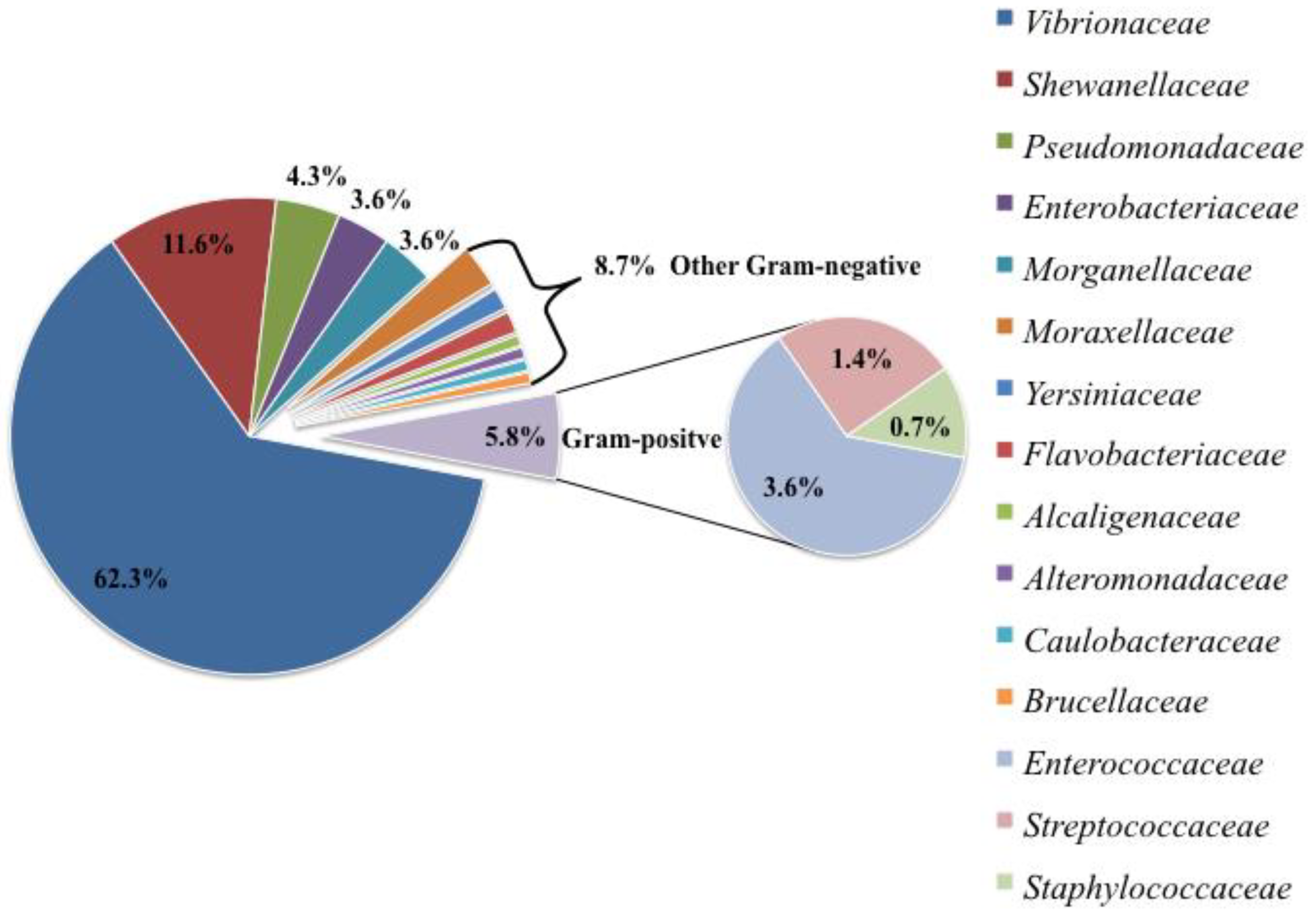
3.1. Bacterial Isolation and Identification

This study included a total of 138 bacterial strains isolated from 60 sea turtles, 50 of which were live turtles and 10 of which were dead. The vast majority (123/138; 89.1%) of bacteria were isolated from clinical samples (n = 63), whereas the remaining ones (15/138; 10.9%) were isolated from organs (n = 23). The different sampling sites that resulted positive to bacterial isolation are summarised in Figure 1, distinguished between clinical samples (Figure 1A) and organs (Figure 1B). It is important to mention that a bacterial strain could have been cultured from different samples of the same turtle, and, vice versa, multiple bacterial strains could have been cultured from the same sample.
Gram-negative organisms represented almost the entirety of the isolated bacteria (130/138; 94.2%), whereas Gram-positive ones accounted for a small portion of the total amount of strains (8/138; 5.8%). In particular, 15 bacterial families were cultured and identified, 12 of which belonged to Gram-negative and 3 to Gram-positive organisms. Within Gram-negative organisms, the most represented families were Vibrionaceae (86/138; 62.3%) and Shewanellaceae (16/138; 11.6%), followed by Pseudomonadaceae (6/138; 4.3%), Enterobacteriaceae (5/138; 3.6%) and Morganellaceae (5/138; 3.6%). The other Gram-negative families (i.e., Alcaligenaceae, Alteromonadaceae, Brucellaceae, Caulobacteraceae, Flavobacteriaceae, Moraxellaceae, Yersiniaceae) were less represented (each <3%) and together constituted only 8.7% of the total amount of strains (12/138), as summarised in Figure 2.
Within Gram-positive organisms, Enterococcaceae (5/138; 3.6%), Streptococcaceae (2/138; 1.4%) and Staphylococcaceae (1/138; 0.7%) were the only represented families (Figure 2).
Bacterial families were isolated with different frequencies from clinical samples and organs. In particular, Vibrionaceae, Pseudomonadaceae and Enterococcaceae were more frequently isolated from clinical samples (94.2%, 83.3 and 80%, respectively) than organs (5.8%, 16.7% and 20%, respectively). Contrarily, Enterobacteriaceae and Morganellaceae were more frequently isolated from organs (80%) than clinical samples (20%). The remaining bacterial families, including Shewanellaceae, were exclusively isolated from clinical samples.
Focusing on the sampling sites (Figure 3), swabs from the oropharynx, flipper wounds, necrotic tissue and carapace fractures resulted in the isolation of numerous strains, predominantly from the Vibrionaceae family. Similarly, Shewanellaceae and Enterococcaceae were mainly isolated from flipper wound swabs, whereas Pseudomonadaceae were slightly more frequent from cloacal swabs. Contrarily, Enterobacteriaceae and Morganellaceae were mainly isolated from the intestine and lung, followed by the liver and spleen.
Regarding bacterial identification (Table S2), 21 species of the Vibrionaceae family were confirmed: Vibrio alginolyticus (n = 26), Vibrio harveyi (n = 22), Vibrio fluvialis (n = 5), Vibrio navarrensis (n = 5), Vibrio diazotrophicus (n = 3), Vibrio anguillarum (n = 1), Vibrio furnissii (n = 1), Vibrio gallicus (n = 1), Vibrio mediterranei (n = 1), Vibrio orientalis (n = 1), Vibrio parahaemolyticus (n = 1), Vibrio pelagius (n = 1), Vibrio pelagius II (n = 1), Vibrio scophthalmi (n = 1), Vibrio splendidus I (n = 1), Vibrio splendidus II (n = 1), Vibrio tubiashii (n = 1), Vibrio xuii (n = 1), Photobacterium damselae subsp. damselae (n = 4), Photobacterium swingsii (n = 1) and Grimontia hollisae (n = 1). In six additional cases, the strains were not successfully identified to the species level; therefore, they were reported as Vibrio spp. (n = 4), Photobacterium spp. (n = 1) and Aliivibrio spp. (n = 1). Within Shewanellaceae, the only two isolated species were Shewanella algae (n = 13) and Shewanella putrefaciens (n = 3). Among Pseudomonadaceae, the isolated species were Pseudomonas aeruginosa (n = 2), Pseudomonas putida (n = 2), Pseudomonas fluorescens (n = 1) and Pseudomonas mendocina (n = 1). The species belonging to the Enterobacteriaceae family were Citrobacter freundii (n = 4) and Enterobacter cancerogenus (n = 1), whereas the only species within Morganellaceae was Morganella morganii subsp. morganii (n = 5).
Among Gram-positive families, Enterococcus faecalis (n = 3), Enterococcus gallinarum (n = 1) and Vagococcus fluvialis (n = 1) were the isolated species within Enterococcaceae, whereas Lactococcus garviae (n = 2) and Staphylococcus sciuri (n = 1) were the only species belonging respectively to Streptococcaceae and Staphylococcaceae.

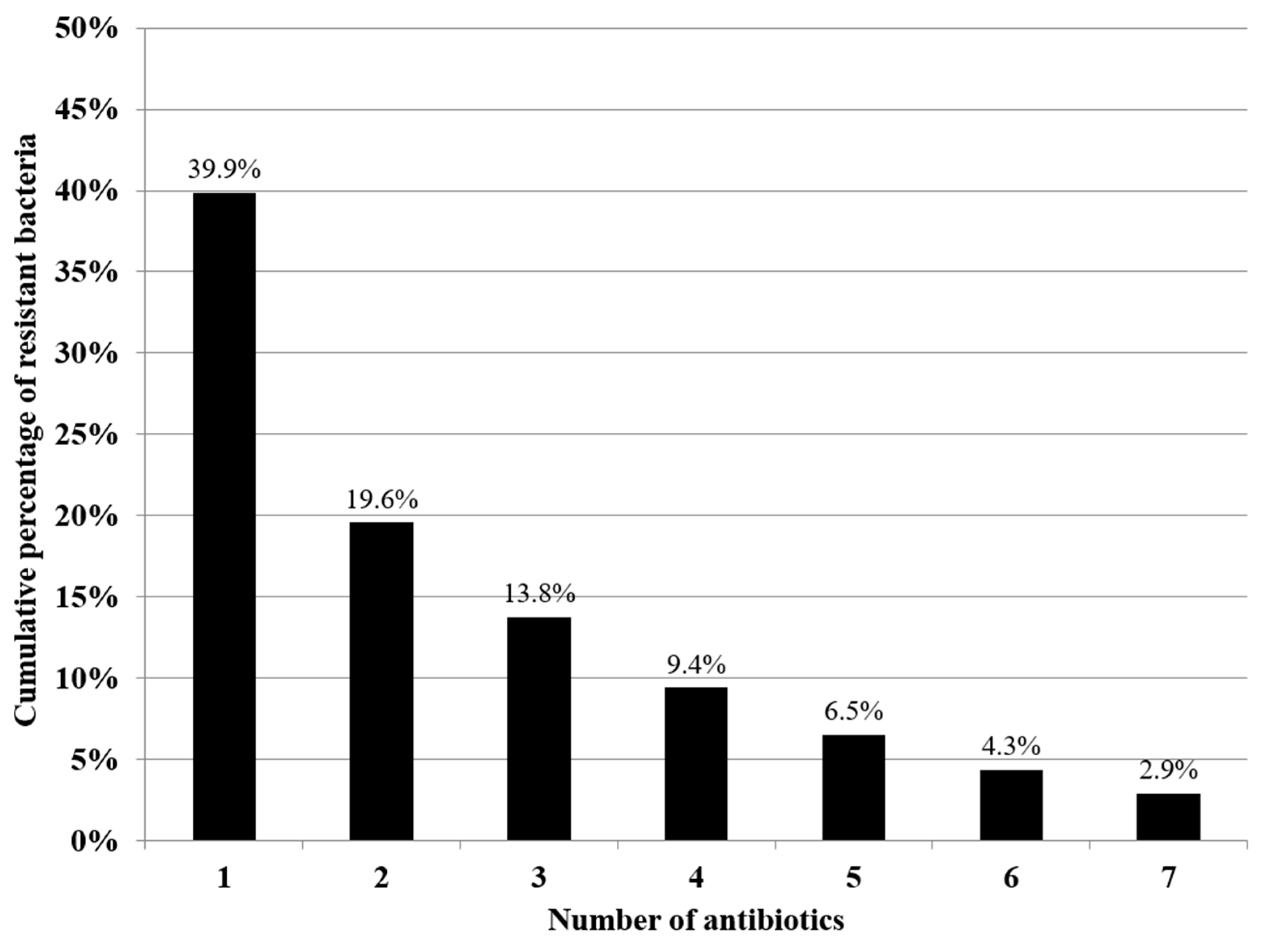
3.2. Antibiotic Susceptibility Testing

The antibiotic resistance profile of all the bacterial strains was evaluated. Less than half of the bacterial strains (55/138; 39.9%) exhibited resistance to one or more of the tested antibiotics, whereas only a small number of strains (4/138; 2.9%) exhibited simultaneous resistance to all of the tested antibiotics (Figure 4).
Bacterial strains exhibited different profiles of antibiotic resistance, which are detailed in Table 1; in particular, the highest rates of resistance were detected towards ceftazidime (17.5% of the strains) and gentamicin (16.8%). For the other tested antibiotics, similar percentages of resistant strains were found for doxycycline and sulfamethoxazole-trimethoprim (respectively, 13.8% and 13.1%) and for enrofloxacin and oxytetracycline (both 12.3%), whereas the lowest percentage was found for flumequine (11.1%). Relatively high percentages of strains (always above 64%) were susceptible to the tested antibiotics, with the highest value (85.4%) detected for sulfamethoxazole-trimethoprim (Table 1).
When considering the main bacterial families (contributing at least 3% of the total amount of strains), Vibrionaceae and Shewanellaceae were those with the lowest percentages of resistant strains to the selected antibiotics, compared to the other families. Specifically, 10.5% of Vibrionaceae strains were resistant to gentamicin and even lower percentages were resistant to other antibiotics: sulfamethoxazole-trimethoprim (8.2%), ceftazidime (8.1%), doxycycline (7%), oxytetracycline (7%), enrofloxacin (5.8%) and flumequine (1.2%). Similarly, 18.8% of Shewanellaceae strains were resistant to doxycycline, while the resistance rates for the other tested antibiotics were between 6.3 and 6.7%. Pseudomonadaceae was the family with the highest rate of resistance to sulfamethoxazole-trimethoprim (66.7% of strains), whereas lower rates of resistance were detected for ceftazidime, doxycycline, enrofloxacin, oxytetracycline (50%), flumequine and gentamicin (33.3%). As regards Enterobacteriaceae, the resistance rates ranged between 40% (i.e., flumequine, oxytetracycline, sulfamethoxazole-trimethoprim) and 60% (i.e., ceftazidime, doxycycline, enrofloxacin, gentamicin). Morganellaceae was the family with the highest rates of resistance (80% of strains) towards four of the seven selected antibiotics (i.e., doxycycline, enrofloxacin, flumequine and oxytetracycline); relatively lower resistance rates were detected towards sulfamethoxazole-trimethoprim and gentamicin (60.0%), whereas only 20% of strains were resistant to ceftazidime.
Among the Gram-positive families, no strain was resistant to doxycycline or oxytetracycline, and no strain within Enterococcaceae was resistant to sulfamethoxazole-trimethoprim either. On the contrary, all Enterococcaceae were resistant to gentamicin and 75% of strains were resistant to ceftazidime, whereas the resistance rates towards enrofloxacin and flumequine were 20% and 60%, respectively (Table 2).
Concerning simultaneous resistance to multiple antibiotics, the MAR indices ranged from 0 to 1, with a MAR index for all bacterial strains of 0.14, which varied between bacterial families. Specifically, the highest MAR index was attributed to the family Morganellaceae (0.66), followed by Enterobacteriaceae (0.51), Pseudomonadaceae (0.48) and Enterococcaceae (0.31). On the contrary, Vibrionaceae and Shewanellaceae were the families with the lowest MAR indices (respectively, 0.07 and 0.08).
Additionally, eleven strains were defined as MDR: three belonging to Enterobacteriaceae (C. freundii); three to Morganellaceae (M. morganii subsp. morganii), two to Pseudomonadaceae (P. aeruginosa and P. putida); two to Vibrionaceae (V. alginolyticus); and one to Shewanellacae (S. algae).
The MAR indices and the proportions of MDR strains were compared between the two groups of sea turtles classified as treated and untreated. The MAR index in the treated group was significantly higher than the MAR index in the untreated group (respectively, 0.29 and 0.07; χ2 = 77.917; DF = 1; p < 0.01). Similarly, the proportion of MDR strains was significantly higher in the treated group compared to the untreated group (respectively, 20.9% and 2.1%; χ2 = 14.3; DF = 1; p < 0.01).

4. Discussion

Loggerhead sea turtle bacteria have attracted increasing attention, to learn more about their role as part of normal microbiota or as pathogens [27,46,47]. In the current study, the bacterial strains isolated from rescued loggerhead sea turtles were considered opportunistic pathogens, in accordance with Ebani [28]. Indeed, the health status of the examined turtles was compromised for different reasons, including the underlying conditions leading to hospitalisation and the stress related to captivity.
Generally, bacterial entry has been hypothesised from the environment through dermal, oral or cloacal routes [48]. In accordance with this hypothesis, the strains described in the present results were mainly isolated from sites exposed to the external environment, such as open injuries (such as limb wounds or carapace fractures) and mucosae (such as the oropharyngeal cavity and the conjunctiva).
The greater presence of Gram-negative bacteria compared to Gram-positive ones is in agreement with previous studies [25,27,49]. However, the predominance of Vibrionaceae over other bacterial families has not always been reported [48,50], as Enterobacteriaceae have usually been the most commonly isolated [26,51].
V. alginolyticus and V. harveyi were the most frequently isolated Vibrionaceae species, in line with previous studies [48,52]. Both species are normally present in aquatic environments, but have also been associated with lesions in sea turtles, such as dermatitis, enteritis and pneumonia [28,53]. Moreover, these species are considered important pathogens of marine animals and potential zoonotic agents [54,55]. Within Vibrionaceae, it is worth mentioning the isolation of P. damselae damselae and V. parahaemolyticus, despite the limited number of strains, because of their recognised pathogenicity in humans and other animals [49,56,57].
The low rates of antibiotic resistance detected among the isolated Vibrionaceae are in line with some previous studies regarding aquaculture and coastal waters [58,59] but in contrast with others documenting higher resistance rates [60,61]. These variations have been usually related to the strain origins, the nutrient levels in the waters, or the use of antibiotics in different countries. For instance, sulphonamides have been widely used in Asia [59], whereas oxytetracyclines have been the most commonly licensed for European aquaculture, followed by flumequine [58]. Additionally, tetracyclines and fluoroquinolones are the antibiotics of choice to treat severe vibriosis in human and veterinary practice [58,60]. In sea turtles, V. alginolyticus has been described as the Vibrio species with the highest rates of antibiotic resistance [52], as also highlighted by the two MDR strains isolated in the present study.
Shewanellaceae have been frequently reported among the predominant bacterial families in sea turtles, usually as part of the normal microbiota [26,62]. However, the isolation of S. algae, and to a lesser extent of S. putrefaciens, mainly from flipper wounds and necrotic tissue, might underline an opportunistic role in the development of secondary infections [63,64]. Despite the low antibiotic resistance rates of Shewanellaceae, almost 20% of strains were resistant to doxycycline, and one MDR strain was found resistant to five out of seven antibiotics (except for gentamicin and sulfamethoxazole-trimethoprim). Indeed, the genus Shewanella has been increasingly reported as responsible for several infections and the appearance of antibiotic resistance [65]. In particular, immunocompromised individuals are at risk of developing soft tissue infections, and treatment might be limited by the presence of resistance genes to β-lactams and quinolones, which are commonly utilised in human medicine [65].
Pseudomonadaceae have been commonly isolated from sea turtles, in particular from cloacal swabs [25,51,66], similarly to our results. However, Pseudomonas has been found in association with different lesions, including respiratory and ocular infections [28,53]. The antibiotic resistance of the Pseudomonadaceae family, in particular of P. aeruginosa, has always received particular attention and has been investigated in different matrices [67,68,69]. In the present study, Pseudomonadaceae were the third family in terms of rates of resistance and MDR strains, similarly to previous results that reported Pseudomonas species as the ones exhibiting resistance to the highest numbers of antibiotics, both in green and loggerhead sea turtles [70,71]. These resistance properties could also be transferred through mobile elements to other bacterial species, which is particularly concerning, considered the ubiquitous nature of Pseudomonas species and their potential as opportunistic pathogens for humans and animals alike [67].
Although Enterobacteriaceae were not the dominant family within the Gram-negative bacteria here isolated, contrarily to previous reports [26,51], Citrobacter is confirmed as one of the most common genera described within this family [72,73]. Indeed, Citrobacter species are well documented sea turtle pathogens, responsible for several lesions [53,74,75], especially in weak and captive turtles [26,27]. Additionally, high levels of antibiotic resistance have frequently been detected in Citrobacter, and Enterobacteriaceae in general, consistent with the present results [71,72,73]. These findings support the existing literature regarding the role of Enterobacteriaceae in the circulation of antibiotic resistance among environmental, commensal and pathogenic bacteria [72,76]. Enterobacteriaceae have an extraordinary ability to acquire resistance from the environment, especially when exposed to antibiotics (in particular, β-lactams) [77]. Moreover, this bacterial family is considered the main carrier of extended-spectrum β-lactamase (ESBL)-encoding genes, which could be transferred with other resistance genes through large mobile elements, thus promoting the occurrence of MDR [78]. Given the abundance of ESBL-producing Enterobacteriaceae and resistance elements described in aquatic environments (e.g., wastewater, freshwater, estuaries and coastal waters), the exchange of resistance from Enterobacteriaceae to other bacteria, and vice versa, could be enhanced in contaminated waters [17,79]. When this gene flow involves pathogenic species, Enterobacteriaceae might serve as mediators between aquatic and clinical settings, with the consequent risk of treatment failure [76,80].
Morganellaceae are a relatively new family, composed of species formerly included within the Enterobacteriaceae [81]; therefore, some reports still refer to M. morganii as a member of Enterobacteriaceae. In addition to isolation from oropharyngeal and cloacal samples [63,73] M. morganii has been detected in leatherback sea turtles with intestinal diverticulitis [82], and it has been associated with wounded and rehabilitated sea turtles, like those examined in our study [27,72]. Consistent with previous reports from sea turtles in areas subjected to elevated anthropic pressure, such as the Mediterranean Sea and the Great Barrier Reef [51,72], Morganellaceae isolated in this work exhibited the greatest rates of resistance towards the selected antibiotics, with the exception of ceftazidime. Contrarily, Morganellaceae isolated from sea turtles of the North-East Atlantic only exhibited intermediate resistance to carbapenems, probably because of the more pristine environment characterizing the island of Maio [63]. A high proportion of MDR strains was observed within this family, although this finding has been mainly ascribed to Morganellaceae’s intrinsic properties [45,72].
Regarding Gram-positive microorganisms, far fewer reports are currently available in sea turtles, compared to Gram-negative; nevertheless, the strains found in this study belonged to the Enterococcaceae, Streptococcaceae and Staphylococcaceae families, as previously described [28,62]. Similarly to Gram-negative bacteria, studies carried out on Gram-positive bacteria usually refer to many species as part of the normal microbiota of sea turtles [66,83,84]. However, these species have also been reported as potential pathogens, alone or in association with Gram-negative bacteria [53,75]. Among Gram-positive families, Enterococcaceae was the most frequently isolated within this study; in particular, Enterococcus faecalis is confirmed as one of the most common enterococci in sea turtles [28,85]. Although usually present in the marine environment, E. faecalis has been isolated from sea turtles with respiratory and skin infections, osteomyelitis and lesions of the bladder, liver, lungs and muscles [49,86,87]. Antibiotic resistance has often been observed in Enterococcaceae; in particular, the strains isolated in this study were found resistant to cephalosporins, aminoglycosides and quinolones, in accordance with previous reports [86,87,88]. Contrarily, the Enterococcus strains here isolated were not resistant to tetracyclines, as described in different marine species [85,87,89]. The medical management of sea turtles infected by Enterococcus might prove challenging, as it often requires a long-term or combined use of multiple antibiotic classes. In particular, the wide use of cephalosporins in sea turtles might contribute to the emergence of resistant enterococci [86,87]. E. faecalis is also considered responsible for many human enterococcal infections, potentially resulting in lethal systemic disease when improperly treated [90]. Within the other two Gram-positive families detected in our study, Staphylococcus sciuri and Lactococcus garviae (Staphylococcaceae and Steptococcaceae, respectively) have seldom been reported in sea turtles. Specifically, S. sciuri has been detected in blood samples of a green turtle affected by fibropapillomatosis [48], whereas L. garviae has been isolated from the liver and kidney of a loggerhead sea turtle and from the cloacal sample of a green turtle [49,89]. Antibiotic resistance was detected toward ceftazidime (S. sciuri and 1/2 L. garviae), flumequine (2/2 L. garviae) and sulfamethoxazole-trimethoprim (1/2 L. garviae), similarly to previous reports [89,91].
The finding of significantly higher MAR indices and a proportion of MDR strains among the bacteria isolated from sea turtles that received antibiotic treatment, compared to those that did not receive antibiotics (or that initiated treatment following sample collection), might suggest that antibiotic treatment had an influence on antibiotic resistance. Indeed, antibiotic treatment could promote resistance through mutations, resistant genes or strain replacement [92]. In particular, reduced dosages, limited tissue permeability and incomplete treatment schedules are known factors contributing to the appearance of antibiotic resistance [93,94]. However, even when antibiotic therapy is accurately prescribed and performed, it could induce the emergence of resistance, predominantly by the selection of resistant strains within the microbial communities of the host, instead of de novo evolution [92]. This is especially true for wound pathogens, such as the ones that were mainly isolated in the present study. On the other hand, some antibiotics, especially quinolones and β-lactams, are involved in the emergence of antibiotic resistance through different mechanisms of mutagenesis, leading to mutations that might be maintained in the host microbiota or be transferred to pathogenic agents [94,95,96]. Additionally, quinolones and cephalosporins are among the most commonly used antibiotics in sea turtles; however these classes include molecules of the Watch group in the AWaRe classification, which are first- and second-choice antibiotics indicated for specific infections and more susceptible to be the target of antibiotic resistance [97].
Therefore, the presence of ARB in sea turtles represents a risk to their health, as it could restrict antibiotic options to treat bacterial infections and potentially compromise the rehabilitation success [27,98]. From a wider perspective, this risk might be extended to humans and other animals, since sea turtles are a potential carrier of zoonotic agents [99,100,101,102]. Zoonoses have been reported to spill over from terrestrial to marine species and back, outlining marine animals not only as victims but also as vectors [52,91,103,104]. Ecotourism and conservational efforts might contribute to the expansion of sea turtle zoonoses, although the major concerns derive from interactions with captive or dead sea turtles and the consumption of turtle products (still practiced in some areas) [52,104,105]. Therefore, some populations (marine animal researchers, rehabilitators, trainers, veterinarians and volunteers) might be at greater risk of transmission through occupational exposure [91,103]. The staff working at the TP was not tested for the presence of ARB; however, biosecurity measures are commonly adopted in the centre, and to our knowledge there were no reports of bacterial infections through occupational exposure during the period of study. The dissemination of antibiotic resistance in pathogenic bacteria poses additional risks for public health, and it becomes particularly alarming when it involves antibiotics commonly used in human medicine or last-resort molecules for the treatment of serious infections, as it reduces their effectiveness and thus any chance of a favourable outcome [10,29,63].

5. Conclusions

Bacterial infections affecting sea turtles are mainly caused by opportunistic agents, usually present in the surrounding environment or as commensal microorganisms. The results of this study provide valuable information on antibiotic resistance in bacteria affecting hospitalised sea turtles. Even if this study does not assess possible variations in antibiotic resistance over time, the present findings cover almost a decade and could represent an appropriate baseline for future investigations. Moreover, these data could be useful to organisations and healthcare and research bodies involved in the rehabilitation and management of sea turtles and potentially other aquatic species.
It is of fundamental importance to improve antimicrobial stewardship in wildlife facilities: susceptibility testing should be performed before initiating antibiotic therapy; narrow-spectrum antibiotics should be preferred to broad-spectrum antibiotics; Access group antibiotics should be prioritised over other groups; and a treatment schedule should be strictly followed [106].
Additional measures to prevent and treat infections in sea turtles and other animals include ensuring the adequate hygienic conditions of the surrounding environment, such as the rearing water [107], and investigating alternative options to antibiotics, such as phage therapy in sea turtles [108,109].
Given the presence of potentially resistant zoonotic bacteria, the adoption of these recommendations and the continuous monitoring of antibiotic resistance will be beneficial in preserving these endangered species, as well as public health and the environment, within a wider One Health perspective. Further research and a close collaboration among different professional figures are strongly needed to clarify the possibilities and mechanisms of antibiotic resistance dissemination across environments, animals and humans. Moreover, molecular methods, which were not applied in the present study, could contribute to the determination of resistant genes, the identification of their sources and the elucidation of their transmission dynamics, in order to infer the potential effects on all the sectors involved.

Supplementary Materials

The following supporting information can be downloaded at: https://www.mdpi.com/article/10.3390/ani14142103/s1, Table S1: Hospitalisation information of 60 loggerhead sea turtles admitted at Turtle Point between 2015 and 2023 and subjected to bacteriological analysis; Table S2: Species identification within each of the main six bacterial families isolated from the clinical samples and organs of 60 loggerhead sea turtles.

Author Contributions

Conceptualisation, E.E. and A.P.; methodology, E.E., A.P. and D.I.; formal analysis, E.E. and A.P.; investigation, E.E., A.P., A.A., D.I., M.O. and G.P.; resources, G.F., E.D.C., S.H. and F.D.N.; data curation, E.E., A.P., A.A. and D.I.; writing—original draft preparation, E.E. and A.P.; writing—review and editing, A.A., M.O., D.I., G.P., F.M., G.F., E.D.C., S.H. and F.D.N.; visualisation, E.E., A.P. and F.M.; supervision, S.H. and F.D.N.; project administration, A.A., S.H. and F.D.N.; All authors have read and agreed to the published version of the manuscript.

Funding

This research received no external funding.

Institutional Review Board Statement

This study analysed hospitalisation records, and no living animal was handled for the purpose of this study. All procedures were included in the clinical examination and diagnostic investigations of the rescued sea turtles, in accordance with the authorisation by the Ministry of the Environment (Protocols n. 0042848/PNM 9 August 2013, n. 0024471/PNM 22 November 2016, n. 0000992/PNM 22 January 2020, n. 0020551/PNM 13 February 2023).

Informed Consent Statement

Not applicable.

Data Availability Statement

The data presented in this study are available within the article.

Acknowledgments

The authors would like to acknowledge the staff of Turtle Point and all the operators and volunteers within the network that contribute to sea turtle rescue and rehabilitation.

Conflicts of Interest

The authors declare no conflicts of interest.

References

  1. Martinez, J.L. General principles of antibiotic resistance in bacteria. Drug Discov. Today Technol. 2014, 11, 33–39. [Google Scholar] [CrossRef] [PubMed]
  2. Reygaert, W.C. An overview of the antimicrobial resistance mechanisms of bacteria. AIMS Microbiol. 2018, 4, 482–501. [Google Scholar] [CrossRef] [PubMed]
  3. D’Costa, V.M.; King, C.E.; Kalan, L.; Morar, M.; Sung, W.W.L.; Schwarz, C.; Froese, D.; Zazula, G.; Calmels, F.; Beruyne, R.; et al. Antibiotic resistance is ancient. Nature 2011, 477, 457–461. [Google Scholar] [CrossRef] [PubMed]
  4. Munita, J.M.; Arias, C.A. Mechanisms of Antibiotic Resistance. Microbiol. Spectr. 2016, 4, 481–511. [Google Scholar] [CrossRef] [PubMed]
  5. Dadgostar, P. Antimicrobial Resistance: Implications and Costs. Infect. Drug Resist. 2019, 12, 3903–3910. [Google Scholar] [CrossRef]
  6. Despotovic, M.; de Nies, L.; Bhanu Busi, S.; Wilmes, P. Reservoirs of antimicrobial resistance in the context of One Health. Curr. Opin. Microbiol. 2023, 73, 102291. [Google Scholar] [CrossRef] [PubMed]
  7. Friedman, N.D.; Temkin, E.; Carmeli, Y. The negative impact of antibiotic resistance. Clin. Microbiol. Infect. 2016, 22, 416–422. [Google Scholar] [CrossRef] [PubMed]
  8. Report World Health Organization. Antimicrobial Resistance: An Emerging Water, Sanitation and Hygiene Issue; World Health Organization: Geneva, Switzerland, 2014.
  9. Bengtsson, B.; Greko, C. Antibiotic resistance—Consequences for animal health, welfare, and food production. Ups J. Med. Sci. 2014, 119, 96–102. [Google Scholar] [CrossRef]
  10. Arnold, K.E.; Williams, N.J.; Bennett, M. ‘Disperse abroad in the land’: The role of wildlife in the dissemination of antimicrobial resistance. Biol. Lett. 2016, 12, 20160137. [Google Scholar] [CrossRef]
  11. Radhouani, H.; Silva, N.; Poeta, P.; Torres, C.; Correia, S.; Igrejas, G. Potential impact of antimicrobial resistance in wildlife, environment and human health. Front. Microbiol. 2014, 5, 23. [Google Scholar] [CrossRef]
  12. Allen, H.K.; Donato, J.; Wang, H.H.; Cloud-Hansen, K.A.; Davies, J.; Handelsman, J. Call of the wild: Antibiotic resistance genes in natural environments. Nat. Rev. Microbiol. 2010, 8, 251–259. [Google Scholar] [CrossRef] [PubMed]
  13. Vittecoq, M.; Godreuil, S.; Prugnolle, F.; Durand, P.; Brazier, L.; Renaud, N.; Arnal, A.; Aberkane, S.; Jean-Pierre, H.; Gauthier-Clerc, M.; et al. Antimicrobial resistance in wildlife. J. Appl. Ecol. 2016, 53, 519–529. [Google Scholar] [CrossRef]
  14. Plaza-Rodriíguez, C.; Alt, K.; Grobbel, M.; Hammerl, J.A.; Irrgang, A.; Szabo, I.; Stingl, K.; Schuh, E.; Wiehle, L.; Pfefferkorn, B.; et al. Wildlife as Sentinels of Antimicrobial Resistance in Germany? Front. Vet. Sci. 2021, 7, 627821. [Google Scholar] [CrossRef] [PubMed]
  15. Gambino, D.; Savoca, D.; Sucato, A.; Gargano, V.; Gentile, A.; Pantano, L.; Vicari, D.; Alduina, R. Occurrence of Antibiotic Resistance in the Mediterranean Sea. Antibiotics 2022, 11, 332. [Google Scholar] [CrossRef] [PubMed]
  16. Marangi, M.; Carlucci, R.; Carlino, P.; Fanizza, C.; Cirelli, G.; Maglietta, R.; Beneduce, L. Dolphins and sea turtles may host zoonotic parasites and pathogenic bacteria as indicators of anthropic pressure in the Gulf of Taranto (Northern Ionian Sea, Central-Eastern Mediterranean Sea). Vet. Res. Commun. 2022, 46, 1157–1166. [Google Scholar] [CrossRef] [PubMed]
  17. Tacão, M.; Correia, A.; Henriques, I. Resistance to broad-spectrum antibiotics in aquatic systems: Anthropogenic activities modulate the dissemination of blaCTX-M-like genes. Appl. Environ. Microbiol. 2012, 78, 4134–4140. [Google Scholar] [CrossRef] [PubMed]
  18. Hazen, E.L.; Abrahms, B.; Brodie, S.; Carroll, G.; Jacox, M.G.; Savoca, M.S.; Scales, K.L.; Sydeman, W.J.; Bograd, S.J. Marine top predators as climate and ecosystem sentinels. Front. Ecol. Environ. 2019, 17, 565–574. [Google Scholar] [CrossRef]
  19. Bossart, G.D. Marine mammals as sentinel species for oceans and human health. Vet. Pathol. 2011, 48, 676–690. [Google Scholar] [CrossRef]
  20. Frederiksen, M.; Mavor, R.A.; Wanless, S. Seabirds as environmental indicators: The advantages of combining data sets. Mar. Ecol. Prog. Ser. 2007, 352, 205–211. [Google Scholar] [CrossRef]
  21. Aguirre, A.A.; Lutz, P.L. Marine turtles as sentinels of ecosystem health: Is fibropapillomatosis an indicator? EcoHealth 2004, 1, 275–283. [Google Scholar] [CrossRef]
  22. Mariani, G.; Bellucci, F.; Cocumelli, C.; Raso, C.; Hochscheid, S.; Roncari, C.; Nerone, E.; Recchi, S.; Di Giacinto, F.; Olivieri, V.; et al. Dietary Preferences of Loggerhead Sea Turtles (Caretta caretta) in Two Mediterranean Feeding Grounds: Does Prey Selection Change with Habitat Use throughout Their Life Cycle? Animals 2023, 13, 654. [Google Scholar] [CrossRef]
  23. Casale, P.; Broderick, A.; Camiñas, J.; Cardona, L.; Carreras, C.; Demetropoulos, A.; Fuller, W.; Godley, B.; Hochscheid, S.; Kaska, Y.; et al. Mediterranean Sea turtles: Current knowledge and priorities for conservation and research. Endanger. Species Res. 2018, 36, 229–267. [Google Scholar] [CrossRef]
  24. Al-Bahry, S.N.; Al-Zadjali, M.A.; Mahmoud, I.Y.; Elshafie, A.E. Biomonitoring marine habitats in reference to antibiotic resistant bacteria and ampicillin resistance determinants from oviductal fluid of the nesting green sea turtle, Chelonia mydas. Chemosphere 2012, 87, 1308–1315. [Google Scholar] [CrossRef]
  25. Alduina, R.; Gambino, D.; Presentato, A.; Gentile, A.; Sucato, A.; Savoca, D.; Filippello, S.; Visconti, G.; Caracappa, G.; Vicari, D.; et al. Is “Caretta Caretta” a Carrier of Antibiotic Resistance in the Mediterranean Sea? Antibiotics 2020, 9, 116. [Google Scholar] [CrossRef]
  26. Blasi, M.F.; Migliore, L.; Mattei, D.; Rotini, A.; Thaller, M.C.; Alduina, R. Antibiotic Resistance of Gram-Negative Bacteria from Wild Captured Loggerhead Sea Turtles. Antibiotics 2020, 9, 162. [Google Scholar] [CrossRef]
  27. Trotta, A.; Marinaro, M.; Sposato, A.; Galgano, M.; Ciccarelli, S.; Paci, S.; Corrente, M. Antimicrobial Resistance in Loggerhead Sea Turtles (Caretta caretta): A Comparison between Clinical and Commensal Bacterial Isolates. Animals 2021, 11, 2435. [Google Scholar] [CrossRef]
  28. Ebani, V.V. Bacterial Infections in Sea Turtles. Vet. Sci. 2023, 10, 333. [Google Scholar] [CrossRef]
  29. Frazzon, A.P.G. Antibiotic-resistant bacteria in free-living marine species. Vet. Rec. 2016, 179, 648–649. [Google Scholar] [CrossRef]
  30. Mazaris, A.D.; Broder, B.; Matsinos, Y.G. An individual based model of a sea turtle population to analyze effects of age dependent mortality. Ecol. Model. 2006, 198, 174–182. [Google Scholar] [CrossRef]
  31. Oroís, J.; Montesdeoca, N.; Camacho, M.; Arenciba, A.; Calabuig, P. Causes of Stranding and Mortality, and Final Disposition of Loggerhead Sea Turtles (Caretta caretta) Admitted to a wildlife Rehabillitation Center in Gran Canaria Island, Spain (1998–2014): A Long-Term Retrospective Study. PLoS ONE 2016, 11, e0149398. [Google Scholar] [CrossRef]
  32. Ullmann, J.; Stachowitsch, M. A Critical review of the Mediterranean sea turtle rescue network: A web looking for a weaver. Nat. Conserv. 2015, 10, 45–69. [Google Scholar] [CrossRef]
  33. Wallace, B.P.; Heppell, S.S.; Lewison, R.L.; Kelez, S.; Crowder, L.B. Impacts of fisheries bycatch on loggerhead turtles worldwide inferred from reproductive value analyses. J. Appl. Ecol. 2008, 45, 1076–1085. [Google Scholar] [CrossRef]
  34. DiMatteo, A.; Canñadas, A.; Roberts, J.; Sparks, L.; Panigada, S.; Boisseau, O.; Moscrop, A.; Fortuna, C.M.; Lauriano, G.; Holcer, D.; et al. Basin-wide estimates of loggerhead turtle abundance in the Mediterranean Sea derived from line transect surveys. Front. Mar. Sci. 2022, 9, 930412. [Google Scholar] [CrossRef]
  35. Maffucci, F.; Corrado, R.; Palatella, L.; Borra, M.; Marullo, S.; Hochscheid, S.; Lacorata, G.; Iudicone, D. Seasonal heterogeneity of ocean warming: A mortality sink for ectotherm colonizers. Sci. Rep. 2016, 6, 23983. [Google Scholar] [CrossRef] [PubMed]
  36. Wyneken, J. The Anatomy of Sea Turtles; NMFS-SEFSC-470; US Department of Commerce, NOAA Technical Memorandum: Washington, DC, USA, 2001; p. 172.
  37. Poppi, L.; Marchiori, E. Standard Protocol for Post-Mortem Examination on Sea Turtles. Guidelines Produced within the Adriatic project “Network for the Conservation of Cetaceans and Sea Turtles in the Adriatic (NETCET). 2013. Available online: https://www.plavi-svijet.org/bw/wp-content/uploads/2017/05/NETCET_Standard-protocols-for-post-mortem-examination-of-sea-turtles (accessed on 16 July 2024).
  38. Cowan, S.T.; Steel, K.J. Manual for the Identification of Medical Bacteria, 3rd ed.; Barrow, G.I., Feltham, R.K.A., Eds.; Cambridge University Press: Cambridge, UK, 1965. [Google Scholar]
  39. Hucker, G.J.; Conn, H.J. Methods of Gram Staining; New York Agricultural Experiment Station: New York, NY, USA, 1923. [Google Scholar]
  40. Clinical and Laboratory Standards Institute. Methods for Antimicrobial Disk Susceptibility Testing of Bacteria Isolated from Aquatic Animals; Approved Guideline; CLSI Document VET03-A; Clinical&Laboratory Standards Institute: Wayne, PA, USA, 2006. [Google Scholar]
  41. Clinical and Laboratory Standards Institute. Performance Standards for Antimicrobial Disk and Dilution Susceptibility Tests for Bacteria Isolated from Animals, 5th ed.; CLSI Standard VET01; Clinical&Laboratory Standards Institute: Wayne, PA, USA, 2018. [Google Scholar]
  42. Alderman, D.J.; Smith, P. Development of draft protocols of standard reference methods for antimicrobial agent susceptibility testing of bacteria associated with fish diseases. Aquaculture 2001, 196, 211–243. [Google Scholar] [CrossRef]
  43. Smith, P.; Christofilogiannis, P. Application of Normalised Resistance Interpretation to the detection of multiple low-level resistance in strains of Vibrio anguillarum obtained from Greek fish farms. Aquaculture 2007, 272, 223–230. [Google Scholar] [CrossRef]
  44. Krumperman, P.H. Multiple antibiotic resistance indexing of Escherichia coli to identify high-risk sources of fecal contamination of foods. Appl. Environ. Microbiol. 1983, 46, 165–170. [Google Scholar] [CrossRef]
  45. Magiorakos, A.P.; Srinivasan, A.; Carey, R.B.; Carmeli, Y.; Falagas, M.E.; Giske, C.G.; Harbarth, S.; Hindler, J.F.; Kahlmeter, G.; Olsson-Liljequist, B.; et al. Multidrug-resistant, extensively drug-resistant and pandrug-resistant bacteria: An international expert proposal for interim standard definitions for acquired resistance. Clin. Microbiol. Infect. 2012, 18, 268–281. [Google Scholar] [CrossRef] [PubMed]
  46. Arizza, V.; Vecchioni, L.; Caracappa, S.; Sciurba, G.; Berlinghieri, F.; Gentile, A.; Persichetti, M.F.; Arculeo, M.; Alduina, R. New insight into the gut microbiome in loggerhead sea turtles Caretta caretta stranded on the Mediterranean coast. PLoS ONE 2019, 14, e0220329. [Google Scholar] [CrossRef]
  47. Biagi, E.; D’Amico, F.; Soverini, M.; Angelini, V.; Barone, M.; Turroni, S.; Rampelli, S.; Pari, S.; Brigidi, P.; Candela, M. Faecal bacterial communities from Mediterranean loggerhead sea turtles (Caretta caretta). Environ. Microbiol. Rep. 2018, 11, 361–371. [Google Scholar] [CrossRef]
  48. Work, T.M.; Balazs, G.H.; Wolcott, M.; Morris, R. Bacteraemia in free-ranging Hawaiian green turtles Chelonia mydas with fibropapillomatosis. Dis. Aquat. Org. 2003, 53, 41–46. [Google Scholar] [CrossRef]
  49. Fichi, G.; Cardeti, A.; Cersini, C.; Mancusi, M.; Guarducci, G.; Di Guardo, G.; Terracciano, G. Bacterial and viral pathogens detected in sea turtles stranded along the coast of Tuscany, Italy. Vet. Microbiol. 2016, 185, 56–61. [Google Scholar] [CrossRef] [PubMed]
  50. Tsai, M.; Chang, C.; Li, T. Antimicrobial-resistance profiles of gram-negative bacteria isolated from green turtles (Chelonia mydas) in Taiwan. Environ. Pollut. 2021, 277, 116870. [Google Scholar] [CrossRef] [PubMed]
  51. Pace, A.; Dipineto, L.; Fioretti, A.; Hochscheid, S. Loggerhead Sea turtles as sentinels in the western Mediterranean: Antibiotic resistance and environment-related modifications of Gram-negative bacteria. Mar. Pollut. Bull. 2019, 149, 110575. [Google Scholar] [CrossRef]
  52. Zavala-Norzagaray, A.A.; Aguirre, A.A.; Velazquez-Roman, J.; Flores-Villaseñor, H.; León-Sicairos, N.; Ley-Quiñonez, C.P.; Hernández-Díaz, L.D.J.; Canizalez-Roman, A. Isolation, characterization, and antibiotic resistance of Vibrio spp. in sea turtles from Northwestern Mexico. Front. Microbiol. 2015, 6, 635. [Google Scholar] [CrossRef] [PubMed]
  53. Orós, J.; Torrent, A.; Calabuig, P.; Déniz, S. Disease sand causes of mortality among sea turtles stranded in the Canary Islands, Spain (1998-2001). Dis. Aquat. Org. 2005, 63, 13–24. [Google Scholar] [CrossRef]
  54. Sheikh, H.I.; Alhamadin, N.I.I.; Liew, H.J.; Fadhlina, A.; Wahid, M.E.A.; Musa, N.; Jalal, K.C.A. Virulence Factors of the Zoonotic Pathogen Vibrio alginolyticus: A Review and Bibliometric Analysis. Appl. Biochem. Microbiol. 2024, 60, 514–531. [Google Scholar] [CrossRef]
  55. Zhang, X.H.; Austin, B. Pathogenicity of Vibrio harveyi to salmonids. J. Fish Dis. 2000, 23, 93–102. [Google Scholar] [CrossRef]
  56. Osorio, C.R.; Vences, A.; Matanza, X.M.; Terceti, M.S. Photobacterium damselae subsp. damselae, a generalist pathogen with unique virulence factors and high genetic diversity. J. Bacteriol. 2018, 200, e00002-18. [Google Scholar] [CrossRef]
  57. Velazquez-Roman, J.; Leon-Sicairos, N.; De Jesus Hernandez-Diaz, L.; Canizalez-Roman, A. Pandemic Vibrio parahaemolyticus O3:K6 on the American continent. Front. Cell Infect. Microbiol. 2014, 3, 110. [Google Scholar] [CrossRef]
  58. Scarano, C.; Spanu, C.; Ziino, G.; Pedonese, F.; Dalmasso, A.; Spanu, V.; Virdis, S.; De Santis, E.P.L. Antibiotic resistance of Vibrio species isolated from Sparus aurata reared in Italian mariculture. New Microbiol. 2014, 37, 329–337. [Google Scholar] [PubMed]
  59. You, K.G.; Bong, C.W.; Lee, C.W. Antibiotic resistance and plasmid profiling of Vibrio spp. in tropical waters of Peninsular Malaysia. Environ. Monit. Assess 2016, 188, 171. [Google Scholar] [CrossRef]
  60. Elmahdi, S.; DaSilva, L.V.; Parveen, S. Antibiotic resistance of Vibrio parahaemolyticus and Vibrio vulnificus in various countries: A review. Food. Microbiol. 2016, 57, 128–134. [Google Scholar] [CrossRef] [PubMed]
  61. Snoussi, M.; Hajlaoui, H.; Noumi, E.; Zanetti, S.; Bakhrouf, A. Phenotypic and molecular characterization of Vibrio alginolyticus strains recovered from juveniles and older Sparus aurata reared in a Tunisian marine farm. Ann. Microbiol. 2008, 58, 141–146. [Google Scholar] [CrossRef]
  62. Kuschke, S.G. What lives on and in the sea turtle? A literature review of sea turtle bacterial microbiota. Anim. Microbiome 2022, 4, 52. [Google Scholar] [CrossRef]
  63. Fernandes, M.; Grilo, M.L.; Carneiro, C.; Cunha, E.; Tavares, L.; Patino-Martinez, J.; Oliveira, M. Antibiotic Resistance and Virulence Profiles of Gram-Negative Bacteria Isolated from Loggerhead Sea Turtles (Caretta caretta) of the Island of Maio, Cape Verde. Antibiotics 2021, 10, 771. [Google Scholar] [CrossRef] [PubMed]
  64. Holt, H.M.; Gahrn-Hansen, B.; Bruun, B. Shewanella algae and Shewanella putrefaciens: Clinical and Microbiological Characteristics. Clin. Microbiol. Infect. 2005, 11, 347–352. [Google Scholar] [CrossRef]
  65. Yousfi, K.; Bekal, S.; Usongo, V.; Touati, A. Current trends of human infections and antibiotic resistance of the genus Shewanella. Eur. J. Clin. Microbiol. Infect. Dis. 2017, 36, 1353–1362. [Google Scholar] [CrossRef] [PubMed]
  66. Santoro, M.; Hernandez, G.; Caballero, M.; Garcia, F. Potential bacterial pathogens carried by nesting leatherback turtles (Dermochelys coriacea) in Costa Rica. Chelonian Conserv. Biol. 2008, 7, 104–108. [Google Scholar] [CrossRef]
  67. Oliveira, M.; Serrano, I.; Santos, J.P.; Bilocq, F.; Pereira, N.; de Santos Loureiro, N.; Tavares, L.; Pirnay, J.P.; De Vos, D. Pseudomonads from Wild Free-Living Sea Turtles in Príncipe Island, Gulf of Guinea. Ecol. Indic. 2017, 81, 260–264. [Google Scholar] [CrossRef]
  68. Zavala-Norzagaray, A.A.; Aguirre, A.A.; Angulo-Zamudio, U.A.; Ley-Quiñonez, C.P.; Flores-Villaseñor, H.; León-Sicairos, N.; Velázquez-Román, J.; Elorriaga-Verplancken, F.R.; Zavala-Félix, K.A.; Hart, C.E.; et al. Isolation, characterization, and antimicrobial susceptibility of bacteria isolated from sea lion (Zalophus californianus) pups in northwestern Mexico. J. Wildl. Dis. 2022, 58, 500–511. [Google Scholar] [CrossRef] [PubMed]
  69. Al-Bahry, S.; Mahmoud, I.; Elshafie, A.; Al-Harthy, A.; Al-Ghafri, S.; Al-Amri, I.; Alkindi, A. Bacterial flora and antibiotic resistance from eggs of green turtles Chelonia mydas: An indication of polluted effluents. Mar. Pollut. Bull. 2009, 58, 720–725. [Google Scholar] [CrossRef] [PubMed]
  70. Al-Bahry, S.N.; Mahmoud, I.Y.; Al-Zadjali, M.; Elshafie, A.; Al-Harthy, A.; Al-Alawi, W. Antibiotic resistant bacteria as bio-indicator of polluted effluent in the green turtles, Chelonia mydas in Oman. Mar. Environ. Res. 2011, 71, 139–144. [Google Scholar] [CrossRef] [PubMed]
  71. Foti, M.; Giacopello, C.; Bottari, T.; Fisichella, V.; Rinaldo, D.; Mammina, C. Antibiotic Resistance of Gram Negatives isolates from loggerhead sea turtles (Caretta caretta) in the central Mediterranean Sea. Mar. Pollut. Bull. 2009, 58, 1363–1366. [Google Scholar] [CrossRef] [PubMed]
  72. Ahasan, M.S.; Picard, J.; Elliott, L.; Kinobe, R.; Owens, L.; Ariel, E. Evidence of antibiotic resistance in Enterobacteriales isolated from green sea turtles, Chelonia mydas on the Great Barrier Reef. Mar. Pollut. Bull. 2017, 120, 18–27. [Google Scholar] [CrossRef] [PubMed]
  73. Pace, A.; Rinaldi, L.; Ianniello, D.; Borrelli, L.; Cringoli, G.; Fioretti, A.; Hochscheid, S.; Dipineto, L. Gastrointestinal investigation of parasites and Enterobacteriaceae in loggerhead sea turtles from Italian coasts. BMC Vet. Res. 2019, 15, 370. [Google Scholar] [CrossRef] [PubMed]
  74. Inurria, A.; Suárez-Pérez, A.; Calabuig, P.; Orós, J. Citrobacter freundii-associated lesions in stranded loggerhead sea turtles (Caretta caretta). Vet. Pathol. 2024, 61, 140–144. [Google Scholar] [CrossRef]
  75. Vega-Manriquez, D.X.; Dávila-Arrellano, R.P.; Eslava-Campos, C.A.; Salazar Jiménez, E.; Negrete-Philippe, A.C.; Raigoza-Figueras, R.; Muñoz-Tenería, F.A. Identification of bacteria present in ulcerative stomatitis lesions of captive sea turtles Chelonia mydas. Vet. Res. Commun. 2018, 42, 251–254. [Google Scholar] [CrossRef] [PubMed]
  76. Maravic, A.; Skocibusic, M.; Cvjetan, S.; Samanic, I.; Fredotovic, Z.; Puizina, J. Prevalence and diversity of extended-spectrum-beta-lactamase-producing Enterobacteriaceae from marine beach waters. Mar. Pollut. Bull. 2015, 90, 60–67. [Google Scholar] [CrossRef]
  77. Gootz, T.D.; Jackson, D.B.; Sherris, J.C. Development of resistance to cephalosporins in clinical strains of Citrobacter spp. Antimicrob. Agents Chemother. 1984, 25, 591–595. [Google Scholar] [CrossRef]
  78. Literacka, E.; Bedenic, B.; Baraniak, A.; Fiett, J.; Tonkic, M.; Jajic-Bencic, I.; Gniadkowski, M. blaCTX-M genes in Escherichia coli strains from Croatian hospitals are located in new (blaCTX-M-3a) and widely spread (blaCTX-M-3a, blaCTX-M-15) genetic structures. Antimicrob. Agents Chemother. 2009, 53, 1630–1635. [Google Scholar] [CrossRef]
  79. Ojer-Usoz, E.; Gonzalez, D.; García-Jalon, I.; Vitas, A.I. High dissemination of extended-spectrum b-lactamase-producing Enterobacteriaceae in effluents from wastewater treatment plants. Water Res. 2014, 56, 37–47. [Google Scholar] [CrossRef] [PubMed]
  80. Yang, H.; Chen, H.; Yang, Q.; Chen, M.; Wang, H. High prevalence of plasmid-mediated quinolone resistance genes qnr and aac(60)-Ib-cr in clinical isolates of Enterobacteriaceae from nine teaching hospitals in China. Antimicrob. Agents Chemother. 2008, 52, 4268–4273. [Google Scholar] [CrossRef] [PubMed]
  81. Adeolu, M.; Alnajar, S.; Naushad, S.; Gupta, R.S. Genome-based phylogeny and taxonomy of the ‘Enterobacteriales’: Proposal for Enterobacterales ord. nov. divided into the families Enterobacteriaceae, Erwiniaceae fam. nov., Pectobacteriaceae fam. nov., Yersiniaceae fam. nov., Hafniaceae fam. nov., Morganellaceae fam. nov., and Budviciaceae fam. nov. Int. J. Syst. Evol. Microbiol. 2016, 66, 5575–5599. [Google Scholar] [CrossRef]
  82. Orós, J.; Camacho, M.; Calabuig, P.; Rial-Berriel, C.; Montesdeoca, N.; Déniz, S.; Luzardo, O.P. Postmortem investigations on leatherback sea turtles (Dermochelys coriacea) stranded in the Canary Islands (Spain) (1998–2017): Evidence of anthropogenic impacts. Mar. Pollut. Bull 2021, 167, 112340. [Google Scholar] [CrossRef] [PubMed]
  83. Cardoso-Brito, V.; Raposo, A.C.S.; Pires, T.T.; Pinna, M.H.; Oria, A.P. Conjunctival bacterial flora and antimicrobial susceptibility of captive and free-living sea turtles in brazil. Vet. Ophthalmol. 2019, 22, 246–255. [Google Scholar] [CrossRef] [PubMed]
  84. Santoro, M.; Hernández, G.; Caballero, M. Aerobic bacterial flora of nesting green turtles (Chelonia mydas) from Tortuguero National Park, Costa Rica. J. Zoo. Wildl. Med. 2006, 37, 549–552. [Google Scholar] [CrossRef] [PubMed]
  85. Prichula, J.; Pereira, R.I.; Wachholz, G.R.; Cardoso, L.A.; Tolfo, N.C.; Santestevan, N.A.; Medeiros, A.W.; Tavares, M.; Frazzon, J.; d’Azevedo, P.A.; et al. Resistance to antimicrobial agents among enterococci isolated from fecal samples of wild marine species in the southern coast of Brazil. Mar. Pollut. Bull. 2016, 105, 51–57. [Google Scholar] [CrossRef] [PubMed]
  86. Innis, C.J.; Braverman, H.; Cavin, J.M.; Ceresia, M.L.; Baden, L.R.; Kuhn, D.M.; Frasca, S., Jr.; McGowan, J.P.; Hirokawa, K.; Weber, E.S., 3rd; et al. Diagnosis and management of Enterococcus spp infections during rehabilitation of cold-stunned Kemp’s ridley turtles (Lepidochelys kempii): 50 cases (2006–2012). J. Am. Vet. Med. Assoc. 2014, 245, 315–323. [Google Scholar] [CrossRef]
  87. Tsai, M.; Chang, C.; Li, T. Multiple-antibiotic resistance of Enterococcus faecalis in an endangered olive ridley sea turtle (Lepidochelys olivacea): A case report. Indian J. Anim. Res. 2019, 1064, 1–6. [Google Scholar] [CrossRef]
  88. Kristich, C.J.; Rice, L.B.; Arias, C.A. Enterococcal Infection—Treatment and Antibiotic Resistance. In Enterococci: From Commensals to Leading Causes of Drug Resistant Infection; Gilmore, M.S., Clewell, D.B., Ike, Y., Shankar, N., Eds.; Massachusetts Eye and Ear Infirmary: Boston, MA, USA, 2014. [Google Scholar] [PubMed]
  89. Short, F.S.; Lôbo-Hajdu, G.; Guimarães, S.M.; Laport, M.S.; Silva, R. Antimicrobial-Resistant Bacteria from Free-Living Green Turtles (Chelonia mydas). Antibiotics 2023, 12, 1268. [Google Scholar] [CrossRef] [PubMed]
  90. Aung, M.S.; Urushibara, N.; Kawaguchiya, M.; Ohashi, N.; Hirose, M.; Kudo, K.; Tsukamoto, N.; Ito, M.; Kobayashi, N. Antimicrobial Resistance, Virulence Factors, and Genotypes of Enterococcus Faecalis and Enterococcus Faecium Clinical Isolates in Northern Japan: Identification of OptrA in ST480 E. Faecalis. Antibiotics 2023, 12, 108. [Google Scholar] [CrossRef] [PubMed]
  91. Stewart, J.R.; Townsend, F.I.; Lane, S.M.; Dyar, E.; Hohn, A.A.; Rowles, T.K.; Staggs, L.A.; Wells, R.S.; Balmer, B.C.; Schwacke, L.H. Survey of antibiotic-resistant bacteria isolated from bottlenose dolphins Tursiops truncatus in the southeastern USA. Dis. Aquat. Organ 2014, 108, 91–102. [Google Scholar] [CrossRef] [PubMed]
  92. Stracy, M.; Snitser, O.; Yelin, I.; Amer, Y.; Parizade, M.; Katz, R.; Rimler, G.; Wolf, T.; Herzel, E.; Koren, G.; et al. Minimizing treatment-induced emergence of antibiotic resistance in bacterial infections. Science 2022, 375, 889–894. [Google Scholar] [CrossRef] [PubMed]
  93. Bassetti, S.; Tschudin-Sutter, S.; Egli, A.; Osthoff, M. Optimizing antibiotic therapies to reduce the risk of bacterial resistance. Eur. J. Intern. Med. 2022, 99, 7–12. [Google Scholar] [CrossRef] [PubMed]
  94. Kohanski, M.A.; DePristo, M.A.; Collins, J.J. Sublethal antibiotic treatment leads to multidrug resistance via radical-induced mutagenesis. Mol. Cell 2010, 37, 311–320. [Google Scholar] [CrossRef] [PubMed]
  95. Beaber, J.W.; Hochhut, B.; Waldor, M.K. SOS response promotes horizontal dissemination of antibiotic resistance genes. Nature 2004, 427, 72–74. [Google Scholar] [CrossRef] [PubMed]
  96. López, E.; Elez, M.; Matic, I.; Blázquez, J. Antibiotic-mediated recombination: Ciprofloxacin stimulates SOS-independent recombination of divergent sequences in Escherichia coli. Mol. Microbiol. 2007, 64, 83–93. [Google Scholar] [CrossRef]
  97. WHO. The WHO AWaRe (Access, Watch, Reserve) Antibiotic Book; World Health Organization: Geneva, Switzerland, 2022.
  98. Laborda, P.; Sanz-García, F.; Ochoa-Sánchez, L.E.; Gil-Gil, T.; Hernando-Amado, S.; Martínez, J.L. Wildlife and Antibiotic Resistance. Front. Cell Infect. Microbiol. 2022, 12, 873989. [Google Scholar] [CrossRef]
  99. Di Renzo, L.; De Angelis, M.E.; Torresi, M.; Di Lollo, V.; Di Teodoro, G.; Averaimo, D.; Defourny, S.V.P.; Di Giacinto, F.; Profico, C.; Olivieri, V.; et al. First Report of Septicaemic Listeriosis in a Loggerhead Sea Turtle (Caretta caretta) Stranded along the Adriatic Coast: Strain Detection and Sequencing. Animals 2022, 12, 2364. [Google Scholar] [CrossRef]
  100. Esposito, E.; Paduano, G.; Iaccarino, D. First Detection of Listeria monocytogenes in Stranded Loggerhead Sea Turtle (Caretta caretta) along the Coast of Campania Region (Southern Italy). In Proceedings of the 7th Mediterranean Conference on Marine Turtles, Tetouan, Morocco, 18–21 October 2022. [Google Scholar]
  101. Pace, A.; Vicari, N.; Rigamonti, S.; Magnino, S.; Borrelli, L.; Dipineto, L.; Fioretti, A.; Hochscheid, S.; Tavares, L.; Duarte, A. Detection of Chlamydial DNA from Mediterranean Loggerhead Sea Turtles in Southern Italy. Animals 2022, 12, 715. [Google Scholar] [CrossRef] [PubMed]
  102. Rubini, S.; Baruffaldi, M.; Taddei, R.; D’Annunzio, G.; Scaltriti, E.; Tambassi, M.; Menozzi, I.; Bondesan, G.; Mazzariol, S.; Centelleghe, C.; et al. Loggerhead Sea Turtle as Possible Source of Transmission for Zoonotic Listeriosis in the Marine Environment. Vet. Sci. 2023, 10, 344. [Google Scholar] [CrossRef] [PubMed]
  103. Waltzek, T.B.; Cortes-Hinojosa, G.; Wellehan, J.F.X., Jr.; Gray, G.C. Marine mammal zoonoses: A review of disease manifestations. Zoonoses Public Health 2012, 59, 521–535. [Google Scholar] [CrossRef]
  104. Aguirre, A.A.; Gardner, S.C.; Marsh, J.C.; Delgado, S.G.; Limpus, C.J.; Nichols, W.J. Hazards associated with the consumption of sea turtles meat and eggs: A review for health care workers and the general public. Ecohealth 2006, 3, 141–153. [Google Scholar] [CrossRef]
  105. Warwick, C.; Arena, P.C.; Steedman, C. Health implications associated with exposure to farmed and wild sea turtles. J. R. Soc. Med. Sh. Rep. 2013, 4, 1–7. [Google Scholar] [CrossRef] [PubMed]
  106. WHO. Antimicrobial Stewardship Interventions: A Practical Guide; WHO Regional Office for Europe: Copenhagen, Denmark, 2021.
  107. Chuen-Im, T.; Suriyant, D.; Sawetsuwannakun, K.; Kitkumthorn, N. The Occurrence of Vibrionaceae, Staphylococcaceae, and Enterobacteriaceae in Green Turtle Chelonia mydas Rearing Seawater. J. Aquat. Anim. Health 2019, 31, 303–310. [Google Scholar] [CrossRef]
  108. Ahasan, M.S.; Kinobe, R.; Elliott, L.; Owens, L.; Scott, J.; Picard, J.; Huerlimann, R.; Ariel, E. Bacteriophage versus antibiotic therapy on gut bacterial communities of juvenile green turtle, Chelonia mydas. Environ. Microbiol. 2019, 21, 2871–2885. [Google Scholar] [CrossRef]
  109. Delli Paoli Carini, A.; Ariel, E.; Picard, J.; Elliott, L. Antibiotic Resistant Bacterial Isolates from Captive Green Turtles and In Vitro Sensitivity to Bacteriophages. Int. J. Microbiol. 2017, 2017, 5798161. [Google Scholar] [CrossRef]
Figure 1. Sampling sites resulting in bacterial isolation, expressed as percentages over the total of 63 clinical samples (A) and 23 organs (B), collected from 60 loggerhead sea turtles.
Animals 14 02103 g001
Figure 2. Bacterial families isolated from clinical samples and organs of 60 loggerhead sea turtles.
Animals 14 02103 g002
Figure 3. Number of strains in clinical samples and organs collected from 60 loggerhead sea turtles. Reported in brackets is the number of samples examined for each site.
Animals 14 02103 g003
Figure 4. Cumulative percentages of resistance to one or more antibiotics of 138 bacterial strains isolated from clinical samples and organs of 60 loggerhead sea turtles.
Animals 14 02103 g004
Table 1. Antibiotic resistance of 138 bacterial strains isolated from clinical samples and organs of 60 loggerhead sea turtles.
AntibioticsPhenotypic Profile of Bacterial Strains (%)
SusceptibleIntermediateResistant
CAZ78.1%4.4%17.5%
DO76.8%9.4%13.8%
ENR74.6%13.0%12.3%
UB75.6%13.3%11.1%
CN66.4%16.8%16.8%
OXY64.5%23.2%12.3%
SXT85.4%1.5%13.1%
CAZ = ceftazidime (30 µg); DO = doxycycline (30 µg); ENR = enrofloxacin (5 µg); UB = flumequine (UB, 30 µg); CN = gentamicin (10 µg); OXY = oxytetracycline (30 µg); SXT = sulfamethoxazole-trimethoprim (25 µg).
Table 2. Antibiotic resistance rates within each of the main six bacterial families isolated from the clinical samples and organs of 60 loggerhead sea turtles.
Bacterial FamilyPercentage of Resistant Strains
CAZDOENRUBCNOXYSXT
Vibrionaceae8.1%7%5.8%1.2%10.5%7%8.2%
Shewanellaceae6.3%18.8%6.3%6.7%6.3%6.3%6.3%
Pseudomonadaceae50%50%50%33.3%33.3%50%66.7%
Enterobacteriaceae60%60%60%40%60%40%40%
Morganellaceae20%80%80%80%60%80%60%
Enterococcaceae75%0%20%60%100%0%0%
CAZ = ceftazidime (30 µg); DO = doxycycline (30 µg); ENR = enrofloxacin (5 µg); UB = flumequine (UB, 30 µg); CN = gentamicin (10 µg); OXY = oxytetracycline (30 µg); SXT = sulfamethoxazole-trimethoprim (25 µg).
Disclaimer/Publisher’s Note: The statements, opinions and data contained in all publications are solely those of the individual author(s) and contributor(s) and not of MDPI and/or the editor(s). MDPI and/or the editor(s) disclaim responsibility for any injury to people or property resulting from any ideas, methods, instructions or products referred to in the content.

