
High Levels of Heavy Metal(loid)s Related to Biliary Hyperplasia in Hedgehogs (Erinaceus europaeus)

 , ,
, ,  ,
,  , and
, and 
Abstract
Simple Summary
Abstract
1. Introduction
2. Materials and Methods
2.1. Necropsies and Liver Samples’ Collection
2.2. Histopathology
2.3. Metal(loid)s Determination
2.4. Statistical Analysis
3. Results
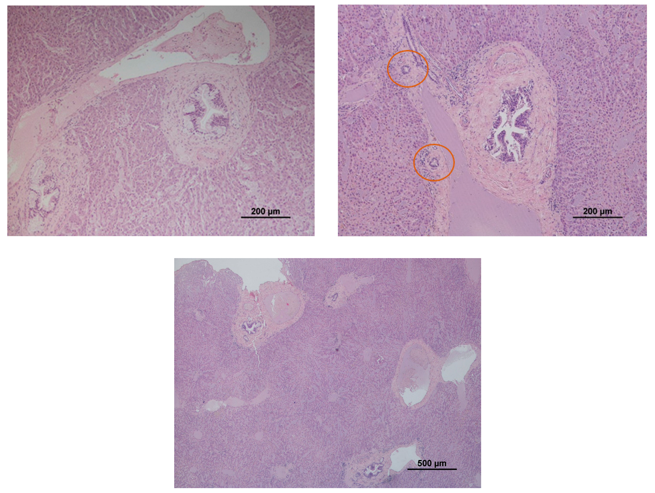
3.1. Histopathology
3.2. Metal(loid)s Determination
4. Discussion
5. Conclusions
Author Contributions
Funding
Institutional Review Board Statement
Informed Consent Statement
Data Availability Statement
Acknowledgments
Conflicts of Interest
Appendix A. Binary Logistic Regression Model Results

| Observed | Predicted | ||||
|---|---|---|---|---|---|
| BH | Percentage Correct | ||||
| No | Yes | ||||
| Step 1 | BH | No | 21 | 3 | 87.5 |
| Yes | 6 | 7 | 53.8 | ||
| Overall Percentage | 75.7 | ||||
| Step | Chi-Square | df | Sig. |
|---|---|---|---|
| 1 | 1.077 | 7 | 0.993 |
| B | S.E. | Wald | df | Sig. | Exp(B) | 95% C.I.for EXP(B) | |||
|---|---|---|---|---|---|---|---|---|---|
| Lower | Upper | ||||||||
| Step 1 | sex(1) | −2.700 | 1.402 | 3.706 | 1 | 0.054 | 0.067 | 0.004 | 1.050 |
| age | 0.019 | 2 | 0.990 | ||||||
| age(1) | −0.212 | 1.708 | 0.015 | 1 | 0.901 | 0.809 | 0.028 | 23.016 | |
| age(2) | −0.174 | 1.458 | 0.014 | 1 | 0.905 | 0.840 | 0.048 | 14.652 | |
| Log10As | −1.057 | 1.665 | 0.403 | 1 | 0.525 | 0.347 | 0.013 | 9.082 | |
| Log10Cd | 0.337 | 1.189 | 0.080 | 1 | 0.777 | 1.401 | 0.136 | 14.416 | |
| Log10Co | 2.797 | 1.930 | 2.100 | 1 | 0.147 | 16.392 | 0.373 | 720.297 | |
| Log10Cr | 0.267 | 2.011 | 0.018 | 1 | 0.894 | 1.306 | 0.025 | 67.179 | |
| Log10Cu | 1.905 | 2.557 | 0.555 | 1 | 0.456 | 6.722 | 0.045 | 1009.238 | |
| Log10Pb | 2.224 | 1.685 | 1.742 | 1 | 0.187 | 9.248 | 0.340 | 251.525 | |
| Constant | −0.243 | 4.242 | 0.003 | 1 | 0.954 | 0.784 | |||
References
- Tovar-Sánchez, E.; Hernández-Plata, I.; Santoyo Martínez, M.; Valencia-Cuevas, L.; Galante, P.M. Heavy Metal Pollution as a Biodiversity Threat. Heavy Met. 2018, 383. [Google Scholar] [CrossRef]
- Ali, H.; Khan, E. Trophic Transfer, Bioaccumulation, and Biomagnification of Non-Essential Hazardous Heavy Metals and Metalloids in Food Chains/Webs—Concepts and Implications for Wildlife and Human Health. Hum. Ecol. Risk Assess. 2019, 25, 1353–1376. [Google Scholar] [CrossRef]
- United Nations Environment Programme. Heavy Metals. Available online: https://www.unep.org/cep/heavy-metals (accessed on 7 December 2021).
- Ding, C.; Chen, J.; Zhu, F.; Chai, L.; Lin, Z.; Zhang, K.; Shi, Y. Biological Toxicity of Heavy Metal(Loid)s in Natural Environments: From Microbes to Humans. Front. Environ. Sci. 2022, 10, 681. [Google Scholar] [CrossRef]
- Rautio, A.; Kunnasranta, M.; Valtonen, A.; Ikonen, M.; Hyvärinen, H.; Holopainen, I.J.; Kukkonen, J.V.K. Sex, Age, and Tissue Specific Accumulation of Eight Metals, Arsenic, and Selenium in the European Hedgehog (Erinaceus europaeus). Arch. Environ. Contam. Toxicol. 2010, 59, 642–651. [Google Scholar] [CrossRef]
- D’Havé, H.; Scheirs, J.; Mubiana, V.K.; Verhagen, R.; Blust, R.; de Coen, W. Non-Destructive Pollution Exposure Assessment in the European Hedgehog (Erinaceus europaeus): II. Hair and Spines as Indicators of Endogenous Metal and As Concentrations. Environ. Pollut. 2006, 142, 438–448. [Google Scholar] [CrossRef] [PubMed]
- Vermeulen, F.; D’Havé, H.; Mubiana, V.K.; van den Brink, N.W.; Blust, R.; Bervoets, L.; de Coen, W. Relevance of Hair and Spines of the European Hedgehog (Erinaceus europaeus) as Biomonitoring Tissues for Arsenic and Metals in Relation to Blood. Sci. Total Environ. 2009, 407, 1775–1783. [Google Scholar] [CrossRef] [PubMed]
- Jota Baptista, C.; Seixas, F.; Gonzalo-Orden, J.M.; Oliveira, P.A. Can the European Hedgehog (Erinaceus europaeus) Be a Sentinel for One Health Concerns? Biologics 2021, 1, 4. [Google Scholar] [CrossRef]
- National Research Council. Animals as Sentinels of Environmental Health Hazards; National Academies Press: Washington, DC, USA, 1991; ISBN 978-0-309-04046-4. [Google Scholar]
- Rabinowitz, P.; Scotch, M.; Conti, L. Human and Animal Sentinels for Shared Health Risks. Vet. Ital. 2009, 45, 23. [Google Scholar] [PubMed]
- van der Schalie, W.H.; Gardner, H.S.; Bantle, J.A.; de Rosa, C.T.; Finch, R.A.; Reif, J.S.; Reuter, R.H.; Backer, L.C.; Burger, J.; Folmar, L.C.; et al. Animals as Sentinels of Human Health Hazards of Environmental Chemicals. Environ. Health Perspect. 1999, 107, 309–315. [Google Scholar] [CrossRef]
- Renu, K.; Chakraborty, R.; Myakala, H.; Koti, R.; Famurewa, A.C.; Madhyastha, H.; Vellingiri, B.; George, A.; Valsala Gopalakrishnan, A. Molecular Mechanism of Heavy Metals (Lead, Chromium, Arsenic, Mercury, Nickel and Cadmium)—Induced Hepatotoxicity—A Review. Chemosphere 2021, 271, 129735. [Google Scholar] [CrossRef]
- Baptista, C.J.; Seixas, F.; Gonzalo-Orden, J.M.; Oliveira, P.A. Biomonitoring of Heavy Metals and Metalloids with Wild Mammals in the Iberian Peninsula: A Systematic Review. Environ. Sci. Pollut. Res. Int. 2022, 29, 18398–18407. [Google Scholar] [CrossRef]
- Hasanein, P.; Emamjomeh, A. Beneficial Effects of Natural Compounds on Heavy Metal-Induced Hepatotoxicity. In Dietary Interventions in Liver Disease: Foods, Nutrients, and Dietary Supplements; Elsevier: Amsterdam, The Netherlands, 2019; pp. 345–355. ISBN 9780128144671. [Google Scholar]
- Balali-Mood, M.; Naseri, K.; Tahergorabi, Z.; Khazdair, M.R.; Sadeghi, M. Toxic Mechanisms of Five Heavy Metals: Mercury, Lead, Chromium, Cadmium, and Arsenic. Front. Pharmacol. 2021, 12, 643972. [Google Scholar] [CrossRef]
- Teschke, R. Aluminum, Arsenic, Beryllium, Cadmium, Chromium, Cobalt, Copper, Iron, Lead, Mercury, Molybdenum, Nickel, Platinum, Thallium, Titanium, Vanadium, and Zinc: Molecular Aspects in Experimental Liver Injury. Int. J. Mol. Sci. 2022, 23, 12213. [Google Scholar] [CrossRef]
- Bethesda, M.D. Trace Elements and Metals. Available online: https://www.ncbi.nlm.nih.gov/books/NBK548854/ (accessed on 22 December 2022).
- Schrank, C.S.; Cormier, S.M.; Blazer, V.S. Contaminant Exposure, Biochemical, and Histopathological Biomarkers in White Suckers from Contaminated and Reference Sites in the Sheboygan River, Wisconsin. J. Great Lakes Res. 1997, 23, 119–130. [Google Scholar] [CrossRef]
- Greenberg, S.R. The Histopathology of Tissue Lead Retention. Histol. Histopathol. 1990, 5, 451–456. [Google Scholar] [PubMed]
- Bexton, S.; Robinson, I. Hedgehogs. In BSAVA Manual of Wildlife Casualities; Mullineux, E., Best, D., Cooper, J.E., Eds.; British Small Animal Veterinary Association: Quedgeley, UK, 2003; pp. 49–65. [Google Scholar]
- West, R.M. Best Practice in Statistics: The Use of Log Transformation. Ann. Clin. Biochem. 2022, 59, 162. [Google Scholar] [CrossRef]
- Eisler, R. Arsenic Hazards to Fish, Wildlife, and Invertebrates: A Synoptic Review; Biological Report 85 (1.12)—Contaminant Hazard Reviews; U.S. Fish and Wildlife Service, Patuxent Wildlife Research Center: Laurel, MD, USA, 1988. [Google Scholar]
- Cooke, J.A. Cadmium in Small Mammals. In Environmental Contaminants in Biota; Beyer, W.N., Meador, J.P., Eds.; CRC Press: Boca Raton, FL, USA, 2011; pp. 627–639. [Google Scholar]
- Capello, V.; Lennox, A.M. Small Mammal Dentistry. In Ferrets, Rabbits, and Rodents: Clinical Medicine and Surgery; Quesenberry, K.E., Carpenter, J.W., Eds.; W.B. Saunders: Philadelphia, PA, USA, 2012; pp. 452–471. ISBN 9781416066217. [Google Scholar]
- Gluhcheva, Y.G.; Atanasov, V.N.; Ivanova, J.M.; Pavlova, E.H. Chronic Exposure to Cobalt Compounds—an in Vivo Study. Cent. Eur. J. Biol. 2014, 9, 973–981. [Google Scholar] [CrossRef]
- Danieli, P.P.; Serrani, F.; Primi, R.; Ponzetta, M.P.; Ronchi, B.; Amici, A. Cadmium, Lead, and Chromium in Large Game: A Local-Scale Exposure Assessment for Hunters Consuming Meat and Liver of Wild Boar. Arch. Environ. Contam. Toxicol. 2012, 63, 612–627. [Google Scholar] [CrossRef]
- Sánchez-Chardi, A.; Ribeiro, C.A.O.; Nadal, J. Metals in Liver and Kidneys and the Effects of Chronic Exposure to Pyrite Mine Pollution in the Shrew Crocidura Russula Inhabiting the Protected Wetland of Doñana. Chemosphere 2009, 76, 387–394. [Google Scholar] [CrossRef]
- Tovar-Sanchez, A.; Huerta-Diaz, M.A.; Negro, J.J.; Bravo, M.A.; Sañudo-Wilhelmy, S.A. Metal Contamination in Interstitial Waters of Doñana Park. J. Environ. Manag. 2006, 78, 286–293. [Google Scholar] [CrossRef] [PubMed]
- Read, H.J.; Martin, M.H. The Effect of Heavy Metals on Populations of Small Mammals from Woodlands in Avon (England); with Particular Emphasis on Metal Concentrations in Sorex araneus L. and Sorex minutus L. Chemosphere 1993, 27, 2197–2211. [Google Scholar] [CrossRef]
- Komarnicki, G.J.K. Tissue, Sex and Age Specific Accumulation of Heavy Metals (Zn, Cu, Pb, Cd) by Populations of the Mole (Talpa europaea L.) in a Central Urban Area. Chemosphere 2000, 41, 1593–1602. [Google Scholar] [CrossRef] [PubMed]
- Ramirez, C.J.; Kim, D.Y.; Hanks, B.C.; Evans, T.J. Copper Toxicosis in New Zealand White Rabbits (Oryctolagus Cuniculus). Vet. Pathol. 2013, 50, 1135–1138. [Google Scholar] [CrossRef] [PubMed]
- Ma, W.C. Lead in Mammals. In Environmental Contaminants in Biota; Beyer, W.N., Meador, J.P., Eds.; CRC Press: Boca Raton, FL, USA, 2011; pp. 595–608. ISBN 9781420084061. [Google Scholar]
- Mehana, E.E.; Meki, A.R.M.A.; Fazili, K.M. Ameliorated Effects of Green Tea Extract on Lead Induced Liver Toxicity in Rats. Exp. Toxicol. Pathol. 2012, 64, 291–295. [Google Scholar] [CrossRef] [PubMed]
- Pankakoski, E.; Hyvärinen, H.; Jalkanen, M.; Koivisto, I. Accumulation of Heavy Metals in the Mole in Finland. Environ. Pollut. 1993, 80, 9–16. [Google Scholar] [CrossRef]
- García-Niño, W.R.; Pedraza-Chaverrí, J. Protective Effect of Curcumin against Heavy Metals-Induced Liver Damage. Food Chem. Toxicol. 2014, 69, 182–201. [Google Scholar] [CrossRef] [PubMed]
- Byron, W.R.; Bierbower, G.W.; Brouwer, J.B.; Hansen, W.H. Pathologic changes in rats and dogs from two-year feeding of sodium arsenite or sodium arsenate. Toxicol. Appl. Pharmacol. 1967, 10, 132–147. [Google Scholar] [CrossRef] [PubMed]
- Tandon, S.K.; Saxena, D.K.; Gaur, J.S.; Chandra, S.V. Comparative toxicity of trivalent and hexavalent chromium: Alterations in blood and liver. Environ. Res. 1978, 15, 90–99. [Google Scholar] [CrossRef] [PubMed]
- Richardson, M.E.; Fox, M.R.S.; Fry, B.E. Pathological Changes Produced in Japanese Quail by Ingestion of Cadmium. J. Nutr. 1974, 104, 323–338. [Google Scholar] [CrossRef]
- Silva, G.F.; Rêma, A.; Teixeira, S.; Pires, M.D.A.; Taulescu, M.; Amorim, I. Pathological Findings in African Pygmy Hedgehogs Admitted into a Portuguese Rehabilitation Center. Animals 2022, 12, 1361. [Google Scholar] [CrossRef]
- Haschek, W.M.; Rousseaux, C.G.; Wallig, M.A. The Liver. In Fundamentals of Toxicologic Pathology; Haschek, W.M., Rousseaux, C.G., Wallig, M.A., Eds.; Academic Press: Cambridge, MA, USA, 2010; pp. 197–235. ISBN 978-0-12-370469-6. [Google Scholar]
- Maronpont, R.; Liver, Bile Duct—Hyperplasia. NTP Nonneoplastic Lesion Atlas 2014, National Toxicology Program. Available online: https://ntp.niehs.nih.gov/nnl/hepatobiliary/liver/bdhyperp/index.htm (accessed on 23 December 2022).
- Liptak, J.M.; Withrow, S.J.; Selting, K.A.; Turek, M.M. Cancer of the Gastrointestinal Tract. In Withrow and MacEwen’s Small Animal Clinical Oncology; Withrow, S.J., Vail, D.M., Eds.; W.B. Saunders: Philadelphia, PA, USA, 2007; pp. 455–510. ISBN 9781455734290. [Google Scholar]

| n | Mean | SD | Min. | Max. | ||
|---|---|---|---|---|---|---|
| As | without BH | 27 | 0.13 | 0.16 | 0.00 | 0.69 |
| with BH | 14 | 0.13 | 0.11 | 0.00 | 0.44 | |
| Total | 41 | 0.13 | 0.14 | 0.00 | 0.69 | |
| Cd | without BH | 27 | 0.46 | 0.59 | 0.00 | 1.84 |
| with BH | 14 | 1.57 | 1.85 | 0.00 | 6.07 | |
| Total | 41 | 0.84 | 1.27 | 0.00 | 6.07 | |
| Co | without BH | 27 | 0.19 | 0.13 | 0.05 | 0.46 |
| with BH | 14 | 0.40 | 0.42 | 0.06 | 1.66 | |
| Total | 41 | 0.27 | 0.28 | 0.05 | 1.66 | |
| Cr | without BH | 27 | 0.12 | 0.13 | 0.02 | 0.70 |
| with BH | 14 | 0.13 | 0.07 | 0.03 | 0.29 | |
| Total | 41 | 0.12 | 0.11 | 0.02 | 0.70 | |
| Cu | without BH | 27 | 33.06 | 21.42 | 12.11 | 102.91 |
| with BH | 14 | 40.68 | 16.09 | 13.60 | 65.75 | |
| Total | 41 | 35.66 | 19.89 | 12.11 | 102.91 | |
| Pb | without BH | 27 | 0.40 | 0.25 | 0.11 | 1.16 |
| with BH | 14 | 0.80 | 1.12 | 0.09 | 4.46 | |
| Total | 41 | 0.54 | 0.70 | 0.09 | 4.46 | |
| N | Mean | SD | Min. | Max | ANOVA | |||||||
|---|---|---|---|---|---|---|---|---|---|---|---|---|
| Sum of Squares | df | Mean Square | F | Sig. | ||||||||
| Log10(As) | without BH | 24 | −0.98 | 0.37 | −1.72 | −0.16 | Between Groups | 0.003 | 1 | 0.003 | 0.026 | 0.873 |
| with BH | 13 | −0.96 | 0.31 | −1.47 | −0.36 | Within Groups | 4.344 | 35 | 0.124 | |||
| Total | 37 | −0.98 | 0.35 | −1.72 | −0.16 | Total | 4.348 | 36 | ||||
| Log10(Cd) | without BH | 26 | −0.81 | 0.74 | −1.90 | 0.26 | Between Groups | 4.137 | 1 | 4.137 | 8.122 | 0.007 ** |
| with BH | 13 | −0.11 | 0.65 | −1.37 | 0.78 | Within Groups | 18.846 | 37 | 0.509 | |||
| Total | 39 | −0.58 | 0.78 | −1.90 | 0.78 | Total | 22.983 | 38 | ||||
| Log10(Co) | without BH | 27 | −0.80 | 0.29 | −1.27 | −0.34 | Between Groups | 0.607 | 1 | 0.607 | 6.001 | 0.019 * |
| with BH | 14 | −0.55 | 0.37 | −1.19 | 0.22 | Within Groups | 3.944 | 39 | 0.101 | |||
| Total | 41 | −0.72 | 0.34 | −1.27 | 0.22 | Total | 4.551 | 40 | ||||
| Log10(Cr) | without BH | 27 | −1.06 | 0.32 | −1.71 | −0.16 | Between Groups | 0.065 | 1 | 0.065 | 0.704 | 0.407 |
| with BH | 14 | −0.97 | 0.27 | −1.53 | −0.54 | Within Groups | 3.594 | 39 | 0.092 | |||
| Total | 41 | −1.03 | 0.30 | −1.71 | −0.16 | Total | 3.658 | 40 | ||||
| Log10(Cu) | without BH | 27 | 1.46 | 0.21 | 1.08 | 2.01 | Between Groups | 0.115 | 1 | 0.115 | 2.733 | 0.106 |
| with BH | 14 | 1.57 | 0.19 | 1.13 | 1.82 | Within Groups | 1.639 | 39 | 0.042 | |||
| Total | 41 | 1.50 | 0.21 | 1.08 | 2.01 | Total | 1.754 | 40 | ||||
| Log10(Pb) | without BH | 27 | −0.47 | 0.25 | −0.95 | 0.06 | Between Groups | 0.161 | 1 | 0.161 | 1.459 | 0.234 |
| with BH | 14 | −0.34 | 0.45 | −1.03 | 0.65 | Within Groups | 4.308 | 39 | 0.110 | |||
| Total | 41 | −0.42 | 0.33 | −1.03 | 0.65 | Total | 4.470 | 40 | ||||
Disclaimer/Publisher’s Note: The statements, opinions and data contained in all publications are solely those of the individual author(s) and contributor(s) and not of MDPI and/or the editor(s). MDPI and/or the editor(s) disclaim responsibility for any injury to people or property resulting from any ideas, methods, instructions or products referred to in the content. |
© 2023 by the authors. Licensee MDPI, Basel, Switzerland. This article is an open access article distributed under the terms and conditions of the Creative Commons Attribution (CC BY) license (https://creativecommons.org/licenses/by/4.0/).
Share and Cite
Jota Baptista, C.; Seixas, F.; Gonzalo-Orden, J.M.; Patinha, C.; Pato, P.; Ferreira da Silva, E.; Casero, M.; Brazio, E.; Brandão, R.; Costa, D.; et al. High Levels of Heavy Metal(loid)s Related to Biliary Hyperplasia in Hedgehogs (Erinaceus europaeus). Animals 2023, 13, 1359. https://doi.org/10.3390/ani13081359
Jota Baptista C, Seixas F, Gonzalo-Orden JM, Patinha C, Pato P, Ferreira da Silva E, Casero M, Brazio E, Brandão R, Costa D, et al. High Levels of Heavy Metal(loid)s Related to Biliary Hyperplasia in Hedgehogs (Erinaceus europaeus). Animals. 2023; 13(8):1359. https://doi.org/10.3390/ani13081359
Chicago/Turabian StyleJota Baptista, Catarina, Fernanda Seixas, José M. Gonzalo-Orden, Carla Patinha, Pedro Pato, Eduardo Ferreira da Silva, María Casero, Erica Brazio, Ricardo Brandão, Daniela Costa, and et al. 2023. "High Levels of Heavy Metal(loid)s Related to Biliary Hyperplasia in Hedgehogs (Erinaceus europaeus)" Animals 13, no. 8: 1359. https://doi.org/10.3390/ani13081359
APA StyleJota Baptista, C., Seixas, F., Gonzalo-Orden, J. M., Patinha, C., Pato, P., Ferreira da Silva, E., Casero, M., Brazio, E., Brandão, R., Costa, D., Mateus, T. L., & Oliveira, P. A. (2023). High Levels of Heavy Metal(loid)s Related to Biliary Hyperplasia in Hedgehogs (Erinaceus europaeus). Animals, 13(8), 1359. https://doi.org/10.3390/ani13081359