Article

IEg67 kDa Bovine Hydatid Cyst Antigen: A Candidate for Developing Sero-Diagnostic Assays for Cystic Echinococcosis, a Disease of One Health Importance

1
Department of Parasitology, Faculty of Veterinary Sciences, University of Veterinary and Animals Sciences, Lahore 54000, Pakistan
2
School of Biosciences, Cardiff University, Cardiff CF10 3AX, UK
3
Department of Pathobiology, College of Veterinary and Animal Sciences, Narowal, Sub-Campus, University of Veterinary and Animals Sciences, Lahore 54000, Pakistan
*
Author to whom correspondence should be addressed.
Animals 2023, 13(5), 866; https://doi.org/10.3390/ani13050866
Submission received: 17 December 2022 / Revised: 6 February 2023 / Accepted: 15 February 2023 / Published: 27 February 2023
(This article belongs to the Section Veterinary Clinical Studies)

Abstract

:

Simple Summary

Cystic echinococcosis is a world-wide zoonotic disease of food animals and humans. The disease negatively impacts food production, causes socio-economic hardship and is an animal welfare concern. Here, we identified and deployed a potential candidate antigen, iEg67 kDa crude antigen, for ELISA kit development that improved diagnostics for cystic echinococcosis in both cattle and buffalo, with 100% sensitivity and 93.8% specificity. The next steps are to validate and optimize this 67 kDa protein-based ELISA kit for sero-diagnosis of cystic echinococcosis in other species of food animals.

Abstract

Cystic echinococcosis (hydatidosis) is a world-wide zoonotic disease of mainly humans, livestock and dogs, caused by Echinococcus granulosus. The disease can negatively impact food production and animal welfare and causes socio-economic hardship. Here, we aimed to identify the local bovine hydatid cyst fluid (BHCF) antigen for developing a sero-diagnostic assay to be used for the pre-slaughter screening of food animals. In total, 264 bovines approved for slaughter in Pakistan were subjected to serum collection and post-mortem screening for hydatid cysts. These cysts were assessed microscopically to assess fertility and viability, and by PCR for molecular confirmation of species. A BHCF antigen was identified from positive sera via SDS-PAGE, confirmed by Western blot, and quantified via a bicinchoninic acid (BCA) assay. The quantified crude BHCF antigen (iEg67 kDa) was then used in ELISA screening to test all sera collected from known positive and negative animals based on hydatid cyst presence/absence. Of the 264 bovines examined, 38 (14.4%) showed hydatid cysts during post-mortem examination. All of these individuals, plus an additional 14 (total: 52; 19.6%) tested positive based on less time-consuming ELISA examination. Based on ELISA, occurrence in females (18.8%) was significantly higher than in males (9.2%) and was higher in cattle (19.5%) compared to buffalo (9.5%). The infection rate increased with age in both host species: cumulatively, 3.6% in animals aged 2–3 years, 14.6% in 4–5-year-olds and 25.6% in 6–7-year-olds. The occurrence of cysts in cattle was significantly higher in the lungs (14.1%) compared to their livers (5.5%), whereas the opposite was true in buffalo (6.6% livers, 2.9% lungs). For both host species, most cysts in the lungs were fertile (65%), while the majority in the liver were sterile (71.4%). We conclude that the identified iEg67 kDa antigen is a strong candidate for the development of a sero-diagnostic screening assay for the pre-slaughter diagnosis of hydatidosis.

1. Introduction

Pakistan has a large proportion of farmers entirely dependent on livestock for their livelihood. Livestock has a major role in the economy of Pakistan, contributing >60% to agriculture, and showing an annual growth rate of 3.06% per year and 11.5% for gross domestic product (GDP) (Pakistan economic survey 2020–2021). The livestock are affected by a range of infectious diseases that adversely reduce growth rates and overall production, particularly affecting food animals. Cystic echinococcosis (hydatidosis) is one such zoonotic disease, caused by the cestode Echinococcus granulosus that infects both humans and animals [1] and has a cosmopolitan distribution [2]. The disease occurs in South America, Eastern Europe, Russia, East Africa, Central Asia, Iran, Iraq, Syria, Jordan, Saudi Arabia and Pakistan [3]. According to the Global Burden of Disease (GBD), 9186 symptomatic cystic echinococcosis patients were reported in Pakistan in 2016, and new cases reported in the same year totalled 2932 per 100,000 population (Global Burden of Disease Results Tool 2021). In many parts of the world, e.g., Mediterranean regions, the Americas, Asia, Australia, Africa, and Europe [4], echinococcosis is categorised as a neglected tropical disease [5].
Two types of hosts are responsible for the transmission of Echinococcus spp.: ruminants and canines. Transmission to intermediate hosts (sheep, cattle, buffalo and goats) occurs through the ingestion of cestode eggs expelled by the definitive host [6]. Dogs and other canines serve as definite hosts when they ingest offal contaminated with Echinococcus granulosus larvae [7]. Humans are accidental hosts for E. granulosus, acquiring the infection through close contact with dogs or by ingesting water or food contaminated with parasitic eggs [8]. As accidental or dead-end hosts, humans cannot transmit the infection. In the intermediate hosts, the parasite develops in a fluid filled hydatid cyst in soft organs, often the liver and lungs, and rarely in the brain, kidney and bone marrow, resulting in morbidity and mortality [9,10,11]. In humans, the infection causes bile duct obstruction and pleural fistula disorders [12]. To prevent and control cystic echinococcosis, methods applied to break the life cycle between the definitive and intermediate hosts include the deworming of dogs, meat inspection, not feeding infected offal to dogs and public awareness of the risks to humans [13]. Despite this, cystic echinococcosis is highly prevalent in ruminants within herd-keeping areas of the world [14], ranging from 53.9% in China [15] and 22% in Ethiopia [16] to 13.9% in Iran [17] and 12% in India [18]. The prevalence of echinococcosis in Pakistan ranges from 2.4 to 35% in different host species [1,19,20], and here the estimated economic loss caused by cystic echinococcosis is USD 1.65 per organ [21].
To control cystic echinococcosis, early diagnosis is essential [22]. The diagnosis of Echinococcus typically relies on post-mortem extraction of the parasites, followed by microscopy and PCR. Echinococcosis cannot be confirmed through clinical signs, and in many countries, there are limited or no microscopy and molecular facilities available for ante-mortem examination. Additionally, radiography and ultrasonography are ineffective in identifying newly formed cysts. Serological assays, on the other hand, could provide a powerful pre-slaughter diagnostic screening tool for cystic echinococcosis. This could reduce the spread of E. granulosus on farms and in the environment, important for the prevention of human infections.
Bovine and sheep hydatid cyst fluid (8, 16, 24 and 38 kDa) and goat hydatid cyst fluid antigens (60 and 245 kDa) have been previously studied for their potential usage in sero-diagnostic assays [23,24,25]. In Pakistan, hydatid cyst fluid antigens of goat, sheep, cattle and buffalo have been characterised [26], but so far have not been tested for use in sero-diagnostic assays. Hence, here we isolate the unique antigen/s from bovine hydatid cysts to assess their usefulness in developing an ELISA for the pre-slaughter sero-diagnosis of cystic echinococcosis in bovines.

2. Materials and Methods

2.1. Study Site and Selection of Animals for Sera and Cyst Collection

Sampling was conducted at a slaughterhouse in the district of Sheikhupura (Pakistan) in December 2021, where 960 bovines (cattle and buffalo, mostly dairy animals) were slaughtered during that month. Animals with the lowest body condition score were targeted in this study. A total of 264 cattle and buffalo individuals were tagged for collection of sera and for post-slaughter examination for hydatid cysts. Blood samples were collected from the jugular vein using sterilised 10 mL syringes (22G needle; Shifa) and stored in serum vacutainers. The tubes were transported on ice to the Department of Parasitology, University of Veterinary and Animal Sciences, Lahore, Pakistan, for further processing the next day. The serum was separated by centrifuging the samples at 3000× g for 20 min. The supernatant (serum) was collected and stored at −20 °C until further use.

2.2. Hydatid Cyst Collection

Following the slaughter of tagged animals, the carcasses were examined for hydatid cysts in the liver, lungs and kidneys. Cysts removed from the liver or lungs were placed individually in sterilised containers for transport to the laboratory and subsequent microscopic examination. No cysts were detected in the kidneys. The meat was not tested for the presence of Echinococcus granulosus.

2.3. Microscopy of Hydatid Cysts

Cysts with viable protoscoleces were classified as fertile and those with no protoscoleces were classified as sterile. To check fertility, fluid from each hydatid cyst was gently aspirated through a 1 mL sterilised syringe, and 0.2 mL was dropped onto a microscopic slide with a cover slip [27]. To assess the viability of protoscoleces, the fluid (0.1 mL) from each hydatid cyst was mixed with 0.1% eosin stain for microscopic examination at 400×, with only viable protoscoleces taking up the stain [28]. All slides were examined with an Olympus CX21FS1 microscope at 400× magnification.

2.4. Molecular Identification of Echinococcus Granulosus

DNA was extracted from the germinal layer and fluid of the hydatid cyst using the WizPrep™ gDNA Tissue Kit (Wizbiosolutions, South Korea, W71060-300). Echinococcus granulosus mtDNA ND1 primers were used: Eg1F81 5′ GTT TTT GGC TGC CGC CAGAAC 3′ and Eg1R83 5′ AAT TAA TGG AAA TAA TAACAA ACT TAA TCA ACA AT3’, which generate a 226 bp amplicon [29]. The PCR was performed in a T100 Thermal Cycler (Bio-Rad, Hercules, CA, USA) as described previously [30]. Briefly, a total reaction volume of 20 μL included 2 μL of genomic cyst DNA (11.5 ng/μL), 10 μL master mix 2X AmpMaster™ Taq (GeneAll®, Exgene™, catalogue number 541-001), 6 µL of Ultrapure™ DEPC water (Invitrogen, 750023) and 1 μL of each forward and reverse primer solution (50 μM). PCR conditions were as follows: initial denaturation step at 94 °C for 3 min, followed by 28 cycles of denaturation at 94 °C for 30 s, annealing at 59.8 °C for 30 s and extension for 1 min at 72 °C, and a final extension step for 5 min at 72 °C. PCR products underwent electrophoresis in 2% agarose gels (1 h at 120 V, current 400 mA) using an SYBR safe DNA gel stain (catalogue no. 2291850; Invitrogen, Waltham, MA, USA alongside 7 μL of DNA ladder (100 bp DNA ladder Gene-direx, Catalogue # DM001-R500), and were observed under a GelDoc 100 imaging system (BioRad, Hercules, CA, USA).

2.5. Preparation of Bovine Hydatid Cyst Fluid (BHCF) Antigen

Fluid was aseptically aspirated using a sterilised syringe (Shifa) from a hydatid cyst present in the liver of a buffalo, transferred to a 50 mL falcon tube, according to [23], and centrifuged at 7000× g for 30 min. Acetate buffer (0.005 M; pH 5) was used to dialyse the supernatant, and then the falcon tube was incubated overnight at 4 °C before centrifuging at 20,000× g for 30 min at 4 °C (ultra-high-speed centrifuge, Sigma 3K30). The precipitates were dissolved in 10 mL of phosphate buffer (0.2 M, pH 8) saturated with 40% ammonium sulphate and were centrifuged at 3000× g for 25 min. The supernatant was then incubated for 10 min at 60 °C and centrifuged again at 20,000× g for 1 h. The supernatant containing the soluble antigens of HCF was filtered with a 0.2 µm filter and stored at −20 °C [31,32].

2.6. SDS-PAGE and Western Blotting

The native antigens separated from the bovine hydatid cyst fluid (BHCF) were analysed via SDS-PAGE and Western blotting according to [33]. Briefly, each lane of a 12% w/v polyacrylamide gel was loaded with 30 μL native antigen (11.65 μ g/μL), protein-loading (Laemmli) buffer and protein marker (PageRulerTm Cat# 26614, thermo scientific), separated by 12% w/v polyacrylamide gel and transferred to the nitrocellulose membrane (NCM, 0.22 µm, Trans-Blot® Turbo™ Midi nitrocellulose transfer packs # 1704159, Bio-Rad, USA) using the Trans-Blot® Turbo™ transfer system (Bio-Rad, USA). The blots were cut into strips, labelled and blocked with (5% w/v) skimmed milk in tris buffer saline (20 mM Tris-HCL, pH 7.2, 150 mM NaCl). The positive serum collected from infected tagged animals (three buffalo, each with 2–3 hydatid cysts in the liver, confirmed after post-mortem) was used as the primary antibody. The strips were charged with the anti-bovine antibody (Anti-bovine IgG-alkaline phosphatase conjugate Sigma Cat # A0705-25ML) as the secondary antibody (1:20,000) after three washings. A chromogenic substrate (NBT/BCIP bio-WORLD, Dublin, OH, USA) was used as a colour marker.

2.7. Quantification of Native Antigen

The bovine hydatid cyst fluid protein was quantified using bicinchoninic acid (BCA) assay according to the manufacturer’s instructions (Cat. 786-570, G-Biosciences, St. Louis, MO, USA). OD values were recorded at 650 nm using a multiskan sky microplate (1530-800580) spectrophotometer.

2.8. ELISA

ELISA was performed according to [34] with minor modifications. Hydatid cyst fluid antigen (100 µL/well in 50 mM Na2CO3 coating buffer at the concentration of 5 μg/mL) was coated on to a 96-well flat-bottom polystyrene plate (JET BioFil, Hong Kong, China, Code # TCP011096) and incubated at 37 °C overnight. The plate was washed 5 times with 250 μL/well 0.001 M PBS/0.05% Tween-20, then blocked with 4% BSA in 0.01 M PBS (200 μL/well), incubated for 2 h at 37 °C and washed 5 times with washing buffers. The primary antibody (serum) was added to the plate at a dilution of 1:99. Two wells were coated with positive sera (confirmed after slaughtering and PCR) and two wells were coated with negative sera. The plate was incubated at 37 °C for 2 h and re-washed 5 times. The plate was then coated with anti-bovine antibody 100 μL/well (Anti-bovine IgG-alkaline phosphatase conjugate Sigma Cat # A0705-25ML) as the secondary antibody (1:5000), incubated for 2 h at 37 °C and then re-washed 5 times. The substrate p-nitrophenyl phosphate (pNPP) (Thermo Scientific, Waltham, MA, USA, Ref # 34045) was added with 1 mg/mL of DAE substrate buffer (ThermoFisher, Waltham, MA, USA, Cat # 34064) at 100 μL/well. After 15 min, the stop solution (1 M NaOH at 100 μL/well) was added to stop the reaction. The microplate ELISA reader (ELX-800, Tennessee, BioTek, Winooski, VT, USA) recorded OD values at 405 nm.

2.9. ELISA Sensitivity, Specificity, Positive Predictive Value (PPV), Negative Predictive Value (NPV), Likelihood Ratio Positive (LR+), Likelihood Ratio Negative (LR−) and Diagnostic Odds Ratio (DOR)

Sensitivity, specificity, positive predictive value (PPV), negative predictive value (NPV), likelihood ratio for a positive test result (LR+), likelihood ratio for a negative test result (LR−) and diagnostic odds ratio (DOR) were calculated according to the formulae in Table 1.

2.10. Statistical Analyses

All statistical analyses were performed using R-Studio version 1.0.143 (R Development Core Team, 2015). We conducted a generalised linear model (GLM) with a Gaussian error family with loglink function with the dependent variable being the percentage of ELISA-positive animals within the following categories (independent variables): species, sex and age. A separate GLM for each species, both with binomial error families and logit functions, was then used to test whether the percentage of ELISA-positive animals varied significantly in terms of age, sex, location of cyst and cyst viability. Chi-square tests determined the association of the disease with species, sex, location of cyst and age groups.
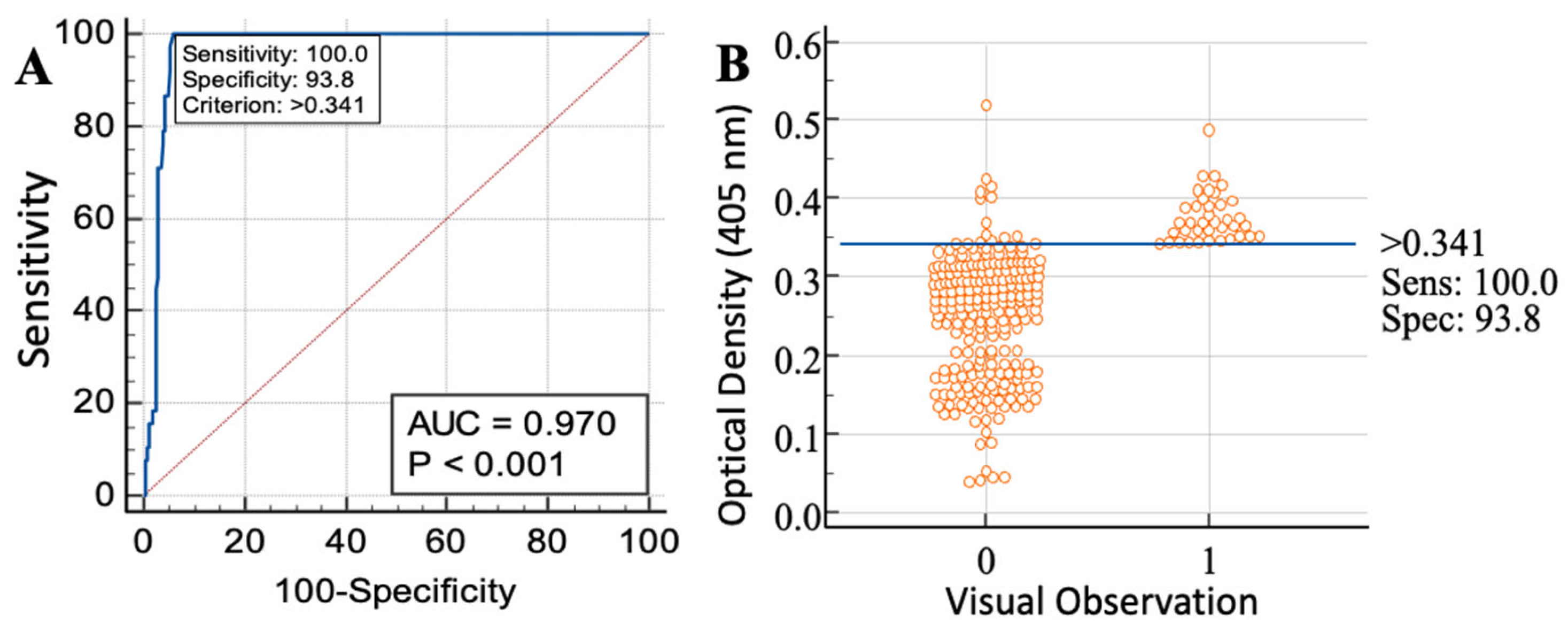
The Medcalc software (version 11.4.4.0) was used for the ROC curve line and interactive dot diagram. The ROC curve shows the sensitivity, specificity and criterion value (cut-off value) on the basis of which samples are declared positive or negative. The dot diagram displays (i) the separation of samples into positive (indicated as 1) and negative (indicated as 0) based on the gold standard (here, post-mortem examination); (ii) the cut-off value (indicated by a horizontal line) of ELISA separating ELISA-positive (above the line) and ELISA-negative (below the line) [36]. For comparing data in Medcalc software, we used “1” to indicate post-mortem positive samples (n = 38) and “0” to indicate post-mortem negative samples (n = 226). On the basis of two negative control samples (negative in post-mortem), we calculated the cut-off value to differentiate between sero-negative and sero-positive samples [37]. The mean OD value (0.136) of two negative controls was multiplied by 2.5 according to [38,39] to obtain the cut-off value of 0.340. In parallel, the OD values of all 264 samples were put into the Medcalc software while telling the software either positive and negative based on post-mortem examination. Based on this, the software also determined the cut-off value as being 0.341.

3. Results

Out of 264 bovines (128 cattle and 136 buffalo) examined at post-mortem, we found that 38 out of 264 (14.4%) contained hydatid cysts. All of these animals were subsequently confirmed as positive via serological examination using an ELISA kit based on the crude antigen (67 kDa) of BHCF. This ELISA approach also identified an additional 14 bovines, increasing the cystic echinococcosis infection rate to 52 out of 264 (19.6%).

3.1. Molecular Confirmation, Identification of Echinococcus-Specific Immunogen and BHCF ELISA

For the antigen characterisation of BHCF, the hydatid cyst fluid collected from buffalo was confirmed to be Echinococcus granulosus based on species-specific primers targeting the mtDNA ND1 gene. The BHCF recovered from animals positive for echinococcosis, analysed via SDS-PAGE and stained with Coomassie blue, revealed an infected E. granulosus 67 kDa (IEg67 kDa) band and this was confirmed via WB analysis (Figure 1A,B). There was also a 130 kDa band present in the SDS-PAGE, potentially representing a dimer (Figure 1A) that was not explored further.
The sensitivity and specificity of ELISA based on the crude antigen was 100% and 93.8%, respectively (Figure 2A,B).
For ELISA, the positive predictive value (PPV), negative predictive value (NPV), LR+, LR− and DOR are given in Table 2.

3.2. Echinococcus Occurrence

Based on ELISA, cattle had significantly higher infection levels than buffalo (GLM: std.err = 0.26, t = 2.5, p = 0.03). For both species, the number of infected individuals was positively associated with age (GLM: std.err = 0.09, t = 3.3, p = 0.009), and fewer males were infected than females (GLM: std.err = 0.25, t = −2.2, p = 0.05). The occurrence of cysts in cattle was significantly higher in the lungs (14.1%) compared to the liver (5.5%), whereas the reverse was true for buffalo (6.6% in liver and 2.9% in the lungs; χ2 = 10.69, p = 0.005). In each host species, neither location of cyst nor cyst viability were significantly related to host age nor gender (GLMs).
From post-mortem examination, the occurrence of cysts was higher in the lungs of cattle (72%) compared to the liver (28%), whereas the reverse was true for buffalo (30.7% in lungs and 69.2% in the livers; χ2 = 10.69, p = 0.005). Fertile cysts were more prevalent in lungs (64.5%) than in livers (28.6%), whereas sterile cysts were more frequently observed in the liver (71.4%) than in lungs (35.5%) (χ2 = 285.028 and p < 0.001). The percentage of fertile cysts was higher in buffalo (84.6%) than in cattle (80%), and there were significantly more sterile cysts in cattle (20%) than in buffalo (15.4%; χ2 = 285.028 and p = 0.06). The distribution of Echinococcus-positive animal sera is shown in Table 3.

4. Discussion

Cystic echinococcosis is a major disease in livestock and humans [40]. Its diagnosis in intermediate hosts is mainly based on necropsy procedures and in most cases the disease is asymptomatic in the early stages [41], so there is an urgency to develop alternative diagnostic methods [42], such as antibody detection through ELISA. Here, we identified and successfully deployed a potential candidate antigen, the iEg67 kDa crude antigen (from cystic fluid collected from buffalo liver) in a sero-diagnostic assay (Ab-detection ELISA) for the diagnosis of cystic echinococcosis in cattle and buffalo with much improved sensitivity and specificity. This antigen showed highly promising results as a candidate for ELISA kit development, and similar to previous studies (e.g., [43,44]) was more accurate than post-mortem examination, with very high sensitivity (100%) and specificity (93.8%) (Table 4). We also observed a 130 kDa band, possibly a dimer of iEg67 kDa, consistent with the previously reported dimerisation of Echinococcus antigens [45,46].
Previous studies have characterised the antigenic profile of hydatid cysts from cows, buffalo, sheep, goats and camels, and of humans infected with cystic echinococcosis, in different geographical areas to identify the potential diagnostic or vaccine candidate proteins [42,47,48]. The sensitivity and specificity of diagnostic tests used to include or exclude a disease (i.e., ‘rule in’ or ‘rule out’, respectively), for previous isolates of the HCF crude antigen, partially purified the crude antigen and recombinant antigens are compared in Table 5.
Factors influencing the sensitivity and specificity of ELISA include the source and quality of cyst fluid, the antigen purification process, laboratory techniques used for diagnosis and the target species/strains. The low sensitivity detected by Ibrahem et al. [49] might be related to the smaller size of their recombinant antigen potentially having fewer antigenic epitopes [49] compared to the native one. This notion is strengthened by Ibrahem et al. [49] who found that the sensitivity of recombinant-protein-based ELISA (25%) was lower compared to crude/native antigens from camel and sheep (see Table 5). Lower specificity might also arise due to the presence of other infections such as Taenia hydatigena, which can be mistaken as hydatid cysts, but would not have been detectable in an HCF-antigen-based ELISA [41]. Gatti et al. [37] used three forms of antigens (LHT, S2B and B) in their enzyme immunoassay (EIA) and recorded the sensitivity and specificity of each (see Table 4), concluding that either LHT or S2B should be used for the sero-diagnosis of cystic echinococcosis. Kandil et al. [50] used two types of antigens: protoscolex antigen and the germinal layer antigen of the hydatid cyst. They observed higher sensitivity (83%) and specificity (70.3%) in the protoscolex antigen than the germinal layer antigen [50] (Table 5). We used total hydatid cyst fluid for our crude HCF antigen preparation and obtained higher sensitivity and specificity values than in previous work; the reasons for this could be numerous.
The occurrence of hydatid cysts in Pakistan was determined in the current study to be 14.4% through post-mortem examination, and 19.7% when screened through the ELISA, similar to the 21% prevalence reported previously in cattle and buffalo from Punjab, India [51]. The increase in the detection rate of ELISA relative to the post-mortem between the current study and Yakubu et al. [43] can be explained by two complementary factors. Firstly, a known limitation of post-mortem lies in the lower detection probability for small developing cysts, possibly occurring more frequently when overall infection rate is higher in a population, as in [43]. Secondly, our ELISA approach (IgG Detection ELISA) did not allow us to differentiate between present and past infections, likely leading to the detection of recovered animals that would have remained undetected in PMs. ELISA can be further developed by targeting IgM for acute infections [52] and by the detection of the IgG titer in paired sampling [53] to have a clearer picture about active and past infections. The gap between the detection rate of ELISA and PM may increase when aged animals (with a higher expected proportion of recovered animals) are sacrificed and examined for the presence of cysts.
As previously reported by [54], infection prevalence was higher in female (18.75% post-mortem and 25% ELISA) than in male (9.2 and 13.3%) hosts. Pregnancy, parturition and lactation seem to enhance the risk of female infections [55]. Risk of infection also increased with age [56], presumably related to the increased exposure of older individuals. The current study also suggests that cattle are more prone to hydatidosis than buffalo (similar to [57], but in contrast to [54]). Apart from geographical location and temperature variation, the infection rate observed by [54] was influenced by the higher number of cattle amongst the slaughtered animals. In the current study, cattle had a higher occurrence of cysts in the lungs rather than the liver, with the reverse found in buffalo (as reported by [57,58,59], but in contrast to [60,61]. This difference may be due to host specificity, with one organ being more permissive to the development of cysts than the other, but this finding could potentially also be influenced by different strains of the parasite. With regard to hydatid cyst fertility and sterility, most studies on livestock populations in Punjab, Pakistan, have reported higher numbers of fertile than sterile cysts [29,47,58]. Variation in fertility may be due to differences in Echinococcus strain, time post-infection, development rate of the cysts and host species.

5. Conclusions

Cystic echinococcosis in food animals reduces their suitability for human consumption globally. Here, we show that an iEg67 kDa antigen identified from bovine hydatid cyst fluid is a suitable candidate for developing sero-diagnostic assays for cystic echinococcosis. An ELISA based on the use of the iEg67kDa antigen improved diagnosis accuracy for cystic echinococcosis in bovines compared to post-mortem examination. Our study showed 100% sensitivity and 93.8% specificity, an improvement compared with all previous studies. This approach can be used for large-scale sero-epidemiological studies of cystic echinococcosis in food animals (i.e., sheep, goat and camels) from diverse climatic conditions and agro-ecological zones, as well as for the screening of humans. We anticipate that this will provide increased information on the distribution and prevalence of cystic echinococcosis in animals and humans, informing on suitable control strategies and contributing towards the formulation of a One Health approach.

Author Contributions

Conceptualisation, H.A., M.I.R. and M.Y.; methodology, S.K., H.A. and J.C.; validation, J.C. and H.A.; data analysis, S.K. and J.C.; investigation, S.K.; resources, H.A.; data curation, S.K.; writing—original draft preparation, S.K., H.A. and J.C.; writing—review and editing, H.A., J.C. and F.H.; supervision, H.A.; project administration, H.A.; funding acquisition, H.A. and M.I.R. All authors have read and agreed to the published version of the manuscript.

Funding

S.K. received funding from Higher Education Commission (HEC) under the International Research Support Initiative Program (1-8/HEC/HRD/2022/12612/IRSIP 51 Agri 20); M.Y. received funding from HEC-National Research Program for Universities (NRPU-7018); H.A. and M.I.R. received funding from HEC-Grand Challenge Fund (GCF-273), Pakistan Agriculture Research Board (PARB-18-476) and the Punjab Higher Education Commission (PHEC/ARA/PIRCA/2020-6/8).

Institutional Review Board Statement

All of the experimental procedures used were approved by the Institutional Guidelines of the Ethical Review Committee of UVAS, Lahore, vide letter no. 939-1, dated 05-09-2019.

Informed Consent Statement

Not applicable.

Data Availability Statement

The data will be available from the corresponding author upon request.

Acknowledgments

We thank Shafqat Shabir and Sarfraz Ur Rahman, PhD scholars at the Department of Parasitology, UVAS, Lahore, for technical help, and Numair Masud for statistical advice.

Conflicts of Interest

The authors have declared no conflict of interest.

References

  1. Ali, I.; Panni, M.K.; Iqbal, A.; Munir, I.; Ahmad, S.; Ali, A. Molecular characterization of Echinococcus species in Khyber Pakhtunkhwa, Pakistan. Acta Sci. Vet. 2015, 43, 1–7. [Google Scholar]
  2. Rokni, M. Echinococcosis/hydatidosis in Iran. Iran. J. Parasitol. 2009, 4, 1–16. [Google Scholar]
  3. Khan, S.N.; Ali, R.; Khan, S.; Norin, S.; Rooman, M.; Akbar, N.; Khan, T.A.; Haleem, S.; Khan, M.A.; Ali, I. Cystic echinococcosis: An emerging zoonosis in southern regions of Khyber Pakhtunkhwa, Pakistan. BMC Vet. Res. 2021, 17, 139. [Google Scholar] [CrossRef] [PubMed]
  4. Khan, A.; Naz, K.; Ahmed, H.; Simsek, S.; Afzal, M.S.; Haider, W.; Ahmad, S.S.; Farrakh, S.; Weiping, W.; Yayi, G. Knowledge, attitudes and practices related to cystic echinococcosis endemicity in Pakistan. Infect. Dis. Poverty 2018, 7, 4. [Google Scholar] [CrossRef] [PubMed] [Green Version]
  5. Ahmed, H.; Ali, K.A.; Afzal, M.S.; Khan, A.A.; Raza, H.; Shah, Z.H.; Simsek, S. Why more research needs to be done on echinococcosis in Pakistan. Infect. Dis. Poverty 2017, 6, 90. [Google Scholar] [CrossRef] [PubMed]
  6. Lawson, J.R.; Gemmell, M. Hydatidosis and Cysticercosis: The Dynamics of Transmission. Adv. Parasitol. 1983, 22, 261–308. [Google Scholar] [CrossRef]
  7. Bourée, P. Hydatidosis: Dynamics of transmission. World J. Surg. 2001, 25, 4. [Google Scholar] [CrossRef] [PubMed]
  8. Nakao, M.; Yanagida, T.; Okamoto, M.; Knapp, J.; Nkouawa, A.; Sako, Y.; Ito, A. State-of-the-art Echinococcus and Taenia: Phylogenetic taxonomy of human-pathogenic tapeworms and its application to molecular diagnosis. Infect. Genet. Evol. 2010, 10, 444–452. [Google Scholar] [CrossRef] [PubMed]
  9. Battelli, G. Echinococcosis: Costs, losses and social consequences of a neglected zoonosis. Veter Res. Commun. 2009, 33, 47–52. [Google Scholar] [CrossRef]
  10. Sarkar, M.; Pathania, M.; Jhobta, A.; Thakur, B.R.; Chopra, R. Cystic pulmonary hydatidosis. Lung India Off. Organ Indian Chest Soc. 2016, 33, 179. [Google Scholar] [CrossRef]
  11. Ali, R.; Khan, S.; Khan, M.; Adnan, M.; Ali, I.; Khan, T.A.; Haleem, S.; Rooman, M.; Norin, S.; Khan, S.N. A systematic review of medicinal plants used against Echinococcus granulosus. PLoS ONE 2020, 15, e0240456. [Google Scholar] [CrossRef] [PubMed]
  12. Daali, M.; Fakir, Y.; Hssaida, R.; Hajji, A.; Had, A. Hydatid cysts of the liver opening in the biliary tract. Report of 64 cases. Ann. Chir. 2001, 126, 242–245. [Google Scholar] [CrossRef] [PubMed]
  13. Gessese, A.T. Review on Epidemiology and Public Health Significance of Hydatidosis. Veter Med. Int. 2020, 2020, 8859116. [Google Scholar] [CrossRef] [PubMed]
  14. Bekele, J.; Butako, B. Occurrence and financial loss assessment of cystic echinococcosis (hydatidosis) in cattle slaughtered at Wolayita Sodo municipal abattoir, Southern Ethiopia. Trop. Anim. Health Prod. 2011, 43, 221–228. [Google Scholar] [CrossRef]
  15. Fan, S.; Dong, H.; Ma, H.; Wang, B.; Iqbal, M.; Zou, M.; Qi, M.; Cao, Z. Meta-analysis on the prevalence of bovine hydatid disease in China from 2000 to 2021. Microb. Pathog. 2022, 168, 105586. [Google Scholar] [CrossRef]
  16. Shumuye, N.A.; Ohiolei, J.A.; Gebremedhin, M.B.; Yan, H.-B.; Li, L.; Li, W.-H.; Zhang, N.-Z.; Fu, B.-Q.; Jia, W.-Z. A systematic review and meta-analysis on prevalence and distribution of Taenia and Echinococcus infections in Ethiopia. Parasites Vectors 2021, 14, 447. [Google Scholar] [CrossRef]
  17. Vaisi-Raygani, A.; Mohammadi, M.; Jalali, R.; Salari, N.; Hosseinian-Far, M. Prevalence of cystic echinococcosis in slaughtered livestock in Iran: A systematic review and meta-analysis. BMC Infect. Dis. 2021, 21, 429. [Google Scholar] [CrossRef]
  18. Grakh, K.; Prakash, A.; Mittal, D.; Kumar, P.; Kumar, R. Epidemiology, Risk Factors and Economics of Echinococcosis in India: A Review. Int. J. Livest. Res. 2020, 10. [Google Scholar] [CrossRef]
  19. Sarıözkan, S.; Yalçın, C. Estimating the production losses due to cystic echinococcosis in ruminants in Turkey. Vet. Parasitol. 2009, 163, 330–334. [Google Scholar] [CrossRef]
  20. Tasawar, Z.; Naz, F.; Lashari, M. The prevalence of hydatidosis in sheep and buffaloes at Multan, Punjab, Pakistan. Glob. Vet. 2014, 12, 332–335. [Google Scholar]
  21. Shafiq, M.T.; Athar, M. Epidemiology and Economical Aspects of Hydatidosis in Different Animals, Man and Its Control in Sheep with Indigenous Plants. Ph.D. Thesis, University of the Punjab, Punjab, Pakistan, 2004; pp. 1–653. [Google Scholar]
  22. Burgu, A.; Doğanay, A.; Gönenç, B.; Sarimehmetoğlu, H.O.; Kalinbacak, F. Analysis of fluids of hydatid cysts from sheep by SDS-PAGE, and determination of specific antigens in protein structure by western blotting. Turk. J. Vet. Anim. Sci. 2000, 24, 493–500. [Google Scholar]
  23. Pagnozzi, D.; Biosa, G.; Addis, M.F.; Mastrandrea, S.; Masala, G.; Uzzau, S. An Easy and Efficient Method for Native and Immunoreactive Echinococcus granulosus Antigen 5 Enrichment from Hydatid Cyst Fluid. PLoS ONE 2014, 9, e104962. [Google Scholar] [CrossRef] [PubMed] [Green Version]
  24. Golassa, L.; Abebe, T.; Hailu, A. Evaluation of crude hydatid cyst fluid antigens for the serological diagnosis of hydatidosis in cattle. J. Helminthol. 2010, 85, 100–108. [Google Scholar] [CrossRef] [PubMed]
  25. Kumar, B.S.; Batabyal, S.; Chattopadhyay, U.K.; Raj, A.; Vinod, U.; Banik, A.; Bhattacharya, S.; Yogendra, M. Isolation, purification and immunobiochemical characterization of goat hydatid cyst fluid antigen. J. Entomol. Zool. Stud. 2020, 8, 1056–1060. [Google Scholar]
  26. Latif, A.; Tanveer, A.; Anjum, A.A.; Ali, M.A.; Rana, M.S.; Khan, M.R.; Ahmad, M.S. Characterization of hydatid cyst fluid from ruminants and humans by SDS-PAGE in Punjab, Pakistan. J. Anim. Plant Sci. 2013, 23, 405–410. [Google Scholar]
  27. Singh, B.B.; Sharma, J.K.; Tuli, A.; Sharma, R.; Bal, M.S.; Aulakh, R.S.; Gill, J.P.S. Prevalence and morphological characterisation of Echinococcus granulosus from north India. J. Parasit. Dis. 2012, 38, 36–40. [Google Scholar] [CrossRef] [Green Version]
  28. Guduro, G.G.; Desta, A.H. Cyst Viability and Economic Significance of Hydatidosis in Southern Ethiopia. J. Parasitol. Res. 2019, 2019, 2038628. [Google Scholar] [CrossRef] [Green Version]
  29. Boufana, B.; Boué, F.; Qiu, J.; Jenkins, D.; Chen, X.; Craig, P.; Umhang, G.; Lahmar, S. Development of Three PCR Assays for the Differentiation between Echinococcus shiquicus, E. granulosus (G1 genotype), and E. multilocularis DNA in the Co-Endemic Region of Qinghai-Tibet plateau, China. Am. J. Trop. Med. Hyg. 2013, 88, 795–802. [Google Scholar] [CrossRef] [Green Version]
  30. Bowles, J.; Blair, D.; McManus, D. Genetic variants within the genus Echinococcus identified by mitochondrial DNA sequencing. Mol. Biochem. Parasitol. 1992, 54, 165–173. [Google Scholar] [CrossRef]
  31. Mohammadzadeh, T.; Sako, Y.; Sadjjadi, S.M.; Sarkari, B.; Ito, A. Comparison of the usefulness of hydatid cyst fluid, native antigen B and recombinant antigen B8/1 for serological diagnosis of cystic echinococcosis. Trans. R. Soc. Trop. Med. Hyg. 2012, 106, 371–375. [Google Scholar] [CrossRef]
  32. Asghari, M.; Mohebali, M.; Kia, E.B.; Farahnak, A.; Aryaeipour, M.; Asadian, S.; Rokni, M.B. Seroepidemiology of human hydatidosis using AgB-ELISA test in Arak, central Iran. Iran. J. Public Health 2013, 42, 391. [Google Scholar]
  33. Nabi, H.; Rashid, I.; Ahmad, N.; Durrani, A.; Akbar, H.; Islam, S.; Bajwa, A.A.; Shehzad, W.; Ashraf, K.; Imran, N. Induction of specific humoral immune response in mice immunized with ROP18 nanospheres from Toxoplasma gondii. Parasitol. Res. 2017, 116, 359–370. [Google Scholar] [CrossRef]
  34. Rahman, S.U.; Akbar, H.; Shabbir, M.Z.; Ullah, U.; Rashid, M.I. Development of Human Toxo IgG ELISA Kit, and False-Positivity of Latex Agglutination Test for the Diagnosis of Toxoplasmosis. Pathogens 2021, 10, 1111. [Google Scholar] [CrossRef] [PubMed]
  35. Glas, A.S.; Lijmer, J.G.; Prins, M.H.; Bonsel, G.J.; Bossuyt, P.M.M. The diagnostic odds ratio: A single indicator of test performance. J. Clin. Epidemiol. 2003, 56, 1129–1135. [Google Scholar] [CrossRef] [PubMed]
  36. Bel-Ochi, N.C.; Bouratbine, A.; Mousli, M. Enzyme-Linked Immunosorbent Assay Using Recombinant SAG1 Antigen to Detect Toxoplasma gondii-Specific Immunoglobulin G Antibodies in Human Sera and Saliva. Clin. Vaccine Immunol. 2013, 20, 468–473. [Google Scholar] [CrossRef] [PubMed] [Green Version]
  37. Gatti, A.; Alvarez, A.R.; Araya, D.; Mancini, S.; Herrero, E.; Santillan, G.; Larrieu, E. Ovine echinococcosis: I. Immunological diagnosis by enzyme immunoassay. Vet. Parasitol. 2007, 143, 112–121. [Google Scholar] [CrossRef]
  38. Akbar, H.; Germon, S.; Berthon, P.; Dimier-Poisson, I.; Moiré, N. Depletion of CD25+ cells during acute toxoplasmosis does not significantly increase mortality in Swiss OF1 mice. Memórias Inst. Oswaldo Cruz 2012, 107, 155–162. [Google Scholar] [CrossRef] [Green Version]
  39. Ismael, A.B.; Sekkai, D.; Collin, C.; Bout, D.; Mévélec, M.-N. The MIC3 Gene of Toxoplasma gondii Is a Novel Potent Vaccine Candidate against Toxoplasmosis. Infect. Immun. 2003, 71, 6222–6228. [Google Scholar] [CrossRef] [Green Version]
  40. Mathewos, M.; Dawa, D.; Yirgalem, M.; Denano, T.; Fesseha, H. Cystic echinococcosis in cattle slaughtered at a slaughterhouse in Gessa, southern Ethiopia. Parasite Epidemiol. Control. 2022, 18, e00262. [Google Scholar] [CrossRef]
  41. El-Kattan, A.M.; Abdel-Ra’Ouf, A.M.; Yousef, R.R.; AbouElnga, T.R.; Mousa, W. Sensitivity and specificity of Indirect Enzyme Linked Immuno sorbent Assay (ELISA) for diagnosis of hydatidosis in dromedary camels using hydatid cyst fluid antigens. J. Vet. Med. Res. 2020, 27, 66–75. [Google Scholar] [CrossRef] [Green Version]
  42. Rahimi, H.; Sadjjadi, S.; Sarkari, B. Performance of Antigen B Isolated from Different Hosts and Cyst Locations in Diagnosis of Cystic Echinococcosis. Iran. J. Parasitol. 2011, 6, 12–19. [Google Scholar] [PubMed]
  43. Yakubu, R.A.; Nock, I.H.; Ndams, I.S.; Luka, S.A.; Yaro, C.A.; Alkazmi, L.; Batiha, G.E.-S. Detection of Echinococcus granulosus sensu lato cysts and seroprevalence of cystic echinococcosis in cattle and camels in Maiduguri Abattoir. J. Parasit. Dis. 2022, 46, 876–888. [Google Scholar] [CrossRef] [PubMed]
  44. Osman, F. Comparative study between serological analysis and visual meat inspection for hydatid cyst in camels. Assiut Vet. Med. J. 2014, 60, 1–6. [Google Scholar] [CrossRef]
  45. Kittelberger, R.; Reichel, M.P.; Jenner, J.; Heath, D.D.; Lightowlers, M.W.; Moro, P.; Ibrahem, M.M.; Craig, P.S.; O’Keefe, J.S. Evaluation of three enzyme-linked immunosorbent assays (ELISAs) for the detection of serum antibodies in sheep infected with Echinococcus granulosus. Vet. Parasitol. 2002, 110, 57–76. [Google Scholar] [CrossRef]
  46. Shepherd, J.C. Antigens for the Immunodiagnosis of Hydatid Disease. Ph.D. Thesis, University of London, Imperial College of Science and Technology, London, UK, 1988. [Google Scholar]
  47. Latif, A.A.; Tanveer, A.; Maqbool, A.; Siddiqi, N.; Kyaw-Tanner, M.; Traub, R.J. Morphological and molecular characterisation of Echinococcus granulosus in livestock and humans in Punjab, Pakistan. Vet. Parasitol. 2010, 170, 44–49. [Google Scholar] [CrossRef]
  48. Omrani, V.F.; Rouhani, S.; Kazemi, B.; Seyyedtabaei, S.J.; Kheirandish, F.; Rezapour, M. Seroprevalence of IgG Antibodies against Echinococcus granulosus by ELISA Method Using Recombinant Agb in Lorestan Province, Western Iran. Iran. J. Public Health 2017, 46, 1132–1138. [Google Scholar]
  49. Ibrahem, M.; Craig, P.; McVie, A.; Ersfeld, K.; Rogan, M. Echinococcus granulosus antigen B and seroreactivity in natural ovine hydatidosis. Res. Vet. Sci. 1996, 61, 102–106. [Google Scholar] [CrossRef]
  50. Kandil, O.; Hassan, N.; Sedky, D.; Ata, E.B. Studies on the specific immunodiagnosis of cystic echinococcosis in camels using enzyme-linked immunosorbent assay. Bulg. J. Vet. Med. 2019, 22, 305–313. [Google Scholar] [CrossRef]
  51. Singh, H.; Aulakh, R.S.; Sharma, R.; Singh, B.B. Prevalence and molecular characterisation of Echinococcus granulosus in disposed of bovine carcasses in Punjab, India. J. Parasit. Dis. 2020, 44, 521–527. [Google Scholar] [CrossRef]
  52. Craig, P.S. Detection of specific circulating antigen, immune complexes and antibodies in human hydatidosis from Turkana (Kenya) and Great Britain, by enzyme-immunoassay. Parasite Immunol. 1986, 8, 171–188. [Google Scholar] [CrossRef]
  53. Bellanger, A.; Wang, J.; Gbaguidi-Haore, H.; Barrera, C.; Bresson-Hadni, S.; Zlobec, I.; Lachenmayer, A.; Richou, C.; Turco, C.; Gottstein, B.; et al. Investigating new serological and tissue markers for the follow-up of patients operated for alveolar echinococcosis. Parasite Immunol. 2021, 43, e12827. [Google Scholar] [CrossRef] [PubMed]
  54. Haleem, S.; Niaz, S.; Qureshi, N.A.; Ullah, R.; Alsaid, M.S.; Alqahtani, A.S.; Shahat, A.A. Incidence, Risk Factors, and Epidemiology of Cystic Echinococcosis: A Complex Socioecological Emerging Infectious Disease in Khyber Pakhtunkhwa, Province of Pakistan. BioMed Res. Int. 2018, 2018, 5042430. [Google Scholar] [CrossRef] [PubMed] [Green Version]
  55. Asrat, M. Prevalence and economic significance of cystic hydatidosis: Bovine at Kombolcha Elfora Industrial Abattoir, North Wollo, Ethiopia. J. Anim. Res. 2015, 5, 707–711. [Google Scholar] [CrossRef]
  56. Azlaf, R.; Dakkak, A. Epidemiological study of the cystic echinococcosis in Morocco. Vet. Parasitol. 2006, 137, 83–93. [Google Scholar] [CrossRef] [PubMed]
  57. Khan, J.; Basharat, N.; Khan, S.; Jamal, S.M.; Rahman, S.U.; Shah, A.A.; Khan, S.; Ali, R.; Khan, S.N.; Ali, I. Prevalence and Molecular Characterization of Cystic Echinococcosis in Livestock Population of the Malakand Division, Khyber Pakhtunkhwa, Pakistan. Front. Vet. Sci. 2021, 8, 757800. [Google Scholar] [CrossRef]
  58. Anwar, A.; Shamim, H.; Rana, H.; Khan, M.; Qudoos, A. Hydatidosis: Prevalence and biometrical studies in cattle (Bos indicus). Pak. J. Agric. Sci. 2000, 37, 29–32. [Google Scholar]
  59. Melaku, A.; Lukas, B.; Bogale, B. Cyst Viability, Organ Distribution and Financial Losses due to Hydatidosis in Cattle Slaughtered at Dessie Municipal Abattoir, North-eastern Ethiopia. Vet. World 2012, 5, 213–218. [Google Scholar] [CrossRef]
  60. Getaw, A.; Beyene, D.; Ayana, D.; Megersa, B.; Abunna, F. Hydatidosis: Prevalence and its economic importance in ruminants slaughtered at Adama municipal abattoir, Central Oromia, Ethiopia. Acta Trop. 2010, 113, 221–225. [Google Scholar] [CrossRef]
  61. Ahmadi, N.; Meshkehkar, M. An abattoir-based study on the prevalence and economic losses due to cystic echinococcosis in slaughtered herbivores in Ahwaz, south-western Iran. J. Helminthol. 2011, 85, 33–39. [Google Scholar] [CrossRef]
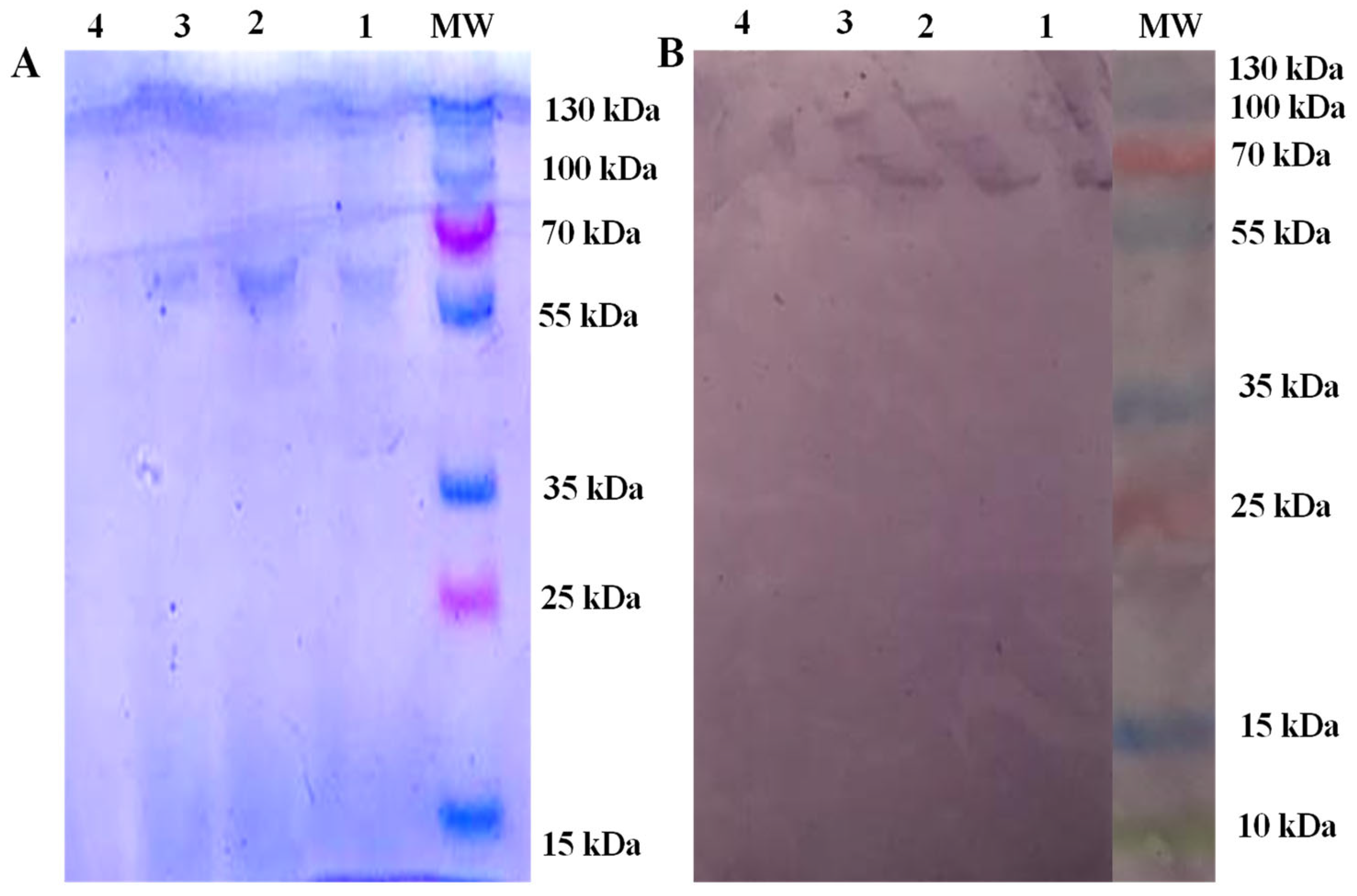
Figure 1. (A) Coomassie-blue-stained SDS-PAGE showing protein separation and identification of Echinococcus granulosus native antigen. Lane MW = protein marker, lanes 1–3 = samples, lane 4 = empty well. (B) Western blot of hydatid cyst fluids from slaughtered buffalo using sera from naturally infected buffalo and developed by an anti-bovine antibody conjugated with alkaline phosphatase. Lane MW = protein marker (run on the same gel as the test samples, but image cropped to remove samples unrelated to the current study), lanes 1, 2 and 3 = samples and lane 4 is an empty well.
Animals 13 00866 g001
Figure 2. (A) Sensitivity, specificity and cut-off value of crude-antigen-based detection of Echinococcus-specific IgG antibodies in bovine sera. (B) Interactive dot plot for negative and positive sera.
Animals 13 00866 g002
Table 1. Definition and formulas to calculate sensitivity, specificity, PPV, NPV, LR+, LR− and DOR of ELISA, based on [35].
NameDefinitionFormula
Sensitivity“The capability of a test to correctly identify the patients with disease”a/(a + c) × 100
Specificity“The potential of a screening test to identify the patients without the disease”d/(b + d) × 100
Positive predictive value (PPV)“Prediction of positive patients before test (identifying true positive)”a/a + b
Negative predictive value (NPV)“Prediction of negative samples before test (identifying true negative)”d/c + d
Likelihood ratio positive (LR+)“Ratio of probability that a positive test outcome would be expected in a patient with a disease to the probability that a positive test outcome would be expected in a patient without a disease”(a/a + c)/(b/b + d)
Likelihood ratio negative (LR−)“The ratio of probability of a patient testing negative who has a disease to the probability of a patient testing negative who does not have a disease”(c/a + c)/(d/b + d)
Diagnostic
odd ratio (DOR)
“Odds of a positive test in those with disease relative to the odds of a positive test in those without disease”LR+/LR−
a = true positive, b = false positive, c = false negative and d = true negative.
Table 2. Sensitivity, specificity, PPV, NPV, LR+, LR− and DOR values of iEgELISA.
SensitivitySpecificityPPVNPVLR+LR−DOR
100%93.8%73.0799.9616.330.0121360.83
Table 3. Distribution of Echinococcus-positive sera categorised by host species, sex and age group.
Age ClassBuffaloCattle
FemaleMaleFemaleMale
2–3 years2/21 (9.5%)0/23 (0%)0/11 (0%)4/28 (14.2%)
4–5 years2/31 (6.5%)3/19 (15.7%)12/31 (38.7%)4/22 (18.1%)
6–7 years9/28 (32.1%)1/4 (25%)11/22 (50%)4/14 (28.5%)
Table 4. Comparison of post-mortem and serological examination results among studies.
Host SpeciesSample SizePost-Mortem (%)ELISA (%)Antigen Used in ELISAReferences
Cattle12819.527.3Ammonium sulphate ((NH4)2SO4) precipitated HCFCurrent study
Buffalo1369.512.5Ammonium sulphate ((NH4)2SO4) precipitated HCFCurrent study
Cattle2561.635.5Freeze-dried precipitated HCF[43]
Camel30414.1452.6Freeze-dried precipitated HCF[43]
Camel20068Phosphotungstic acid and 2M magnesium chloride used for HCF precipitation[44]
HCF = hydatid cyst fluid.
Table 5. Comparison of sensitivity and specificity of iEgELISA based on crude antigen (this study) with results from previous work. Arrows denote the direction of change compared with values from the present study.
Sensitivity (%)Specificity (%)Host OriginAntigen Size (kDa)Type of AntigenReferences
10093.8Buffalo67Crude antigenCurrent study
82.8 ↓62.5 ↓CamelNot mentionedCrude antigen[41]
79.3 ↓75 ↓CamelNot mentionedPartially purified crude antigen[41]
36 ↓93 ↓Sheep8–24Crude antigen[49]
25 ↓99 ↑Sheep6Recombinant antigen[49]
96% ↓71% ↓CamelNot mentionedCrude antigen[49]
99% ↓90% ↓CamelNot mentionedCamel antigen B[49]
89.2 ↓89.5 ↓Sheep8–12Total hydatid liquid (LHT)[37]
80.0 ↓93.9 ↑Sheep16Purified portion of total hydatid liquid (S2B)[37]
86.4 ↓92.8 ↓Sheep20–24Purified antigen (B)[37]
83 ↓70.3 ↓CamelNot mentionedProtoscolex antigen[50]
46.5 ↓41.7 ↓CamelNot mentionedGerminal layer antigen[50]
LHT = total hydatid liquid, S2B = purified portion of total hydatid liquid and B = purified antigen.
Disclaimer/Publisher’s Note: The statements, opinions and data contained in all publications are solely those of the individual author(s) and contributor(s) and not of MDPI and/or the editor(s). MDPI and/or the editor(s) disclaim responsibility for any injury to people or property resulting from any ideas, methods, instructions or products referred to in the content.